Leukocyte adhesion defect: An uncommon immunodeficiency
4
Abstract Leukocyte adhesion deficiency (LAD) is a rare primary immunodeficiency disorder with autosomal recessive inheritance which is characterized by presence of a defect of phagocytic function resulting from a lack of leukocyte cell surface expression of b2 integrin molecules (CD11 and CD18) that are essential for chemotaxis. The classic symptoms of the disease are failure of separation of the umbilical cord and recurrent bacterial infections, which continue throughout life. We describe here two cases of infants who presented with characteristic history of recurrent infections, delayed separation of umbilical cord and marked leukocytosis. Keywords: Leukocyte adhesion defect. Introduction Three leukocyte adhesion deficiency (LAD) disorders have been well recognized, caused by genetic defects in Beta 2 integrin family consist of LFA-1 (CD11a/CD18), Mac-1 (CD11b/CD18), Complement receptor 4 (CD11c/CD18), and (CD11d/CD18). 1 LAD-I is caused by mutations in the ITGB2 gene coding b2 subunit of beta 2 integrin family. This mutation results in formation of b2 subunit that cannot bind with other subunits to form b2 integrin. Leukocytes that lack these integrin fail to attach to blood vessel wall to contribute in immune response. 1 LAD-II is a rare disorder characterized by deficiency in a guanosine diphosphate-fucose transporter in the Golgi apparatus, which is essential for fucosylating the ligands of the selectin adhesion receptors. 4,5 Thus leukocytes fail to roll along the vasculature and integrin-mediated adhesion stabilization does not take place. 2 LAD-III, the most recently described type in which the integrins on bone marrow-derived leukocytes and platelets fail to function due to mutation in the FERMT3 gene coding for the kindlin-3 protein, thus introducing a premature stop codon resulting in a non-functioning protein. 3,4 Case-1 An 8 months old female child was brought to Shifa International Hospital in January 2016, with history of fever, and cough for 3 days. Fever was high grade, associated with vomiting and loose stools. Vomiting was non greenish, non-projectile, and loose stools were semi solid and greenish in colour, not associated with runny nose and runny eyes. She had multiple episodes of pyrexia from the age of 2 months, every 3-4 weeks that required multiple hospitalizations. She was the only child of consanguineous parents born at term by spontaneous vaginal delivery following an uncomplicated pregnancy. The umbilical cord separated on 41st postnatal day. There was no history suggestive of sepsis or umbilical infection in the neonatal period. She was developmentally normal and received her routine immunizations. On examination, she had no dysmorphism, was pale, febrile with no icterus, petechiae, or bruises. Vitals were normal. Her weight was 7500 gram and height was 67 cm. There was no lymphadenopathy. Throat was congested with no exudates. Chest examination showed bilateral equal air entry with mild crepitation. Abdominal, cardiovascular and nervous examination all was unremarkable. Investigations revealed haemoglobin concentration of 8.5 g/dl, platelet count of 556,000/mm 3 markedly raised leukocyte count of 54000/mm3 (neutrophils 80%, lymphocytes 13%, and monocytes 7%). Chest x-ray showed bilateral infiltrates. Her serum immunoglobulin profile and flow cytometry analysis revealed diagnosis of LAD 1 type (Figure-1). The child was managed with antibiotics and discharged on trimethoprim/ sulfamethoxazole and fluconazole prophylaxis. Since the day of discharge she had no follow- up. Case-2 A three and half months old male child was brought to Shifa International Hospital in 2015, with history of fever for 4-5 days. Fever was high grade, associated with cough. There was no history of vomiting, loose stools, runny nose and runny eyes. He was the third child of consanguineous parents born at term by spontaneous vaginal delivery Vol. 68, No. 1, January 2018 119 CASE REPORT Leukocyte adhesion defect: An uncommon immunodeficiency Sehar Nigar, Ejaz Ahmed Khan, Tahir Aziz Ahmad Shifa International Hospital, Islamabad. Correspondence: Ejaz Ahmed Khan. Email: [email protected]
Transcript of Leukocyte adhesion defect: An uncommon immunodeficiency

AbstractLeukocyte adhesion deficiency (LAD) is a rare primaryimmunodeficiency disorder with autosomal recessiveinheritance which is characterized by presence of a defectof phagocytic function resulting from a lack of leukocytecell surface expression of b2 integrin molecules (CD11and CD18) that are essential for chemotaxis. The classicsymptoms of the disease are failure of separation of theumbilical cord and recurrent bacterial infections, whichcontinue throughout life.
We describe here two cases of infants who presented withcharacteristic history of recurrent infections, delayedseparation of umbilical cord and marked leukocytosis.
Keywords: Leukocyte adhesion defect.
IntroductionThree leukocyte adhesion deficiency (LAD) disorders havebeen well recognized, caused by genetic defects in Beta 2integrin family consist of LFA-1 (CD11a/CD18), Mac-1(CD11b/CD18), Complement receptor 4 (CD11c/CD18),and (CD11d/CD18).1
LAD-I is caused by mutations in the ITGB2 gene coding b2subunit of beta 2 integrin family. This mutation results information of b2 subunit that cannot bind with othersubunits to form b2 integrin. Leukocytes that lack theseintegrin fail to attach to blood vessel wall to contribute inimmune response.1
LAD-II is a rare disorder characterized by deficiency in aguanosine diphosphate-fucose transporter in the Golgiapparatus, which is essential for fucosylating the ligandsof the selectin adhesion receptors.4,5 Thus leukocytes failto roll along the vasculature and integrin-mediatedadhesion stabilization does not take place.2
LAD-III, the most recently described type in which theintegrins on bone marrow-derived leukocytes andplatelets fail to function due to mutation in the FERMT3gene coding for the kindlin-3 protein, thus introducing apremature stop codon resulting in a non-functioningprotein.3,4
Case-1An 8 months old female child was brought to ShifaInternational Hospital in January 2016, with history offever, and cough for 3 days. Fever was high grade,associated with vomiting and loose stools. Vomiting wasnon greenish, non-projectile, and loose stools were semisolid and greenish in colour, not associated with runnynose and runny eyes. She had multiple episodes ofpyrexia from the age of 2 months, every 3-4 weeks thatrequired multiple hospitalizations. She was the only childof consanguineous parents born at term by spontaneousvaginal delivery following an uncomplicated pregnancy.The umbilical cord separated on 41st postnatal day. Therewas no history suggestive of sepsis or umbilical infectionin the neonatal period. She was developmentally normaland received her routine immunizations.
On examination, she had no dysmorphism, was pale,febrile with no icterus, petechiae, or bruises. Vitals werenormal. Her weight was 7500 gram and height was 67 cm.There was no lymphadenopathy. Throat was congestedwith no exudates. Chest examination showed bilateralequal air entry with mild crepitation. Abdominal,cardiovascular and nervous examination all wasunremarkable.
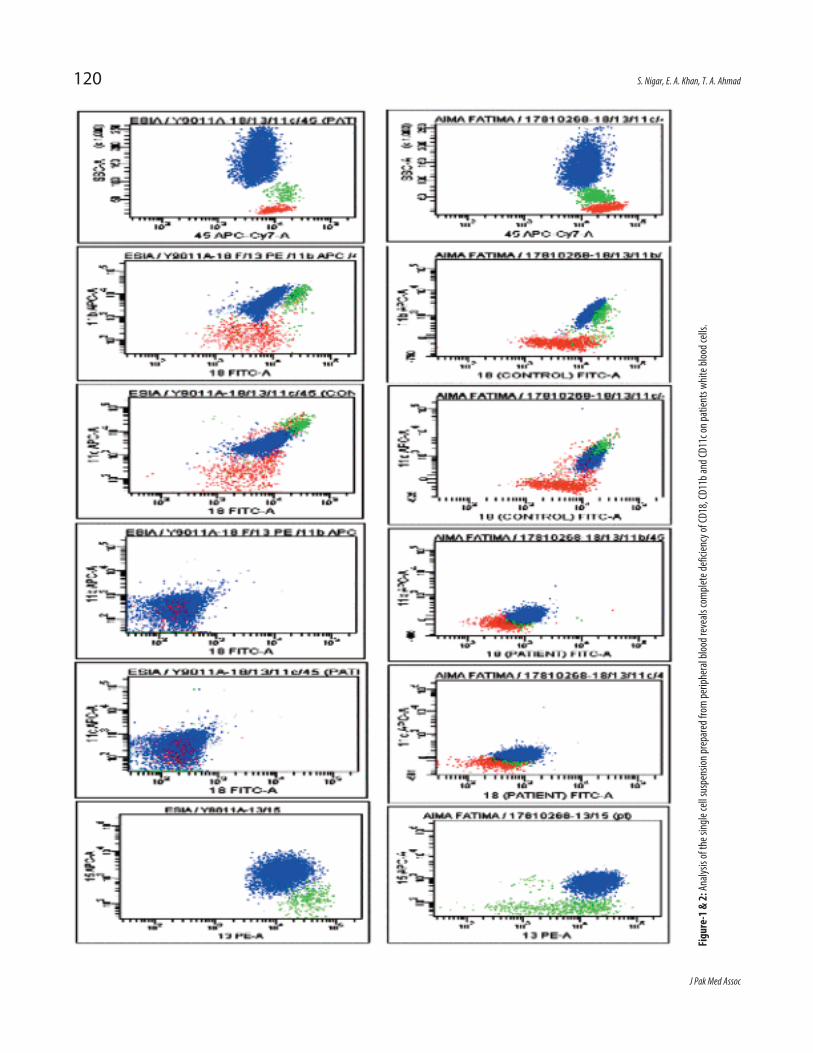
Investigations revealed haemoglobin concentration of 8.5g/dl, platelet count of 556,000/mm3 markedly raisedleukocyte count of 54000/mm3 (neutrophils 80%,lymphocytes 13%, and monocytes 7%). Chest x-rayshowed bilateral infiltrates. Her serum immunoglobulinprofile and flow cytometry analysis revealed diagnosis ofLAD 1 type (Figure-1).
The child was managed with antibiotics and dischargedon trimethoprim/ sulfamethoxazole and fluconazoleprophylaxis. Since the day of discharge she had no follow-up.
Case-2A three and half months old male child was brought toShifa International Hospital in 2015, with history of feverfor 4-5 days. Fever was high grade, associated with cough.There was no history of vomiting, loose stools, runny noseand runny eyes. He was the third child of consanguineousparents born at term by spontaneous vaginal delivery
Vol. 68, No. 1, January 2018
119
CASE REPORT
Leukocyte adhesion defect: An uncommon immunodeficiencySehar Nigar, Ejaz Ahmed Khan, Tahir Aziz Ahmad
Shifa International Hospital, Islamabad.Correspondence: Ejaz Ahmed Khan. Email: [email protected]
J Pak Med Assoc
120 S. Nigar, E. A. Khan, T. A. Ahmad
Figu
re-1
& 2:
Analysis of the sing
le cell suspension
prepared from
peripheral bloo
d reveals comple
te de
ficien
cy of CD
18, CD11b an
d CD11c on
patients w
hite b
lood cells.

following an uncomplicated pregnancy. The umbilicalcord separated on 22nd postnatal day. There was historyof omphalitis in neonatal period. He was developmentallynormal. One of his siblings died in infancy due torecurrent infections, while other siblings are healthy.
On examination, he was pale, febrile with nodysmorphism. His weight was 3600 gram which wasbelow 5th percentile and height was 58 cm. Vitals werenormal. There was no icterus, petechiae, or bruises orlymphadenopathy. Chest examination showed bilateralequal air entry. Abdomen was soft, slightly distended withliver 3 cm below right costal margin, soft in consistency,non-tender with smooth surface. There was no ascitesand bowel sounds were audible. Cardiovascular andnervous system examination was unremarkable.
Investigations revealed haemoglobin concentration of 8.1g/dl, platelet count of 611,000/mm3, markedly raisedleukocyte count of 87400/mm3 (neutrophils 86%,lymphocytes 10%, monocytes 3%). Blood culture did notyield any organism. Serum immunoglobulins levels werenormal for age. Flow cytometry was done (Figure-2).Based on his clinical presentation, family history and flowcytometric analysis, the diagnosis of LAD type 1 wasestablished. Since the time of diagnosis, he had 2-3 timeshospitalization for oral ulcer, otitis media, fungal infectionand fevers. Bone marrow transplant has not been doneyet, due to non-availability of donor.
Verbal consent was taken from father on telephone forpublication of the case.
DiscussionLeukocyte adhesion deficiency type I (LAD-1) is anautosomal recessive disorder characterized by severe andrecurrent bacterial infections, altered wound healing andsignificant morbidity that is caused by absent ordiminished expression of integrins b2 class.6
In severely deficient b2 expression (less than 1% of thenormal amount of cell surface expression of the b2
integrin heterodimers) patients often die within the firstyear while moderately deficient b2 expression (5%-10% ofthe normal level) results in a milder form of disease.7 Thecardinal features of classical form of disease includesdelayed detachment of umbilical cord, non-purulentbacterial infections in the presence of markedgranulocytosis and impaired wound healing.8 Both casespresented had these classical features.
The umbilical cord usually sloughs by the end of 2ndweek of life and mean time of separation is 7.4 days.However causes of delayed separation of cord such asurachal anomalies, antibiotic administration, prematurityand low birth weight must be excluded.11,12
In our cases, both infants had the characteristic clinicalfeatures of delayed separation of cord, recurrentinfections and marked leukocytosis. With suspicion ofLAD it was decided to investigate it and diagnosis wasconfirmed by flow cytometry showing absence of CD18,CD11b, CD11c on neutrophils. Over 300 cases of LAD 1have been reported worldwide, while for LAD II and LADIII, there are less than 10 cases each.5 Tipu et al havereported a patient to be suffering from this disease fromPakistan.5
LAD is an attractive disease for human gene therapy asthe conventional therapy with antibiotics andgranulocytes transfusions improves the symptoms onlybut does not correct the phenotype.10 The only definitetreatment is stem cell or bone marrow transplantationfrom histocompatible relatives and should be consideredas an early therapeutic option if a suitable HLA-matchedstem-cell donation is available.9 Our cases emphasize thatas physicians we should investigate any child presentingwith delayed umbilical cord separation and markedlyraised leukocyte count. Detail immunological assessmentis also needed with management aimed at treatinginfections, hygiene, and antibiotic prophylaxis with ageappropriate immunizations. Definite therapy requiresstem cell transplant or gene therapy that should be
Vol. 68, No. 1, January 2018
Leukocyte adhesion defect: An uncommon immunodeficiency 121
Table: Laboratory results of both cases.
Reports Case 1 Case 2 Reference Values
Hemoglobin (g/dl) 8.5 8.1 16-NovLeukocytes count/microliter 54000 87400 4000-11000 N = 80% , L = 13% N = 86% , L = 10% Platelets/UL 556000 611000 150,000 - 400,000IgA g/L 1.03 0.1 0.08-0.91IgG g/L 10.7 8.5 2.03-9.48IgM g/L 2.83 0.97 0.17-1.50Flow cytometry Absence of CD 18, CD 11b, CD 11c Absence of CD 18, CD 11b, CD 11c

explored early.
ConclusionLAD is a rare immune deficiency disorder and should besuspected in an infant with typical presentation andconfirmed by flow cytometric analysis for the expression ofdeficient integrin molecules on leukocytes.
Disclaimer: The abstract has not been published orpresented in any conference.
Conflict of Interest: None.
Funding Sources: None.
References1. van de Vijver E, Maddalena A, Sanal Ö, Holland SM, Uzel G, Madkaikar
M, et al. Hematologically important mutations: leukocyte adhesiondeficiency (first update). Blood Cells Mol Dis. 2012; 48:53-61.
2. Simpson BN, Hogg N, Svensson LM, McDowall A, Daley W, YarbroughK. A New Leukocyte Hyper adhesion Syndrome of Delayed CordSeparation, Skin Infection, and Nephrosis. Pediatrics. 2014; 133: e257-62.
3. Svensson L, Howarth K, McDowall A, Patzak I, Evans R, Ussar S, et al.Leukocyte adhesion deficiency-III is caused by mutations in KINDLIN3affecting integrin activation. Nat Med. 2009; 15: 306-12.
4. Malinin NL, Zhang L, Choi J, Ciocea A, Razorenova O, Ma YQ, et al. Apoint mutation in KINDLIN3 ablates activation of three integrinsubfamilies in humans. Nat Med. 2009; 15: 313-8.
5. Tipu HN, Tahir A, Ahmed TA, Hazir T, Waqar MA. Leukocyte AdhesionDefect. J Pak Med Assoc. 2008; 58: 643-5.
6. Harris ES, Shigeoka AO, Li W, Adams RH, Prescott SM, McIntyre TM, et
al. Anovel syndrome of variant leukocyte adhesion deficiency involvingdefects in adhesion mediated by beta1 and beta2 integrins. Blood.2001; 97: 767-76.
7. Gallin JI, Snyderman RKishimoto TK, Baldwin ET, Anderson DC, GallinJI, Snyderman R. The role of beta2 integrins in inflammation.Inflammation: Basic Principles and Clinical Correlates. 3rd ed.Philadelphia: Williams and Wilkins, 1999; pp- 537-69.
8. Kuijpers TW, Van Lier RA, Hamann D, de Boer M, Thung LY, WeeningRS, et al. Leukocyte adhesion deficiency type 1 (LAD-1)/variant. Anovel immunodeficiency syndrome characterized by dysfunctionalbeta2 integrins. J Clin Invest. 1997; 100: 1725-33.
9. Waseem Qasim, Marina Cavazzana-Calvo, E. Graham Davies, JefferyDavis, Michel Duval, et al. Allogeneic Hematopoietic Stem-CellTransplantation for Leukocyte Adhesion Deficiency. Pediatrics. 2009;123: 836-40.
10. Movahedi M, Entezari N, Pourpak Z, Mamishi S, Chavoshzadeh Z,Gharagozlou M, et al. Clinical and Laboratory Findings in IranianPatients with Leukocyte Adhesion Deficiency. J Clin Immunol. 2007;27: 302-7.
11. Kliegman RM, Stoll BJ. The fetus and the neonatal infant. In: BehrmanRE, Kliegman RM, Jenson HB, eds. Nelson Textbook of Pediatrics 16thed. Philadelphia: WB Saunders, 2000; pp-257.
12. Razvi S, Murphy R, Shlasko E, Cunningham-Rundles C. delayedseparation of the umbilical cord attributable to urachal anomalies.Pediatrics. 2001; 108: 493-4.
J Pak Med Assoc
122 S. Nigar, E. A. Khan, T. A. Ahmad