Lessons from a 2 decade journey through customizing ablations...
63
A. John Kanellopoulos, MD Kanellopoulos MD 1 Lessons from a 2 decade journey through customizing ablations and cornea biomechanics with CXL (CXL plus part I) A. John Kanellopoulos, MD President, the ISRS Director, Laservision.gr Institute, Athens, Greece Clinical Professor NYU Medical School, NY
Transcript of Lessons from a 2 decade journey through customizing ablations...
A. John Kanellopoulos, MDKanellopoulosMD 1
Lessons from a 2 decade journey through
customizing ablations and cornea biomechanics with CXL
(CXL plus part I) A. John Kanellopoulos, MD
President, the ISRS Director, Laservision.gr Institute, Athens, Greece Clinical Professor NYU Medical School, NY

A. John Kanellopoulos, MD
Financial interests (D) consultant for:
AJKMD events Alcon Allergan Avedro KeraMed i-Optics ISP Surgical, LLC Optovue
Zeiss

A. John Kanellopoulos, MD
For the things we have to learn before we can do, we learn by doing.
— Aristotle

A. John Kanellopoulos, MD
CXL efficacy and safety Decrease of UV-intensity
courtesy E. Spoel MD
3.00 mW/cm²
1.49 mW/cm²
0.74 mW/cm²
0.36 mW/cm²
0.18 mW/cm²
0.09 mW/cm²

A. John Kanellopoulos, MD
Our Athens team’s CXL contributions: • Applying topo-guided PRK in CXLed ectatic corneas 2004 • Combining same-day CXL with topo-guided reshaping of irregular corneas Athens
Protocol: 2005 • Higher fluence: 2006 (6mW, 10mW)
• Intra-stromal treatments through femto-pocket: 2007 • LASIK+CXL( Xtra ): 2008 (ESCRS) • LASIK Xtra for hyperopia: 2011 (ASCRS)
• PiXL CXL corneal differentials: 2013 (AAO) • CXL in Boston Kpro (Cornea resistance to melt) • Athens Protocol with PiXL CXL 2015
• TMR: Topography-modified refraction 2016

A. John Kanellopoulos, MD
Zurich 2011
• LASIK combined with CXL• CXL or bullus keratopathy• In-pocket CXL• The Athens Protocol

A. John Kanellopoulos, MD 7
introduced: Higher fluence CXL: 6, 7, 9, 10 and 12mW/cm2
AAO 2008: CXL for 15 minutes utilizing 7mW/cm2 fluence

A. John Kanellopoulos, MD
2007: Introduction of riboflavin in a femto-pocket
8
N E W T E C H N I Q U E
Collagen Cross-linking in Early Keratoconus With Ribofl avin in a Femtosecond Laser-created Pocket: Initial Clinical ResultsAnastasios John Kanellopoulos, MD

A. John Kanellopoulos, MD
Introduced Prophylactic CXL in PRK and LASIK 2008
Clinical Ophthalmology Dovepress
O R I G I N A L R E S E A R C H
open access to scientific and medical research
Open Access Full Text Article
Comparison of prophylactic higher fluence corneal cross-linking to control, in myopic LASIK, one year results
Anastasios John Kanellopoulos1,2
George Asimellis1
Costas Karabatsas1
1LaserVision.gr Clinical and Research Eye Institute, Athens, Greece; 2New York University Medical School, New York, NY, USA
Purpose: To compare 1-year results: safety, efficacy, refractive and keratometric stability, of
femtosecond myopic laser-assisted in situ keratomileusis (LASIK) with and without concurrent
prophylactic high-fluence cross-linking (CXL) (LASIK-CXL).
Methods: We studied a total of 155 consecutive eyes planned for LASIK myopic correction.
Group A represented 73 eyes that were treated additionally with concurrent prophylactic high-
fluence CXL; group B included 82 eyes subjected to the stand-alone LASIK procedure. The
following parameters were evaluated preoperatively and up to 1-year postoperatively: manifest
refractive spherical equivalent (MRSE), refractive astigmatism, visual acuity, corneal keratom-
t d d th li l ll t W l tt d k t t t ti l d
Ker
atom
etric
read
ings
(D)
Time after surgery (months)35
K-flatPreoperative43.9445.17
37.6438.32
37.6938.35
37.6638.36
37.6738.37
1-month 3-months 6-months 12-months
K-steep
37
39
41
43
45
47LASIK-CXL group,65 eyes up to 12-months post-op
A
Ker
atom
etric
read
ings
(D)
Time after surgery (months)35
Preoperative43.15K-flat
K-steep 44.07
1-month37.4538.12
3-months37.6538.32
6-months37.8938.57
12-months38.0238.66
37
39
41
43
45
47Stand-alone LASIK group,75 eyes up to 12-months post-op
B
Figure 8 Stability of corneal keratometry for (A) the LASIK-CXL group and (B) the stand-alone LASIK group, expressed in diopters (D), up to 1-year postoperatively.Abbreviations: CXL, cross-linking; LASIK, laser-assisted in situ keratomileusis.

A. John Kanellopoulos, MD
Athens Protocol:Topo-guided partial PRK + CXL 1-Topolyzer:Placido disc topography 2-Pentacam (Oculyzer) 3-Pentacam HD (oculyzer II)-Refractive suite 4-Vario (placido disc +pupil sensor+iris recognition+limbal landmarks recognition)
WaveLight® FS200Femtosecond Laser
WaveLight® EX500Excimer Laser
WaveLight®Refrac<veSuiteSimilartechnologies:Zeiss,Schwind,Ivis

A. John Kanellopoulos, MD
J Cornea 2007
CXL followed 6 months later by a partial tPRK
2004: Over the last 12 years we have introduced and treated over 3000 cases of KCN and ectasia with CXL combined
with a topo-guided excimer normalization: the “Athens Protoco” now practiced globally!!!

A. John Kanellopoulos, MD
TheAthensProtocol4steps:same day PTK > topoPRK > MMC > CXL (6mW/cm2 x 15 min)
6
ê
è12
O R I G I N A L A R T I C L E
Comparison of Sequential vs Same-day Simultaneous Collagen Cross-linking and Topography-guided PRK for Treatment of KeratoconusAnastasios John Kanellopoulos, MD
ABSTRACT
PURPOSE Th f d ffi f l ll
K eratoconus is a bilateral, non-symmetric, noninfl am-matory progressive corneal degeneration that fre-quently manifests in post-pubescent young adults
Sequential vs Simultaneous Topography-guided PRK and CXL/Kanellopoulos
Figure 2. Cornea optical coherence tomog-raphy demonstrates hyper-reflective intra-corneal stromal “lines” at 2/3 depth (arrows) corresponding with the clinical presence of the corneal collagen cross-linking (CXL) demarcation line in a patient from the simultaneous group 3 years following the combined topography-guided photorefrac-tive keratectomy and CXL procedure.

A. John Kanellopoulos, MD 13
Kanellopoulos AJ: JRS Sept 09: 358 cases with over 2 year follow-up: 160 cases Sequential (left) Vs 198 cases same-day Combined (right)
PreOp PostOpUCVA LogMar 0.9 ±0.3 0.49 ±0.25
BSCVA LogMar 0.41 ±0.25 0.16 ±0.22
Mean Decrease MRSE
2.50±1.2
Mean K Decrease
2.75±1.3
Mean Haze Score
1.2±0.5
Mean CCT 465±45 395±25
Pre-op Post-opUCVA LogMar 0.96 ±0.2 0.3 ±0.2
BSCVA LogMar 0.39 ±0.3 0.11 ±0.16 (p<0.001)
Mean Decrease MRSE
3.2±1.4 (p<0.005)
Mean K Decrease
3.50±1.3 (p<0.005)
Mean Haze Score
0.5±0.3 (p<0.0052
Mean CCT 475±55 405±35
9
Sequential CXL and after TCAT Combined TCAT + CXL: The Athens Protocol

A. John Kanellopoulos, MD 14
O R I G I N A L A R T I C L E S
Management of Corneal Ectasia After LASIK With Combined, Same-day, Topography-guided Partial Transepithelial PRK and Collagen Cross-linking: The Athens ProtocolAnastasios John Kanellopoulos, MD; Perry S. Binder, MS, MD
327Journal of Refractive Surgery • Vol. 27, No. 5, 2011
Management of Corneal Ectasia After LASIK/Kanellopoulos & Binder
ectasia and was offered Intacs (Addition Technology Inc, Des Plaines, Illinois) or a corneal transplant.
He presented to our institution in September 2007, 3 years after LASIK. Uncorrected distance visual acu-ity was 20/40 in the right eye and 20/15 in the left eye. Manifest refraction was �1.50 �2.00 � 65 (20/20) in
the right eye and plano (20/15) in the left eye. Kera-tometry was 41.62@65/43.62@155 in the right eye and 41.75/42.12@10 in the left eye. Central ultrasound pachymetry was 476 µm in the right eye and 490 µm in the left eye.
On September 13, 2007, 39 months after LASIK,
Figure 2. Case 2. Topography on the left shows marked inferior steepening before topography-guided PRK/CXL treatment. The topography on the right shows the same cornea 18 months after topography-guided PRK/CXL with marked flattening of the corneal ectasia and normalization of the cornea.
Figure 3. Case 3. Clinical course of the right eye. A) Topography 3 years after LASIK demonstrates irregular astigmatism and marked inferior corneal steepen-ing. Uncorrected distance visual acu-ity was 20/40 and corrected distance visual acuity was 20/20 with refraction of �1.50 �2.00 � 65. B) Topography 3 months after topography-guided PRK/CXL procedure demonstrates a flatter and normalized cornea. Uncorrected distance visual acuity was 20/15. C) Topographic reproduction of the topography-guided PRK treatment plan with the WaveLight platform. This platform plans to remove tissue in an irregular fashion to normalize the corneal ectasia seen in Figure 3A. D) Comparison map, derived from subtract-ing image B from A, represents the topo-graphic difference in this case 3 months after the combined treatment. The para-central flattening is self-explanatory, as the PRK and CXL have flattened the cone apex. The superior nasal arcuate flatten-ing represents the actual part-hyperopic correction, which the topography-guided treatment has achieved, to accomplish steepening in the area central to this arc. Thus, the topography-guided treatment has normalized the ectatic cornea by flat-tening the cone apex and at the same time by “steepening” the remainder of the central cornea.
Management of Corneal Ectasia After LASIK/Kanellopoulos & Binder
Figure 4. Case 4. Pentacam comparison of the right eye. The left column shows the data and topography before topography-guided PRK/CXL. The center column shows the postoperative data and topography. The right column shows the difference (pre- minus postoperative).
JRS 2011
A. John Kanellopoulos, MD
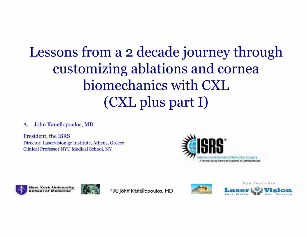
TheAthensProtocol4steps:same day partialPRK > PTK > MMC > CXL (6mW/cm2 x 15 min)
2-PTK
1- topo -guided
PRK 3- 30” MMC
4-: CXL
6
15

A. John Kanellopoulos, MD
Surgical Procedure 1. Partial topography-guided excimer-
laser ablation, employing photorefractive keratectomy (PRK) in combination with the T-CAT procedure. Optical zone 5.00 to 5.50 mm.
2. Excimer-laser ablation (uniform 50 µm over a 7.00 mm zone), employing the PTK mode.
3. CXL with UV-A irradiance of 6 mW/cm2, applied for 15΄ employing the KXL I or II system (Avedro Inc., Waltham, MA).

A. John Kanellopoulos, MD
Step 4: attempted Rx to 0, OZ to 5 or 5.5mm, cyl axis to match topo axis not refractive axis
17

A. John Kanellopoulos, MD
Post LASIK ectasia: 26y/o pilot, from UCVA 20/60 to 20/15
18

A. John Kanellopoulos, MD

A. John Kanellopoulos, MD
768 Copyright © SLACK Incorporated
O R I G I N A L A R T I C L E
Novel Placido-derived Topography-guided Excimer Corneal Normalization With Cyclorotation Adjustment: Enhanced Athens Protocol for KeratoconusAnastasios John Kanellopoulos, MD; George Asimellis, PhD
ABSTRACT
PURPOSE: To comparatively investigate the efficacy of the enhanced Athens Protocol procedure guided by novel Placido-derived topography with cyclorotation compensation (the cyclorotation adjusted group) to similar cases guided by Scheimpflug-derived tomogra-phy without cyclorotation compensation (the non-cyclo-rotation adjusted group).
METHODS: Two groups were evaluated: the cyclorota-tion adjusted group (n = 110 eyes) and the non-cyclo-rotation adjusted group (n = 110 eyes). Analysis was based on digital processing of Scheimpflug imaging derived curvature difference maps preoperatively and 3 months postoperatively. The vector (r, �) corresponding to the steepest corneal point (cone) on the preopera-tive surgical planning map (rp, �p) and on the curvature difference map (rd, �d) were computed. The differences between the peak topographic angular data (�� = |�p – �d|) and weighted angular difference (W�� = �� � �r) were calculated.
RESULTS: For the cyclorotation adjusted group, �� was 7.18° ± 7.53° (range: 0° to 34) and W�� was 3.43 ± 4.76 mm (range: 0.00 to 21.41 mm). For the non-cyclorotation adjusted group, �� was 14.50° ± 12.65° (range: 0° to 49°) and W�� was 10.23 ± 15.15 mm (range: 0.00 to 80.56 mm). The cyclorotation adjusted group appeared superior to the non-cyclorotation ad-justed group, in both the smaller average angular differ-ence between attempted to achieved irregular curvature normalization and in weighted angular difference, by a statistically significant margin (��: P = .0058; W��: P = .015).
CONCLUSIONS: This study suggests that employment of the novel Placido-derived topographic data of highly irregular corneas, such as in keratoconus, treated with topography-guided profile with cyclorotation compensa-tion leads to markedly improved cornea normalization.
[J Refract Surg. 2015;31(11):768-773.]
orneal cross-linking (CXL) is considered a valid option for progressive keratoconus/corneal ectasia treatment.1 By increasing corneal biomechanical
strength, CXL results in keratectasia arrest.2 In addition, CXL has also been shown to improve corneal irregularity and re-duce central anterior corneal steepening.3
Combined with CXL, partial anterior surface normalization via topography-guided customized partial excimer laser abla-tion may offer, in addition to keratectasia arrest, improved topographic and refractive outcomes.3,4 The Athens Protocol comprises phototherapeutic keratectomy (PTK) of 50 µm, a partial photorefractive keratectomy (PRK) for the topography-guided customized anterior surface normalization, and high-fluence CXL for corneal stabilization.5 Long-term results6 and anterior segment optical coherence tomography quantitative findings7 have demonstrated the stability of the procedure in large cohorts of patients. Variations of this technique have been applied and reported globally.8-14
Because the topography-guided ablation step of the pro-cedure bears a high degree of customization, the impact of effective alignment between treatment planning based on the topography-derived data and surgically applied ablation pat-tern is pivotal for a successful outcome. Critical parameters affecting alignment are horizontal and vertical eye move-ments, eye pupil centroid shift, and possible cyclorotation. The significance of these principles has been reported pre-operatively and intraoperatively in refractive procedures.15 High-speed active eye tracking along with cyclorotational topographic adjustment (CTA) has been introduced during the past 2 years in refractive lasers such as the EX500 ex-cimer laser (Alcon Laboratories, Inc., Fort Worth, TX), which
From Laservision.gr Clinical and Research Eye Institute, Athens, Greece (AJK); and NYU Medical School, Department of Ophthalmology, New York, New York (GA).
Submitted: April 28, 2015; Accepted: August 11, 2015
Dr. Kanellopoulos is a consultant for Alcon/WaveLight, Allegran, Avedro, and i-Optics. Dr. Asimellis has no financial or proprietary interest in the materials presented herein.
Correspondence: Anastasios John Kanellopoulos, MD, Laservision.gr Clinical and Research Eye Institute, 17 Tsocha Street, Athens, 115 21 Greece. E-mail: [email protected]
doi: 10.3928/1081597X-20151021-06
C
Figure B. The ‘compare 2 exams’ output from the Scheimpflug imaging device. (Left) The preoperative sagittal curvature map, (middle) the postopera-tive map, and (right) the difference of the two maps.

A. John Kanellopoulos, MD21
Average K from 48.5 to 44 Refraction -2.5-4.5@155 (20/70) to -1-1.5@10 (20/20)
21

A. John Kanellopoulos, MD22
Caution: marked refractive effect with the 3mW protocols
22

A. John Kanellopoulos, MD

A. John Kanellopoulos, MD
Conclusions • The Athens Protocol (partial topo-guided PTK
combined with CXL) appears to be safe and effective in ectasia stabilization, and visual rehab
over 12 years later.
• Alternative treatments are CXL alone
• Contact lenses: RGPs and/or Scleral lenses
• ICRS
• Lamellar keratoplasty
• Penetrating keratoplasty
O R I G I N A L A R T I C L E
Corneal Refractive Power and Symmetry Changes Following Normalization of Ectasias Treated With Partial Topography-Guided PTK Combined With Higher-Fluence CXL (The Athens Protocol)Anastasios John Kanellopoulos, MD; George Asimellis, PhD
344 Copyright © SLACK Incorporated
Corneal Refractive Power and Symmetry Changes/Kanellopoulos & Asimellis
treatment, r2 was 0.407 and P value less than .001 (Table 2).
Data analysis indicates that the mean of the paired differences regarding the flat anterior keratometric val-
ues was reduced (flattened) postoperatively by -3.09 ± 2.69 D, or -6.56%, and was statistically significant (P < .05) (Table 2). The uncertainty associated with esti-mating the difference from sample data indicated, with a 95% confidence interval, that the true difference was between -2.76 and -3.41 D. The steep anterior keratomet-ric values showed a postoperative flattening by -4.19 ± 2.96 D, or -8.19%, again statistically significant (P < .05). The true 95% difference was between -3.84 and -4.55 D.
The mean of the paired differences regarding the flat posterior keratometric values showed an increase of +0.12 ± 0.61 D, or -1.76% (considering the negative sign of the posterior keratometric values). The 95% true difference was between -0.191 and -0.0449 D. The analysis for the steep posterior keratometric val-ues showed a postoperative change of -0.02 ± 0.55 D or +0.04% (95% true difference between -0.0485 and +0.0829 D). These differences were not statistically significant (flat P = .135, steep P = .606).
DISCUSSIONIn this study, a rotating Scheimpflug camera was
employed to measure both the anterior and posterior corneal curvature in a large number (267) of kerato-conic cases, before and after (1 year postoperatively) a combined CXL and anterior surface excimer laser nor-malization. In the case of keratoconus, highly irregu-lar keratometric values were present. For example, the simultaneous investigation of anterior and posterior corneal keratometric values has indicated statistically significant differences between normal and keratoco-nus-suspect eyes.16 In a study evaluating keratometric values in keratoconic compared to normal eyes, the
TABLE 1Anterior and Posterior Corneal Surface Keratometry and Astigmatism as Measured in the 8-mm Diameter Zone Before and After Treatment
Before Treatment After Treatment
Parameter Average (D) SD (D) Max (D) Min (D) Average (D) SD (D) Max (D) Min (D)
Anterior cornea
Flat 47.06 ±6.02 78.50 33.70 43.97 ±5.81 73.2 30.1
Steep 51.24 ±6.75 80.70 39.50 47.04 ±6.86 81.3 33.9
Mean 49.03 ±6.21 78.80 38.80 46.37 ±6.73 80.9 31.9
Astigmatism -1.97 ±6.21 11.30 -12.40 -1.56 ±3.80 12.4 -11.5
Posterior cornea
Flat -6.70 ±0.99 -4.60 -9.90 -6.58 ±1.05 -3.3 -10.4
Steep -7.67 ±1.15 -5.60 -11.00 -7.69 ±1.22 -5.2 -13.2
Mean -7.08 ±1.40 8.50 -10.20 -7.08 ±1.06 -4.2 -11.5
Astigmatism +0.53 ±1.02 +4.00 -2.60 +0.45 ±1.29 +4.3 -5.3
SD = standard deviation; D = diopters; Max = maximum; Min = minimum
Figure 1. Scatter and fitted line plots of posterior astigmatism expressed in diopters (D) versus anterior astigmatism (also expressed in D) with 95% confidence intervals (CI) and 95% prediction intervals (PI). (Top) Before and (bottom) after treatment.

A. John Kanellopoulos, MD
Athens Protocol: improved anterior corneal profile, but what about the posterior?
2

A. John Kanellopoulos, MD
Variable Fluence, topo-customized pattern CXL KXL II device (Avedro, Waltham, MA, USA) CE marked 2013

A. John Kanellopoulos, MD
The Athens Protocol evolution topo-guided PTK+variable fluence topo-customized CXL

A. John Kanellopoulos, MD
The Athens Protocol evolution topo-guided PTK+variable fluence topo-customized CXL

A. John Kanellopoulos, MD
The Athens Protocol evolution Minimal cone ablation

A. John Kanellopoulos, MD
Recent FDA topography-guided LASIK data-2013
4.8. Efficacy Outcomes Changes In Manifest Refraction, Refractive Stability, Vector Analyses, Changes In UCVA, Patient-Reported Outcomes
A summary of key efficacy variables at each of the postoperative visits is provided below in Table 16 for the myopia cohort treated with Topo-guided (T-CAT) LASIK.
Month 1 Month 3 Month 6 Month 9 Month 12
EFFICACY VARIABLES
MRSE ± 0.50 D
n/N 220/248 227/247 227/244 221/237 218/230
(%) (88.71%) (91.90%) (93.03%) (93.25%) (94.78%)
(CI) (84.1, 92.4) (87.8, 95.0) 89.1, 95.9) (89.3, 96.1) (91.1, 97.3)
MRSE ± 1.00 D
n/N 244/248 244/247 241/244 235/237 229/230
(%) (98.39%) (98.79%) (98.77%) (99.16%) (99.57%)
(CI) (95.9, 99.6) (96.5, 99.7) (96.4, 99.7) (97.0, 99.9) (97.6,100.0)
MRSE ± 2.00 D
n/N 248/248 247/247 243/244 237/237 230/230
(%) (100.0%) (100.0%) (99.59%) (100.0%) (100.0%)
(CI) (98.5,100.0) (98.5,100.0) (97.7,100.0) (98.5,100.0) (98.4,100.0)
UCVA 20/20 or better
n/N 217/248 229/247 217/244 212/237 213/230
(%) (87.50%) (92.71%) (88.93%) (89.45%) (92.61%)
(CI) (82.7, 91.3) (88.7, 95.6) (84.3, 92.6) (84.8, 93.1) (88.4, 95.6)
UCVA 20/40 or better if BCVA 20/20 or better
preop
n/N 239/242 239/241 235/238 231/232 224/225
(%) (98.76%) (99.17%) (98.74%) (99.57%) (99.56%)
(CI) (96.4, 99.7) (97.0, 99.9) (96.4, 99.7) (97.6,100.0) (97.5,100.0)
Table 16: Summary Of Key Efficacy Parameters After Topo-guided (T-CAT) LASIK
WaveLight ALLEGRETTO WAVE® Eye-Q Excimer Laser - P020050/S012, Summary of Safety and Effectiveness Data (SSED)
http://www.accessdata.fda.gov/cdrh_docs/pdf2/P020050S012b.pdf Accessed August 27, 2014
UCVA 20/20 or better
n/N 217/248 229/247 217/244 212/237 213/230
(%) (87.50%) (92.71%) (88.93%) (89.45%) (92.61%)
(CI) (82.7, 91.3) (88.7, 95.6) (84.3, 92.6) (84.8, 93.1) (88.4, 95.6)
UCVA 20/40 or better if BCVA 20/20 or better
preop
n/N 239/242 239/241 235/238 231/232 224/225
(%) (98.76%) (99.17%) (98.74%) (99.57%) (99.56%)
(CI) (96.4, 99.7) (97.0, 99.9) (96.4, 99.7) (97.6,100.0) (97.5,100.0)
Table 16: Summary Of Key Efficacy Parameters After Topo-guided (T-CAT) LASIK

A. John Kanellopoulos, MD
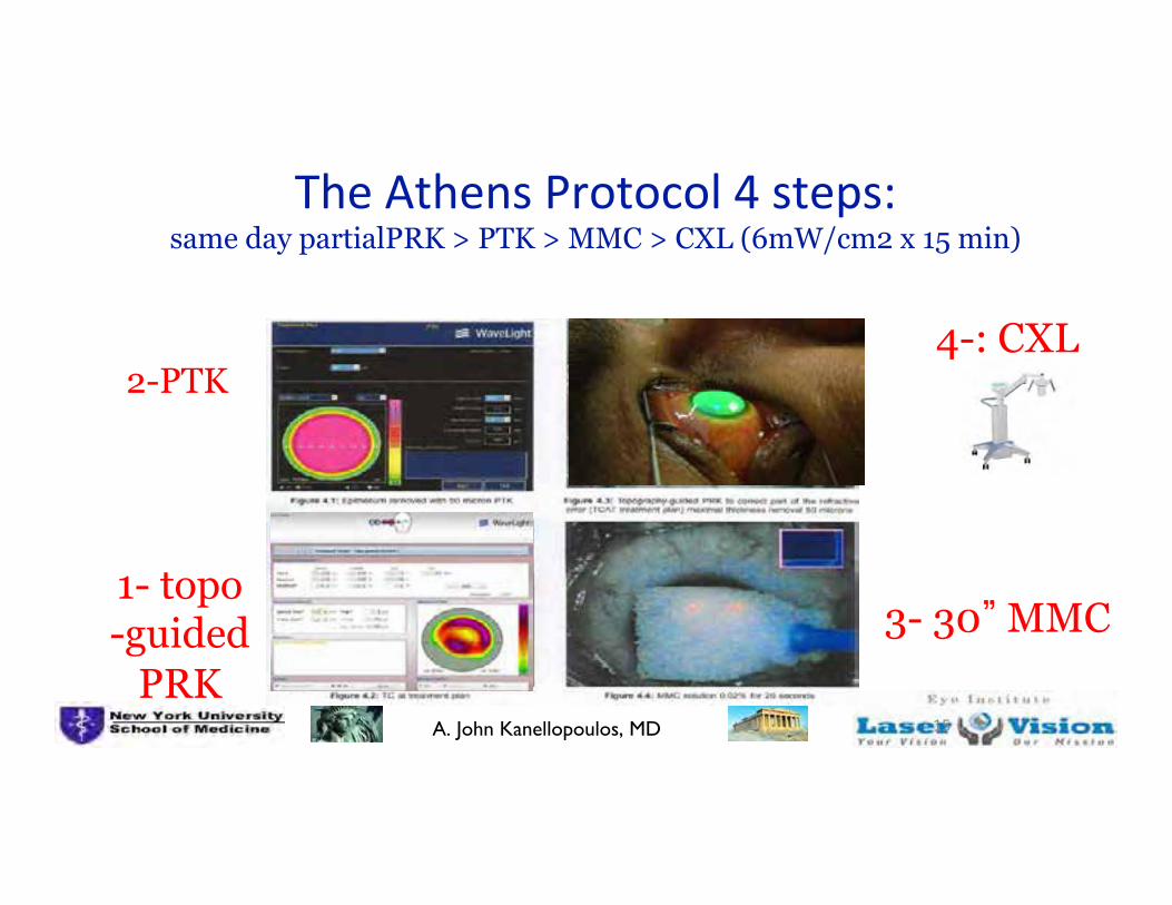
TMR: Topography-modified refraction Astigmatism adjustment
decreased cyl

A. John Kanellopoulos, MD
TMR: Topography-modified refraction Astigmatism adjustment: decreased cyl

A. John Kanellopoulos, MD
TMR : Topography-Modified refraction Astigmatism adjustment
increased cyl

A. John Kanellopoulos, MD
TMR : Topography-Modified refraction Astigmatism adjustment: increased cyl
A. John Kanellopoulos, MD
TMR : Topography-Modified refraction Astigmatism adjustment
AUTHOR
PROOF
COPY
Not for
publication
© 2016 Kanellopoulos. This work is published and licensed by Dove Medical Press Limited. The full terms of this license are available at https://www.dovepress.com/terms.php and incorporate the Creative Commons Attribution – Non Commercial (unported, v3.0) License (http://creativecommons.org/licenses/by-nc/3.0/). By accessing the work you
hereby accept the Terms. Non-commercial uses of the work are permitted without any further permission from Dove Medical Press Limited, provided the work is properly attributed. For permission for commercial use of this work, please see paragraphs 4.2 and 5 of our Terms (https://www.dovepress.com/terms.php).
Clinical Ophthalmology 2016:10 1–9
Clinical Ophthalmology Dovepress
submit your manuscript | www.dovepress.com
Dovepress 1
O R I G I N A L R E S E A R C H
open access to scientific and medical research
Open Access Full Text Article
122345
Topography-modified refraction (TMR): adjustment of treated cylinder amount and axis to the topography versus standard clinical refraction in myopic topography-guided LASIK
Anastasios John Kanellopoulos1,2
1LaserVision Clinical and Research Institute, Athens, Greece; 2Department of Ophthalmology, NYU Medical School, New York, NY, USA
Purpose: To evaluate the safety, efficacy, and contralateral eye comparison of topography-guided
myopic LASIK with two different refraction treatment strategies.
Setting: Private clinical ophthalmology practice.
Patients and methods: A total of 100 eyes (50 patients) in consecutive cases of myopic
topography-guided LASIK procedures with the same refractive platform (FS200 femtosecond
and EX500 excimer lasers) were randomized for treatment as follows: one eye with the standard
clinical refraction (group A) and the contralateral eye with the topographic astigmatic power
and axis (topography-modified treatment refraction; group B). All cases were evaluated pre- and
post-operatively for the following parameters: refractive error, best corrected distance visual
acuity (CDVA), uncorrected distance visual acuity (UDVA), topography (Placido-disk based)
and tomography (Scheimpflug-image based), wavefront analysis, pupillometry, and contrast
sensitivity. Follow-up visits were conducted for at least 12 months.
Results: Mean refractive error was 5.5 D of myopia and 1.75 D of astigmatism. In group A
versus group B, respectively, the average UDVA improved from 20/200 to 20/20 versus 20/16;
postoperative CDVA was 20/20 and 20/13.5; 1 line of vision gained was 27.8% and 55.6%; and
2 lines of vision gained was 5.6% and 11.1%. In group A, 27.8% of eyes had over 0.50 diopters
of residual refractive astigmatism, in comparison to 11.7% in group B (P 0.01). The residual per-
centages in both groups were measured with refractive astigmatism of more than –0.5 diopters.
Conclusion: Topography-modified refraction (TMR): topographic adjustment of the amount
and axis of astigmatism treated, when different from the clinical refraction, may offer superior
outcomes in topography-guided myopic LASIK. These findings may change the current clinical
paradigm of the optimal subjective refraction utilized in laser vision correction.
Keywords: TMR, topography-modified refraction, myopic LASIK, femtosecond laser,
FS200, EX500 excimer laser, long-term stability, regression, astigmatism correction, post-LASIK
refraction
IntroductionLaser vision correction has been established over the last 2 decades as a safe and
effective intervention, with Laser-assisted in situ keratomileusis (LASIK) being one
of the main techniques practiced globally.1,2
Femtosecond laser-assisted LASIK has become a popularized modification
over the last decade and over the standard LASIK technique utilizing mechanical
microkeratomes.3,4
Correspondence: Anastasios John KanellopoulosLaserVision Clinical and Research Institute, 17 Tsocha Street, Athens 11521, GreeceTel 30 210 747 2777Fax 30 210 747 2789Email [email protected]
Clinical Ophthalmology 2016:10 submit your manuscript | www.dovepress.com
Dovepress
Dovepress
3
Comparison of topography cylinder adjustment in LASIK vs standard refraction
Figure 1 Image A shows the topography-guided treatment when refractive error is adjusted by the user to zero sphere and zero cylinder. This image allows the user to visualize the corrections made by the software and added to the excimer correction in order to normalize the anterior cornea curvature to the cornea vertex. Image B illustrates the topography treatment pattern after the refraction has been adjusted by the user to the desired sphere and cylinder and includes the changes noted in image A.Notes: “Clinical” is the clinical refraction dialed by the user into the system; the “measured” refraction is the cylindrical amount and axis calculated by the topography software, to be corrected so the anterior cornea can me maximally normalized in regard to the cornea vertex; “modified” represents the surgeon/user adjusted data.Abbreviations: 3D, three-dimensional; max, maximum, cen, center; trans, transition zone; res, residual; VD, vertex distance.
the right of the top image in Figure 1. The trefoil-like image
is also presented here in correlation with the pupillary aper-
ture, also captured by the Vario topographer, and in this
top window it shows clearly decentration to the trefoil-like
normalization ablation, revealing angle-kappa compensation
by the topography software. This ablation pattern will induce
some myopia and it was calculated that in order to keep it
neutral there is a need to add 0.25 D of myopia to the clinical
Clinical Ophthalmology 2016:10submit your manuscript | www.dovepress.com
Dovepress
Dovepress
6
Kanellopoulos
Figure 4 Safety distance visual acuity graph (percentage of eyes with gain/loss in Snellen lines), at 3-month visit: (A) group A and (B) group B.Notes: Group A: one eye with the standard clinical refraction; Group B: the contralateral eye with the topographic astigmatic power and axis (topography-modified treatment refraction).
Figure 5 Defocus equivalent results at 3-month visit: (A) group A and (B) group B.Notes: Group A: one eye with the standard clinical refraction; Group B: the contralateral eye with the topographic astigmatic power and axis (topography-modified treatment refraction).
Figure 6 Predictability of spherical equivalent (SE) correction, showing achieved SE versus attempted SE: (A) group A and (B) group B.Notes: Group A: one eye with the standard clinical refraction; Group B: the contralateral eye with the topographic astigmatic power and axis (topography-modified treatment refraction). Green marks show outcomes within 0.25 diopters of attempted vs achieved; blue marks show attempted vs achieved of under 0.25 diopters (under-corrections); red marks show attempted vs achieved of over 0.25 diopters (over-corrections).

A. John Kanellopoulos, MD
3
Topography - Guided University
Courses 2016:
Become pro�icient interpreting in cornea diagnostics and designing expert
topography guided laser treatments!
Didactic course and hands-on treatment designing ofmultiple case scenarios (virgin eyes, complicated ther-apeutic treatments: older decentered or irregular abla-tions, cornea scars, ectasia and keratoconus).How to adjust optical zone, transition zone, and accountfor topography spherical neutralization.Each participant will bring his or her clinical cases anddesign a treatment, and/or will be given all of the casescenarios noted above to design a treatment.Anticipate spherical change surprisesModulation of biomechanics with various CXL protocols.
• Outline and 2016 locations • Pre-ESCRS • Pre-AAOCourse Director: A. John Kanellopoulos, MD
www.topo-guided.com
Copenhagen‘16 course logistics
Friday September 9th, 2016 (pre-ESCRS)
8:00 AM to 15:30 PM
At the Crowne Plaza Copenhagen Towers
Ørestads Boulevard 114 - 118 2300 København S, Denmark
[email protected] Tel: +45 8877 6655Fax: +45 8877 6611
Chicago ‘16 course logistics
Thursday October 13th, 2016 (pre-AAO)
8:00 AM to 15:30 PM
At the Hyatt Regency McCormick Place
2233 S Martin Luther King Dr,
Chicago, 60616, IL, USA
Tel: +1 312 567 1234
8:00Breakfast - Registration
8:30-11:30 • Introduction to current cornea diagnostics and their relative differences: Placido Topogra-
phy, Scheimpflug tomography, Anterior segment OCT, LED color reflection topography. • Corneal epithelial mapping and its clinical relevance in diagnosis and treatment • Basic principles in employing topography data (Scheimpflug based and/or Placido-based)
in the customization of an excimer corneal ablation. Technology overview and case presentations, with the Wavelight, Schwind and Ivis platforms.
• Topography astigmatism, centroid and angle kappa considerations for possible revision of the clinical refraction used in each ablation
11:30-12:30Discussion lunch
12:30-15:30 • Topography customized methodology for virgin myopic and hyperopic eyes • Topography customized methodology for irregular corneas (previously treated: RK, decen-
tered and/or irregular ablations, as well as irregular and ectasia cases) • Anticipating asphericity and sphere compensatory nomograms for better spherical correc-
tion and emmetropia. • Participants will gain access to an online database with over 100 cases examples (pre-op
data, treatment design, treatment video, postop data and overview of what went well andwhat potentially went off-target)
• Complications assessment and management.
•Each participant will have the chance to design several treatments on site!
The course will be limited to 30 participants. Advanced registration and information:
http://www.topo-guided.com/
The course will be limited to 30 participants.
http://www.topo-guided.com/
2016 Course’s Mutual Outline (Copenhagen and Chicago)
2016 Course’s Mutual Outline (Copenhagen and Chicago)
Topography - Guided University
Courses 2016:• Correlation of multiple corneal imaging devices may
enhance accuracy of assessment by including pos-sible epithelial remodeling data, and limiting specificlimitations of Placido-based, Scheimflug-based andcolor LED refraction topography.
• When using topography maps in laser corneal abla-tion, all these parameters are considered under amuch more meticulous and critical perspective. Al-though originally designed to treat irregular eyes, ithas recently become apparent, hat topo-guidedtreatments may be superior for routine myopicand/or hyperopic laser vision correction.
• This vigorous didactic course and wet lab on topog-raphy-customized corneal ablations will focus on fa-miliarizing the small number of participants on multipleimaging assessment and interpretation, data acqui-sition and treatment modifications with hands-on thedesign platforms and data present on-site.
• Additionally, the participants will be offered accessto a very large databank with most topo-guidedscenario treatments and outcomes.
Disclaimer:Please be advised that if you are traveling from outside the US for aUS course or within the EU for a European course, you would be responsible for obtaining the appropriate visas on your own.The AJKMD course services will provide a letter of invitation uponcon�irmation of your registration.2Our courses do not provide CME credits.3Our courses are purely instructional. Any medical procedure liability lays with the operating surgeon and the bylaws observed inthe country and state of his/ her practice.
Topography - Guided University
Courses 2016:
Course Director: A. John Kanellopoulos, MD
Topography - Guided University
Courses 2016:
Become pro�icient interpreting in cornea diagnostics and designing expert
topography guided laser treatments!
www.topo-guided.com

A. John Kanellopoulos, MD
Customized CXL for KCN!

A. John Kanellopoulos, MD
Epithelial remodeling after partialtopography-guided normalization
and high-fluence short-duration crosslinking(Athens protocol): Results up to 1 year
Anastasios John Kanellopoulos, MD, George Asimellis, PhD
ARTICLE
The findings in the current study agree with those inour previous study1; that is, although an overall thickerepithelium with large variations can be observed clini-cally and topographically in eyes with keratoconus, ineyes treated with CXL the variability in epitheliumthickness and topographic thickness decreased by astatistically significant margin and was more uniform.We have theorized that epithelial hyperplasia in biome-chanically unstable corneas (ie, increased epithelialregrowth activity) might be associated with a moreelastic cornea.1 The laboratory and clinical findings ofincreased corneal rigidity after CXL are widelyaccepted,23–25 including in studies of accelerated high-fluence CXL.26
In conclusion, we present the results in a compre-hensive study of the postoperative development ofcorneal epithelial thickness distribution after keratoco-nus management using combined anterior cornealnormalization by topography-guided excimer abla-tion and accelerated CXL. The epithelial healing pro-cesses can be monitored by AS-OCT with ease in aclinical setting, expanding the clinical application ofthis technology. Our findings suggest less topographicvariability and overall reduced epithelial thicknessdistribution in keratoconus eyes treated with CXL us-ing the Athens protocol.
WHAT WAS KNOWN
� Postoperative epithelial remodeling after partial anteriorsurface normalization with an excimer laser and high-fluence CXL, assessed with high-frequency scanningUBM, results in reduced overall epithelial thickness andtopographic variability.
WHAT THIS PAPER ADDS
� Detailed follow-up of Athens protocol–treated eyes up to 1year confirmed previous ultrasound findings of the overallthinner and smoother epithelial thickness profilescompared with the profiles of untreated keratoconic eyes.
REFERENCES1. Kanellopoulos AJ, Aslanides IM, Asimellis G. Correlation be-
tween epithelial thickness in normal corneas, untreated ectatic
corneas, and ectatic corneas previously treated with CXL; is
overall epithelial thickness a very early ectasia prognostic fac-
tor? Clin Ophthalmol 2012; 6:789–800. Available at: http://
www.ncbi.nlm.nih.gov/pmc/articles/PMC3373227/pdf/opth-6-789.
pdf. Accessed June 11, 2014
2. Kanellopoulos AJ. Long term results of a prospective random-
ized bilateral eye comparison trial of higher fluence, shorter
duration ultraviolet A radiation, and riboflavin collagen cross
Figure 3. Mean and center epithelial thicknesses in the 3 groups.Error bars correspond to the SD (KCNZ keratoconus, no treatment).
Figure 4. Epithelial thickness variability and range in the 3 groups.Error bars correspond to the SD (KCNZ keratoconus, no treatment).
5EPITHELIAL REMODELING IN KERATOCONIC EYES
J CATARACT REFRACT SURG - VOL -, - 2014
Figure 2. Comparative AS-OCT epithelial thickness (mm) 3-D mapsshows an image from Group A taken 1 year postoperatively andan image fromGroup B (IZ inferior; INZ inferior–nasal; ITZ infe-rior–temporal; NZ nasal; SZ superior; SNZ superior–nasal; STZsuperior–temporal; T Z temporal).
EPITHELIAL REMODELI

A. John Kanellopoulos, MD 3Kanellopoulos,MD
Severe scar of the cornea BCVA 20/200,
Lamellar or PK?
91

A. John Kanellopoulos, MDKanellopoulos,MD 40

A. John Kanellopoulos, MDKanellopoulos,MD 41
From 20/200 to 20/40!

A. John Kanellopoulos, MD
Hyperopic LASIK a drop of 0.1% riboflavin sodium phosphate solution, spread over the exposed stromal bed for 60”

A. John Kanellopoulos, MD
Compelling stability evidence in the contralateral eye hyperopic LASIK + CXL group

A. John Kanellopoulos, MD 44
COMPARISON OF REFRACTIVE AND KERATOMETRIC STABILITY MYOPIC LASIK VS LASIK+ CXL
Kanellopoulos AJ, Asimellis G: Clin. Ophthalmol 2014

A. John Kanellopoulos, MD
Comparison of keratometric stability compelling clinical evidence that LASIK+CXL works!
42
43
44
45
46
47
Pre op 1m 3m 6m 12m 24m Mea
n K
erat
omet
ry (D
)
Lasik Xtra Std Lasik
Kanellopoulos AJ, Kahn J: JRS November 2012

A. John Kanellopoulos, MD 1
Does in situ CXL work? Ex-vivo evidence Two-surface intra-lamellar bed corneal dissections were performed within a 5.5 mm optical zone. The lenticule was extracted through a 3.5 mm wide superior canal. High-fluence CXL was conducted in the pocket created.

A. John Kanellopoulos, MD 1
2-dimentional biomechanical testing

A. John Kanellopoulos, MDKanellopoulosMD 1
Results Substantial (up to +100%) increase in biomechanical strength has been noted when using biaxial stress measurements.
High-irradiance CXL combined with myopic LASIK:flap and residual stroma biomechanical propertiesstudied ex-vivoAnastasios JohnKanellopoulos,1,2 GeorgeAsimellis,1 Joseph BCiolino,3
BorjaSalvador-Culla,3 James Chodosh3
1Laservision.gr Eye Institute,Athens, Greece2Department ofOphthalmology, New YorkUniversity Medical School,New York, New York, USA3Department of Cornea &Refractive Surgery, HarvardMedical School, MassachusettsEye & Ear Infirmary, Boston,Massachusetts, USA
Correspondence to
ABSTRACTBackground/aims To evaluate ex vivo biomechanicaland enzymatic digestion resistance differences betweenstandard myopic laser in-situ keratomileusis (LASIK)compared with LASIK+CXL, in which high-irradiancecross-linking (CXL) is added.Methods Eight human donor corneas were subjectedto femtosecond-assisted myopic LASIK. Group A (n=4)served as a control group (no CXL). The corneas inLASIK+CXL group B were subjected to concurrent
h l i hi h i di CXL ( 4) S li dil d
collagen resistance against enzymatic degradationhas been associated with CXL.6–8
We have introduced an alternative CXL applica-tion, adjuvant to myopic laser in-situ keratomileusis(LASIK+CXL). The application aims to improvelong-term keratometric stability9 and to reduceregression likelihood following moderate and highmyopic LASIK10 by proactively restoring corneal bio-mechanical strength.11 Riboflavin solution is brieflyapplied on the exposed stromal bed at the comple-i f h i bl i h fl i h i
Laboratory science

A. John Kanellopoulos, MD
O R I G I N A L A R T I C L E
Longitudinal Postoperative LASIK Epithelial Thickness Profile Changes in Correlation With Degree of Myopia CorrectionAnastasios John Kanellopoulos, MD; George Asimellis, PhD
Figure 1. Detail from the analysis and report software main report, showing corneal and epithelial three-dimensional pachymetry maps over the 6-mm corneal diameter in a postoperative LASIK examination. The patient (left eye) received treatment for -4.75 diopters of sphere and -0.75 diopters of astigmatism, and was imaged 1 month postoperatively. * = thickness minimum (both corneal and epithelial maps); + = thickness maximum (epithelial map only).
Figure 2. The correlation of increase in epithelial thickness at the center (green dots), on the mean over the 6-mm diameter (blue), and on the 5-mm mid-peripheral zone (yellow) 1 month following myopic LASIK cor-rection. There were 4 cases between -8 and -9 diopters (D), 7 cases between -7 and -8 D, 10 cases between -6 and -7 D, 8 cases between -5 and -6 D, 15 cases between -4 and -5 D, 13 cases between -3 and -4 D, and 6 cases between -2 and -3 D. Error bars indicate standard deviation.

A. John Kanellopoulos, MD
CLINICAL SCIENCE
Epithelial Remodeling After Femtosecond Laser–AssistedHighMyopic LASIK: Comparison of Stand-aloneWith LASIKCombined WithAU2 Prophylactic High-fluence Cross-linking
Anastasios J. Kanellopoulos, MD*† and George Asimellis, PhD*
MS NO: CORNEA-D-13-00497
nth 2014 Topographic Epithelial Profile Thickness Changes
D i i i i li i l i k 24 2 6 5 8 ( 18 42) Th i
FIGURE 1. A, AS-OCT high-resolutioncross-sectional meridional image ofa right eye treated with LASIK-Xtra for28.00 D of sphere and 20.25 D ofastigmatism, and was it imaged6 months postoperatively. There isa clear depiction of the central cornealepithelial layer, Bowman membrane,anterior stroma, Descemet membrane,and anterior chamber. Deep stromalhyperreflective lines may correlate withthe depth of the CXL-effect achievedwith the LASIK-Xtra procedure ac-cording to our previous reported find-ings. B, Detail from of the analysis andreport software main report, showingcorneal and epithelial 3-dimensionalpachymetry maps over the 6-mmcorneal diameter. The symbol * in-dicates thickness minimum (both cor-neal and epithelial maps), andAU14 thesymbol + thickness maximum (epi-thelial map only).
Cornea � Volume 0, Number 0, Month 2014 Topographic Epithelial Profile Thickness Changes
A
Combined laser in situ keratomileusisand prophylactic high-fluence corneal
collagen crosslinking for high myopia: Two-yearsafety and efficacy
Anastasios John Kanellopoulos, MD, George Asimellis, PhD
PURPOSE: To evaluate the safety, efficacy, and refractive and keratometric stability of myopicfemtosecond laser in situ keratomileusis (LASIK) with concurrent prophylactic high-fluencecorneal collagen crosslinking (CXL) compared with the outcomes of standard femtosecond LASIK.
SETTING: Private clinical practice Athens Greece
ARTICLE

A. John Kanellopoulos, MD
CXL of “vehicle” cornea in Boston Keratoprosthesis type I: J. Cornea 2014

A. John Kanellopoulos, MD
Is CXL a refractive procedure? Most investigators speak of “disease reversal” when flattening occurs after CXL in ectasia
This is a simple 3mW CXL-alone case from 2005
No scar developed, Now 2013 has Flattened 12D!!!
52

A. John Kanellopoulos, MD
© 2014 Kanellopoulos. This work is published by Dove Medical Press Limited, and licensed under Creative Commons Attribution – Non Commercial (unported, v3.0) License. The full terms of the License are available at http://creativecommons.org/licenses/by-nc/3.0/. Non-commercial uses of the work are permitted without any further
permission from Dove Medical Press Limited, provided the work is properly attributed. Permissions beyond the scope of the License are administered by Dove Medical Press Limited. Information on how to request permission may be found at: http://www.dovepress.com/permissions.php
Clinical Ophthalmology 2014:8 697–702
Clinical Ophthalmology
Video abstract
Point your SmartPhone at the code above. If you have a QR code reader the video abstract will appear. Or use:
http://dvpr.es/1eiR39A
Dovepress
submit your manuscript | www.dovepress.com
Dovepress 697
O R I G I N A L R E S E A R C H
open access to scientific and medical research
Open Access Full Text Article
http://dx.doi.org/10.2147/OPTH.S59934
Novel myopic refractive correction with transepithelial very high-fluence collagen cross-linking applied in a customized pattern: early clinical results of a feasibility study
Anastasios John KanellopoulosLaserVision.gr Institute, Athens, Greece, and New York Medical School, New York, NY, USA
Correspondence: A John Kanellopoulos LaserVision.gr Institute, 17 Tsocha Street, 115 21 Athens, Greece Tel 30 21 0747 2777 Fax 30 21 0747 2789 Email [email protected]
Background: The purpose of this study is to report the safety and efficacy of a new application
of collagen cross-linking using a novel device to achieve predictable refractive myopic changes
in virgin corneas.
Methods: Four cases were treated with a novel device employing very high-fluence collagen
cross-linking applied in a myopic pattern. Prior to treatment, riboflavin solution was applied to
the intact epithelium. The collagen cross-linking device was then engaged for a total of 12 J/cm2,
to be applied transepithelially in a predetermined pattern. Cornea clarity, corneal keratometry,
and corneal topography were evaluated by both Placido disc and Scheimpflug imaging, along
with cornea anterior segment optical coherence tomography and endothelial cell counts.
Results: An average of 2.3 diopters was achieved in the first week in all four cases treated
with the very high-fluence myopic collagen cross-linking intervention. There was a slight
regression to 1.44 diopters at 1 month, which remained stable at 6-month follow-up. The mean
keratometry change was from 44.90 diopters to 43.46 diopters. There was no significant change
in endothelial cell counts or corneal clarity. There was some mild change in epithelial thickness
distribution, with the treated area showing a slight but homogeneous reduction in mean thick-
ness from 52 m to 44 m.
Conclusion: This report describes the novel application of very high-fluence collagen cross-
linking with a predictable well defined myopic refractive (flattening) corneal effect. This
technique has the advantages of essentially no postoperative morbidity, immediate visual
rehabilitation, and the potential for tapering until the desired result is achieved.
Keywords: myopia, refractive correction, high-fluence collagen cross-linking, clinical results
IntroductionCollagen cross-linking has been used for many years as a means of stabilizing cornea
ectasia.1–5 Although a multitude of treatments and techniques are available, it has been
well documented that the procedure almost invariably results in some central anterior
corneal flattening,1–5 which has often been interpreted as “disease regression.” As our
understanding and the technology available for collagen cross-linking has progressed,
it has been theorized that differential application of collagen cross-linking in specific
areas of the cornea may produce predictable refractive changes. Several aspects of
this theory need further investigation. Is it possible to achieve predictable refractive
changes? Can this be achieved through an intact epithelium? Can the human cornea
tolerate higher fluence of ultraviolet light? This paper describes the use of a novel
Clinical Ophthalmology 2014:8 submit your manuscript | www.dovepress.com
Dovepress
Dovepress
699
High-fluence collagen cross-linking for myopic refractive correction
epithelial thickness over the treated area (Figure 3). The
average epithelial thickness was 49 m preoperatively, which
decreased to 44 m at 1 month postoperatively, and then
increased to 48 m at 5 months postoperatively.
DiscussionA multitude of reports have established the significant
refractive changes that accompany classic collagen
cross-linking1–8 utilizing the classic Dresden protocol
(3 mW/cm2 for 30 minutes), as well as collagen cross-
linking utilizing higher fluence,9 and even cross-linking
delivered in eyes that have had riboflavin placed within a
femtosecond laser-created pocket or intrastromal corneal
ring segment channels.10,11 Over the years, most clinicians
have referred to this process as “flattening,” which has often
been interpreted as “disease regression.” We have long
Figure 1 Placido disc topography for patient 3 preoperatively (left) and 6 months postoperatively (right) depicting the significant and regular central corneal flattening effect.
Figure 2 Scheimpflug imaging data for patient 3 preoperatively (left) and 6 months postoperatively (right) depicting the significant and regular central corneal flattening effect.Abbreviations: D, diopters; OD, right eye.

A. John Kanellopoulos, MD
Case Rep Ophthalmol 2014;5:172–180
DOI: 10.1159/000363371 Published online: June 18, 2014
© 2014 S. Karger AG, Basel 1663‒2699/14/0052‒0172$39.50/0 www.karger.com/cop
This is an Open Access article licensed under the terms of the Creative Commons Attribution-NonCommercial 3.0 Unported license (CC BY-NC) (www.karger.com/OA-license), applicable to the online version of the article only. Distribution permitted for non-commercial purposes only.
A. John Kanellopoulos, MD
Laservision.gr Eye Institute 17 Tsocha Street, GR–115 21 Athens (Greece) E-Mail [email protected]
Toric Topographically Customized Transepithelial, Pulsed, Very High-Fluence, Higher Energy and Higher Riboflavin Concentration Collagen Cross-Linking in Keratoconus Anastasios John Kanellopoulosa, b William J. Duppsc, d Ibrahim Sevene, f George Asimellisa aLaservision.gr Eye Institute, Athens, Greece; bDepartment of Ophthalmology, NYU Medical School, New York, N.Y., Departments of cOphthalmology and dBiomedical Engineering, Cleveland Clinic, eCole Eye Institute, Cleveland Clinic, and fDepartment of Chemical and Biomedical Engineering, Cleveland State University, Cleveland, Ohio, USA
Key Words Topography customizable cross-linking · High-fluence cross-linking · Transepithelial cross-linking · Toric cross-linking · Keratoconus · Photorefractive intrastromal cross-linking · KXL II
Abstract Purpose: To report a novel application of toric topographically customized transepithelial collagen cross-linking (CXL) aiming to achieve refractive astigmatic changes in a keratoconic cornea. Methods: Specially formulated riboflavin transepithelial administration and delivery of high-fluence UVA in a topographically customized pattern was applied in an eye with progressive keratoconus. Visual acuity, cornea clarity, keratometry, topography, and pachymetry with a multitude of modalities, as well as endothelial cell counts were evaluated for >6 months. Results: Uncorrected distance visual acuity changed from preoperative 20/40 to 20/25 at 6 months. A mean astigmatic reduction of 0.8 D, and significant cornea surface normalization was achieved 6 months postoperatively. There was some mild change in the epithelial distribution, with the treated area having a slight normalization in the average epithelial thickness. Conclusions: We introduce herein the novel application of a topograph-
Down
loade
d by:
87.20
3.71.0
- 6/20
/2014
11:17
:32 A
M
Case Rep Ophthalmol 2014;5:172–180
DOI: 10.1159/000363371
© 2014 S. Karger AG, Basel www.karger.com/cop
Kanellopoulos et al.: Topographically Customizable Toric Transepithelial CXL
178
Fig. 1. Customized treatment profile employed in the treatment. Left panel: details of the applied customizable pattern and parameters for UVA exposure; right panel: overlay of the pattern on the sagittal curvature map.
Fig. 2. Placido disk topography data showing sagittal curvature maps depicting significant refractive changes along the axis of the customized cross-linking pattern. Panel 1: 1 day preoperatively. Panel 2: 6 months postoperatively. Panel to the right: difference 2–1.
Down
loade
d by:
87.20
3.71.0
- 6/2
0/201
4 11:1
7:32 A
M
A. John Kanellopoulos, MD
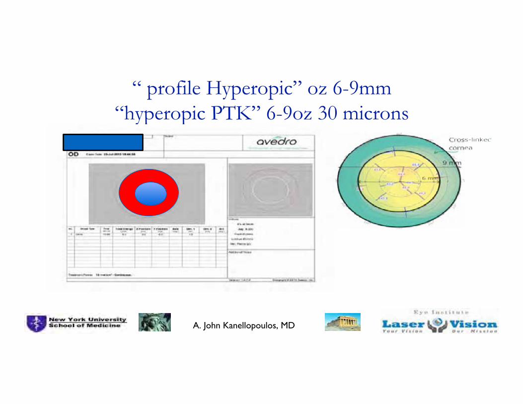
“ profile Hyperopic” oz 6-9mm “hyperopic PTK” 6-9oz 30 microns

A. John Kanellopoulos, MD
Placido topo data

A. John Kanellopoulos, MD
Infectious Keratitis in a 38y/o F MD

A. John Kanellopoulos, MD
Customized infectious keratitis treatment
20mW/cm2, 20 Joules continuous for 10’

A. John Kanellopoulos, MD

A. John Kanellopoulos, MD
CXL of “vehicle” cornea in Boston Keratoprosthesis type I: J. Cornea 2014

A. John Kanellopoulos, MDKanellopoulos MD
6
Conclusions / Our current CXL protocols
1-Athens Protocol: topo partial PRK +15’x 6mw/cm2
or combined with PiXL (4-20 Joules)
2-LASIK Xtra: 1’ soaking and 60” 30mW/cm2 (1.8 Joules) for all hyperopes, 80” for myopes (2.4 Joules)
2-PRK Xtra: 1’ soaking 80” X 30mW/cm2 (2.4 Joules)
5-Transepi CXL: 0.25% ribo + 30mW X 3’
6-Infection: 0.25% riboflavin + 20mW/cm2 /20 Joules
7-PiXL 0.25% ribo + 30mW/cm2 7.2-20 Joules
Prospective randomized trials have yet to establish the comparative efficacy of the multitude of CXL technique available today

A. John Kanellopoulos, MD
6
Topography - Guided University
Courses 2016:
Become pro�icient interpreting in cornea diagnostics and designing expert
topography guided laser treatments!
Didactic course and hands-on treatment designing ofmultiple case scenarios (virgin eyes, complicated ther-apeutic treatments: older decentered or irregular abla-tions, cornea scars, ectasia and keratoconus).How to adjust optical zone, transition zone, and accountfor topography spherical neutralization.Each participant will bring his or her clinical cases anddesign a treatment, and/or will be given all of the casescenarios noted above to design a treatment.Anticipate spherical change surprisesModulation of biomechanics with various CXL protocols.
• Outline and 2016 locations • Pre-ESCRS • Pre-AAOCourse Director: A. John Kanellopoulos, MD
www.topo-guided.com
Copenhagen‘16 course logistics
Friday September 9th, 2016 (pre-ESCRS)
8:00 AM to 15:30 PM
At the Crowne Plaza Copenhagen Towers
Ørestads Boulevard 114 - 118 2300 København S, Denmark
[email protected] Tel: +45 8877 6655Fax: +45 8877 6611
Chicago ‘16 course logistics
Thursday October 13th, 2016 (pre-AAO)
8:00 AM to 15:30 PM
At the Hyatt Regency McCormick Place
2233 S Martin Luther King Dr,
Chicago, 60616, IL, USA
Tel: +1 312 567 1234
8:00Breakfast - Registration
8:30-11:30 • Introduction to current cornea diagnostics and their relative differences: Placido Topogra-
phy, Scheimpflug tomography, Anterior segment OCT, LED color reflection topography. • Corneal epithelial mapping and its clinical relevance in diagnosis and treatment • Basic principles in employing topography data (Scheimpflug based and/or Placido-based)
in the customization of an excimer corneal ablation. Technology overview and case presentations, with the Wavelight, Schwind and Ivis platforms.
• Topography astigmatism, centroid and angle kappa considerations for possible revision of the clinical refraction used in each ablation
11:30-12:30Discussion lunch
12:30-15:30 • Topography customized methodology for virgin myopic and hyperopic eyes • Topography customized methodology for irregular corneas (previously treated: RK, decen-
tered and/or irregular ablations, as well as irregular and ectasia cases) • Anticipating asphericity and sphere compensatory nomograms for better spherical correc-
tion and emmetropia. • Participants will gain access to an online database with over 100 cases examples (pre-op
data, treatment design, treatment video, postop data and overview of what went well andwhat potentially went off-target)
• Complications assessment and management.
•Each participant will have the chance to design several treatments on site!
The course will be limited to 30 participants. Advanced registration and information:
http://www.topo-guided.com/
The course will be limited to 30 participants.
http://www.topo-guided.com/
2016 Course’s Mutual Outline (Copenhagen and Chicago)
2016 Course’s Mutual Outline (Copenhagen and Chicago)
Topography - Guided University
Courses 2016:• Correlation of multiple corneal imaging devices may
enhance accuracy of assessment by including pos-sible epithelial remodeling data, and limiting specificlimitations of Placido-based, Scheimflug-based andcolor LED refraction topography.
• When using topography maps in laser corneal abla-tion, all these parameters are considered under amuch more meticulous and critical perspective. Al-though originally designed to treat irregular eyes, ithas recently become apparent, hat topo-guidedtreatments may be superior for routine myopicand/or hyperopic laser vision correction.
• This vigorous didactic course and wet lab on topog-raphy-customized corneal ablations will focus on fa-miliarizing the small number of participants on multipleimaging assessment and interpretation, data acqui-sition and treatment modifications with hands-on thedesign platforms and data present on-site.
• Additionally, the participants will be offered accessto a very large databank with most topo-guidedscenario treatments and outcomes.
Disclaimer:Please be advised that if you are traveling from outside the US for aUS course or within the EU for a European course, you would be responsible for obtaining the appropriate visas on your own.The AJKMD course services will provide a letter of invitation uponcon�irmation of your registration.2Our courses do not provide CME credits.3Our courses are purely instructional. Any medical procedure liability lays with the operating surgeon and the bylaws observed inthe country and state of his/ her practice.
Topography - Guided University
Courses 2016:
Course Director: A. John Kanellopoulos, MD
Topography - Guided University
Courses 2016:
Become pro�icient interpreting in cornea diagnostics and designing expert
topography guided laser treatments!
www.topo-guided.com

A. John Kanellopoulos, MD
Thank You!