LESIONS OF RHEUMATOID ARTHRITIS - ard.bmj.com · calcaneus andthe calcaneal spur, andthe other the...
10
HEEL LESIONS OF RHEUMATOID ARTHRITIS BY E. G. L. BYWATERS From the Special Ulnit for Juvenile Rheumatism, Canadian Red Cross Memorial Hospital, Taplow, Bucks, and the Department of Medicine, Postgraduate Medical School of London (RECEIVED FOR PUBLICATION DECEMBER 16, 1953) Two lesions affecting the heel in rheumatoid arthritis have received little or no attention of recent years: one involves the plantar surface of the calcaneus and the calcaneal spur, and the other the synovial bursa between the insertion of the Achilles tendon into the calcaneus and the calcaneus itself. This note is designed primarily to draw attention to the sub-Achilles lesion, since it may be the presenting sign of rheumatoid arthritis and is often a major complaint: secondarily, to point out that the plantar surface of the calcaneus is often actively involved in rheumatoid arthritis and to indicate the nature of the involvement. Normal Anatomy.-Normally (Fig. la, opposite), the Achilles tendon is inserted about 2cm. below the upper surface of the calcaneus; the latter is covered with- fibro-cartilage opposite the free tendon (Fig. lb), and the bursal space is lined over the tendon by a thin synovium, and at its margins by synovium-covered fatty pads (Fig. lc); there is a very small amount of viscous fluid. With advancing age, some cartilage degeneration occurs (Fig. 2); this is well described by Rossler (1896) on the basis of 225 Fig. 2.-Bone, showing cartilage degeneration in later life. bursae examined from about 140 cadavers of various ages. The bone, however, remains intact throughout life, and x rays show a smooth strong cortical layer. The plantar surface of the os calcaneus (Fig. 3) is Fig. 3.-Plantar surface of calcaneus (x 7) in normal female aged 36, with x ray inset. Note smooth cortical bone. 42 on 10 June 2019 by guest. Protected by copyright. http://ard.bmj.com/ Ann Rheum Dis: first published as 10.1136/ard.13.1.42 on 1 March 1954. Downloaded from
-
Upload
trinhtuong -
Category
Documents
-
view
215 -
download
0
Transcript of LESIONS OF RHEUMATOID ARTHRITIS - ard.bmj.com · calcaneus andthe calcaneal spur, andthe other the...

HEEL LESIONS OF RHEUMATOID ARTHRITISBY
E. G. L. BYWATERSFrom the Special Ulnit for Juvenile Rheumatism, Canadian Red Cross Memorial Hospital, Taplow, Bucks,
and the Department of Medicine, Postgraduate Medical School ofLondon(RECEIVED FOR PUBLICATION DECEMBER 16, 1953)
Two lesions affecting the heel in rheumatoidarthritis have received little or no attention of recentyears: one involves the plantar surface of thecalcaneus and the calcaneal spur, and the other thesynovial bursa between the insertion of the Achillestendon into the calcaneus and the calcaneus itself.This note is designed primarily to draw attention tothe sub-Achilles lesion, since it may be the presentingsign of rheumatoid arthritis and is often a majorcomplaint: secondarily, to point out that the plantarsurface of the calcaneus is often actively involved inrheumatoid arthritis and to indicate the nature ofthe involvement.
Normal Anatomy.-Normally (Fig. la, opposite),the Achilles tendon is inserted about 2cm. below theupper surface of the calcaneus; the latter is coveredwith- fibro-cartilage opposite the free tendon(Fig. lb), and the bursal space is lined over thetendon by a thin synovium, and at its margins bysynovium-covered fatty pads (Fig. lc); there is a verysmall amount of viscous fluid. With advancing age,some cartilage degeneration occurs (Fig. 2); this iswell described by Rossler (1896) on the basis of 225
Fig. 2.-Bone, showing cartilage degeneration in later life.
bursae examined from about 140 cadavers ofvarious ages. The bone, however, remains intactthroughout life, and x rays show a smooth strongcortical layer.The plantar surface of the os calcaneus (Fig. 3) is
Fig. 3.-Plantar surface of calcaneus (x 7) in normal female aged 36, with x ray inset. Note smooth cortical bone.
42
on 10 June 2019 by guest. Protected by copyright.
http://ard.bmj.com
/A
nn Rheum
Dis: first published as 10.1136/ard.13.1.42 on 1 M
arch 1954. Dow
nloaded from

(a)
i'-~
43HEEL LESIONS OF RHEUMATOID ARTHRITIS%*X NN. X X;~~~~~'A ~~~2Q
7
4d
-*-*~~~~~~~~~~~*w4__Ho~~~~~~~~~~~~~~~~~~~~~~~~~~~~~~~~~~~~~.
9L- -
Fig. 1.-Achilles tendon insertion in female aged 36 (with carcinoma of the cervix).(a) Microphotograph of sagittal section ( x 7) with x rays inset.Note T-shaped bursal cavity.(b) High-power view of marked area showing intra-bursal fat pad and synovial cells covering it (x 130).(c) High-power view of marked area of fibro-cartilage covering bone (x 130).
IN
II -1 i
I-,...- %
- 46.,.. .
lll.. .N, I "
on 10 June 2019 by guest. Protected by copyright.
http://ard.bmj.com
/A
nn Rheum
Dis: first published as 10.1136/ard.13.1.42 on 1 M
arch 1954. Dow
nloaded from

ANNALS OF THE RHEUMATIC DISEASES
covered by dense periosteum into which are insertedposteriorly the Achilles tendon and anteriorly theplantar aponeurosis. In between, the heel restson a cushioned pad of fat interlaced with strongfascial sheets. Plantar spurs are seen in manypeople, the incidence increasing with age, withoutany complaint relating thereto; they consist of anextension of bone into the periosteal insertion of theplantar aponeurosis.
Incidence.-In the past 7 years 22 patients havecomplained of pain or swelling, or both, in the heel;in all except one radiological changes were visible.All, except one with synovial chondromatosis of thesub-Achilles bursa, were suffering from rheumatoidarthritis, as judged either by the actual presence atthat time of other clinical and radiological criteriaor by their development in the next few years. Theincidence is probably of the order of 2-3 per cent.,since, in an unselected series of 250 patients withrheumatoid arthritis, seen between 1939 and 1948and followed to the present time (Dresner andBywaters, 1953), six complained of painful heels andshowed these lesions radiologically.* These six areincluded in the 21 mentioned above. Two others ofthe 21 in the series showed plantar-spur formationand ossification of the Achilles tendon only, withouterosions, and these were not considered further. TheTable (opposite) gives details of the group of nineteencases with rheumatoid arthritis, radiological erosionsbeing seen in eighteen of them. It will be noted thatnine are female and ten male, a reversal of the usualsex-ratio. The pgtients' ages at the time of the heellesion ranged between 21 and 65 years (usually inthe 30s and 40s). The erythrocyte sedimentation ratewas raised at the time in eleven of the nineteen andnormal in eight.
Clinical Features
Sometimes the complaint of heel tenderness camewithin a few months of the onset of rheumatoidarthritis (four patients), and might even be one ofthe presenting symptoms, as in Cases 1 and 2.
Case 2, male, aged 32, complained of pain in back,feet, and ankles in July, 1945. Examination one monthfrom onset revealed pain and swelling in bothankles, the left knee, and the metatarsophalangeal jointof both big toes. There was tenderness over both Achillestendons and soreness of the plantar surface of bothheels. The erythrocyte sedimentation rate was 105 mm./hr(Westergren).One year later he was back at work, with no pain in
the heels but some in the forefoot. Improvement con-tinued and the erythrocyte sedimentation rate fell to
* Those without complaint were not x-rayed.
25 mm. in 1947, and to 18 mm. in 1950, but there wasoccasionally some pain in both heels and tendernessover the os calcis.
Fig. 4 shows the appearance of erosions beneath bothAchilles tendons and beneath the heels ; these hadhealed on the left by 1950, but left spurs not presentoriginally.
Fig. 4.-Case 2, heels: at onset, and during the following 8 years.Note bilateral sub-Achilles and plantar erosions.
The sub-Achilles bursal lesion produces pain onwalking and on pressure of the shoe: tenderness iselicited by pressure on the posterior surface of thecalcanean bone at the insertion of the tendon. Thistendon itself appears to be broader and flatter thanusual or may present a localized swelling near itsinsertion. Beneath it is felt a large fluctuant mass,occupying the space between tendon and ankle andprotruding on either side of the tendon. Attemptsto withdraw fluid by needling were unsuccessful inthree cases. These lesions were bilateral in two cases;Fig. 5 (opposite) shows the appearances in Case 9.In a number of cases the lesions were accompaniedby plantar calcaneal erosions (Table).The plantar lesions manifest themselves with pain
and tenderness, but without swelling.Both types of lesion may remain tender on and
off for years, but more usually the tenderness and the
44
on 10 June 2019 by guest. Protected by copyright.
http://ard.bmj.com
/A
nn Rheum
Dis: first published as 10.1136/ard.13.1.42 on 1 M
arch 1954. Dow
nloaded from

HEEL LESIONS OF RHEUMATOID ARTHRITISTABLE
PARTICULARS OF NINETEEN PATIENTS WITH RHEUMATOID ARTHRITIS
AffectedTarsus or Location of Lesions
Differential Erythrocyte Metatarso- Time sinceCase Hospital Sex Age Sheep-cell Sedimentation Nodules phalangeal Onset of Sub-Achilles PlantarNo. No. (yrs) Agglutination Rate (X or 0) Heel Lesions
Titre (Westergren) R L (yrs) R L R L
1 T. 07468 M140 1 32 31 X R L 0 0 O R L2 H. 57175 M 32 1 8 105 0 R L 1/12 R LR L
*3 T. 27636 F 65 1 32 37 0 O L 3/12 R 0 0 04 T. 20123 M 39 1 8 6 0 R L 4/12 0 L O L5 H. 59756 F 56 1 256 36 X 0 L 6/12 R 0 0 O 06 T. 18367 M 21 15 0 R L I 0 L 0 0O7 T. 17264 M 45 1 8 21 X R L 2 0 L R L8 T. 23117 M 24 1 1 15 0 0 O 2 O R L9 H.101980 F 35 1 16 35 0 R L 2 R O10 010 H. 72396 M 21 10 0 R L 3 R 0 0 011 T. 16352 F 43 1 4 27 0 R L 3 0 L 0 012 H.131886 F 35 1 128 33 X R L 5 R 0 0 L13 H. 54262 F 48 1 32 43 X R L 5 R 0 R L14 H. 33924 M 46 100 X R L 6 0 L 0 015 T. 10551 F 50 1 16 6 0 0 0 9 0 L 0 016 H. 74648 M 39 1 :16 102 X R L 10 ,R 0 R L17 H.148476 F 40 1 2 18 X R L 11 R 0 0 018 T. 24020 M 25 1 4 9 0 RO 12 0 L 0 019 H. 68196 !F 33 1 32 12 X R L 13 IR L 0 0
* No bony x-ray change.
swelling disappear after a few years leaving radio-logical but no functional residua. In this they differfrom the lesions of diarthrodial joints. It may benoted (Table) that, in the three cases where there isunilateral metatarsal or tarsal involvement, the asso-ciated sub-Achilles lesion is also unilateral but onthe opposite side. This may be coincidence, but itmay possibly represent a localization due to extrause; in a similar way we have often noticed thatwhere olecranal nodules are unilateral, the oppositeshoulder is usually affected but not the ipsilateral one.The Table also shows that there is little correlation
between the presence of plantar erosions and eithernodule formation elsewhere or a raised sheep-cellagglutination titre.
Radiology.-The earliest radiological sign of asub-Achilles lesion is in the soft tissues. The tendon
becomes thicker on lateral view and the clear spacebeneath it, normally occupied by radio-translucentfat, becomes opaque owing to the presence of inflam-matory cells, fluid, and blood vessels (Case 5, Fig. 6,overleaf).At the same time, a rarefaction appears in the
subjacent bone, and the sharply defined layer ofsubchondral cortical bone becomes less well definedand fuzzy. Finally, well-marked erosions, maximaljust above the upper end of the insertion of thetendon, appear; this change may be quite rapid, as inCase 11 (Fig. 7, overleaf), where the interval betweenthe first and second x rays was only 4 months, thefirst picture being taken within 4 weeks of the onsetof pain and swelling. After a period of years, healingsets in, which is witnessed radiologically by re-mineralization of the bone and disappearance of theabnormal soft tissue thickening.
Fig. 5.-Case 9, bilateral swelling in, under, and to each side of Achilles tendon, present for 1 month.
45
on 10 June 2019 by guest. Protected by copyright.
http://ard.bmj.com
/A
nn Rheum
Dis: first published as 10.1136/ard.13.1.42 on 1 M
arch 1954. Dow
nloaded from

46 ANNALS OF THE RHEUMATIC DISEASES
Dec. 5) 1951_ ; ;0; QE' ;; z _ B t~~~~~~~~~~~~~A
on 10 June 2019 by guest. Protected by copyright.
http://ard.bmj.com
/A
nn Rheum
Dis: first published as 10.1136/ard.13.1.42 on 1 M
arch 1954. Dow
nloaded from

HEEL LESIONS OF RHEUMATOID ARTHRITIS
behind the spur backwards; they appear first asrarefactions and finally as clear spaces, somewhatmoth-eaten in appearance.The plantar spur is usually involved and lengtheni-
ed (Case 13, Fig. 10), or it may appear for thefirst time as a result of this process.The last figure illustrates well the major destruc-
tion that may occur to a depth of apparenitlyup to I cm. into the bone.
Pathology.-Biopsy has been perforrmed on threepatients and an autopsy examinationi oni one. Thebiopsies showed a mass of rheumatoid graniulationtissue, hyperplastic membrane, and large collectionsof plasma cells and lymphocytes embedded in a massof myxomatous conniective tissue.
In some areas the synovial membrane bore someresemblanice to the later stages of a rheumatoidnodule, where the palisade layer comes to lie on
Fig. 9.-Case 19, erosions of whole su1b-chondral surface and development ofsmatll spurs between May, 1951, and
June. 1952.
Fig. 10.-Case 13. progressive destruc-tion of plantar suirface of bone between
August and November, 1948.
A permanent erosion scar is usuallyleft as in Case 15 (Fig. 8), although insome patients no actual defect remainsand the only sign of previous damage isa roughening of the cortical bony out-line (Fig. 6). In others, defects occur allalong the sub-tendinous surface frominsertion up to the free edge (Case 19),(Fig. 9.)The plantar lesions conisist solely of
an erosion of bonie, often occurringover an area of 1-2 cm. from the joint
47
on 10 June 2019 by guest. Protected by copyright.
http://ard.bmj.com
/A
nn Rheum
Dis: first published as 10.1136/ard.13.1.42 on 1 M
arch 1954. Dow
nloaded from
48 ANNALS OF THE RHEUMATIC DISEASESrei-.4wz_5_ .4
SW''~~~~.
Fig.Case1 5,1. 15 Photomicro- '( kIWgraph of bursal lining cells. Haema-
toxylin and eosin (wx100) ¶ _Fig. 12.-Case 16, Photomicrographdiof sub-Achilles erosion and bursal 4 i.,,.obliteration in healed stage (reti- su-Ahilesftculin x 7) with x ray of same area ,
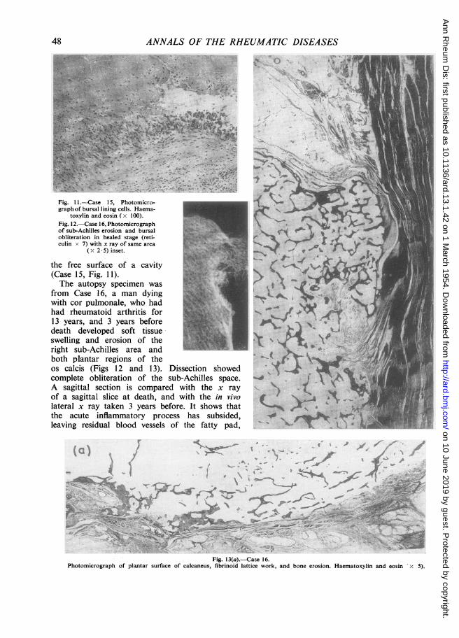
the free surface of a cavityah4l(Case 15, Fig. 11).The autopsy specimen was s , .
from Case 16, a man dyingwith cor pulmonale, who hadhad rheumatoid arthritis for US13 years, and 3 years beforedeath developed soft tissue Jswelling and erosion of theright sub-Achilles area andboth plantar regions of theos calcis (Figs 12 and 13). Dissection showedcomplete obliteration of the sub-Achilles space.A sagittal section is compared with the x ray ~ -of a sagittal slice at death, and with the in vivolateral x ray taken 3 years before. It shows thatthe acute inflammatory process has subsided,leaving residual blood vessels of the fatty pad, . I
-~ ~ 9 ' t ,' ' '5',.
Fig. 13(a).-Case 16.Photomicrograph of plantar surface of calcaneus, fibrinoid lattice work, and bone erosion. Haematoxylin and eosin 'x 5).
on 10 June 2019 by guest. Protected by copyright.
http://ard.bmj.com
/A
nn Rheum
Dis: first published as 10.1136/ard.13.1.42 on 1 M
arch 1954. Dow
nloaded from
HEEL LESIONS OF RHEUMATOID ARTHRITIS
Fig. 13(b).-Case 16, x ray (x 2-5).
obliteration of the space, and a healed erosion ofbone above the upper part of the tendon insertion,with loss of cartilage except at this site.
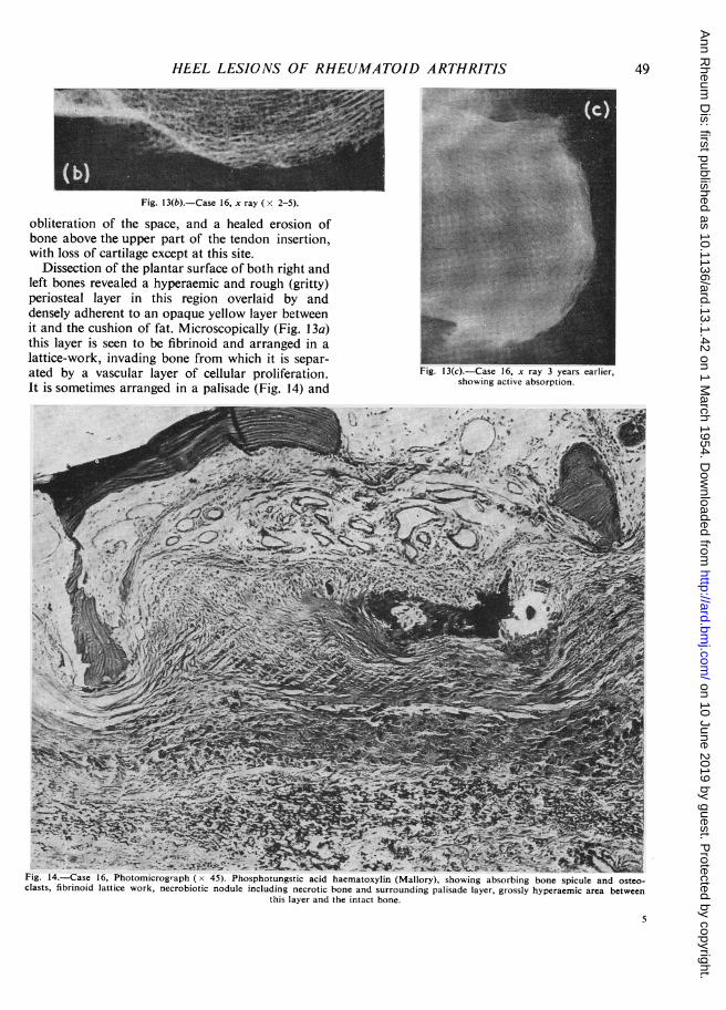
Dissection of the plantar surface of both right andleft bones revealed a hyperaemic and rough (gritty)periosteal layer in this region overlaid by anddensely adherent to an opaque yellow layer betweenit and the cushion of fat. Microscopically (Fig. 1 3a)this layer is seen to be fibrinoid and arranged in alattice-work, invading bone from which it is separ-ated by a vascular layer of cellular proliferation.It is sometimes arranged in a palisade (Fig. 14) and
sho--wing actve rabs3yearseptshowing active absorption.
Fig. 14.-Case 16, Photomicrograph (x 45). Phosphotungstic acid haematoxylin (Mallory), showing absorbing bone spicule and osteo-clasts, fibrinoid lattice work, necrobiotic nodule including necrotic bone and surrounding palisade layer, grossly hyperaemic area between
this layer and the intact bone.
49
on 10 June 2019 by guest. Protected by copyright.
http://ard.bmj.com
/A
nn Rheum
Dis: first published as 10.1136/ard.13.1.42 on 1 M
arch 1954. Dow
nloaded from

ANNALS OF THE RHEUMATIC DISEASESsometimes more loosely, but morphologically itobviously represents nodular tissue, to which itcorresponds tinctorially, when stained with haemato-xylin and eosin, toluidine blue, periodic acid Schiff,silver impregnation, and Mallory's phosphotungsticacid haematoxylin. Bone spicules were largely erodedaway, some remaining partly surrounded by osteo-clasts, and others buttressed by fresh layers of bone.
Differential Diagnosis of Sub-Achilles Swellings
The only patient in this series who did not haverheumatoid arthritis showed calcification radio-logically in the sub-Achilles swelling (Case 20, Fig. 15),and, when this was excised, it proved histologicallyto be a synovial chondromatosis. Other tumoursand infections (e.g. tuberculosis: Blencke, 1908;Wiesinger, 1896) may of course also occur, butwe have not met them. Ossification in the Achillestendon is an occasional cause of swelling and pain(Haglund, 1928). The lesion must also be differen-tiated from the peritendinous cellulitis described byRaynal (1883), which is probably traumatic in origin.
TreatmentTreatment in these patients is directed towards the
generalized joint involvement. Specific local measuresare sometimes necessary, particularly when theselesions are the only ones causing symptoms. In mildcases, the sub-Achilles swelling may be treatedconservatively since it usually heals in a few yearswithout residual damage, but if it causes severepain or disability (as in Cases 5, 11, and 15) it is betterexcised, which procedure led in these three patients toconsiderable improvement in function. The plantarlesions are probably due in part to excessive useof the heels since they are frequently associated withmetatarsal lesions. The provision of a soft padbeneath the heel or a padded ring usually gives thepatient some relief.
Discussion
Sub-Achilles bursitis was first described by Albert(1893), who called it "achillodynia". Nine of hiscases were later described by Rossler (1896),together with another in which the swelling wasextirpated and examined. Only two of these cases
13..~~~~~L: .i, j -
50
on 10 June 2019 by guest. Protected by copyright.
http://ard.bmj.com
/A
nn Rheum
Dis: first published as 10.1136/ard.13.1.42 on 1 M
arch 1954. Dow
nloaded from

HEEL LESIONS OF RHEUMATOID ARTHRITIS
not possible to determine whether their patients hadrheumatoid arthritis, rheumatic fever, gout, orspondylitis as we know these entities to-day. Evenin the most recent publications mentioning achillo-dynia (e.g. Haglund, 1928; Berenwenger, 1930;or Tepper and Haspekov, 1939), the details given areinsufficient to determine the type of rheumatism.In all of these older series the chief aetiologicalfactor was thought to be gonorrhoea, followed bytrauma, and a large number of other causes, includ-ing rheumatism, tuberculosis (Blencke, 1908;Wiesinger, 1896), syphilis (Schirren, 1902), influenza(Franke, 1895), gouty diathesis, etc.The present series of lesions is, with one exception
(a synovial chondromatosis), associated with anddue to rheumatoid arthritis: this is not thought tobe coincidental, or entirely accounted for by selec-tion of patients. It is thought that rheumatoidarthritis is the most common cause of sub-Achillesbursitis and of erosive plantar calcaneal lesions, andthat the two are often associated the one with theother.
Summary
Nineteen cases of rheumatoid arthritis with heellesions are described clinically and radiologically.Pathological observations were made in three cases.The two lesions which not infrequently occurtogether are:
(i) a sub-Achilles bursitis which erodes the oscalcis and finally obliterates the bursa,
(ii) an erosion of the plantar surface of the oscalcis by fibrinoid-containing tissue closelyresembling a rheumatoid nodule.
I am grateful to Mr. Arden who kindly referred severalof these patients and allowed me to use his notes, to
Dr. Glynn and Dr. Harrison for use of pathologicalmaterial, and to Mr. Fiske for the photographs.
REFERENCESAlbert, E. (1893). Wien. med. Pr., 34, 41.Berenwenger, P. (1930). Arch. klin. Chir., 159, 472.Blencke, A. (1908). Z. Orthop. Chir., 20, 363.Dresner, E., and Bywaters, E. G. L. (1953). Unpublished observations.Franke, F. (1895). Arch. klin. Chir., 49, 487.Haglund, P. (1928). Acta Chir. Scand., 63, 292.Konig, F. (1910). Dtsch. med. Wschr., 36, 597.Raynal, E. (1883) Arch. gen. Med., 152, 677.Rossler, A. (18965. Dtsch. Z. Chir., 42, 274.Rolly, F., and Appelt, 0. (1914). Arch. klin. Chir., 105, 358.Schirren, C. (1902). Dtsch. Z. Chir., 67, 132.Tepper, P. A., and Haspekov, G. E. (1939). Acta med. scand., 100, 296.Wiesinger, -. (1896). Dtsch. Z. Chir., 43, 603.
LUsions du talon dans l'arthrite rhumatismaleRtsuMlt
On decrit au point de vue clinique et radiologique19 cas d'arthrite rhumatismale avec des lesions du talon.Dans trois cas on a pu faire une etude anatomo-pathologique. On a trouve deux sortes de lesions sur-venant assez souvent ensemble:
(i) une bursite sous-achilleenne, erodant le calcaneumet finalement obliterant la bourse,
(ii) une erosion de la surface plantaire du calcaneumpar un tissu contenant une substance fibrinoide etressemblant beaucoup a un nodule rhumatismal.
Lesiones del talon en la artritis reumatoideSUMARIO
Se describe clinica y radiol6gicamente 19 casos deartritis reumatoide con lesiones del tal6n. En tres casosse pudo hacer un estudio anatomo-patol6gico. Encon-trAronse dos tipos de lesiones, muchas veces al mismotiempo:
(i) una bursitis sub-aquilea causando la erosi6n delcalcAneo y finalmente obliterando la bursa,
(ii) una erosi6n de la superficie plantar del calcAneopor un tejido conteniendo una substancia fibrinoide ymuy parecida a un n6dulo reumatico.
51
on 10 June 2019 by guest. Protected by copyright.
http://ard.bmj.com
/A
nn Rheum
Dis: first published as 10.1136/ard.13.1.42 on 1 M
arch 1954. Dow
nloaded from