LEISHMANIA - · PDF fileLEISHMANIA MAJOR COMPLEX Lesishmania major: It causes a moist type...
17
GENUS: LEISHMANIA Species parasitic in man: Under the genus Leishmania, there are 2 subgenus: 1. Leishmania 2. Viannia SPECIES PARASITIC IN MEN Under subgenus Leishmania, there are following species: LEISHMANIA DONOVANI COMPLEX SPECIES DISEASE CAUSING VECTOR RESERVOIR HOSTS *Leishmania donovani *Kala-azar (Visceral leishmaniasis) 1. P.argentipes (India)* 2. P.chinesis (China) Man* *Leishmania infantum 1. Infantile kala-azar* 2. ZVL (Zoonotic visceral leishmanias is)* 1. P.perniciosus (Mid asia, Middle east, China) 2. P.chinesis and P.alexandri (China) Dog, fox, Jackal. *Leishmania chagasi ZVL in new world* Lutzomyia longipalpis (New world) Dog, Fox. Leishmania archibaldi Visceral leishmaniasis in Africa 1. P. Orientalis 2. P.martini (Africa) Rodents, carnivorous animals. *Only remember the vector of kala-azar in India (P.argentipes). LEISHMANIA TROPICA COMPLEX Leishmania tropica: It causes a dry type of cutaneous lesion in urban areas of North-West India.
Transcript of LEISHMANIA - · PDF fileLEISHMANIA MAJOR COMPLEX Lesishmania major: It causes a moist type...

GENUS: LEISHMANIA
Species parasitic in man:
Under the genus Leishmania, there are 2 subgenus:
1. Leishmania
2. Viannia
SPECIES PARASITIC IN MEN
Under subgenus Leishmania, there are following species:
LEISHMANIA DONOVANI COMPLEX
SPECIES DISEASE CAUSING VECTOR RESERVOIR
HOSTS
*Leishmania
donovani
*Kala-azar
(Visceral
leishmaniasis)
1. P.argentipes
(India)*
2. P.chinesis
(China)
Man*
*Leishmania
infantum
1. Infantile
kala-azar*
2. ZVL
(Zoonotic
visceral
leishmanias
is)*
1. P.perniciosus
(Mid asia,
Middle east,
China)
2. P.chinesis and
P.alexandri
(China)
Dog,
fox,
Jackal.
*Leishmania
chagasi
ZVL in new
world*
Lutzomyia longipalpis
(New world)
Dog,
Fox.
Leishmania
archibaldi
Visceral
leishmaniasis in
Africa
1. P. Orientalis
2. P.martini
(Africa)
Rodents,
carnivorous
animals.
*Only remember the vector of kala-azar in India (P.argentipes).
LEISHMANIA TROPICA COMPLEX
Leishmania tropica: It causes a dry type of cutaneous lesion in urban
areas of North-West India.

LEISHMANIA MAJOR COMPLEX
Lesishmania major: It causes a moist type of cutaneous lesion in rural
areas of Central asia.
LEISHMANIA MEXICANA COMPLEX
Leishmania Mexicana: It causes a single cutaneous lesion on the face, ear
or hand in countries of South America. (Mexico, Venezuela, Columbia
etc.)
Other species in this group are:
1. Leishmania amazonensis
2. Leishmania venezuelensis
3. Leishmania garnhami etc. (All of them causes local cutaneous
lesion)
Under subgenus viannia, there are following species:
Leishmania braziliensis complex
Leishmania braziliensis:
It causes a malignant type of lesion as a papullopustular swelling of skin
localised on the nostrils, mouth, eyes, face, ears, elbow and knee, which
with time causes destructive and mutilating erosions. It commonly
occurs in countries of South America.
Other complexes are (not needed):
1. Leishmania guyanensis complex
2. Leishmania lainsoni complex
3. Leishmania naiffi complex

CLINICAL CLASSIFICATION OF LEISHMANIASIS
1. VISCERAL LEISHMANIASIS (KALA-AZAR)
Indian kala-azar is caused by Leishmania donovani with a human
reservoir.
2. CUTANEOUS LEISHMANIASIS
• The classical lesion is characterised by a nodule at the site of
inoculation followed by formation of a central crust which may fall
away forming a wet type of ulcer.
• Or it may be present as a papulonodular lesion covered by
superficial scales forming a dry type of ulcer.
• So, a depressed scar and altered pigmentation on healing of the
nodules at the edge of lesion is a characteristic feature of CL.
It is of 3 types as discussed below:
1. Post kala-azar dermal leishmaniasis: (PKDL)
• It occurs mainly in India.
• It is caused by Leishmania donovani.
• It is a late sequel to visceral leishmaniasis. It occurs typically
1-2 years after recovery from VL.
• It is characterized by non ulcerating skin lesion, which is of 3
types:
1) Macular and hypopigmented lesion: on the trunk and
extremities.
2) Erythematous patch: on the nose, cheek and chin. Also
called “butterfly erythema”.
3) Nodules: Soft, painless, yellowish pink nodules appear
mainly on the face. There is absence of ulceration
which can distinguish them from oriental sore (dermal
leishmaniasis).
• They do not heal spontaneously.

2. Old world CL:
• The main causative agents are:
1) Leishmania major
2) Leishmania tropica
3) Leishmania aethiopica.
• It is of 3 types:
1) Urban/ lupoid/ tuberculoid leishmaniasis
2) Rural leishmaniasis
3) Diffuse cutaneous leishmaniasis
• Urban leishmaniasis is caused by L.tropica in North India. It is
characterized by a self healing ulcer staring as a small itching
papule covered with fine whitish scale which gradually
becomes dark and thick and finally falls away and a
depressed scar is formed. It is present on the face, feet, legs
and arms.
• Lupoid/ tuberculoid leishmaniasis is caused by L.tropica. It is
characterized by a non-self healing ulcer with peripheral
activity, which mainly occurs on the face, which is a result of
an incomplete immune response of an earlier episode of
oriental sore.
• Rural leishmaniasis is caused by Rural leishmaniasis is caused
by L.major in North India. It is characterized by multiple
painless lesions on the nose, lips and limbs, which heals
rapidly, but in non-immune person may result in
disfigurement of face.
• DCL of old world is caused by L.aethiopica. It is a result of
specific deficiency of CMI to Leishmania antigen. It is
characterized by appearance of nodular infiltrative lesions
mainly over the face, ears, extremities and buttocks.
• But there is no ulceration or mucosal involvement.
Histologically the nodes consist almost entirely of histocytes
with a relative absence of lymphocytes and plasma cells.
• It has a strong resemblance to lepromatous leprosy, because
of the absence of CMI as in leprosy and that the leishmanin
skin test is negative. Amastigote form is found both in blood
and bone marrow.
3. New World CL:
• It is seen mainly in South and Central Ameraica.
• Causative agents:
� L.peruviana
� L.braziliensis
� L.mexicana
� L.guyanensis
� L.panamensis
• New world DCL is caused by:
� L.amazonesis
� L.mexicana
3.MUCOCUTANEOUS LEISHMANIASIS
• It is mainly caused by L.braziliensis and occasionally by
L.panamensis.
• It is seen in south and central America.
• It consists of two stages: the primary cutaneous lesions,
sometimes followed by secondary mucosal involvement,
which occurs after a variable time of latency.
• It is seen in nasal mucosal membrane, pharynx, larynx and
upper lip, sometime whole nasal septum is destroyed.
• Granulomas develop at mucocutaneous junction followed
by gross destruction of soft tissue and cartilage causing
disfigurement of nose and mouth.
• Death may occur from severe respiratory infections due to
acute obstruction of respiratory passages.
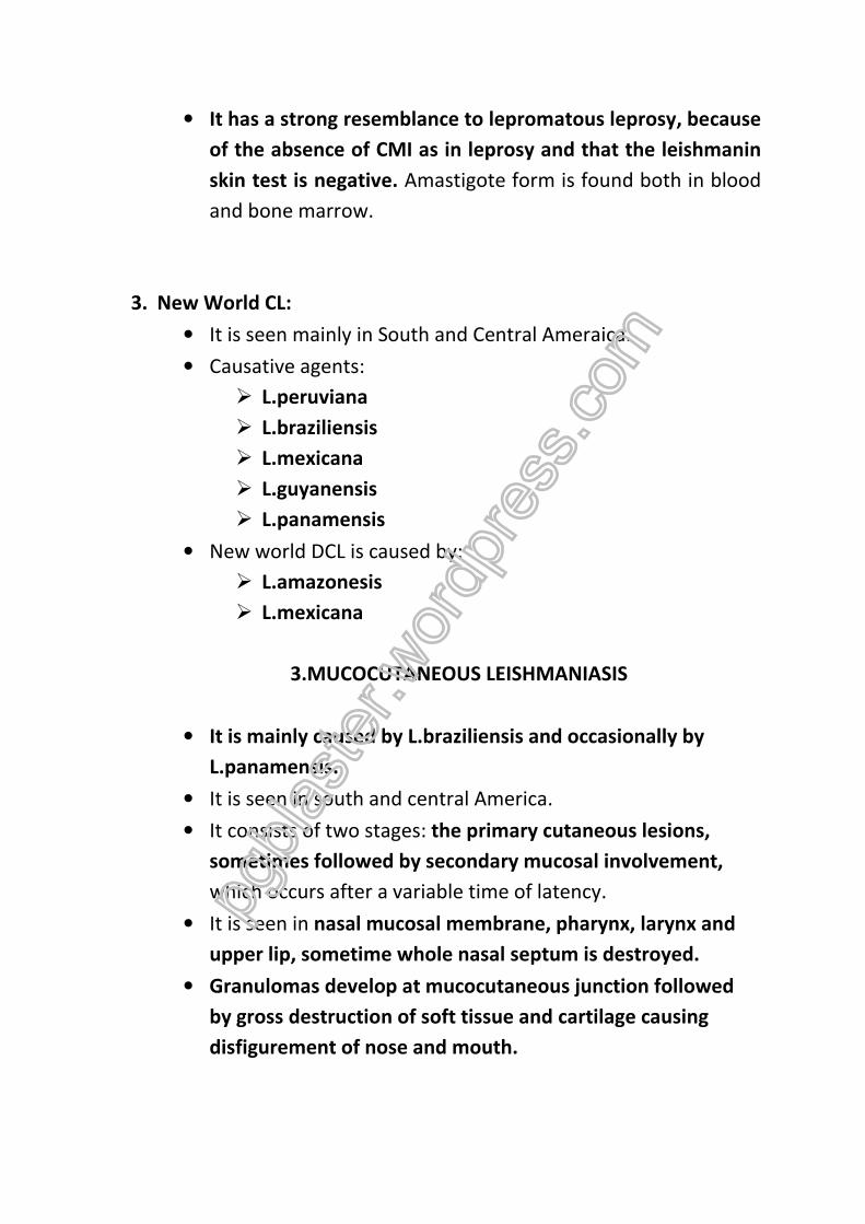
MORPHOLOGY OF LEISHMANIA DONOVANI
The parasite exists in mainly two stages-
1. Amastigote forms: Aflagellar state, appears in man.
2. Promastigote forms: Flagellar state, apperas in gut of sandfly and in
artificial culture.
AMASTIGOTE FORM PROMASTIGOTE FORM
*1st
row is of Giemsa stain, 2nd
row is of SEM. (4th
photo- dividing
promastigote)
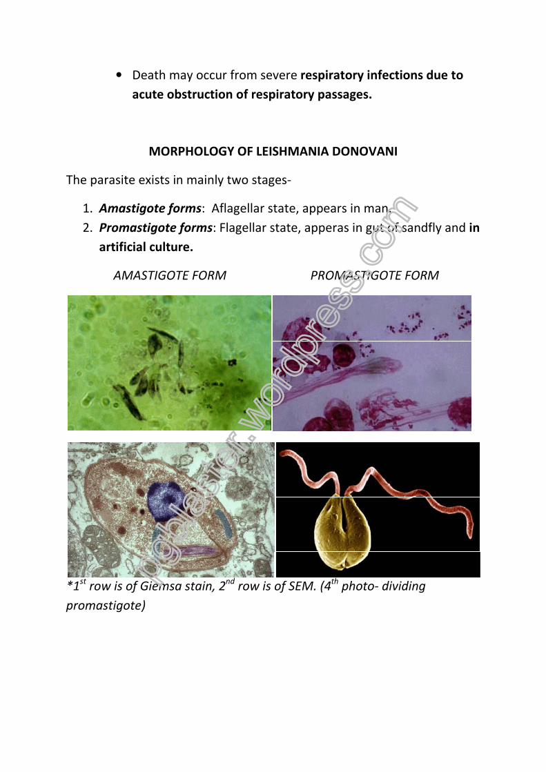
(PROMASTIGOTE FORM)
*GENERAL STRUCTURE OF LEISHHMANIA (WITH FLAGELLA)
FEATURES AMASTIGOTE FORM PROMASTIGOTE FORM
Size and shape Round/ oval measuring 2-4 µm. Earlier stage: Short and oval,
pear shaped.
Mature stage: Long, slender,
spindle shaped.
Cell membrane Delicate. -
Nucleus Oval/ round and situated in the
middle of the cell/ along the side
of the cell wall.
It is situated centrally.
Kinetoplast (It
comprises of a DNA
containing body.)
It is situated tangentially/ at
right angle of nucleus.
It is situated transversely near
the anterior end.
Axoneme A delicate filament extending
from the kinetoplast to the
margin of the body, it represents
Same as that.

the root of flagellum.
Flagellum Not present. Present, projecting from the
front.
Vacuole A clear unstained space lying
along the axoneme.
A light eosinophilic vacuole is
situated infront of the
kinetoplast.
CULTURE OF LEISHMANIA
• The cultivation of Leishmania is done in a special culture medium
named “NNN medium”, which contains salt agar and defibrinated
rabbit’s blood in 2:1 ratio.
• In this medium, the material is inoculated at water of condensation
at 22-24°C and intracellular growth can be maintained at 37°C for
around 32 days.
• In the culture, the amastigote form is converted to promastigote
form, which is divided by longitudinal fission and gives rise to
numerous flagella.
• There should be special measures to control bacterial
contamination because it may cause degeneration and death of
L.donovani.
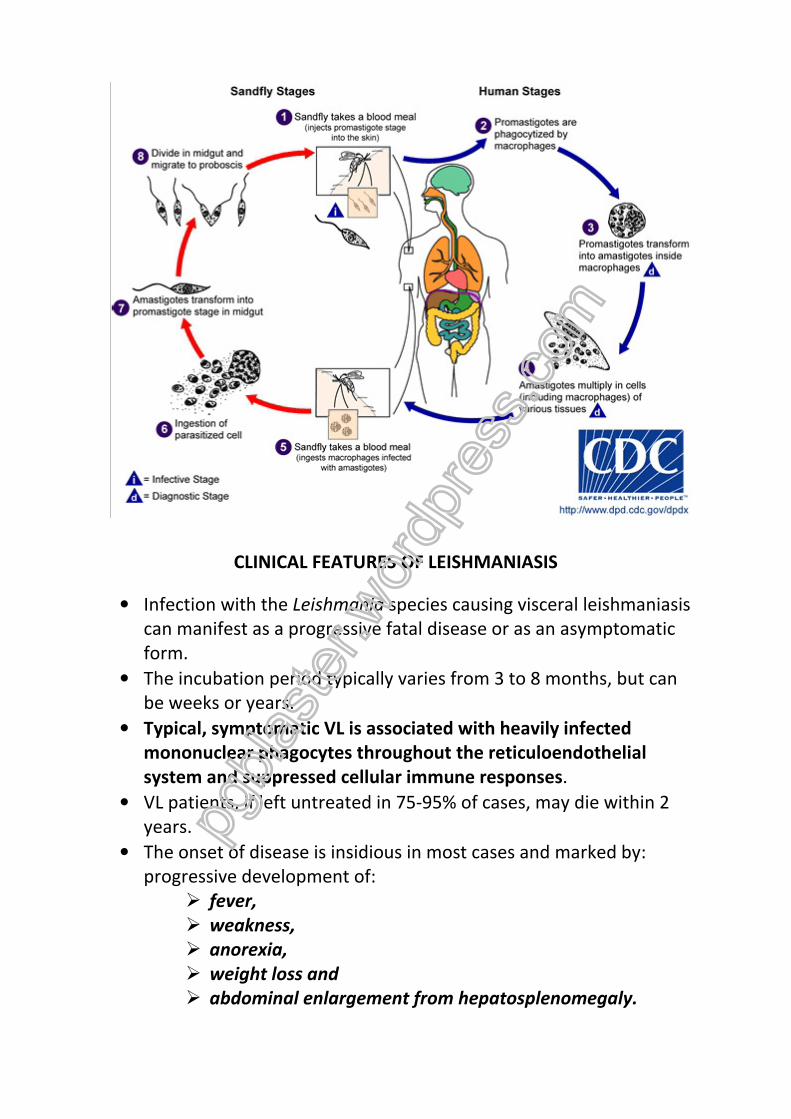
LIFE CYCLE AND PATHOGENESIS
• Leishmania parasite exists in two forms- amastigote and
promastigote.
• When an infected sandfly bites an individual, there is inoculation of
promastigote form into the skin.
• After inoculation into skin by a sandfly, promastigotes are
phagocytosed by dermal macrophages, where they convert to
amastigotes and multiply within acidic vacuoles.
• Additional mononuclear phagocytes are attracted to the site of the
initial lesion and become infected.
• The host cells are enlarged by the burden of multiplicating
amastigotes, and are eventually ruptured.

• Amastigotes then disseminate through the regional lymphatics
and the vascular system to infect mononuclear phagocytes
throughout the RE system.
Bone marrow aspirate from a patient suffering from VL showing
amastigotes in the macrophages.
• Progressive recruitment of amastigote-infected mononuclear
phagocytes and inflammatory cells within organs results in
distortion of the native tissue architecture and often, massive
hepatosplenic enlargement.
• Parasitized reticuloendothelial cells can be found in bone marrow,
lymph nodes, skin and other organs.
• A blood sucking insect draws these free amastigote forms as well
as those in the monocytes during its blood meal.
• In certain species of sandfly, these ingested amastigote forms
develop into promastugote forms which divides by binary fission in
the midgut of sandfly giving rise to numerous flagellates.
• These flagellates tend to spread to the anterior part of the
alimentary canal (buccal cavity and pharynx and finally proboscis)
after 6-9 days of ingestion of blood. This type of development is
called as “Anterior station development”.
• As salivary glands of sandfly are not affected, the transmission is
by bite, not the saliva.
COUNTRY RESERVOIR OF INFECTION
INDIA HUMAN
CHINA AND BRAZIL DOG
EAST AFRICA RODENT
RUSSIA JACKAL
CLINICAL FEATURES OF LEISHMANIASIS
• Infection with the Leishmania species causing visceral leishmaniasis
can manifest as a progressive fatal disease or as an asymptomatic
form.
• The incubation period typically varies from 3 to 8 months, but can
be weeks or years.
• Typical, symptomatic VL is associated with heavily infected
mononuclear phagocytes throughout the reticuloendothelial
system and suppressed cellular immune responses.
• VL patients, if left untreated in 75-95% of cases, may die within 2
years.
• The onset of disease is insidious in most cases and marked by:
progressive development of:
� fever,
� weakness,
� anorexia,
� weight loss and
� abdominal enlargement from hepatosplenomegaly.

• Fever, accompanied by chills is usually intermittent or remittent
with twice-daily temperature spikes.
• During the less common acute cases, fever can be of abrupt onset
and have a periodicity similar to that of malaria.
• Progressive and massive hepatosplenomegaly is characteristic of
VL.
• Infected individuals in the Sudan often also develop
lymphadenopathy.
• In India patients with VL commonly develop hyperpigmentation of
extremities, face and abdomen.
• Hemorrhage can occur from various sites.
• Severe cachexia is a prominent feature of VL, driven in part by
high levels of TNF-α.
• Death from VL occurs either from the:
� primary, multisystem disease causing malnutrition and
bone marrow suppression and/or,
� from secondary bacterial infections such as tuberculosis,
dysentery, pneumonia and measles.
• Important laboratory findings in advanced visceral disease include
profound pancytopenia (due to bone marrow suppression),
eosinopenia, hypoalbuminemia and hypergammaglobulinemia
(mainly IgG).
• The cause of anemia in kala-azar is now thought to be due to
splenic hemolysis of RBC.***
• The ESR is usually elevated.
• Kidneys may show evidence of immune complex deposition, but
renal failure is rare.
• Several infectious and hematologic diseases can mimic visceral
leishmaniasis. These include:
1) Malaria,
2) Schistosomiasis,
3) Miliary tuberculosis,
4) African trypanosomiasis,
5) Typhoid fever,
6) Brucellosis,
7) Histoplasmosis,
8) Bacterial endocarditis,

9) Lymphoma and
LABORATORY DIAGNOSIS OF KALA
DIRECT EVIDENCES (Demonstration of L.donovani)
� Peripheral blood smear:
blood film has to be done to
in the PBS. Because of the small number of parasites present in the
blood, there is often a negative result obtained. The chances of
finding a Leishmania parasite is greatly increased if any of the
following methods are applied:
1. By making a thick
2. By centrifuging citrated blood.
3. By producing a thick leucocytic edge
drawn and just before the blood is totally exhausted, the
spreading slide is abruptly lifted off.)
� Blood culture: It is the least sensitive method
only disadvantage is that the result is obtained slow and after a
long time (almost a month).
aseptically and mixed with 10 ml citrated saline solution.
are then allowed to settle in a cold
cellular deposit is then inoculated in NNN medium and inculbated
DIRECT EVIDENCE
PERIPHERAL BLOOD:
AMASTIGOTE FORM
BLOOD CULTURE IN NNN MEDIUM:
PROMASTIGOTE FORM
ymphoma and Leukemia.
LABORATORY DIAGNOSIS OF KALA-AZAR
DIRECT EVIDENCES (Demonstration of L.donovani)
Peripheral blood smear: A microscopical examination of a stained
blood film has to be done to identify the amastigote forms
in the PBS. Because of the small number of parasites present in the
blood, there is often a negative result obtained. The chances of
finding a Leishmania parasite is greatly increased if any of the
following methods are applied:
By making a thick blood film.
By centrifuging citrated blood.
By producing a thick leucocytic edge (when a thick blood film is
drawn and just before the blood is totally exhausted, the
spreading slide is abruptly lifted off.)
It is the least sensitive method for diagnosis.
only disadvantage is that the result is obtained slow and after a
long time (almost a month). 1-2 ml of blood is taken from a vein
aseptically and mixed with 10 ml citrated saline solution.
are then allowed to settle in a cold incubator (22°C) overnight.
cellular deposit is then inoculated in NNN medium and inculbated
LABORATORY EVIDENCES
DIRECT EVIDENCE
BLOOD CULTURE IN NNN MEDIUM:
PROMASTIGOTE FORM
BIOPSY MATERIAL
STERNAL/ ILIAC CREST MARROW
PUNCTURE
SPLENIC PUNCTURE
INDIRECT EVIDENCE
BLOOD COUNT (ANEMIA,
LEUCOPENIA, RAISED ESR)
ALDEHYDE TEST
DIRECT EVIDENCES (Demonstration of L.donovani)
A microscopical examination of a stained
identify the amastigote forms present
in the PBS. Because of the small number of parasites present in the
blood, there is often a negative result obtained. The chances of
finding a Leishmania parasite is greatly increased if any of the
(when a thick blood film is
drawn and just before the blood is totally exhausted, the
for diagnosis. The
only disadvantage is that the result is obtained slow and after a
2 ml of blood is taken from a vein
aseptically and mixed with 10 ml citrated saline solution. The cells
incubator (22°C) overnight. The
cellular deposit is then inoculated in NNN medium and inculbated
INDIRECT EVIDENCE
SEROLOGICAL TESTS
ALDEHYDE TESTDEMONSTRATION
OF ANTIBODIES

at 22°C for 1-4 weeks. At the end of each week, a drop of
condensation fluid is examined for promastigote forms.
� Biopsy material:
1) SPLENIC PUNCTURE: When spleen is enlarged, it is the most
important clue to diagnosis. Amastigote forms are found in
the stained culture and promastigote forms are found in
culture. The only risk of a splenic puncture is that bleeding
may continue from the punctured wound in patients of
leukaemia and hemorrhagic diathesis.
2) BONE MARROW PUNCTURE FROM STERNUM/ILIAC CREST: It
offers a method of diagnosis particularly in early cases, when
spleen is not so enlarged as to be punctured. Its
disadvantage is that parasites are scanty. As in the splenic
puncture, the amastigote forms are found in the stained
culture and promastigote forms are found in culture.
INDIRECT EVIDENCES
� Blood count:
1) Neutropenia with a relative lymphocytosis and monocytosis
is revealed.
2) Average TC is <3000/mm3, may fall upto below 1000/mm3.
3) RBC is also decreased, the RBC:WBC ratio becomes 1:2000,
when the normal being 1:750.
� Serological tests:
1) Aldehyde test:
� It is the test for rise of γ-globulin.
� 1-2 ml of serum is taken in a glass test tube and 1-2
drops of 40% formalin is added to it.
� A positive result is obtained by the observation of
jellifying/ milk white opacity like a hard-boiled egg in
2-20 minutes.
� It should be remembered that the result will not be
positive until 3 months of disease progression.
� False positive result- African trypanosomiasis, multiple
myeloma and cirrhosis.
� False negative result- cutaneous leishmaniasis.
2) Complement fixation test with WKK antigen:
� The antigen used in this reaction is prepared from
human tubercle (by Witebsky, Klingenstein and Kuhn,
hence WKK antigen) because Leishmania and
Mycobacteria share a common antigen.
� The test is based on the presence of certain immune
bodies in the sera of kala-azar patients.
� This test has distinct advantage of early detection of
the case, becoming positive within 3 weeks of the
disease.
� False positive results:
I. Leprosy
II. Pulmonary TB
III. Tropical pulmonary eoinophilia.
*Other serological tests:
1. IFA (Immuno fluorescence assay)- Most commonly used.
2. ELISA
3. Direct agglutination test
4. Latex particle agglutination test
5. Immunoblotting
6. Countercurrent immunoelectrophoresis etc.
TREATMENT OF LEISHMANIASIS (FOR PHARMACOLOGY)
The following classes of drugs are used in treatment of leishmaniasis:
ANTIMONY COMPOUND
• SSG (Sodium stibogluconate)
DIAMIDINE
• Pentamidine
ANTIFUNGAL DRUGS
• AMB (Amphotericin B)
OTHER CHOICES
• Paromomycin

SODIUM STIBOGLUCONATE (SSG)
It is a first line drug against kala
due to extensive resistance. It is an pentavalent antimonial compound,
which is water soluble and containing 1/3 rd of antimony by weight.
Mechanism of action:
� The drug is in pentavalent form and water soluble.
� It is converted to active trivalent form by an enzyme present in
Leishmania amastigote form.
� This active trivalent form causes
parasite and oxidative damage to the parasite.
Dose and route of administration:
� 20 mg/ kg daily by i.m/ i.v route (Maximum 850 mg) for 20
or more.
� Relapsing cases should immediately
Preparations:
� Abnate
� Stibo.
Adverse effects:
Nausia
Vomitting
Metallic taste
Cough
Pain abdomen
Pain and stiffness of injected muscle
Sterile abscess
Mental syndromes
SODIUM STIBOGLUCONATE (SSG)
It is a first line drug against kala-azar but now it is not used in Bihar (India)
due to extensive resistance. It is an pentavalent antimonial compound,
is water soluble and containing 1/3 rd of antimony by weight.
The drug is in pentavalent form and water soluble.
It is converted to active trivalent form by an enzyme present in
Leishmania amastigote form.
This active trivalent form causes efflux of Glutathione from the
oxidative damage to the parasite.
Dose and route of administration:
20 mg/ kg daily by i.m/ i.v route (Maximum 850 mg) for 20
Relapsing cases should immediately be retreated with same dose.
Pain and stiffness of injected muscle
azar but now it is not used in Bihar (India)
due to extensive resistance. It is an pentavalent antimonial compound,
is water soluble and containing 1/3 rd of antimony by weight.
It is converted to active trivalent form by an enzyme present in
efflux of Glutathione from the
20 mg/ kg daily by i.m/ i.v route (Maximum 850 mg) for 20-30 days
be retreated with same dose.
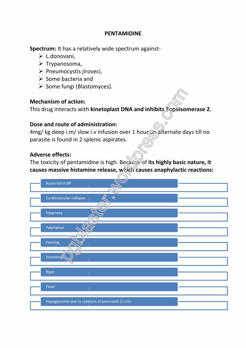
Spectrum: It has a relatively wide spectrum against
� L.donovani,
� Trypanosoma,
� Pneumocystis jiroveci,
� Some bacteria and
� Some fungi (Blastomyces)
Mechanism of action:
This drug interacts with
Dose and route of administration:
4mg/ kg deep i.m/ slow i.v infusion over 1 hour on alternate days till no
parasite is found in 2 splenic aspirates.
Adverse effects:
The toxicity of pentamidine is high. Because of
causes massive histamine release, which causes anaphylactic reactions:
Acute fall in BP
Cardiovascular collapse
Dyspnoea
Palpitation
Fainting
Vomitting
Rigor
Fever
Hypoglycemia due to cytolysis of pancreatic
PENTAMIDINE
It has a relatively wide spectrum against-
Pneumocystis jiroveci,
ome bacteria and
fungi (Blastomyces).
This drug interacts with kinetoplast DNA and inhibits Topoisomerase 2.
Dose and route of administration:
4mg/ kg deep i.m/ slow i.v infusion over 1 hour on alternate days till no
parasite is found in 2 splenic aspirates.
city of pentamidine is high. Because of its highly basic nature, it
causes massive histamine release, which causes anaphylactic reactions:
Hypoglycemia due to cytolysis of pancreatic β cells.
kinetoplast DNA and inhibits Topoisomerase 2.
4mg/ kg deep i.m/ slow i.v infusion over 1 hour on alternate days till no
its highly basic nature, it
causes massive histamine release, which causes anaphylactic reactions:

Use: Only for salvage therapy of antimonial failure cases.
Amphotericin B (AMB)
Mechanism of action:
Like fungi, Leishmania also has high percentage of ergosterol. So,
antifungal agents are highly effective in kala-azar.
Use:
It is extensively used in antimonial resistance. In bihar, it is the standard
treatment due to extensive SSG resistance. But high toxicity and
prolonged hospitalization limits its application.
*Liposomal AMB is particularly suitable for treatment of kala-azar
because it delivers the drug directly inside the RE cells of liver and
spleen where the amastigote live.
PARAMOMYCIN
Type: An aminoglycoside antibiotic.
Dose: 10-15 mg/ kg/ day for 21 days.
Side effects: Ototoxicity, elevated serum transaminase levels, pain at
injection site.
LOCAL TREATMENT OF DERMAL LEISHMANIASIS/ ORIENTAL SORE
1. SSG: 2 ml solution round the sore.
2. Paramomycin ointment: Applied locally.