Starch Phosphorylase Inhibitor fromSweet Potato' - Plant Physiology
HAL Id: pasteur-01674361https://hal-riip.archives-ouvertes.fr/pasteur-01674361
Submitted on 2 Jan 2018
HAL is a multi-disciplinary open accessarchive for the deposit and dissemination of sci-entific research documents, whether they are pub-lished or not. The documents may come fromteaching and research institutions in France orabroad, or from public or private research centers.
L’archive ouverte pluridisciplinaire HAL, estdestinée au dépôt et à la diffusion de documentsscientifiques de niveau recherche, publiés ou non,émanant des établissements d’enseignement et derecherche français ou étrangers, des laboratoirespublics ou privés.
Public Domain
Leishmania infantum 5’-MethylthioadenosinePhosphorylase presents relevant structural divergence to
constitute a potential drug targetHela Abid, Emna Harigua-Souiai, Thouraya Mejri, Mourad Barhoumi, Ikram
Guizani
To cite this version:Hela Abid, Emna Harigua-Souiai, Thouraya Mejri, Mourad Barhoumi, Ikram Guizani. Leishmaniainfantum 5’-Methylthioadenosine Phosphorylase presents relevant structural divergence to constitute apotential drug target. BMC Structural Biology, BioMed Central, 2017, 17 (1), pp.9. �10.1186/s12900-017-0079-7�. �pasteur-01674361�
Abid et al. BMC Structural Biology (2017) 17:9 DOI 10.1186/s12900-017-0079-7
RESEARCH ARTICLE Open Access
Leishmania infantum 5’-MethylthioadenosinePhosphorylase presents relevant structuraldivergence to constitute a potential drugtarget
Hela Abid1,2, Emna Harigua-Souiai1, Thouraya Mejri1, Mourad Barhoumi1 and Ikram Guizani1*Abstract
Background: The 5′-methylthioadenosine phosphorylase (MTAP), an enzyme involved in purine and polyaminemetabolism and in the methionine salvage pathway, is considered as a potential drug target against cancerand trypanosomiasis. In fact, Trypanosoma and Leishmania parasites lack de novo purine pathways and relyon purine salvage pathways to meet their requirements. Herein, we propose the first comprehensive bioinformaticand structural characterization of the putative Leishmania infantum MTAP (LiMTAP), using a comparativecomputational approach.
Results: Sequence analysis showed that LiMTAP shared higher identity rates with the Trypanosoma brucei(TbMTAP) and the human (huMTAP) homologs as compared to the human purine nucleoside phosphorylase(huPNP). Motifs search using MEME identified more common patterns and higher relatedness of the parasiteproteins to the huMTAP than to the huPNP. The 3D structures of LiMTAP and TbMTAP were predicted byhomology modeling and compared to the crystal structure of the huMTAP. These models presented conservedsecondary structures compared to the huMTAP, with a similar topology corresponding to the Rossmann fold.This confirmed that both LiMTAP and TbMTAP are members of the NP-I family. In comparison to the huMTAP, the 3Dmodel of LiMTAP showed an additional α-helix, at the C terminal extremity. One peptide located in this specific region wasused to generate a specific antibody to LiMTAP. In comparison with the active site (AS) of huMTAP, the parasite ASspresented significant differences in the shape and the electrostatic potentials (EPs). Molecular docking of5′-methylthioadenosine (MTA) and 5′-hydroxyethylthio-adenosine (HETA) on the ASs on the three proteins predicteddifferential binding modes and interactions when comparing the parasite proteins to the human orthologue.
Conclusions: This study highlighted significant structural peculiarities, corresponding to functionally relevant sequencedivergence in LiMTAP, making of it a potential drug target against Leishmania.
Keywords: Leishmania, MTAP, Homology modeling, Molecular docking, Antibody
* Correspondence: [email protected]; [email protected] of Molecular Epidemiology and Experimental Pathology(LR11IPT04/ LR16IPT04), Institut Pasteur de Tunis, Université de Tunis ElManar, Tunis, TunisiaFull list of author information is available at the end of the article
© The Author(s). 2017 Open Access This articInternational License (http://creativecommonsreproduction in any medium, provided you gthe Creative Commons license, and indicate if(http://creativecommons.org/publicdomain/ze
le is distributed under the terms of the Creative Commons Attribution 4.0.org/licenses/by/4.0/), which permits unrestricted use, distribution, andive appropriate credit to the original author(s) and the source, provide a link tochanges were made. The Creative Commons Public Domain Dedication waiverro/1.0/) applies to the data made available in this article, unless otherwise stated.
Abid et al. BMC Structural Biology (2017) 17:9 Page 2 of 17
BackgroundNeglected Tropical Diseases (NTDs) correspond to mul-tiple transmissible pathologies that mainly occur in tropi-cal and sub-tropical regions. They affect populationsliving in poverty with more than a billion people in 149countries worldwide [1]. Here, we focus on leishmania-ses, a group of vector-borne diseases caused by differentspecies of protozoan parasites of the genus Leishmania[2]. Three hundred and 50 million people are at risk ofinfection and 2 million cases are reported worldwideeach year [3]. One to 1.5 million cases of cutaneousleishmaniasis (CL) and 0.2–0.5 million cases of visceralleishmaniasis (VL) are reported annually [3]. VL ismainly caused by Leishmania donovani and Leishmaniainfantum (L. infantum) species, with an annual deathtoll of 50,000 cases [3]. Mainstay therapy is based onthe use of toxic pentavalent antimonials in long treat-ment courses [4]. Furthermore, their prolonged use isincreasingly inducing parasite drug resistance [5]. Secondline drugs, such as pentamidine, miltefosine, andamphotericin B also are toxic, costly or have adverseeffects [6]. Therefore, the need for new targets and newdrugs is increasingly important, and constitutes researchpriority.Search of novel potential drug targets mainly focuses
on biochemical and metabolic pathways that show dif-ferences between pathogens and their host. Purine sal-vage, polyamine biosynthesis and thiol metabolism areamong the most important metabolic pathways beingconsidered for drug development against diseases causedby Trypanosomatidae parasites [7, 8]. Some of the moststriking differences between parasites and their mamma-lian host are found in purine metabolism [9]. In mam-mals, the de novo and/or the so-called “salvage”pathways ensure the synthesis of the purine nucleotides.To the contrary, most parasites studied rely on thesalvage pathways for their purine requirement as theylack the pathways for de novo purine biosynthesis [9].Therefore, salvage purine metabolism constitutes poten-tially an excellent target for the rational design ofantiparasitic drugs. Among the enzymes involved in pur-ine metabolism, 5′-methylthioadenosine phosphorylase(MTAP) plays a crucial role in purine and polyaminemetabolism and in the methionine salvage pathway [10].The 5′-methylthioadenosine (MTA), natural substrate ofMTAPs, is generated during polyamine biosynthesis andis then cleaved to adenine and 5′-methylthioribose-1-phosphate [10, 11], which are respectively incorporatedinto the salvage pathways of purine and methionine [12].MTAP, an entry enzyme to methionine salvage pathway,plays an important role to maintain low intracellularlevels of MTA, thus to preserve a proper cellular func-tion. Methionine synthesis, polyamine synthesis, proteintrans-methylation and trans-sulfuration pathways are
excellent targets for chemotherapeutic interventionagainst African trypanosomes, which are phylogenetic-ally close to Leishmania parasites [13]. MTAP wasdescribed as an interesting chemotherapeutic target inAfrican trypanosomes (Trypanosoma brucei brucei), forwhich selective transition-state analogues were developed.We cite the 5′-hydroxyethylthio-adenosine (HETA), ananalogue of MTA, which is highly metabolized by theTrypanosome MTAP in comparison to the mammalianenzyme [10, 14]. Growth inhibition assays showed IC50
values ≤1 μM for HETA, which was selected amongpossible candidates for in vivo evaluations. HETA ex-hibited 70% to 90% cure rates when administered tomice infected with T. brucei brucei [10, 14]. Moreover,Leishmania major MTAP (LmMTAP) and Trypanosomabrucei MTAP (TbMTAP) have high druggability indexes(0.8, range: 0 to 1) according to the TDR (TropicalDisease Research) Targets Database (www.tdrtargets.org).This explains our interest to such proteins as promisingdrug targets against diseases caused by Trypanosomatidaeparasites. However so far, no study targeted MTAP inLeishmania.The enzymes that catalyze the phosphorolytic cleavage
of the glycosidic bond in nucleosides are structurallyclassified in two families called nucleoside phosphorylase-I (NP-I) and nucleoside phosphorylase-II (NP-II) [15].Members of NP-II family share a common two-domainsubunit fold and a dimeric quaternary structure whilemembers of the NP-I family, including MTAP, share acharacteristic subunit topology with a trimeric or ahexameric quaternary structure. They accept a rangeof purine or pyrimidine nucleosides as substrates.Multiple MTAP proteins from archaeal, bacterial andmammalian species were characterized on the enzym-atic or structural levels [16–24]. It was reported thatMTAP functions as a dimer in Mycobacteriumsmegmatis and Mycobacterium tuberculosis [21, 22],as a trimer in human MTAP (huMTAP) [18], inSchistosoma mansoni (SmMTAP) [24] and in a puta-tive bacterial MTAP (PDBids: 4GLF and 4GLJ) [25],and as a hexamer in Sulfolobus solfataricus andPyrococcus furiosus [19, 26]. The first structure ofhuMTAP was solved at 1.7 Ǻ (PDBid: 1CG6) and theenzyme showed a trimeric quaternary structure verysimilar to that seen in mammalian purine nucleosidephosphorylase (huPNP) [18]. Since then, other humanand bacterial MTAPs were crystallized and depositedin the protein database (www.pdb.org). Despite theexistence of multiple quaternary structures within theNP-I family members, the subunit fold is highly con-served [15]. It consists of central β-sheets that form adistorted β-barrel, surrounded by several α-helices,characteristic of the Rossmann fold topology mainlyfound in proteins that bind nucleotides [15].
Abid et al. BMC Structural Biology (2017) 17:9 Page 3 of 17
The active site (AS) in NP-I family members consists ofadjacent phosphate- and nucleoside-binding sites (BSs),mainly constituted by residues from the central β-sheetsand the interconnecting loops from one subunit, and resi-dues from an adjacent subunit [15]. While the phospho-rolysis reaction is common to all enzymes of the NP-Ifamily, there are significant differences in nucleosidespecificity within the family members [15, 27].Characterizing the AS of an enzyme is a key step to-
wards the identification of novel and selective inhibitorsthat may constitute lead molecules. It is further import-ant to understand and elucidate the binding mode of theenzyme substrate. Such knowledge is valuable for thedesign of transition-state analogues, which appear to beinteresting inhibitors in the case of NP-I family members[28–35]. Indeed the co-crystal structure of the huPNPwith acyclovir diphosphate led to the design of a seriesof 9-substituted 9-deazapurine analogues that showedIC50 values ranging from 17 to 120 nM [28]. A compara-tive analysis of the binding mode of MTA versusMethylthio-immucillin-A (MTM), a tight-binding transi-tion state inhibitor of huMTAP, onto the AS of huMTAPrevealed differential interactions between the naturalsubstrate and its analogous inhibitor [34].In this context, we aimed to proceed to a comprehensive
comparative bioinformatics and structural characterizationof the putative L. infantum MTAP (LiMTAP). Primary se-quence alignment (PSA), active site prediction on the PSA,and MEME modeling confirmed closer relationships ofLiMTAP and TbMTAP, its orthologue in T. brucei, to huM-TAP than to huPNP. This was further confirmed by ECnumber predictions using the 3D homology model, thusconfirming the annotation of the parasite proteins as5'-methylthioadenosine phosphorylases. The comparison ofthe 3D homology models of LiMTAP and TbMTAP pro-teins to huMTAP was thus conducted showing a globalconservation of the 2D topology and 3D structure with not-able peculiarities like the occurrence of an extra α helix thatbrings the N- and C- termini to a close proximity, andstrikingly different solvent accessible surface areas (SASAs)and electrostatic potentials (EPs) distributions. Different po-tential binding sites (BSs) were also identified by SOM-BSfinder, save for a cavity corresponding to the active site(AS) that presented a different shape and a larger size onthe parasite proteins. Thus, characterization of the bindingmode of MTA and HETA into the AS and docking simula-tions were performed predicting different binding modesand molecular interactions within the active site cavity. Inspite of the relative conservation of the AS residues, in linewith different EPs and binding modes, these residuesinteract differently with MTA in the predicted structuralalignments. Such differential interactions also explainedthe selective inhibition of TbMTAP by HETA. Struc-tural differences between LiMTAP and huMTAP were
exploited to produce a polyclonal antibody that isspecific to LiMTAP.
MethodsPrimary sequence alignment and motifs identificationMTAP primary sequences of L. infantum (LinJ05.0830)and T. brucei (Tb927.7.704) were extracted from the Tri-Tryp database (TriTrypDB; http://www.tritrypdb.org/tritrypdb), where they were annotated as putative MTAPs.The huMTAP and huPNP sequences were extractedfrom the UniProt Knowledgebase (UniProtKB; http://www.uniprot.org/uniprot), using the accession numbersQ13126 and P00491, respectively. Sequences were alignedunder T-Coffee [36] using the clustalw_msa method.Motifs search was performed using MEME [37]. Theprogram was asked to generate eight motifs having 6–50residues size.
Generation of the 2D and 3D structuresThe secondary structures of LiMTAP, TbMTAP andhuMTAP (PDBid: 1CG6) were detected using theSTRIDE program [38] under the Pro-origami web server(http://munk.csse.unimelb.edu.au/pro-origami/; [39]). Ad-vanced options were used to exclude 310 helices and pihelices. Topology diagrams were then generated andmanually labeled in order to indicate residue numbers foreach secondary structure element.The 3D structure modeling of the parasite MTAP pro-
teins was performed through I-TASSER server [40, 41](http://zhanglab.ccmb.med.umich.edu/I-TASSER). Fivemodels were generated for each submitted protein se-quence, ranked according to their C-scores as an estimateof their quality [42]. The model with the best (highest)C-score was retained and further refined through theModRefiner server [43]. Predictions on functional annota-tions of each protein based on proteins structurally relatedto the predicted 3D model were provided by I-TASSER. Thisincluded Enzyme Commission (EC) numbers and GeneOntology (GO) terms predictions using COACH [44].
Surface mapping and active site predictionsThe electrostatic potential (EP) on protein surfaces wascalculated using the Poisson-Boltzman equation usingthe Adaptive Poisson-Boltzmann Solver plug-in (APBS)implemented in PyMOL (The PyMOL Molecular Graph-ics System, Version 1.8 Schrödinger, LLC). Calculationswere performed on the refined version of I-TASSER 3Dmodels of LiMTAP and TbMTAP, and on the crystalstructure of the huMTAP (PDB ID: 1CG6).An adapted version of SOM-BSfinder [45], a Self-
Organizing Maps-based algorithm [46], was used to identifypotential binding sites (BSs) on the parasite proteins. TheEnamine Golden Fragments (EGF) collection, containingchemically diverse fragments satisfying the Rule of Three
Abid et al. BMC Structural Biology (2017) 17:9 Page 4 of 17
[47] was used as probe library. A 3D Self-Organized Map(SOM) of the atomic coordinates of the docked probes anda Unified Distance Matrix (U-matrix) were generated aspreviously described [45]. Interaction hot spots revealedthrough the presence of areas with high neuron densitiesassociated with low values in the U-matrix (U-values) wereconsidered as “high neuron consensus”- areas. Regions withlow densities associated with high U-value were consideredas barriers separating the docking hot spots. We defined acutoff (tU) on the U-values to distinguish potential BSs(consensual binding regions with U-values ≤ tU) frombarriers between BSs (regions with U-values > tU) as: tu =mu + √ v, where mU and v are respectively the mean andthe variance of the U-values.
Molecular dockingMTA, the natural substrate of MTAP and HETA, an in-hibitor of the TbMTAP (see Additional file 1: Figure S1),were docked targeting the AS of the huMTAP PDBentry 1CG6 and the two parasite refined models forLiMTAP and TbMTAP, obtained from I-TASSER server.Structure data file (SDF) of the MTA and HETA mole-cules were downloaded from the PubChem database(https://pubchem.ncbi.nlm.nih.gov/) under the accessionnumbers 439,176 and CHEMBL191917, respectively.Ligands SDF files and receptors PDB files were con-verted into the PDBQT format using the Open Babelpackage [48], as follows: (i) hydrogen atoms were added,(ii) 3D atomic coordinates were generated and (iii) Gas-teiger atomic partial charges were calculated. Dockingcalculations were performed using AutoDock Vina 1.1.2[49] with its default parameters and up to 20 dockingposes for each ligand were asked to be generated. Pair-wise atomic Euclidean distance was calculated betweeneach protein and MTA or HETA in their best dockingpose, defined as the lowest-energy pose according to thescoring function implemented in AutoDock Vina [49].Distances lower than or equal to a cutoff of 4Ǻ wereconsidered to determine residues involved in protein-ligand interactions. All interactions were examined andirrelevant ones were removed.
Cell extracts preparationLeishmania infantum LV50 (MHOM/TN/94/LV50) par-asites were cultivated in standard RPMI 1640 Mediumsupplemented with 2 μM L-glutamine, 1 U/mL penicil-lin, 0.5 U/mL streptomycin (Gibco BRL, Germany) and10% heat-inactivated fetal calf serum (FCS, Dutscher,France) at 22 °C. Parasites were collected when culturesreached the stationary phase and were then centrifugedat 1600 g for 20 min. The washed dry pellets were storedat −80 °C until use.To extract endogenous proteins, frozen parasite pellets,
kept on ice, were resuspended in 1 mL of lysis buffer
(50 mM Tris-HCl (pH 7.4), 0.1 mM disodium EDTA), con-taining 0.05 mM of Phenylmethanesulfonyl fluoride (PMSF)as inhibitor of proteases. The cells were sonicated (4 × 10 s)to reduce viscosity and were then centrifuged for 15 min at1600 g, at 4 °C. The supernatants were dialyzed during 2 hagainst a buffer containing 50 mM Tris-HCl (pH 7.4),0.1 mM disodium EDTA, at 4 °C, to eliminate the endogen-ous phosphate as described previously [35].Peripheral Blood Mononuclear PBMCs were prepared
from heparinized blood, collected from one consentedhealthy donor (who provided a written consent). The studyprotocol was approved by the local ethical comittee of theInstitut Pasteur de Tunis. The PBMCs were collected bydensity centrifugation through Lymphocyte Separationmedium (Eurobio, France). PBMC were washed two timesin 10 ml (1×) PBS at 500 g for 10 min and lysed on ice bysonication (2 × 10 s), in presence of 0.05 mM of PMSF.Protein concentrations of LV50 and PBMC lysates
were determined by the Bicinchoninic acid (BCA)protein assay kit (Sigma, Germany) with bovine serumalbumin (BSA) as a standard.
Western blotFour putative antigenic LiMTAP peptides were predictedusing Antigenic, a method described previously [50], im-plemented in EMBOSS. The peptide (AIVTKPEHIPAETKQRIAPLVASK), located in the C-terminus of LiM-TAP, was used to produce a polyclonal antibody(Genescript, USA). The specificity of this anti-LiMTAPwas assessed by western blot.PBMC lysates were used as human control in western
blot. Three amounts (3 μg, 7 μg and 15 μg) of LV50 lysatesand 15 μg of PBMC lysates were resolved by electrophor-esis on SDS-polyacrylamide gels (12%), transferred ontoa polyvinylidene difluoride (PVDF) blotting membrane(GE Healthcare life sciences, Amersham, Germany) andprobed by immunoblotting with anti-LiMTAP (d: 1/10000)(Genescript, USA) and anti-horseradish peroxidase (HRP)conjugated secondary (d: 1/5000) antibodies (Promega,Madisson WI). The proteins were visualized by the en-hanced chemiluminescence detection system (ECL, Pierce,Rockford, IL). The same experiment was assessed usingan anti-human β-actin (d: 1/5000) (Cell SignalingTechnology, Danvers, MA) as a positive control forhuman PBMC.
ResultsCloser relationship of the parasite proteins to thehuMTAP and common motif patternsPrimary sequence alignment (PSA) of the two parasiteproteins, huMTAP and huPNP, was performed usingT-Coffee program (Fig. 1). The PSA revealed high di-vergence on the N- and C-termini of the huPNP
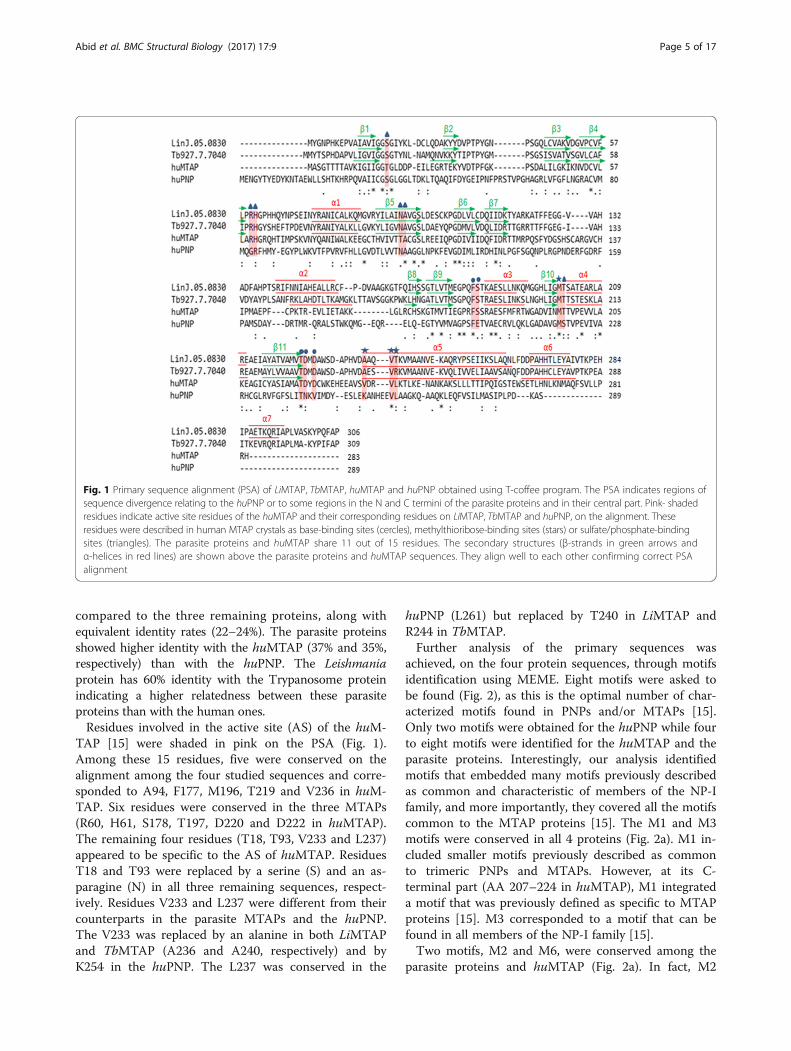
Fig. 1 Primary sequence alignment (PSA) of LiMTAP, TbMTAP, huMTAP and huPNP obtained using T-coffee program. The PSA indicates regions ofsequence divergence relating to the huPNP or to some regions in the N and C termini of the parasite proteins and in their central part. Pink- shadedresidues indicate active site residues of the huMTAP and their corresponding residues on LiMTAP, TbMTAP and huPNP, on the alignment. Theseresidues were described in human MTAP crystals as base-binding sites (cercles), methylthioribose-binding sites (stars) or sulfate/phosphate-bindingsites (triangles). The parasite proteins and huMTAP share 11 out of 15 residues. The secondary structures (β-strands in green arrows andα-helices in red lines) are shown above the parasite proteins and huMTAP sequences. They align well to each other confirming correct PSAalignment
Abid et al. BMC Structural Biology (2017) 17:9 Page 5 of 17
compared to the three remaining proteins, along withequivalent identity rates (22–24%). The parasite proteinsshowed higher identity with the huMTAP (37% and 35%,respectively) than with the huPNP. The Leishmaniaprotein has 60% identity with the Trypanosome proteinindicating a higher relatedness between these parasiteproteins than with the human ones.Residues involved in the active site (AS) of the huM-
TAP [15] were shaded in pink on the PSA (Fig. 1).Among these 15 residues, five were conserved on thealignment among the four studied sequences and corre-sponded to A94, F177, M196, T219 and V236 in huM-TAP. Six residues were conserved in the three MTAPs(R60, H61, S178, T197, D220 and D222 in huMTAP).The remaining four residues (T18, T93, V233 and L237)appeared to be specific to the AS of huMTAP. ResiduesT18 and T93 were replaced by a serine (S) and an as-paragine (N) in all three remaining sequences, respect-ively. Residues V233 and L237 were different from theircounterparts in the parasite MTAPs and the huPNP.The V233 was replaced by an alanine in both LiMTAPand TbMTAP (A236 and A240, respectively) and byK254 in the huPNP. The L237 was conserved in the
huPNP (L261) but replaced by T240 in LiMTAP andR244 in TbMTAP.Further analysis of the primary sequences was
achieved, on the four protein sequences, through motifsidentification using MEME. Eight motifs were asked tobe found (Fig. 2), as this is the optimal number of char-acterized motifs found in PNPs and/or MTAPs [15].Only two motifs were obtained for the huPNP while fourto eight motifs were identified for the huMTAP and theparasite proteins. Interestingly, our analysis identifiedmotifs that embedded many motifs previously describedas common and characteristic of members of the NP-Ifamily, and more importantly, they covered all the motifscommon to the MTAP proteins [15]. The M1 and M3motifs were conserved in all 4 proteins (Fig. 2a). M1 in-cluded smaller motifs previously described as commonto trimeric PNPs and MTAPs. However, at its C-terminal part (AA 207–224 in huMTAP), M1 integrateda motif that was previously defined as specific to MTAPproteins [15]. M3 corresponded to a motif that can befound in all members of the NP-I family [15].Two motifs, M2 and M6, were conserved among the
parasite proteins and huMTAP (Fig. 2a). In fact, M2
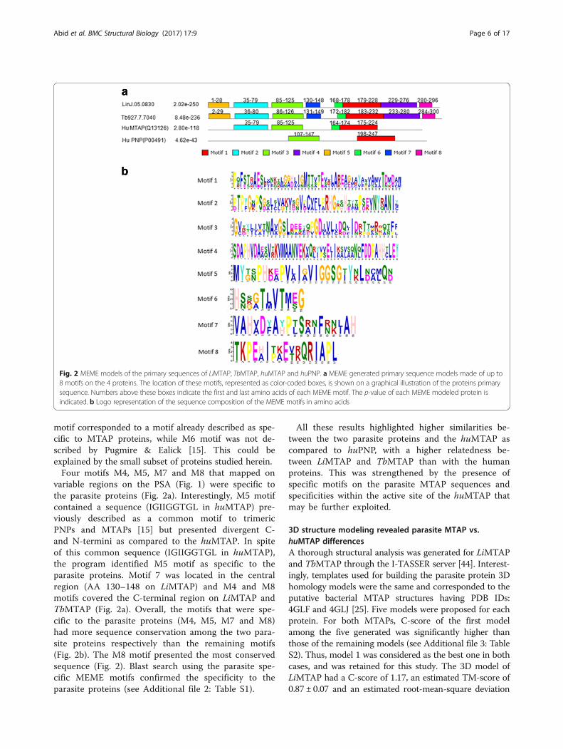
Fig. 2 MEME models of the primary sequences of LiMTAP, TbMTAP, huMTAP and huPNP. a MEME generated primary sequence models made of up to8 motifs on the 4 proteins. The location of these motifs, represented as color-coded boxes, is shown on a graphical illustration of the proteins primarysequence. Numbers above these boxes indicate the first and last amino acids of each MEME motif. The p-value of each MEME modeled protein isindicated. b Logo representation of the sequence composition of the MEME motifs in amino acids
Abid et al. BMC Structural Biology (2017) 17:9 Page 6 of 17
motif corresponded to a motif already described as spe-cific to MTAP proteins, while M6 motif was not de-scribed by Pugmire & Ealick [15]. This could beexplained by the small subset of proteins studied herein.Four motifs M4, M5, M7 and M8 that mapped on
variable regions on the PSA (Fig. 1) were specific tothe parasite proteins (Fig. 2a). Interestingly, M5 motifcontained a sequence (IGIIGGTGL in huMTAP) pre-viously described as a common motif to trimericPNPs and MTAPs [15] but presented divergent C-and N-termini as compared to the huMTAP. In spiteof this common sequence (IGIIGGTGL in huMTAP),the program identified M5 motif as specific to theparasite proteins. Motif 7 was located in the centralregion (AA 130–148 on LiMTAP) and M4 and M8motifs covered the C-terminal region on LiMTAP andTbMTAP (Fig. 2a). Overall, the motifs that were spe-cific to the parasite proteins (M4, M5, M7 and M8)had more sequence conservation among the two para-site proteins respectively than the remaining motifs(Fig. 2b). The M8 motif presented the most conservedsequence (Fig. 2). Blast search using the parasite spe-cific MEME motifs confirmed the specificity to theparasite proteins (see Additional file 2: Table S1).
All these results highlighted higher similarities be-tween the two parasite proteins and the huMTAP ascompared to huPNP, with a higher relatedness be-tween LiMTAP and TbMTAP than with the humanproteins. This was strengthened by the presence ofspecific motifs on the parasite MTAP sequences andspecificities within the active site of the huMTAP thatmay be further exploited.
3D structure modeling revealed parasite MTAP vs.huMTAP differencesA thorough structural analysis was generated for LiMTAPand TbMTAP through the I-TASSER server [44]. Interest-ingly, templates used for building the parasite protein 3Dhomology models were the same and corresponded to theputative bacterial MTAP structures having PDB IDs:4GLF and 4GLJ [25]. Five models were proposed for eachprotein. For both MTAPs, C-score of the first modelamong the five generated was significantly higher thanthose of the remaining models (see Additional file 3: TableS2). Thus, model 1 was considered as the best one in bothcases, and was retained for this study. The 3D model ofLiMTAP had a C-score of 1.17, an estimated TM-score of0.87 ± 0.07 and an estimated root-mean-square deviation
Abid et al. BMC Structural Biology (2017) 17:9 Page 7 of 17
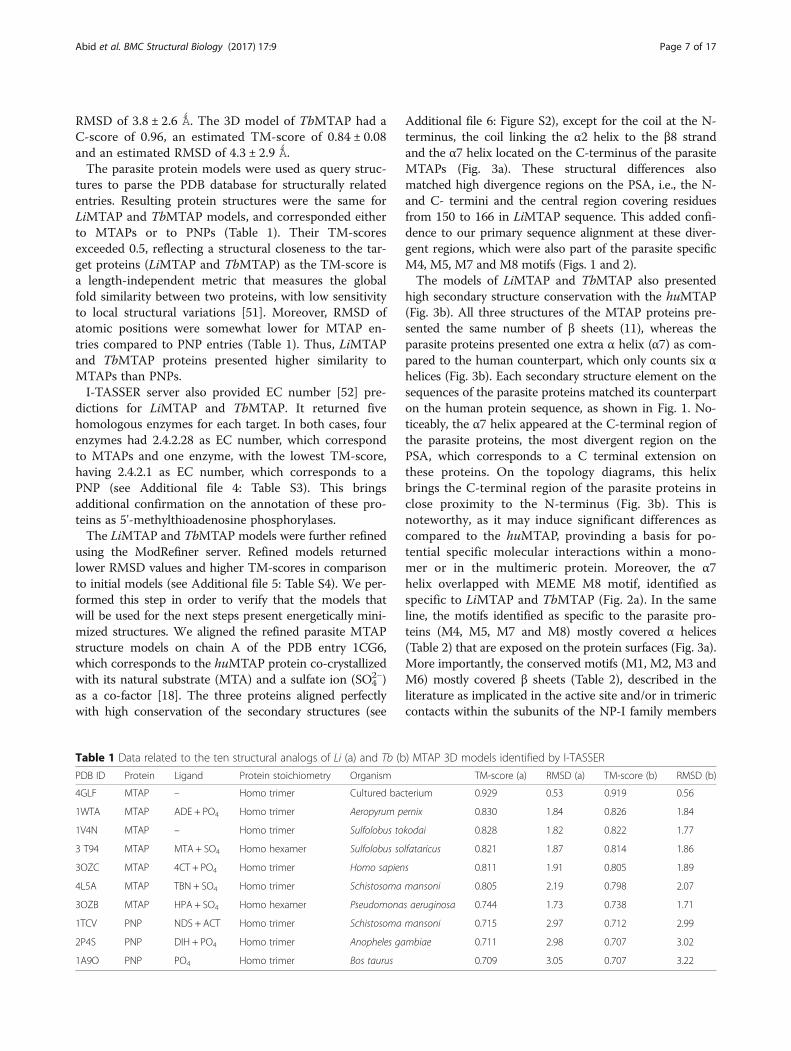
RMSD of 3.8 ± 2.6 Ǻ. The 3D model of TbMTAP had aC-score of 0.96, an estimated TM-score of 0.84 ± 0.08and an estimated RMSD of 4.3 ± 2.9 Ǻ.The parasite protein models were used as query struc-
tures to parse the PDB database for structurally relatedentries. Resulting protein structures were the same forLiMTAP and TbMTAP models, and corresponded eitherto MTAPs or to PNPs (Table 1). Their TM-scoresexceeded 0.5, reflecting a structural closeness to the tar-get proteins (LiMTAP and TbMTAP) as the TM-score isa length-independent metric that measures the globalfold similarity between two proteins, with low sensitivityto local structural variations [51]. Moreover, RMSD ofatomic positions were somewhat lower for MTAP en-tries compared to PNP entries (Table 1). Thus, LiMTAPand TbMTAP proteins presented higher similarity toMTAPs than PNPs.I-TASSER server also provided EC number [52] pre-
dictions for LiMTAP and TbMTAP. It returned fivehomologous enzymes for each target. In both cases, fourenzymes had 2.4.2.28 as EC number, which correspondto MTAPs and one enzyme, with the lowest TM-score,having 2.4.2.1 as EC number, which corresponds to aPNP (see Additional file 4: Table S3). This bringsadditional confirmation on the annotation of these pro-teins as 5'-methylthioadenosine phosphorylases.The LiMTAP and TbMTAP models were further refined
using the ModRefiner server. Refined models returnedlower RMSD values and higher TM-scores in comparisonto initial models (see Additional file 5: Table S4). We per-formed this step in order to verify that the models thatwill be used for the next steps present energetically mini-mized structures. We aligned the refined parasite MTAPstructure models on chain A of the PDB entry 1CG6,which corresponds to the huMTAP protein co-crystallizedwith its natural substrate (MTA) and a sulfate ion (SO4
2−)as a co-factor [18]. The three proteins aligned perfectlywith high conservation of the secondary structures (see
Table 1 Data related to the ten structural analogs of Li (a) and Tb (b
PDB ID Protein Ligand Protein stoichiometry Organism
4GLF MTAP – Homo trimer Cultured bac
1WTA MTAP ADE + PO4 Homo trimer Aeropyrum p
1V4N MTAP – Homo trimer Sulfolobus to
3 T94 MTAP MTA + SO4 Homo hexamer Sulfolobus so
3OZC MTAP 4CT + PO4 Homo trimer Homo sapien
4L5A MTAP TBN + SO4 Homo trimer Schistosoma
3OZB MTAP HPA + SO4 Homo hexamer Pseudomona
1TCV PNP NDS + ACT Homo trimer Schistosoma
2P4S PNP DIH + PO4 Homo trimer Anopheles ga
1A9O PNP PO4 Homo trimer Bos taurus
Additional file 6: Figure S2), except for the coil at the N-terminus, the coil linking the α2 helix to the β8 strandand the α7 helix located on the C-terminus of the parasiteMTAPs (Fig. 3a). These structural differences alsomatched high divergence regions on the PSA, i.e., the N-and C- termini and the central region covering residuesfrom 150 to 166 in LiMTAP sequence. This added confi-dence to our primary sequence alignment at these diver-gent regions, which were also part of the parasite specificM4, M5, M7 and M8 motifs (Figs. 1 and 2).The models of LiMTAP and TbMTAP also presented
high secondary structure conservation with the huMTAP(Fig. 3b). All three structures of the MTAP proteins pre-sented the same number of β sheets (11), whereas theparasite proteins presented one extra α helix (α7) as com-pared to the human counterpart, which only counts six αhelices (Fig. 3b). Each secondary structure element on thesequences of the parasite proteins matched its counterparton the human protein sequence, as shown in Fig. 1. No-ticeably, the α7 helix appeared at the C-terminal region ofthe parasite proteins, the most divergent region on thePSA, which corresponds to a C terminal extension onthese proteins. On the topology diagrams, this helixbrings the C-terminal region of the parasite proteins inclose proximity to the N-terminus (Fig. 3b). This isnoteworthy, as it may induce significant differences ascompared to the huMTAP, provinding a basis for po-tential specific molecular interactions within a mono-mer or in the multimeric protein. Moreover, the α7helix overlapped with MEME M8 motif, identified asspecific to LiMTAP and TbMTAP (Fig. 2a). In the sameline, the motifs identified as specific to the parasite pro-teins (M4, M5, M7 and M8) mostly covered α helices(Table 2) that are exposed on the protein surfaces (Fig. 3a).More importantly, the conserved motifs (M1, M2, M3 andM6) mostly covered β sheets (Table 2), described in theliterature as implicated in the active site and/or in trimericcontacts within the subunits of the NP-I family members
) MTAP 3D models identified by I-TASSER
TM-score (a) RMSD (a) TM-score (b) RMSD (b)
terium 0.929 0.53 0.919 0.56
ernix 0.830 1.84 0.826 1.84
kodai 0.828 1.82 0.822 1.77
lfataricus 0.821 1.87 0.814 1.86
s 0.811 1.91 0.805 1.89
mansoni 0.805 2.19 0.798 2.07
s aeruginosa 0.744 1.73 0.738 1.71
mansoni 0.715 2.97 0.712 2.99
mbiae 0.711 2.98 0.707 3.02
0.709 3.05 0.707 3.22
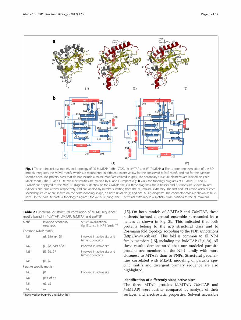
Table 2 Functional or structural correlation of MEME sequencemotifs found in huMTAP, LiMTAP, TbMTAP and huPNP
Motif Involved secondarystructures
Structural/functionalsignificance in NP-I family (a)
Common MTAP motifs
M1 α3, β10, α4, β11 Involved in active site andtrimeric contacts
M2 β3, β4, part of α1 Involved in active site
M3 β5, β6, β7 Involved in active site andtrimeric contacts
M6 β8, β9
Parasite specific motifs
M5 β1 Involved in active site
M7 part of α2
M4 α5, α6
M8 α7(a)Reviewed by Pugmire and Ealick [15]
Fig. 3 Three- dimensional models and topology of (1) huMTAP (pdb: 1CG6), (2) LiMTAP and (3) TbMTAP. a The cartoon representation of the 3Dmodels integrates the MEME motifs, which are represented in different colors: yellow for the conserved MEME motifs and red for the parasitespecific ones. The protein parts that do not include a MEME motif are colored in grey. The secondary structure elements are labeled on eachMTAP model. The N- and C- terminal extremities are marked by N and C, respectively. b Only the topology diagrams of (1) huMTAP and (2)LiMTAP are displayed as the TbMTAP diagram is identical to the LiMTAP one. On these diagrams, the α-helices and β-strands are shown by redcylinders and blue arrows, respectively, and are labeled by numbers starting from the N- terminal extremity. The first and last amino acids of eachsecondary structure are shown on the corresponding shape, on both huMTAP (1) and LiMTAP (2) diagrams. The connector coils are shown as bluelines. On the parasite protein topology diagrams, the α7 helix brings the C- terminal extremity in a spatially close position to the N- terminus
Abid et al. BMC Structural Biology (2017) 17:9 Page 8 of 17
[15]. On both models of LiMTAP and TbMTAP, theseβ sheets formed a central ensemble surrounded by αhelices as shown in Fig. 3b. This indicated that bothproteins belong to the α/β structural class and toRossmann fold topology according to the PDB annotations(http://www.rcsb.org). This fold is common to all NP-Ifamily members [15], including the huMTAP (Fig. 3a). Allthese results demonstrated that our modeled parasiteproteins are members of the NP-I family with morecloseness to MTAPs than to PNPs. Structural peculiar-ities correlated with MEME modeling of parasite spe-cific motifs and divergent primary sequence are alsohighlighted.
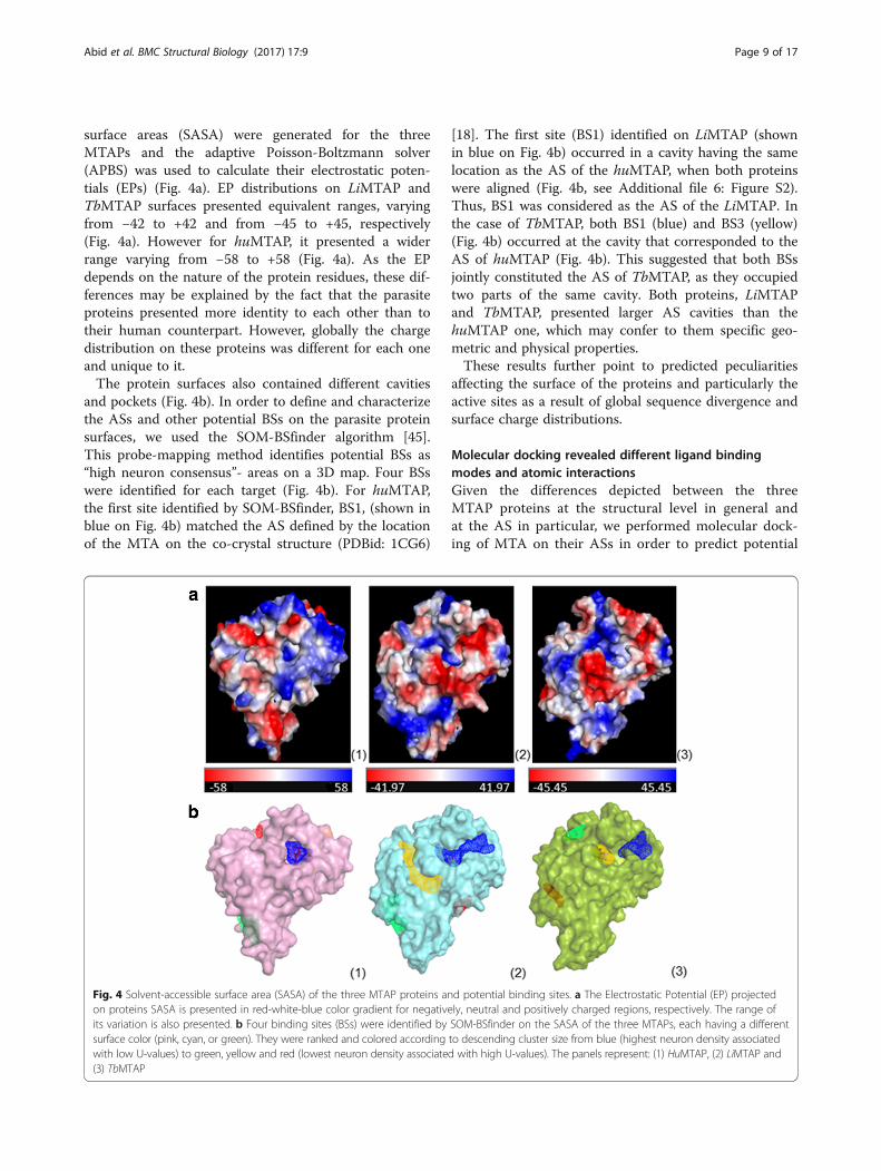
Identification of differently sized active sitesThe three MTAP proteins (LiMTAP, TbMTAP andhuMTAP) were further compared by analysis of theirsurfaces and electrostatic properties. Solvent accessible
Abid et al. BMC Structural Biology (2017) 17:9 Page 9 of 17
surface areas (SASA) were generated for the threeMTAPs and the adaptive Poisson-Boltzmann solver(APBS) was used to calculate their electrostatic poten-tials (EPs) (Fig. 4a). EP distributions on LiMTAP andTbMTAP surfaces presented equivalent ranges, varyingfrom −42 to +42 and from −45 to +45, respectively(Fig. 4a). However for huMTAP, it presented a widerrange varying from −58 to +58 (Fig. 4a). As the EPdepends on the nature of the protein residues, these dif-ferences may be explained by the fact that the parasiteproteins presented more identity to each other than totheir human counterpart. However, globally the chargedistribution on these proteins was different for each oneand unique to it.The protein surfaces also contained different cavities
and pockets (Fig. 4b). In order to define and characterizethe ASs and other potential BSs on the parasite proteinsurfaces, we used the SOM-BSfinder algorithm [45].This probe-mapping method identifies potential BSs as“high neuron consensus”- areas on a 3D map. Four BSswere identified for each target (Fig. 4b). For huMTAP,the first site identified by SOM-BSfinder, BS1, (shown inblue on Fig. 4b) matched the AS defined by the locationof the MTA on the co-crystal structure (PDBid: 1CG6)
Fig. 4 Solvent-accessible surface area (SASA) of the three MTAP proteins anon proteins SASA is presented in red-white-blue color gradient for negativeits variation is also presented. b Four binding sites (BSs) were identified bysurface color (pink, cyan, or green). They were ranked and colored accordingwith low U-values) to green, yellow and red (lowest neuron density associate(3) TbMTAP
[18]. The first site (BS1) identified on LiMTAP (shownin blue on Fig. 4b) occurred in a cavity having the samelocation as the AS of the huMTAP, when both proteinswere aligned (Fig. 4b, see Additional file 6: Figure S2).Thus, BS1 was considered as the AS of the LiMTAP. Inthe case of TbMTAP, both BS1 (blue) and BS3 (yellow)(Fig. 4b) occurred at the cavity that corresponded to theAS of huMTAP (Fig. 4b). This suggested that both BSsjointly constituted the AS of TbMTAP, as they occupiedtwo parts of the same cavity. Both proteins, LiMTAPand TbMTAP, presented larger AS cavities than thehuMTAP one, which may confer to them specific geo-metric and physical properties.These results further point to predicted peculiarities
affecting the surface of the proteins and particularly theactive sites as a result of global sequence divergence andsurface charge distributions.
Molecular docking revealed different ligand bindingmodes and atomic interactionsGiven the differences depicted between the threeMTAP proteins at the structural level in general andat the AS in particular, we performed molecular dock-ing of MTA on their ASs in order to predict potential
d potential binding sites. a The Electrostatic Potential (EP) projectedly, neutral and positively charged regions, respectively. The range ofSOM-BSfinder on the SASA of the three MTAPs, each having a differentto descending cluster size from blue (highest neuron density associatedd with high U-values). The panels represent: (1) HuMTAP, (2) LiMTAP and
Abid et al. BMC Structural Biology (2017) 17:9 Page 10 of 17
impact on the binding modes and interactions withligands.Docking of MTA on the huMTAP was performed on
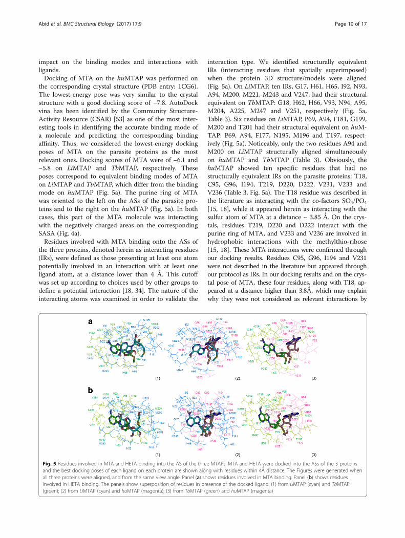
the corresponding crystal structure (PDB entry: 1CG6).The lowest-energy pose was very similar to the crystalstructure with a good docking score of −7.8. AutoDockvina has been identified by the Community Structure-Activity Resource (CSAR) [53] as one of the most inter-esting tools in identifying the accurate binding mode ofa molecule and predicting the corresponding bindingaffinity. Thus, we considered the lowest-energy dockingposes of MTA on the parasite proteins as the mostrelevant ones. Docking scores of MTA were of −6.1 and−5.8 on LiMTAP and TbMTAP, respectively. Theseposes correspond to equivalent binding modes of MTAon LiMTAP and TbMTAP, which differ from the bindingmode on huMTAP (Fig. 5a). The purine ring of MTAwas oriented to the left on the ASs of the parasite pro-teins and to the right on the huMTAP (Fig. 5a). In bothcases, this part of the MTA molecule was interactingwith the negatively charged areas on the correspondingSASA (Fig. 4a).Residues involved with MTA binding onto the ASs of
the three proteins, denoted herein as interacting residues(IRs), were defined as those presenting at least one atompotentially involved in an interaction with at least oneligand atom, at a distance lower than 4 Ǻ. This cutoffwas set up according to choices used by other groups todefine a potential interaction [18, 34]. The nature of theinteracting atoms was examined in order to validate the
Fig. 5 Residues involved in MTA and HETA binding into the AS of the threand the best docking poses of each ligand on each protein are shown aloall three proteins were aligned, and from the same view angle. Panel (a) shinvolved in HETA binding. The panels show superposition of residues in pr(green); (2) from LiMTAP (cyan) and huMTAP (magenta); (3) from TbMTAP (g
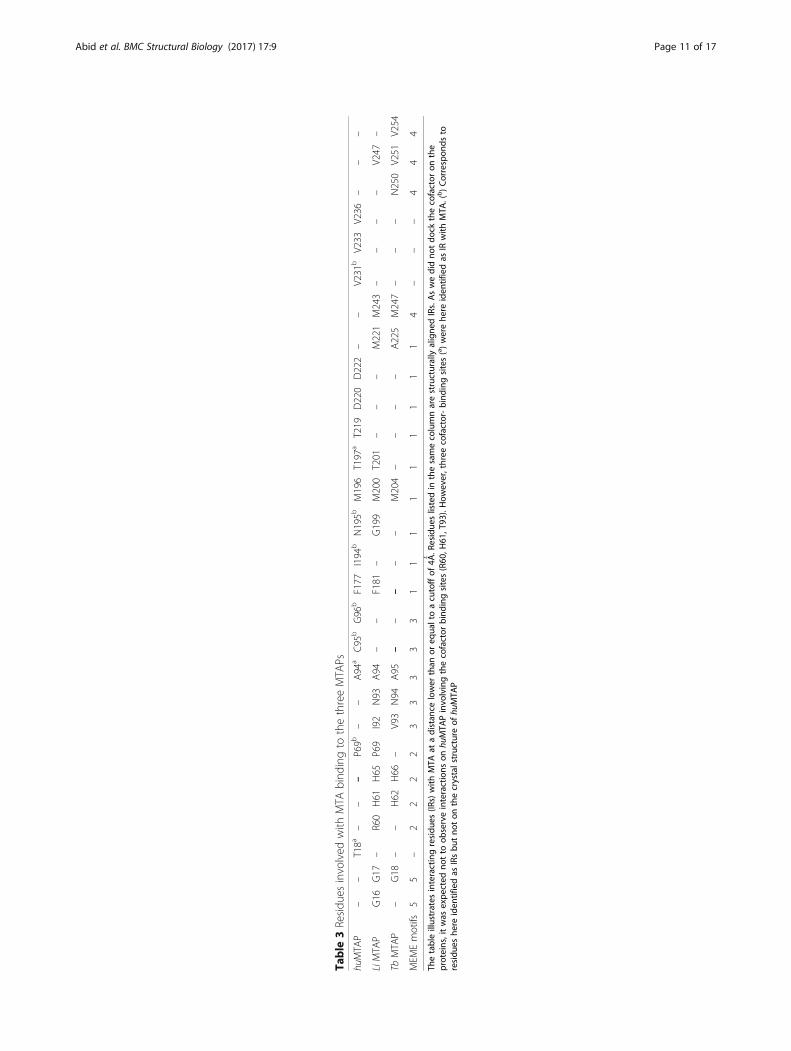
interaction type. We identified structurally equivalentIRs (interacting residues that spatially superimposed)when the protein 3D structure/models were aligned(Fig. 5a). On LiMTAP, ten IRs, G17, H61, H65, I92, N93,A94, M200, M221, M243 and V247, had their structuralequivalent on TbMTAP: G18, H62, H66, V93, N94, A95,M204, A225, M247 and V251, respectively (Fig. 5a,Table 3). Six residues on LiMTAP, P69, A94, F181, G199,M200 and T201 had their structural equivalent on huM-TAP: P69, A94, F177, N195, M196 and T197, respect-ively (Fig. 5a). Noticeably, only the two residues A94 andM200 on LiMTAP structurally aligned simultaneouslyon huMTAP and TbMTAP (Table 3). Obviously, thehuMTAP showed ten specific residues that had nostructurally equivalent IRs on the parasite proteins: T18,C95, G96, I194, T219, D220, D222, V231, V233 andV236 (Table 3, Fig. 5a). The T18 residue was described inthe literature as interacting with the co-factors SO4/PO4
[15, 18], while it appeared herein as interacting with thesulfur atom of MTA at a distance ~ 3.85 Ǻ. On the crys-tals, residues T219, D220 and D222 interact with thepurine ring of MTA, and V233 and V236 are involved inhydrophobic interactions with the methylthio-ribose[15, 18]. These MTA interactions were confirmed throughour docking results. Residues C95, G96, I194 and V231were not described in the literature but appeared throughour protocol as IRs. In our docking results and on the crys-tal pose of MTA, these four residues, along with T18, ap-peared at a distance higher than 3.8Ǻ, which may explainwhy they were not considered as relevant interactions by
e MTAPs. MTA and HETA were docked into the ASs of the 3 proteinsng with residues within 4Ǻ distance. The Figures were generated whenows residues involved in MTA binding. Panel (b) shows residuesesence of the docked ligand: (1) from LiMTAP (cyan) and TbMTAPreen) and huMTAP (magenta)
Table
3Residu
esinvolved
with
MTA
bind
ingto
thethreeMTA
Ps
huMTA
P–
–T18a
––
–P69b
––
A94
aC95
bG96
bF177
I194
bN195b
M196
T197
aT219
D220
D222
––
V231
bV233
V236
––
–
LiMTA
PG16
G17
–R60
H61
H65
P69
I92
N93
A94
––
F181
–G199
M200
T201
––
–M221
M243
––
––
V247
–
TbMTA
P–
G18
––
H62
H66
–V93
N94
A95
––
––
–M204
––
––
A225
M247
––
–N250
V251
V254
MEM
Emotifs
55
–2
22
23
33
33
11
11
11
11
14
––
–4
44
Thetableillustrates
interactingresidu
es(IR
s)with
MTA
atadistan
celower
than
oreq
ualtoacutoffof
4Ǻ.R
esidue
slistedin
thesamecolumnarestructurally
aligne
dIRs.Aswedidno
tdo
ckthecofactor
onthe
proteins,itwas
expe
cted
notto
observeinteractions
onhu
MTA
Pinvo
lvingthecofactor
bind
ingsites(R60
,H61
,T93
).How
ever,three
cofactor-bind
ingsites(a)werehe
reiden
tifiedas
IRwith
MTA
.(b)Correspon
dsto
residu
eshe
reiden
tifiedas
IRsbu
tno
ton
thecrystalstructure
ofhu
MTA
P
Abid et al. BMC Structural Biology (2017) 17:9 Page 11 of 17
Abid et al. BMC Structural Biology (2017) 17:9 Page 12 of 17
the crystallographers [15, 18]. Herein, we considered allpossible interactions as relevant in order to minimize falsenegatives.On our model, the residues of huMTAP interacting
with the purine ring of MTA were F177, T219, D220and D222, which is consistent with literature [18]. Ex-cept for F177, these residues had no structurally equiva-lent IRs on the parasite proteins (Table 3) although theymatched perfectly well on the primary sequence align-ment. The residues interacting with the purine ring ofMTA were G16, G17, R60, H65, I92, N93, A94, M221,M243 and V247, in LiMTAP and G18, V93, N94, A95,A225, M247, N250, V251 and V254, in TbMTAP(Fig. 5a). Of these residues, I92, M221, M243 and V247on LiMTAP and their counterparts (V93, A225, M247and V251) on TbMTAP appeared to establish hydropho-bic interactions with the two rings of the purine moietyof MTA (Fig. 5a). This type of hydrophobic pocket/inter-actions have been depicted with other NP-I family mem-bers, namely E. coli Uridine Phosphorylase [54, 55].Interestingly, the residue M196 in huMTAP was inter-acting with the ribose ring of MTA [18]. Its structuralcounterparts in LiMTAP (M200) similarly established ahydrogen bond with the 2′- hydroxyl of the ribose(Fig. 5a). However, their trypanosomal counterpart(M204) was involved in hydrophobic interactions withthe 5′-methylthioribose part of MTA (Fig. 5a).In a second step, we docked HETA, a specific in-
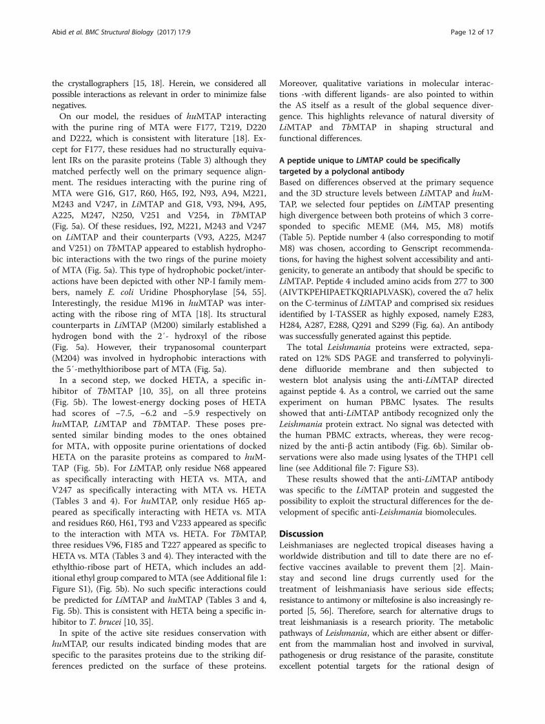
hibitor of TbMTAP [10, 35], on all three proteins(Fig. 5b). The lowest-energy docking poses of HETAhad scores of −7.5, −6.2 and −5.9 respectively onhuMTAP, LiMTAP and TbMTAP. These poses pre-sented similar binding modes to the ones obtainedfor MTA, with opposite purine orientations of dockedHETA on the parasite proteins as compared to huM-TAP (Fig. 5b). For LiMTAP, only residue N68 appearedas specifically interacting with HETA vs. MTA, andV247 as specifically interacting with MTA vs. HETA(Tables 3 and 4). For huMTAP, only residue H65 ap-peared as specifically interacting with HETA vs. MTAand residues R60, H61, T93 and V233 appeared as specificto the interaction with MTA vs. HETA. For TbMTAP,three residues V96, F185 and T227 appeared as specific toHETA vs. MTA (Tables 3 and 4). They interacted with theethylthio-ribose part of HETA, which includes an add-itional ethyl group compared toMTA (see Additional file 1:Figure S1), (Fig. 5b). No such specific interactions couldbe predicted for LiMTAP and huMTAP (Tables 3 and 4,Fig. 5b). This is consistent with HETA being a specific in-hibitor to T. brucei [10, 35].In spite of the active site residues conservation with
huMTAP, our results indicated binding modes that arespecific to the parasites proteins due to the striking dif-ferences predicted on the surface of these proteins.
Moreover, qualitative variations in molecular interac-tions -with different ligands- are also pointed to withinthe AS itself as a result of the global sequence diver-gence. This highlights relevance of natural diversity ofLiMTAP and TbMTAP in shaping structural andfunctional differences.
A peptide unique to LiMTAP could be specificallytargeted by a polyclonal antibodyBased on differences observed at the primary sequenceand the 3D structure levels between LiMTAP and huM-TAP, we selected four peptides on LiMTAP presentinghigh divergence between both proteins of which 3 corre-sponded to specific MEME (M4, M5, M8) motifs(Table 5). Peptide number 4 (also corresponding to motifM8) was chosen, according to Genscript recommenda-tions, for having the highest solvent accessibility and anti-genicity, to generate an antibody that should be specific toLiMTAP. Peptide 4 included amino acids from 277 to 300(AIVTKPEHIPAETKQRIAPLVASK), covered the α7 helixon the C-terminus of LiMTAP and comprised six residuesidentified by I-TASSER as highly exposed, namely E283,H284, A287, E288, Q291 and S299 (Fig. 6a). An antibodywas successfully generated against this peptide.The total Leishmania proteins were extracted, sepa-
rated on 12% SDS PAGE and transferred to polyvinyli-dene difluoride membrane and then subjected towestern blot analysis using the anti-LiMTAP directedagainst peptide 4. As a control, we carried out the sameexperiment on human PBMC lysates. The resultsshowed that anti-LiMTAP antibody recognized only theLeishmania protein extract. No signal was detected withthe human PBMC extracts, whereas, they were recog-nized by the anti-β actin antibody (Fig. 6b). Similar ob-servations were also made using lysates of the THP1 cellline (see Additional file 7: Figure S3).These results showed that the anti-LiMTAP antibody
was specific to the LiMTAP protein and suggested thepossibility to exploit the structural differences for the de-velopment of specific anti-Leishmania biomolecules.
DiscussionLeishmaniases are neglected tropical diseases having aworldwide distribution and till to date there are no ef-fective vaccines available to prevent them [2]. Main-stay and second line drugs currently used for thetreatment of leishmaniasis have serious side effects;resistance to antimony or miltefosine is also increasingly re-ported [5, 56]. Therefore, search for alternative drugs totreat leishmaniasis is a research priority. The metabolicpathways of Leishmania, which are either absent or differ-ent from the mammalian host and involved in survival,pathogenesis or drug resistance of the parasite, constituteexcellent potential targets for the rational design of
Table
4Residu
esinvolved
with
HETAbind
ingto
thethreeMTA
Ps
huMTA
P–
–T18
––
H65
–P69
––
A94
C95
G96
F177
I194
N195
M196
T197
T219
D220
D222
––
–V231
V236
––
–
LiMTA
PG16
G17
–R60
H61
H65
N68
P69
I92
N93
A94
––
F181
–G199
M200
T201
––
–M221
–M243
––
––
–
TbMTA
P–
G18
––
H62
H66
––
V93
N94
A95
V96
–F1
85–
–M204
––
––
A225
T227
M247
––
N250
V251
V254
MEM
Emotifs
55
–2
22
22
33
33
31
11
11
11
11
14
––
44
4
Thetableillustrates
interactingresidu
eswith
HETAat
adistan
celower
than
oreq
ualtoacutoffof
4Ǻ.R
esidue
slistedin
thesamecolumnarestructurally
aligne
dIRs.Re
sidu
eswereob
served
that
interact
inthethree
MTA
Pswith
both
MTA
andHETA.R
esidue
sspecifically
invo
lved
with
HETAbind
ing(vs.MTA
)areshow
nin
bold
Abid et al. BMC Structural Biology (2017) 17:9 Page 13 of 17

Table 5 List of the four peptides selected in silico on LiMTAPand their correlation to MEME motifs
Peptides Amino acids sequence (position) MEME motifs
1 MYGNPHKEPVAIAV (1–14) M5
2 HEALLRCFPDVAAGKGTFQIH (148–168) –
3 DAPHVDAAQVTKV (230–242) M4
4 AIVTKPEHIPAETKQRIAPLVASK (277–300) M8
Abid et al. BMC Structural Biology (2017) 17:9 Page 14 of 17
antiparasitic drugs [8, 57]. Several reports have shown thattargeting the polyamine biosynthesis and purine salvage en-zymes against Trypanosomatidae have yielded promisingresults [10, 14, 35, 58]. Among these enzymes, MTAP playsa crucial role in purine and polyamine metabolism and inthe methionine salvage pathway [10]. Selective inhibitors ofthe T. brucei enzyme have been described, and theyshowed an in vitro cytotoxicity (IC50) of 10 nM for the 5′-deoxy-5′-(hydroxyethylthio)-tubercidin and high curerates (70% to 90%) of HETA when administrated to miceinfected with T. brucei [10, 35]. However, to our know-ledge, in Leishmania, an organism phylogenetically closeto Trypanosoma, the protein MTAP was not consideredso far as a potential drug target and was not characterizedyet. Given the role of this protein in humans and its puta-tive functional conservation in the studied parasites, itseemed important to characterize the protein in Leish-mania infantum, a pathogen that causes visceral leish-maniasis in North Africa, Europe, Asian countries andLatin America. Indeed the knowledge of the 3D structureof a drug target protein is of great importance to conductstructure-based drug discovery. Such characterization al-lows confirming putative roles and identification of
Fig. 6 Characterization of the polyclonal antibody directed against an antigeantigenic peptide unique to LiMTAP. The peptide includes amino acids 277 tothrough I-TASSER (E283, H284, A287, E288, Q291, S299) are colored in brown.respectively the LiMTAP antibody, and the β-actin antibody as a control for huhuman PBMC were resolved on 12% SDS-PAGE gel, transferred to PVDF mem(1/10000) or anti-β-actin (1/5000) antibodies. The Figure is representative of th(Vivantis, CA, USA); (2) Human PBMC lysates; (3), (4) and (5): 3, 7 and 15 microg
commonalities as well as specific features to an organism,and thus assessing whether natural diversity of such con-served proteins could make them potentially good candi-dates for drug design. When a protein structure is not orcould not be resolved experimentally, homology modelingis one of the most powerful tools to obtain robust modelsof a protein structure [59, 60].Bioinformatics approaches were used to bring insights
into the trypanosomal sirtuin structure and functionfrom L. major, L. infantum, T. brucei and T. cruzi. Struc-ture comparisons with the human protein and moleculardocking permitted to highlight specificities that were ofinterest in predicting specific/selective inhibitors [61].The Leishmania elongation factor alpha, sharing 82% ofidentity with its mammalian orthologue, was also suc-cessfully used in identifying novel anti-Leishmaniamolecules in silico [62]. In spite of this high identity rate,selective inhibitors could be identified in silico that tar-geted a unique structural feature on the parasite protein,resulting from a 12 amino acids long deletion [62].Herein, we used homology modeling to generate the 3Dstructure models of LiMTAP and TbMTAP and comparethem to other MTAP structures of relevance like inhumans. For this purpose, 3D models of the parasiteMTAPs were performed by I-TASSER, matching struc-ture predictions with known functional templates [41].This homology modeling server was already used byother groups for instance to understand functions ofhuman thiol dioxygenase enzymes [63] and to assess sta-bility of the Rabies Virus G protein trimer through mo-lecular dynamics [64].Through sequence and structure comparison of the
putative L. infantum MTAP protein with the human and
nic C-terminal peptide of LiMTAP. a C-terminal location of the surface300, colored in yellow or brown. Residues identified as highly exposed
b Specificity of the C-terminal Li-peptide was tested by western blot usingman PBMC lysates. The total proteins extracted from Leishmania andbrane and then subjected to western blot analysis using anti-LiMTAPree independent experiments. Lanes: (1) Prestained marker MW in kDarams of L. infantum (LV50) promastigote lysates, respectively
Abid et al. BMC Structural Biology (2017) 17:9 Page 15 of 17
T. brucei counterparts, we could depict significant globalsimilarities. Important sequence identity rates, level ofactive site residues conservation, presence of commonMEME motifs to NP-I family members, similar 3Dtopology, and I-TASSER functional annotations andEC number predictions (2.4.2.28) consolidated the hy-pothesis that the Leishmania protein is a 5′-methylthioadenosine phosphorylase. However, in spiteof these commonalities shared by the three proteins,it was possible to identify specific structural featuresthat were congruent with divergence on the primarysequence, itself underlying specific MEME modeledmotifs. Relevance of such structural specificities wasfurther confirmed by the identification of a highlyantigenic and exposed peptide among four structurallydivergent regions/peptides corresponding to one ofthese specific motifs. Notably, a polyclonal antibodydirected against this peptide at the C-terminus provedto recognize specifically the Leishmania protein (andso did not react with human cell extracts).Primary sequence divergence, reflected on surface EP
and BS predictions, brought additional information aboutpeculiarities of the parasite protein models. Notably, thepredicted active sites (located on similar parts of the pro-teins) presented different shapes and volumes thatprompted looking at the protein-ligand interactions. In-deed, these are of high importance towards a comprehen-sive study of enzymes in general and drug targets inparticular [24, 61, 65]. Molecular docking of MTA, thenatural substrate, and HETA, a well characterized inhibi-tor in T. brucei, into the AS of the three proteins showedequivalent docking scores between MTA and HETA andlower docking scores (better free energy of binding) onthe human target as compared to the parasitic counter-parts. This could be due to the fact that docking simula-tions were performed on a crystal structure of thehuMTAP, which corresponds to the biologically optimalconformation for ligand binding. Molecular docking ofboth MTA and HETA highlighted differential bindingmodes where the purine ring occupied opposite positionin parasite MTAPs to the one in the human protein. Theinteractions into these pockets defined within a 4 Ǻ range,corresponding to hydrogen bonds or hydrophobic interac-tions, also revealed qualitative differences at differentlevels in each comparison. Notably, the docking of MTAmolecule predicted specific IRs in huMTAP that had nostructural equivalent IRs on the parasite MTAPs. More-over, the docked MTA into both LiMTAP and TbMTAPinvolved less hydrophobic IRs with the methylthio partand much more interactions with the purine than thoseseen in huMTAP. In addition, the interactions with theethyl group of HETA were characterized by three residues(V96, F185 and T227), only seen in TbMTAP and not inits Leishmania and human counterparts. Noticeably, most
residues that are unique to Leishmania or Trypanosomaprotein belong to kinetoplastid specific MEME motifs.These were essentially mapping on the surface of the pro-tein and embedding α helices, while the ones shared withthe huMTAP were mostly within the central β core. Im-portantly, our analysis also brought structural explanationto the specific inhibitory effect of TbMTAP by HETA [10],through the presence of specific HETA IRs within theTbMTAP AS. This approach could constitute a basis forthe design of non-active mutants and/or the design oftransition-state inhibitors [24, 35]. In line with this, a re-cent structural study of the MTAP of Schistosomamansoni (Sm) highlighted structural features that differen-tiated this protein from human MTAP, bringing basis forintelligent design of novel SmMTAP inhibitors [24].
ConclusionsIn conclusion, our study highlights commonalities andpeculiarities among human, L. infantum and T. bruceiMTAP proteins. Primary, secondary and tertiary align-ments correlated well to each other in spite of local se-quence divergence. Herein, we put an emphasis on suchdivergence as it has a functional relevance among natur-ally occurring MTAPs. The study predicts structural dif-ferences that may impact enzymatic activities of theLeishmania protein in presence of the natural substrateor other ligands. It also refers that sequence peculiaritiescould be targeted to design Leishmania specific biomole-cules. This is a first step towards selection of Leish-mania MTAP as a potential drug target.
Additional files
Additional file 1: Figure S1. Chemical structure of MTA and HETA,docked in HuMTAP, LiMTAP and TbMTAP active sites. (PPTX 66 kb)
Additional file 2: Table S1. Taxonomy blast reports for MEME motifs.Organism, blast name, score, number of hits and organism descriptionwere provided for each MEME motif report. (a) M5 motif, (b) M7 motif, (c)M4 motif, (d) M8 motif, (e) M6 motif. (PPTX 974 kb)
Additional file 3: Table S2. C-scores of the five models generated byI-TASSER for LiMTAP and TbMTAP. The first model was retained for eachprotein (LiMTAP and TbMTAP) as it presented the highest C-score. (PPTX 44 kb)
Additional file 4: Table S3. EC number predictions provided byI-TASSER for LiMTAP and TbMTAP 3D models. Four hits among the fivereturned had the highest TM-scores and 2.4.2.28 as EC number, whichcorresponds to 5’-Methylthioadenosine phosphorylase. (PPTX 86 kb)
Additional file 5: Table S4. RMSD (Ǻ) and TM-score of the refined 3Dmodels of LiMTAP and TbMTAP. Refined models returned low RMSDvalues and high TM-scores. (PPTX 39 kb)
Additional file 6: Figure S2. Alignment of LiMTAP and TbMTAP 3Dmodels on the Human crystal structure (PDB: 1CG6). HuMTAP, LiMTAP andTbMTAP were represented by cartoons and colored in violet, cyan and yellow,respectively. The three MTAP models aligned perfectly. (PPTX 151 kb)
Additional file 7: Figure S3. Characterization of the polyclonal antibodydirected against an antigenic C-terminal peptide of LiMTAP. The totalproteins extracted from L. infantum and human THP1 cells were resolved on12% SDS-PAGE gel, transferred to PVDF membrane and then subjected towestern blot analysis using anti-LiMTAP (1/10000) antibody. The Figure is
Abid et al. BMC Structural Biology (2017) 17:9 Page 16 of 17
representative of three independent experiments. Lanes: (1) Prestainedmarker MW in kDa (Vivantis, CA, USA); (2) THP1 lysates; (3) Fifteenmicrograms of L. infantum (LV50) promastigote lysates. (PPTX 45 kb)
Abbreviations3D: Three dimensional; APBS: Adaptive Poisson-Boltzmann Solver; AS: Activesite; BS: Binding site; CL: Cutaneous leishmaniasis; EC: Enzyme commission;EP: Electrostatic potential; GO: Gene ontology; HETA: 5′-hydroxyethylthio-adenosine;Hu: Human; IR: Interacting residues; L. major: Leishmania major; Li: Leishmaniainfantum; MTA: 5′-methylthioadenosine; MTAP: 5′-methylthioadenosinephosphorylase; MTM: Methylthio-immucillin-A; NP-I: Nucleoside phosphorylase I;NP-II: Nucleoside phosphorylase II; NTD: Neglected tropical disease;PBMC: Peripheral blood mononuclear cells; PNP: Purine nucleoside phosphorylase;PSA: primary sequence alignment; SASA: Solvent accessible surface area;SOM: Self-Organized Map; Tb: Trypanosoma brucei; TDR: Tropical Disease Research;VL: Visceral leishmaniasis
AcknowledgementsWe thank Dr. Khadija Essafi-Benkhadir for rewarding advice and for providing usthe β-actin antibody, Dr. Ons Zakraoui and Rafeh Oualha for technical help. Weare grateful to Dr. Yosser Zina Abdelkrim for advice and fruitful discussion.
FundingThis work was supported by the Tunisian Ministry of Higher Education andScientific Research (LR00SP04, LR11IPT04 & LR16IPT04).
Availability of data and materialsAll data generated or analyzed during this study are included in this publishedarticle (and its additional files).
Authors’ contributionsThe experiments were conceived and designed by HA, EHS, TM, MB and IG. Theexperiments were performed by HA, EHS and TM. The data were analyzed byHA, EHS, TM, MB and IG. The manuscript was drafted and written by HA, EHS,MB and IG. All authors read and approved the final version of the manuscript.
Ethics approval and consent to participateBlood was collected from one healthy donor who provided written informedconsent. The study protocol was approved by the local ethical comittee ofthe Institut Pasteur de Tunis.
Consent for publicationNot applicable.
Competing interestsThe authors declare that they have no competing interests.
Publisher’s NoteSpringer Nature remains neutral with regard to jurisdictional claims inpublished maps and institutional affiliations.
Author details1Laboratory of Molecular Epidemiology and Experimental Pathology(LR11IPT04/ LR16IPT04), Institut Pasteur de Tunis, Université de Tunis ElManar, Tunis, Tunisia. 2Faculté des Sciences de Bizerte, Université deCarthage, Tunis, Tunisie.
Received: 11 July 2017 Accepted: 21 November 2017
References1. Hotez PJ, Bottazzi ME, Strych U, Chang LY, Lim YA, Goodenow MM,
AbuBakar S. Neglected tropical diseases among the Association ofSoutheast Asian Nations (ASEAN): overview and update. PLoS Negl Trop Dis.2015;9(4):e0003575.
2. Guizani I, Mukhtar M, Alvar J, Ben Abderrazak S, Shaw J. Leishmaniases. In:Encyclopedia of environmental health, vol. 3. Burlington: Elsevier; 2011. p. 453–80.
3. Alvar J, Velez ID, Bern C, Herrero M, Desjeux P, Cano J, Jannin J, den Boer M,Team WHOLC. Leishmaniasis worldwide and global estimates of its incidence.PLoS One. 2012;7(5):e35671.
4. Croft SL, Coombs GH. Leishmaniasis–current chemotherapy and recentadvances in the search for novel drugs. Trends Parasitol. 2003;19(11):502–8.
5. Chakravarty J, Sundar S. Drug resistance in leishmaniasis. J Glob Infect Dis.2010;2(2):167–76.
6. Caldeira LR, Fernandes FR, Costa DF, Frezard F, Afonso LC, Ferreira LA.Nanoemulsions loaded with amphotericin B: a new approach for thetreatment of leishmaniasis. Eur J Pharm Sci. 2015;70:125–31.
7. Datta AK, Datta R, Sen B. Antiparasitic chemotherapy: tinkering with thepurine salvage pathway. Adv Exp Med Biol. 2008;625:116–32.
8. Singh N, Kumar M, Singh RK. Leishmaniasis: current status of available drugsand new potential drug targets. Asian Pac J Trop Med. 2012;5(6):485–97.
9. el Kouni MH. Potential chemotherapeutic targets in the purine metabolismof parasites. Pharmacol Ther. 2003;99(3):283–309.
10. Bacchi CJ, Sufrin JR, Nathan HC, Spiess AJ, Hannan T, Garofalo J, Alecia K,Katz L, Yarlett N. 5′-alkyl-substituted analogs of 5′-methylthioadenosine astrypanocides. Antimicrob Agents Chemother. 1991;35(7):1315–20.
11. Bertino JR, Waud WR, Parker WB, Lubin M. Targeting tumors that lackmethylthioadenosine phosphorylase (MTAP) activity: current strategies.Cancer Biol Ther. 2011;11(7):627–32.
12. Backlund PS Jr, Smith RA. Methionine synthesis from 5′-methylthioadenosine inrat liver. J Biol Chem. 1981;256(4):1533–5.
13. Goldberg B, Rattendi D, Lloyd D, Yarlett N, Bacchi CJ. Kinetics of methioninetransport and metabolism by Trypanosoma brucei brucei and Trypanosomabrucei rhodesiense. Arch Biochem Biophys. 2000;377(1):49–57.
14. Sufrin JR, Spiess AJ, Kramer DL, Libby PR, Miller JT, Bernacki RJ, Lee YH,Borchardt RT, Porter CW. Targeting 5′-deoxy-5′-(methylthio)adenosinephosphorylase by 5′-haloalkyl analogues of 5′-deoxy-5′-(methylthio)adenosine. J Med Chem. 1991;34(8):2600–6.
15. Pugmire MJ, Ealick SE. Structural analyses reveal two distinct families ofnucleoside phosphorylases. Biochem J. 2002;361(Pt 1):1–25.
16. Della Ragione F, Carteni-Farina M, Gragnaniello V, Schettino MI, Zappia V.Purification and characterization of 5′-deoxy-5′-methylthioadenosinephosphorylase from human placenta. J Biol Chem. 1986;261(26):12324–9.
17. Della Ragione F, Oliva A, Gragnaniello V, Russo GL, Palumbo R, ZappiaV. Physicochemical and immunological studies on mammalian 5′-deoxy-5′-methylthioadenosine phosphorylase. J Biol Chem.1990;265(11):6241–6.
18. Appleby TC, Erion MD, Ealick SE. The structure of human 5′-deoxy-5′-methylthioadenosine phosphorylase at 1.7 a resolution provides insightsinto substrate binding and catalysis. Structure. 1999;7(6):629–41.
19. Cacciapuoti G, Bertoldo C, Brio A, Zappia V, Porcelli M. Purification andcharacterization of 5′-methylthioadenosine phosphorylase from thehyperthermophilic archaeon Pyrococcus furiosus: substrate specificity andprimary structure analysis. Extremophiles. 2003;7(2):159–68.
20. Guan R, Ho MC, Almo SC, Schramm VL. Methylthioinosine phosphorylasefrom Pseudomonas Aeruginosa. Structure and annotation of a novelenzyme in quorum sensing. Biochemistry. 2011;50(7):1247–54.
21. Buckoreelall K, Wilson L, Parker WB. Identification and characterization oftwo adenosine phosphorylase activities in mycobacterium smegmatis. JBacteriol. 2011;193(20):5668–74.
22. Buckoreelall K, Sun Y, Hobrath JV, Wilson L, Parker WB. Identification ofRv0535 as methylthioadenosine phosphorylase from mycobacteriumtuberculosis. Tuberculosis (Edinb). 2012;92(2):139–47.
23. Zhang Y, Zwart PH, Ealick SE. A corrected space group for Sulfolobussulfataricus 5′-deoxy-5′-methylthioadenosine phosphorylase II. ActaCrystallogr D Biol Crystallogr. 2012;68(Pt 3):249–52.
24. Torini JR, Brandao-Neto J, DeMarco R, Pereira HD. Crystal structure ofSchistosoma Mansoni adenosine Phosphorylase/5′-MethylthioadenosinePhosphorylase and its importance on adenosine salvage pathway. PLoSNegl Trop Dis. 2016;10(12):e0005178.
25. Bartasun P, Cieslinski H, Bujacz A, Wierzbicka-Wos A, Kur J. A study on theinteraction of rhodamine B with methylthioadenosine phosphorylaseprotein sourced from an Antarctic soil metagenomic library. PLoS One.2013;8(1):e55697.
26. Zhang Y, Porcelli M, Cacciapuoti G, Ealick SE. The crystal structure of 5′-deoxy-5′-methylthioadenosine phosphorylase II from Sulfolobus solfataricus, athermophilic enzyme stabilized by intramolecular disulfide bonds. J Mol Biol.2006;357(1):252–62.
27. Cacciapuoti G, Forte S, Moretti MA, Brio A, Zappia V, Porcelli M. A novelhyperthermostable 5′-deoxy-5′-methylthioadenosine phosphorylase fromthe archaeon Sulfolobus solfataricus. FEBS J. 2005;272(8):1886–99.
Abid et al. BMC Structural Biology (2017) 17:9 Page 17 of 17
28. Montgomery JA, Niwas S, Rose JD, Secrist JA 3rd, Babu YS, Bugg CE, ErionMD, Guida WC, Ealick SE. Structure-based design of inhibitors of purinenucleoside phosphorylase. 1. 9-(arylmethyl) derivatives of 9-deazaguanine.J Med Chem. 1993;36(1):55–69.
29. Secrist JA 3rd, Comber RN, Gray RJ, Gilroy RB, Montgomery JA. Syntheses of5′-substituted analogues of carbocyclic 3-deazaadenosine as potentialantivirals. J Med Chem. 1993;36(15):2102–6.
30. Erion MD, Niwas S, Rose JD, Ananthan S, Allen M, Secrist JA 3rd, Babu YS,Bugg CE, Guida WC, Ealick SE, et al. Structure-based design of inhibitors ofpurine nucleoside phosphorylase. 3. 9-Arylmethyl derivatives of 9-deazaguanine substituted on the methylene group. J Med Chem.1993;36(24):3771–83.
31. Guida WC, Elliott RD, Thomas HJ, Secrist JA 3rd, Babu YS, Bugg CE, ErionMD, Ealick SE, Montgomery JA. Structure-based design of inhibitors ofpurine nucleoside phosphorylase. 4. A study of phosphate mimics. J MedChem. 1994;37(8):1109–14.
32. Kelly JA, Kuzin AP. The refined crystallographic structure of a DD-peptidasepenicillin-target enzyme at 1.6 a resolution. J Mol Biol. 1995;254(2):223–36.
33. Miles RW, Tyler PC, Furneaux RH, Bagdassarian CK, Schramm VL. One-third-the-sites transition-state inhibitors for purine nucleoside phosphorylase.Biochemistry. 1998;37(24):8615–21.
34. Singh V, Shi W, Evans GB, Tyler PC, Furneaux RH, Almo SC, Schramm VL.Picomolar transition state analogue inhibitors of human 5′-methylthioadenosinephosphorylase and X-ray structure with MT-immucillin-A. Biochemistry.2004;43(1):9–18.
35. Sufrin JR, Spiess AJ, Marasco CJ Jr, Rattendi D, Bacchi CJ. Novel trypanocidalanalogs of 5′-(methylthio)-adenosine. Antimicrob Agents Chemother.2008;52(1):211–9.
36. Notredame C, Higgins DG, Heringa J. T-coffee: a novel method for fast andaccurate multiple sequence alignment. J Mol Biol. 2000;302(1):205–17.
37. Bailey TL, Elkan C. Fitting a mixture model by expectation maximization todiscover motifs in biopolymers. Proc Int Conf Intell Syst Mol Biol. 1994;2:28–36.
38. Martin J, Letellier G, Marin A, Taly JF, de Brevern AG, Gibrat JF. Proteinsecondary structure assignment revisited: a detailed analysis of differentassignment methods. BMC Struct Biol. 2005;5:17.
39. Stivala A, Wybrow M, Wirth A, Whisstock JC, Stuckey PJ. Automatic generation ofprotein structure cartoons with pro-origami. Bioinformatics. 2011;27(23):3315–6.
40. Zhang Y. I-TASSER server for protein 3D structure prediction. BMC Bioinformatics.2008;9:40.
41. Roy A, Kucukural A, Zhang Y. I-TASSER: a unified platform for automatedprotein structure and function prediction. Nat Protoc. 2010;5(4):725–38.
42. Zhang Y, Skolnick J. Automated structure prediction of weakly homologousproteins on a genomic scale. Proc Natl Acad Sci U S A. 2004;101(20):7594–9.
43. Xu D, Zhang Y. Improving the physical realism and structural accuracy ofprotein models by a two-step atomic-level energy minimization. Biophys J.2011;101(10):2525–34.
44. Roy A, Xu D, Poisson J, Zhang Y. A protocol for computer-based proteinstructure and function prediction. J Vis Exp. 2011;57:e3259.
45. Harigua-Souiai E, Cortes-Ciriano I, Desdouits N, Malliavin TE, Guizani I, NilgesM, Blondel A, Bouvier G. Identification of binding sites and favorable ligandbinding moieties by virtual screening and self-organizing map analysis. BMCBioinformatics. 2015;16:93.
46. Bouvier G, Evrard-Todeschi N, Girault JP, Bertho G. Automatic clustering ofdocking poses in virtual screening process using self-organizing map.Bioinformatics. 2010;26(1):53–60.
47. Congreve M, Carr R, Murray C, Jhoti H. A ‘rule of three’ for fragment-basedlead discovery? Drug Discov Today. 2003;8(19):876–7.
48. O'Boyle NM, Banck M, James CA, Morley C, Vandermeersch T, Hutchison GR.Open Babel: an open chemical toolbox. J Cheminform. 2011;3:33.
49. Trott O, Olson AJ. AutoDock Vina: improving the speed and accuracy ofdocking with a new scoring function, efficient optimization, andmultithreading. J Comput Chem. 2010;31(2):455–61.
50. Kolaskar AS, Tongaonkar PC. A semi-empirical method for prediction ofantigenic determinants on protein antigens. FEBS Lett. 1990;276(1–2):172–4.
51. Xu J, Zhang Y. How significant is a protein structure similarity with TM-score= 0.5? Bioinformatics. 2010;26(7):889–95.
52. Barrett AJ. Nomenclature Committee of the International Union ofbiochemistry and molecular biology (NC-IUBMB). Enzyme nomenclature.Recommendations 1992. Supplement 4: corrections and additions (1997).Eur J Biochem. 1997;250(1):1–6.
53. Baumgartner MP, Camacho CJ. Choosing the optimal rigid receptor fordocking and scoring in the CSAR 2013/2014 experiment. J Chem Inf Model.2016;56(6):1004–12.
54. Morgunova E, Mikhailov AM, Popov AN, Blagova EV, Smirnova EA,Vainshtein BK, Mao C, Armstrong Sh R, Ealick SE, Komissarov AA, et al.Atomic structure at 2.5 a resolution of uridine phosphorylase from E. Coli asrefined in the monoclinic crystal lattice. FEBS Lett. 1995;367(2):183–7.
55. Caradoc-Davies TT, Cutfield SM, Lamont IL, Cutfield JF. Crystal structures ofEscherichia Coli uridine phosphorylase in two native and three complexedforms reveal basis of substrate specificity, induced conformational changesand influence of potassium. J Mol Biol. 2004;337(2):337–54.
56. Hendrickx S, Van den Kerkhof M, Mabille D, Cos P, Delputte P, Maes L,Caljon G. Combined treatment of miltefosine and paromomycin delays theonset of experimental drug resistance in Leishmania infantum. PLoS NeglTrop Dis. 2017;11(5):e0005620.
57. Chawla B, Madhubala R. Drug targets in Leishmania. J Parasit Dis.2010;34(1):1–13.
58. Heby O, Persson L, Rentala M. Targeting the polyamine biosyntheticenzymes: a promising approach to therapy of African sleeping sickness,Chagas’ disease, and leishmaniasis. Amino Acids. 2007;33(2):359–66.
59. Moult J, Fidelis K, Kryshtafovych A, Rost B, Hubbard T, Tramontano A. Criticalassessment of methods of protein structure prediction-round VII. Proteins.2007;69(Suppl 8):3–9.
60. Kryshtafovych A, Fidelis K, Tramontano A. Evaluation of model qualitypredictions in CASP9. Proteins. 2011;79(Suppl 10):91–106.
61. Kaur S, Shivange AV, Roy N. Structural analysis of trypanosomal sirtuin: aninsight for selective drug design. Mol Divers. 2010;14(1):169–78.
62. Nandan D, Lopez M, Ban F, Huang M, Li Y, Reiner NE, Cherkasov A. Indel-based targeting of essential proteins in human pathogens that have closehost orthologue(s): discovery of selective inhibitors for Leishmania donovanielongation factor-1alpha. Proteins. 2007;67(1):53–64.
63. Sarkar B, Kulharia M, Mantha AK. Understanding human thiol dioxygenaseenzymes: structure to function, and biology to pathology. Int J Exp Pathol.2017;98:52–66.
64. Fernando BG, Yersin CT, Jose CB, Paola ZS. Predicted 3D model of the rabiesvirus glycoprotein Trimer. Biomed Res Int. 2016;2016:1674580.
65. Brogi S, Giovani S, Brindisi M, Gemma S, Novellino E, Campiani G, BlackmanMJ, Butini S. In silico study of subtilisin-like protease 1 (SUB1) from differentplasmodium species in complex with peptidyl-difluorostatones andcharacterization of potent pan-SUB1 inhibitors. J Mol Graph Model.2016;64:121–30.
• We accept pre-submission inquiries
• Our selector tool helps you to find the most relevant journal
• We provide round the clock customer support
• Convenient online submission
• Thorough peer review
• Inclusion in PubMed and all major indexing services
• Maximum visibility for your research
Submit your manuscript atwww.biomedcentral.com/submit
Submit your next manuscript to BioMed Central and we will help you at every step: