Leica_MM_AF-Brochure_en - Leica Microsystems MM AF...Leica MM AF is an integrated system solution...
12
Leica MM AF powered by MetaMorph ® Integrated System for Bioimaging
-
Upload
truongcong -
Category
Documents
-
view
216 -
download
0
Transcript of Leica_MM_AF-Brochure_en - Leica Microsystems MM AF...Leica MM AF is an integrated system solution...
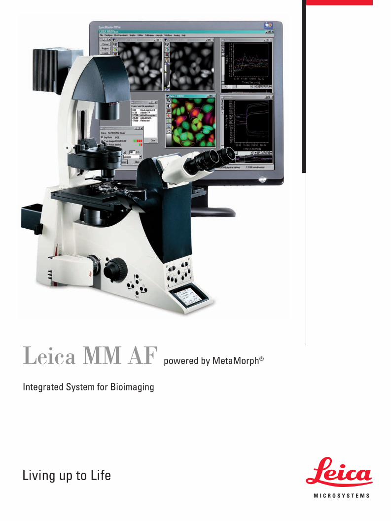
Leica MM AF powered by MetaMorph®
Integrated System for Bioimaging
Bioimaging techniques contribute to a growing number of scien-tifi c breakthroughs. The Leica MM AF Imaging System powered by MetaMorph, plays a large role in this revolution. With its image acquisition, processing and analysis capabilities, and complete set of tools for automation, Leica MM AF opens the door for new insights into cellular function.
Leica MM AF’s fl exibility and versatility make it a powerful system for performing operations such as time lapse, multi-dimensional acquisition and 3D reconstruction, and for making measurements such as morphometry, colocalization and brightness. In biological experiments using live cell imaging, Leica MM AF combines the speed, fl exibility and unmatched customer support required to get better results, faster.
2
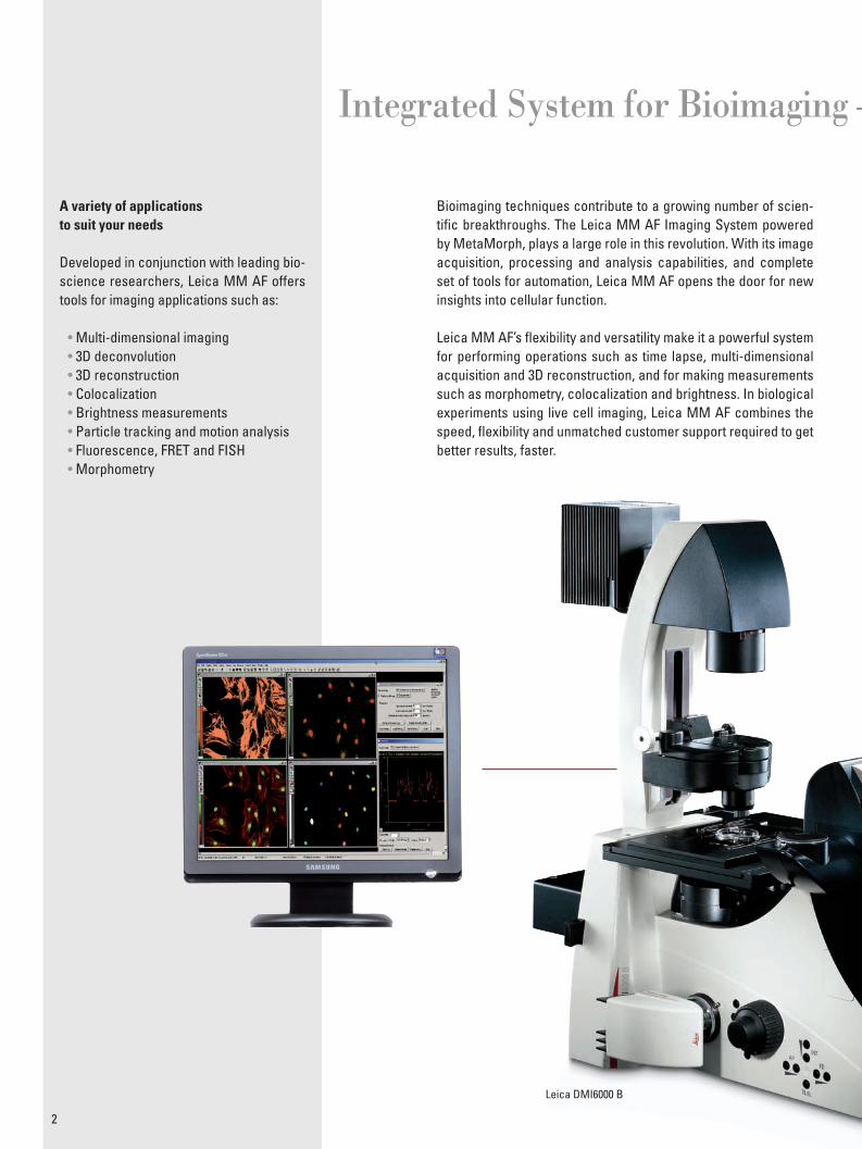
A variety of applicationsto suit your needs
Developed in conjunction with leading bio-science researchers, Leica MM AF offers tools for imaging applications such as:
• Multi-dimensional imaging• 3D deconvolution• 3D reconstruction• Colocalization• Brightness measurements• Particle tracking and motion analysis• Fluorescence, FRET and FISH• Morphometry
Leica DMI6000 B
Integrated System for Bioimaging –
3
Custom confi gured for you
Leica MM AF is available in two custom confi gurations:
• Leica MM AF Acquisition and Analysis Complete software package for control and integration of all automated Leica research microscopes, peripherals and Leica cameras. Multi-dimensional image acquisition, image processing and analysis. Drivers for other cameras suppliers optional.
• Leica MM AF Offl ineComplete software package for image measurement, processing and analysis. Includes all the analysis capabilities of Leica MM AF Acquisition and Analysis without devices control. Perfect for multi-user facilities.
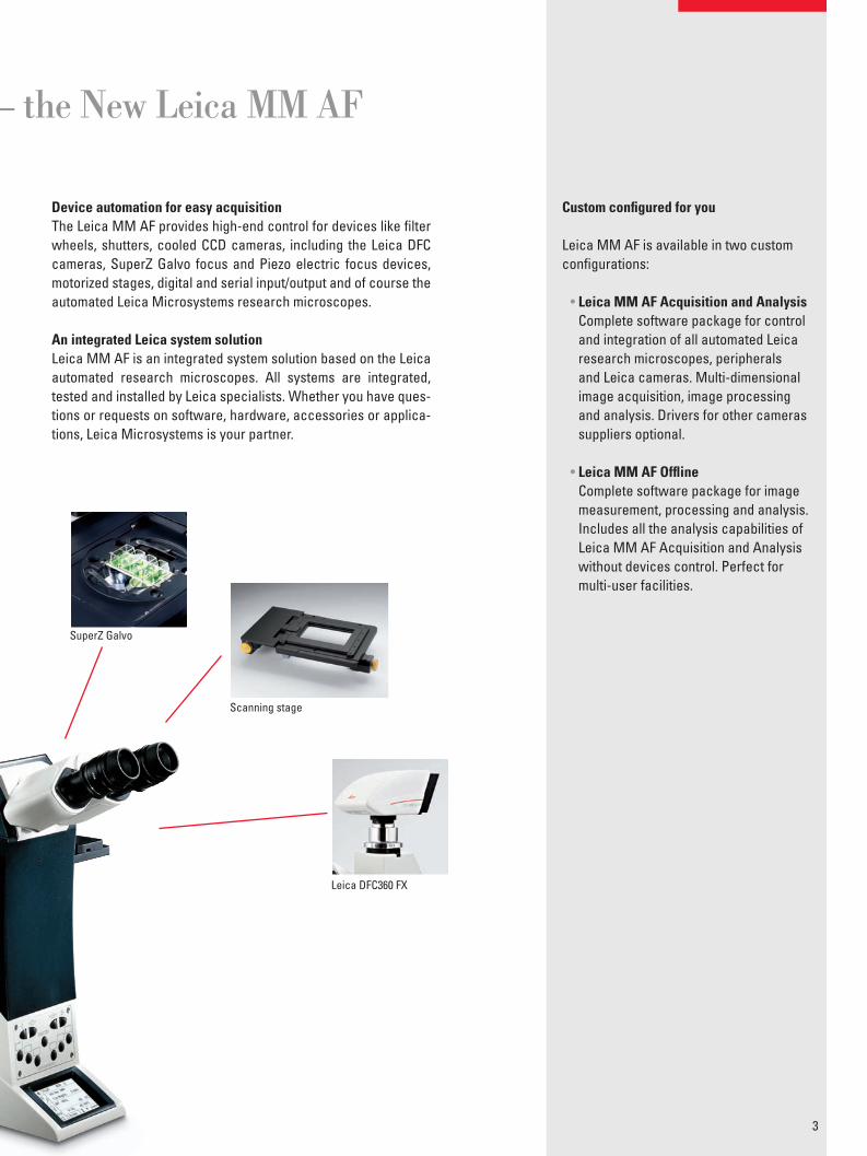
Device automation for easy acquisitionThe Leica MM AF provides high-end control for devices like fi lter wheels, shutters, cooled CCD cameras, including the Leica DFC cameras, SuperZ Galvo focus and Piezo electric focus devices, motorized stages, digital and serial input/output and of course the automated Leica Microsystems research microscopes.
An integrated Leica system solutionLeica MM AF is an integrated system solution based on the Leica automated research microscopes. All systems are integrated, tested and installed by Leica specialists. Whether you have ques-tions or requests on software, hardware, accessories or applica-tions, Leica Microsystems is your partner.
SuperZ Galvo
Scanning stage
Leica DFC360 FX
– the New Leica MM AF
A simple interface guides you through each dimension and settings can be modifi ed after acquisition is initiated. The Leica micro-scope peripheral controls are integrated in the Leica MM AF tool-bar, displaying current illumination, magnifi cation and XYZ loca-tion settings. Leica MM AF’s customizable auto-focus capabilities keep lengthy timedependent events in focus.
A Powerful Multi-dimensional Imaging Tool
4
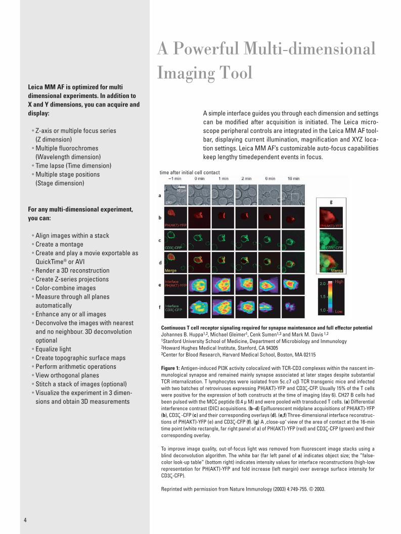
Continuous T cell receptor signaling required for synapse maintenance and full effector potentialJohannes B. Huppa1,2, Michael Gleimer1, Cenk Sumen1,3 and Mark M. Davis 1,2
1Stanford University School of Medicine, Department of Microbiology and Immunology2Howard Hughes Medical Institute, Stanford, CA 943053Center for Blood Research, Harvard Medical School, Boston, MA 02115
Figure 1: Antigen-induced PI3K activity colocalized with TCR-CD3 complexes within the nascent im-munological synapse and remained mainly synapse associated at later stages despite substantial TCR internalization. T lymphocytes were isolated from 5c.c7 αβ TCR transgenic mice and infected with two batches of retroviruses expressing PH(AKT)-YFP and CD3ζ-CFP. Usually 15% of the T cells were positive for the expression of both constructs at the time of imaging (day 6). CH27 B cells had been pulsed with the MCC peptide (0.4 μ M) and were pooled with transduced T cells. (a) Differential interference contrast (DIC) acquisitions. (b–d) Epifl uorescent midplane acquisitions of PH(AKT)-YFP (b), CD3ζ -CFP (c) and their corresponding overlays (d). (e,f) Three-dimensional interface reconstruc-tions of PH(AKT)-YFP (e) and CD3ζ-CFP (f). (g) A ‚close-up‘ view of the area of contact at the 16-min time point (white rectangle, far right panel of a) of PH(AKT)-YFP (red) and CD3ζ-CFP (green) and their corresponding overlay.
To improve image quality, out-of-focus light was removed from fl uorescent image stacks using a blind deconvolution algorithm. The white bar (far left panel of a) indicates object size; the “false-color look-up table” (bottom right) indicates intensity values for interface reconstructions (high-low representation for PH(AKT)-YFP and fold increase (left margin) over average surface intensity for CD3ζ-CFP).
Reprinted with permission from Nature Immunology (2003) 4:749-755. © 2003.
Leica MM AF is optimized for multi dimensional experiments. In addition to X and Y dimensions, you can acquire and display:
• Z-axis or multiple focus series(Z dimension)
• Multiple fl uorochromes(Wavelength dimension)
• Time lapse (Time dimension)• Multiple stage positions
(Stage dimension)
For any multi-dimensional experiment, you can:
• Align images within a stack• Create a montage• Create and play a movie exportable as
QuickTime® or AVI• Render a 3D reconstruction• Create Z-series projections• Color-combine images• Measure through all planes
automatically• Enhance any or all images• Deconvolve the images with nearest
and no neighbour. 3D deconvolutionoptional
• Equalize light• Create topographic surface maps• Perform arithmetic operations• View orthogonal planes• Stitch a stack of images (optional)• Visualize the experiment in 3 dimen-
sions and obtain 3D measurements
time after initial cell contact
Intensity over time measurements are important in studies such as protein motility, FRET and protein-protein interactions. Leica MM AF facilitates time lapse acquisition by offering streaming as an ac-quisition option. With the appropriate devices, streaming allows you to acquire at the maximum rate of the camera (patented).
Meeting advanced requirementsAnother feature for time lapse is the Live Replay option. With ap-propriate devices, and when viewing live images, you can press a key when an interesting event occurs and capture a stack con-taining some past history of the event as well as some data after the event happened.
Customization through journalingJournals are sophisticated, customizable and powerful macros that record and perform a series of tasks without the need for a programming language. The software‘s Journal Editor allows you to create functions to simplify system operations, automate acqui-sition and device control, and sequence events. User-defi nable taskbars and custom menus make it easy to achieve one-button control of your system.
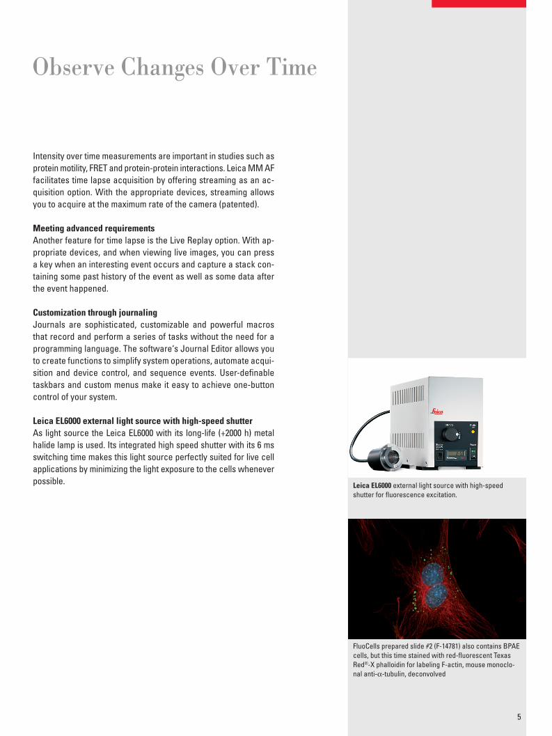
Leica EL6000 external light source with high-speed shutterAs light source the Leica EL6000 with its long-life (+2000 h) metal halide lamp is used. Its integrated high speed shutter with its 6 ms switching time makes this light source perfectly suited for live cell applications by minimizing the light exposure to the cells whenever possible.
Observe Changes Over Time
5
Leica EL6000 external light source with high-speedshutter for fl uorescence excitation.
FluoCells prepared slide #2 (F-14781) also contains BPAEcells, but this time stained with red-fl uorescent Texas Red®-X phalloidin for labeling F-actin, mouse monoclo-nal anti-α-tubulin, deconvolved
While good experiment data can be obtained by analyzing a single fl uorescent probe, you often get better results by examining more complex interactions. Leica MM AF‘s colocalization tools provide a higher level of detail, with quantitative data regarding regions of overlap between two fl uorescent probes.
These tools enable you to graphically represent the intensities of each probe on a pixel-by-pixel basis and calculate a correlation coeffi cient to give a measure of both positive and negative colo-calization. Your data can then be exported to a spreadsheet or text fi le.
Measure brightness over timeMany fl uorescence experiments depend on measuring bright-ness parameters and Leica MM AF excels at providing this type of information. With Leica MM AF, you can log intensity data from selected regions in an image stack or live video image over time and choose which parameters to capture.
Plot Colocalization and Brightness Measurements for Visual Representations
6
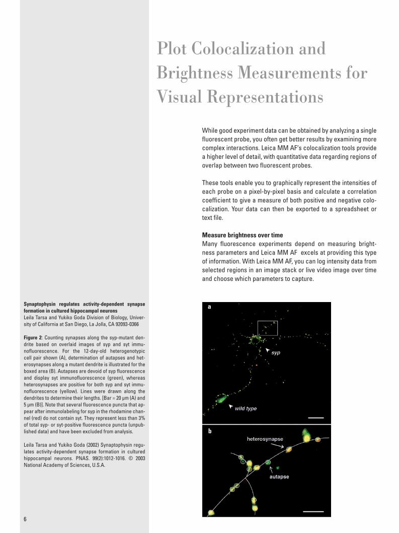
Synaptophysin regulates activity-dependent synapse formation in cultured hippocampal neuronsLeila Tarsa and Yukiko Goda Division of Biology, Univer-sity of California at San Diego, La Jolla, CA 92093-0366
Figure 2: Counting synapses along the syp-mutant den-drite based on overlaid images of syp and syt immu-nofl uorescence. For the 12-day-old heterogenotypic cell pair shown (A), determination of autapses and het-erosynapses along a mutant dendrite is illustrated for the boxed area (B). Autapses are devoid of syp fl uorescence and display syt immunofl uorescence (green), whereas heterosynapses are positive for both syp and syt immu-nofl uorescence (yellow). Lines were drawn along the dendrites to determine their lengths. [Bar = 20 μm (A) and 5 μm (B)]. Note that several fl uorescence puncta that ap-pear after immunolabeling for syp in the rhodamine chan-nel (red) do not contain syt. They represent less than 3% of total syp- or syt-positive fl uorescence puncta (unpub-lished data) and have been excluded from analysis.
Leila Tarsa and Yukiko Goda (2002) Synaptophysin regu-lates activity-dependent synapse formation in cultured hippocampal neurons. PNAS. 99(2):1012-1016. © 2003 National Academy of Sciences, U.S.A.
a
b
Follow the movement of tagged particles over time such as fl u-orescently-labeled cell surface molecules, microtubules, nucleic acids, lipids and other objects with sub-pixel resolution.
Leica MM AF facilitates your analysis with features for spatial calibration, point-to-point measurements, automated time stamp-ing of images and tracking of objects.
Measure X and Y coordinates, velocity, mean displacement, mean vector length and more, then plot your measurements onto print-able and custom-confi gurable graphs for easy visualization.
Algorithms for ParticleTracking and Motion Analysis
7
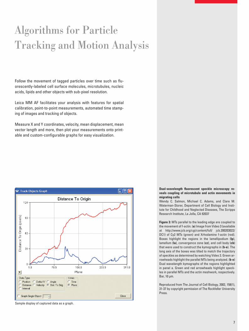
Dual-wavelength fl uorescent speckle microscopy re-veals coupling of microtubule and actin movements in migrating cellsWendy C. Salmon, Michael C. Adams, and Clare M. Waterman-Storer, Department of Cell Biology and Insti-tute for Childhood and Neglected Diseases, The Scripps Research Institute, La Jolla, CA 92037
Figure 3: MTs parallel to the leading edge are coupled to the movement of f-actin. (a) Image from Video 3 (available at http://www.jcb.org/cgi/content/full/ jcb.200203022/DC1) of Cy2 MTs (green) and Xrhodamine f-actin (red). Boxes highlight the regions in the lamellipodium (lp), lamellum (la), convergence zone (cz), and cell body (cb) that were used to construct the kymographs in (b-e). The long axis of the boxes was tilted to match the trajectory of speckles as determined by watching Video 3. Green ar-rowheads highlight the parallel MTs being analyzed. (b-e) Dual wavelength kymographs of the regions highlighted in panel a. Green and red arrowheads highlight speck-les in parallel MTs and the actin meshwork, respectively. Bar, 10 μm.
Reproduced from The Journal of Cell Biology, 2002, 158(1), 31-37 by copyright permission of The Rockfeller University Press.
Sample display of captured data as a graph.
Common applications of fl uorescent-based methods are providing new insights into protein dynamics and the biological processes they regulate.
With a typical system confi guration, Leica MM AF easily auto-mates and simplifi es the process of acquiring, color-combining and visualizing multiple fl uorophores.
Live cell studies demand the rapid acquisition and low-light level imaging of highly sensitive, cooled CCD cameras with high quan-tum effi ciency, low noise and fast readout rates.
FRET Applications with Leica MM AFThe Leica MM AF makes it easy to handle automated wavelength devices and automatically aligns multiple images. A FRET-specifi c dialog box automates the complex arithmetic needed to account for and correct fl uorescent background and bleed through in your images.
The Speed and PrecisionNeeded for Fluorescence
8
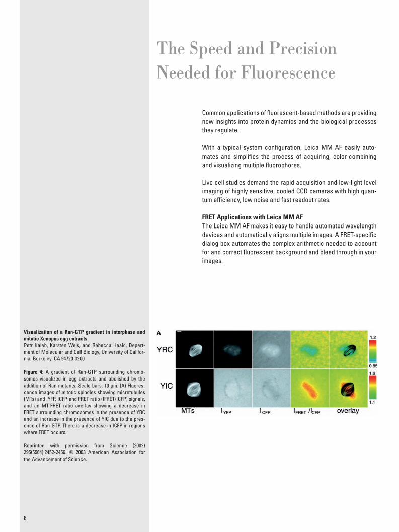
Visualization of a Ran-GTP gradient in interphase and mitotic Xenopus egg extractsPetr Kalab, Karsten Weis, and Rebecca Heald, Depart-ment of Molecular and Cell Biology, University of Califor-nia, Berkeley, CA 94720-3200
Figure 4: A gradient of Ran-GTP surrounding chromo-somes visualized in egg extracts and abolished by the addition of Ran mutants. Scale bars, 10 μm. (A) Fluores-cence images of mitotic spindles showing microtubules (MTs) and IYFP, ICFP, and FRET ratio (IFRET/ICFP) signals, and an MT-FRET ratio overlay showing a decrease in FRET surrounding chromosomes in the presence of YRC and an increase in the presence of YIC due to the pres-ence of Ran-GTP. There is a decrease in ICFP in regions where FRET occurs.
Reprinted with permission from Science (2002) 295(5564):2452-2456. © 2003 American Association for the Advancement of Science.
9
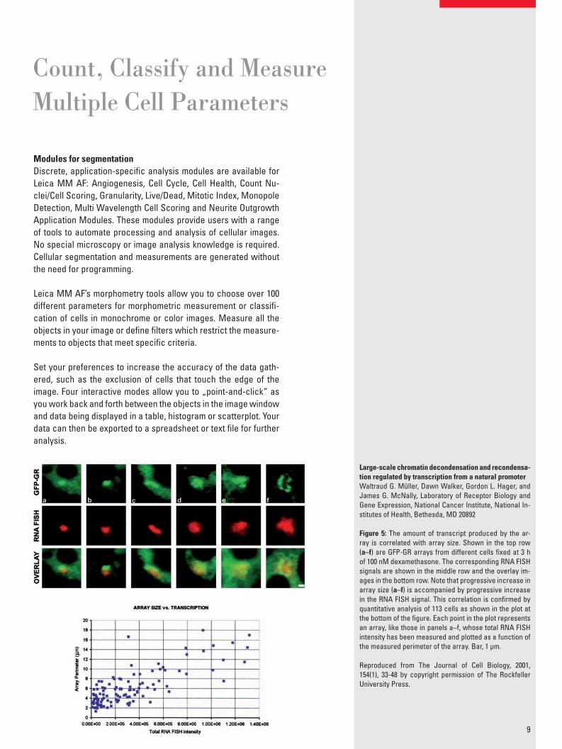
Modules for segmentationDiscrete, application-specifi c analysis modules are available for Leica MM AF: Angiogenesis, Cell Cycle, Cell Health, Count Nu-clei/Cell Scoring, Granularity, Live/Dead, Mitotic Index, Monopole Detection, Multi Wavelength Cell Scoring and Neurite Outgrowth Application Modules. These modules provide users with a range of tools to automate processing and analysis of cellular images. No special microscopy or image analysis knowledge is required. Cellular segmentation and measurements are generated without the need for programming.
Leica MM AF’s morphometry tools allow you to choose over 100 different parameters for morphometric measurement or classifi -cation of cells in monochrome or color images. Measure all the objects in your image or defi ne fi lters which restrict the measure-ments to objects that meet specifi c criteria.
Set your preferences to increase the accuracy of the data gath-ered, such as the exclusion of cells that touch the edge of the image. Four interactive modes allow you to „point-and-click“ as you work back and forth between the objects in the image window and data being displayed in a table, histogram or scatterplot. Your data can then be exported to a spreadsheet or text fi le for further analysis.
Count, Classify and Measure Multiple Cell Parameters
Large-scale chromatin decondensation and recondensa-tion regulated by transcription from a natural promoterWaltraud G. Müller, Dawn Walker, Gordon L. Hager, and James G. McNally, Laboratory of Receptor Biology and Gene Expression, National Cancer Institute, National In-stitutes of Health, Bethesda, MD 20892
Figure 5: The amount of transcript produced by the ar-ray is correlated with array size. Shown in the top row (a–f) are GFP-GR arrays from different cells fi xed at 3 h of 100 nM dexamethasone. The corresponding RNA FISH signals are shown in the middle row and the overlay im-ages in the bottom row. Note that progressive increase in array size (a–f) is accompanied by progressive increase in the RNA FISH signal. This correlation is confi rmed by quantitative analysis of 113 cells as shown in the plot at the bottom of the fi gure. Each point in the plot represents an array, like those in panels a–f, whose total RNA FISH intensity has been measured and plotted as a function of the measured perimeter of the array. Bar, 1 μm.
Reproduced from The Journal of Cell Biology, 2001, 154(1), 33-48 by copyright permission of The Rockfeller University Press.
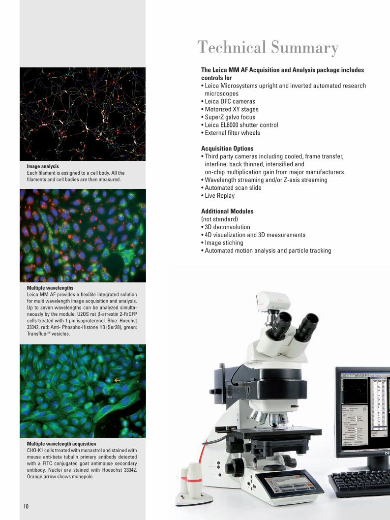
The Leica MM AF Acquisition and Analysis package includes controls for• Leica Microsystems upright and inverted automated research
microscopes• Leica DFC cameras• Motorized XY stages• SuperZ galvo focus• Leica EL6000 shutter control• External fi lter wheels
Acquisition Options• Third party cameras including cooled, frame transfer,
interline, back thinned, intensifi ed andon-chip multiplication gain from major manufacturers
• Wavelength streaming and/or Z-axis streaming• Automated scan slide• Live Replay
Additional Modules(not standard)• 3D deconvolution• 4D visualization and 3D measurements• Image stiching• Automated motion analysis and particle tracking
Technical Summary
10
Image analysisEach fi lament is assigned to a cell body. All thefi laments and cell bodies are then measured.
Multiple wavelengthsLeica MM AF provides a fl exible integrated solution for multi wavelength image acquisition and analysis. Up to seven wavelengths can be analyzed simulta-neously by the module. U2OS rat β-arrestin 2-RrGFP cells treated with 1 μm isoproterenol. Blue: Hoechst 33342, red: Anti- Phospho-Histone H3 (Ser28), green: Transfl uor® vesicles.
Multiple wavelength acquisitionCHO-K1 cells treated with monastrol and stained with mouse anti-beta tubulin primary antibody detected with a FITC conjugated goat antimouse secondary antibody. Nuclei are stained with Hoeschst 33342. Orange arrow shows monopole.
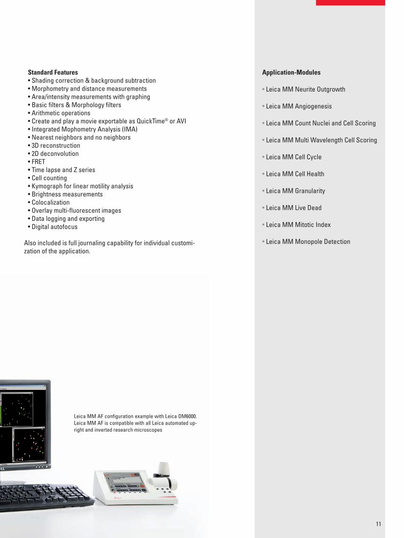
Standard Features• Shading correction & background subtraction• Morphometry and distance measurements• Area/intensity measurements with graphing• Basic fi lters & Morphology fi lters• Arithmetic operations• Create and play a movie exportable as QuickTime® or AVI• Integrated Mophometry Analysis (IMA)• Nearest neighbors and no neighbors• 3D reconstruction• 2D deconvolution• FRET• Time lapse and Z series• Cell counting• Kymograph for linear motility analysis• Brightness measurements• Colocalization• Overlay multi-fl uorescent images• Data logging and exporting• Digital autofocus
Also included is full journaling capability for individual customi-zation of the application.
11
Leica MM AF confi guration example with Leica DM6000.Leica MM AF is compatible with all Leica automated up-right and inverted research microscopes
Application-Modules
• Leica MM Neurite Outgrowth
• Leica MM Angiogenesis
• Leica MM Count Nuclei and Cell Scoring
• Leica MM Multi Wavelength Cell Scoring
• Leica MM Cell Cycle
• Leica MM Cell Health
• Leica MM Granularity
• Leica MM Live Dead
• Leica MM Mitotic Index
• Leica MM Monopole Detection
The statement by Ernst Leitz in 1907, “with the user, for the user,” describes the fruitful collaboration with end users and driving force of innovation at Leica Microsystems. We have developed fi ve brand values to live up to this tradition: Pioneering, High-end Quality, Team Spirit, Dedication to Science, and Continuous Improvement. For us, living up to these values means: Living up to Life.
Active worldwide Australia: North Ryde Tel. +61 2 8870 3500 Fax +61 2 9878 1055
Austria: Vienna Tel. +43 1 486 80 50 0 Fax +43 1 486 80 50 30
Belgium: Groot Bijgaarden Tel. +32 2 790 98 50 Fax +32 2 790 98 68
Canada: Richmond Hill/Ontario Tel. +1 905 762 2000 Fax +1 905 762 8937
Denmark: Ballerup Tel. +45 4454 0101 Fax +45 4454 0111
France: Nanterre Cedex Tel. +33 811 000 664 Fax +33 1 56 05 23 23
Germany: Wetzlar Tel. +49 64 41 29 40 00 Fax +49 64 41 29 41 55
Italy: Milan Tel. +39 02 574 861 Fax +39 02 574 03392
Japan: Tokyo Tel. +81 3 5421 2800 Fax +81 3 5421 2896
Korea: Seoul Tel. +82 2 514 65 43 Fax +82 2 514 65 48
Netherlands: Rijswijk Tel. +31 70 4132 100 Fax +31 70 4132 109
People’s Rep. of China: Hong Kong Tel. +852 2564 6699 Fax +852 2564 4163
Portugal: Lisbon Tel. +351 21 388 9112 Fax +351 21 385 4668
Singapore Tel. +65 6779 7823 Fax +65 6773 0628
Spain: Barcelona Tel. +34 93 494 95 30 Fax +34 93 494 95 32
Sweden: Kista Tel. +46 8 625 45 45 Fax +46 8 625 45 10
Switzerland: Heerbrugg Tel. +41 71 726 34 34 Fax +41 71 726 34 44
United Kingdom: Milton Keynes Tel. +44 1908 246 246 Fax +44 1908 609 992
USA: Bannockburn/lllinois Tel. +1 847 405 0123 Fax +1 847 405 0164 and representatives in more than 100 countries
Leica Microsystems operates globally in four divi sions, where we rank with the market leaders.
• Life Science DivisionThe Leica Microsystems Life Science Division supports the imaging needs of the scientifi c community with advanced innovation and technical expertise for the visualization, measurement, and analysis of microstructures. Our strong focus on understanding scientifi c applications puts Leica Microsystems’ customers at the leading edge of science.
• Industry DivisionThe Leica Microsystems Industry Division’s focus is to support customers’ pursuit of the highest quality end result. Leica Microsystems provide the best and most innovative imaging systems to see, measure, and analyze the micro-structures in routine and research industrial applications, materials science, quality control, forensic science inves-tigation, and educational applications.
• Biosystems DivisionThe Leica Microsystems Biosystems Division brings his-topathology labs and researchers the highest-quality, most comprehensive product range. From patient to pa-thologist, the range includes the ideal product for each histology step and high-productivity workfl ow solutions for the entire lab. With complete histology systems fea-turing innovative automation and Novocastra™ reagents, Leica Microsystems creates better patient care through rapid turnaround, diagnostic confi dence, and close cus-tomer collaboration.
• Medical DivisionThe Leica Microsystems Medical Division’s focus is to partner with and support surgeons and their care of pa-tients with the highest-quality, most innovative surgi cal microscope technology today and into the future.
“With the user, for the user”Leica Microsystems
www.leica-microsystems.com
Orde
r no.
: Eng
lish
914
673
• II/1
1/DX
/Br.H
. • C
opyr
ight
© b
y Le
ica
Mic
rosy
stem
s CM
S Gm
bH, W
etzla
r, Ge
rman
y, 2
011
LEIC
A an
d th
e Le
ica
Logo
are
regi
ster
ed tr
adem
arks
of L
eica
Mic
rosy
stem
s IR
Gm
bH.