Legionella protection and vaccination mediated by … · Centre for Animal Biotechnology, ......
43
1 Title: Legionella protection and vaccination mediated by Mucosal 1 Associated Invariant T (MAIT) cells 2 3 Authors: Huimeng Wang 1 , Criselle D’Souza 1,2 , Xin Yi Lim 1 , Lyudmila Kostenko 1 , Troi 4 J Pediongco 1 , Sidonia BG Eckle 1 , Bronwyn S Meehan 1 , Nancy Wang 1 , Shihan Li 1 , 5 Ligong Liu 3,4 , Jeffrey YW Mak 3,4 , David P Fairlie 3,4 , Yoichiro Iwakura 5 , Jennifer M 6 Gunnersen 10 , Andrew W Stent 11 , Jamie Rossjohn 6,7,8 , Glen P Westall 9 , Lars Kjer- 7 Nielsen 1 , Richard A, Strugnell 1 , James McCluskey 1* , Alexandra J Corbett 1 , and Timothy 8 SC Hinks 1,12 †, Zhenjun Chen 1 † 9 Affiliations 10 1. Department of Microbiology and Immunology, Peter Doherty Institute for Infection 11 and Immunity, University of Melbourne, Parkville, Victoria 3000, Australia. 12 2. Centre for Animal Biotechnology, Faculty of Veterinary and Agricultural Sciences, 13 University of Melbourne, Parkville, Victoria 3000, Australia. 14 3. Division of Chemistry and Structural Biology, Institute for Molecular Bioscience, The 15 University of Queensland, Brisbane, Queensland 4067, Australia. 16 4. Australian Research Council Centre of Excellence in Advanced Molecular Imaging, 17 The University of Queensland, Brisbane, Queensland 4067, Australia. 18 5. Center for Animal Disease Models, Research Institute for Biomedical Sciences, Tokyo 19 University of Science, Chiba-ken 278-8510, Japan. 20 peer-reviewed) is the author/funder. All rights reserved. No reuse allowed without permission. The copyright holder for this preprint (which was not . http://dx.doi.org/10.1101/231472 doi: bioRxiv preprint first posted online Dec. 9, 2017;
Transcript of Legionella protection and vaccination mediated by … · Centre for Animal Biotechnology, ......
1
Title: Legionella protection and vaccination mediated by Mucosal 1
Associated Invariant T (MAIT) cells 2
3
Authors: Huimeng Wang1, Criselle D’Souza1,2, Xin Yi Lim1, Lyudmila Kostenko1, Troi 4
J Pediongco1, Sidonia BG Eckle1, Bronwyn S Meehan1, Nancy Wang1, Shihan Li1, 5
Ligong Liu3,4, Jeffrey YW Mak3,4, David P Fairlie3,4, Yoichiro Iwakura5, Jennifer M 6
Gunnersen10, Andrew W Stent11, Jamie Rossjohn6,7,8, Glen P Westall9, Lars Kjer-7
Nielsen1, Richard A, Strugnell1, James McCluskey1*, Alexandra J Corbett1, and Timothy 8
SC Hinks1,12†, Zhenjun Chen1† 9
Affiliations 10
1. Department of Microbiology and Immunology, Peter Doherty Institute for Infection 11
and Immunity, University of Melbourne, Parkville, Victoria 3000, Australia. 12
2. Centre for Animal Biotechnology, Faculty of Veterinary and Agricultural Sciences, 13
University of Melbourne, Parkville, Victoria 3000, Australia. 14
3. Division of Chemistry and Structural Biology, Institute for Molecular Bioscience, The 15
University of Queensland, Brisbane, Queensland 4067, Australia. 16
4. Australian Research Council Centre of Excellence in Advanced Molecular Imaging, 17
The University of Queensland, Brisbane, Queensland 4067, Australia. 18
5. Center for Animal Disease Models, Research Institute for Biomedical Sciences, Tokyo 19
University of Science, Chiba-ken 278-8510, Japan. 20
peer-reviewed) is the author/funder. All rights reserved. No reuse allowed without permission. The copyright holder for this preprint (which was not. http://dx.doi.org/10.1101/231472doi: bioRxiv preprint first posted online Dec. 9, 2017;

2
6. Infection and Immunity Program and The Department of Biochemistry and Molecular 21
Biology, Biomedicine Discovery Institute, Monash University, Clayton, Australia. 22
7. Australian Research Council Centre of Excellence in Advanced Molecular Imaging, 23
Monash University, Clayton, Victoria 3800, Australia 24
8. Institute of Infection and Immunity, Cardiff University, School of Medicine, Heath 25
Park, CF14 4XN, Wales, UK 26
9. Allergy Immunology and Respiratory Medicine, Alfred Hospital, Melbourne, Victoria 27
3004, Australia. 28
10. University of Melbourne, Anatomy and Neuroscience Department, Victoria 3000, 29
Australia. 30
11. Faculty of Veterinary and Agricultural Sciences, University of Melbourne, 31
Melbourne, Victoria 3000, Australia. 32
12. Respiratory Medicine Unit, Nuffield Department of Medicine Experimental 33
Medicine, University of Oxford, OX3 9DU, Oxfordshire, UK 34
Contact information: 35
* Corresponding author and lead contact: J McCluskey ([email protected]) 36
Additional title page footnotes: 37
† These authors contributed equally to this work as joint senior authors. 38
39
40
peer-reviewed) is the author/funder. All rights reserved. No reuse allowed without permission. The copyright holder for this preprint (which was not. http://dx.doi.org/10.1101/231472doi: bioRxiv preprint first posted online Dec. 9, 2017;

3
Abstract: 41
Mucosal associated invariant T (MAIT) cells recognize conserved microbial metabolites 42
from riboflavin synthesis. Striking evolutionary conservation and pulmonary abundance 43
implicate them in antibacterial host defense, yet their roles in protection against clinically 44
significant pathogens are unknown. Murine Legionella infection induced MR1-dependent 45
MAIT cell activation and rapid pulmonary accumulation of MAIT cells associated with 46
immune protection detectable in fully immunocompetent host animals. MAIT cell 47
protection was more evident in mice lacking CD4+ cells, whilst profoundly 48
immunodeficient RAG2-/-gC-/- mice were substantially rescued from uniformly lethal 49
Legionella infection by adoptively-transferred MAIT cells. This protection was 50
dependent on MR1, IFN-g and GM-CSF, but not IL-17, TNF-a or perforin. Protection 51
was enhanced and observed earlier post-infection in mice that were Ag-primed to boost 52
MAIT cells before infection. Our findings define a significant role for MAIT cells in 53
protection against a major human pathogen and indicate a potential role for vaccination to 54
enhance MAIT cell immunity. 55
Key words 56
Mucosal associated invariant T cell, T cell, infection, Legionella, MHC-related protein 1, 57
IFN-gamma, intracellular, lung, human, mouse, riboflavin. 58
59
peer-reviewed) is the author/funder. All rights reserved. No reuse allowed without permission. The copyright holder for this preprint (which was not. http://dx.doi.org/10.1101/231472doi: bioRxiv preprint first posted online Dec. 9, 2017;

4
Mucosal-associated invariant T (MAIT) cells are innate-like lymphocytes with the 60
potential to recognize a broad range of microbial pathogens. MAIT cells express a ‘semi-61
invariant’ ab T cell receptor (TCR) and recognize small molecules presented by the 62
major histocompatibility complex (MHC) class I-related molecule (MR1)1,2. These 63
molecules comprise derivatives of the riboflavin biosynthetic pathway3-5, which is 64
conserved between a wide variety of bacteria, mycobacteria and yeasts3,6, but is absent 65
from mammals, and therefore provides an elegant mechanism to discriminate host and 66
pathogen. Indeed the enzymatic pathway required for riboflavin synthesis has been 67
identified in all microbes shown to activate MAIT cells, and is absent in those that do 68
not3. 69
70
A striking feature of MAIT cell immunity is the high level of conservation of MR1 across 71
150 million years of mammalian evolution7-9, implying a strong evolutionary pressure to 72
maintain the MAIT cell compartment. Furthermore, MAIT cells have a strong pro-73
inflammatory phenotype10 and are abundant in humans in blood and lung tissue11, whilst 74
in C57BL/6 mice are found in greater abundance in the lungs than any other organs12. 75
Together these features implicate MAIT cells in a critical role in respiratory host defense. 76
However, very few pathogens have been demonstrated in vivo to cause activation and 77
proliferation of MAIT cells13,14. In studies implicating a role for MAIT cells in protective 78
immunity against pathogens, the definition of these cells was limited by the lack of MR1-79
Ag tetramers14. To date no studies have clearly defined a functional role for MAIT cells 80
in protection against a clinically important human pathogen. 81
82
peer-reviewed) is the author/funder. All rights reserved. No reuse allowed without permission. The copyright holder for this preprint (which was not. http://dx.doi.org/10.1101/231472doi: bioRxiv preprint first posted online Dec. 9, 2017;

5
Using a model of bacterial lung infection with the intracellular bacteria Salmonella 83
enterica serovar Typhimurium we have previously shown that riboflavin gene-competent 84
bacteria can cause rapid activation and proliferation of MAIT cells13. We therefore 85
hypothesized that this response could also be elicited with an authentic human lung 86
pathogen and would contribute to protection against disease. 87
Legionella spp are facultative intracellular pathogens, gram-negative, flagellated bacteria 88
which, when inhaled, cause a spectrum of disease from self-limiting Pontiac fever to 89
severe, necrotic pneumonia: Legionnaire’s disease15. Incidence of Legionnaire’s has 90
nearly trebled since 2000, with >5000 cases/year in the USA, inflicting a 10% mortality 91
despite best treatment16. In North America and Europe16 the predominant pathogen is L. 92
pneumophila whilst in Australasia and Thailand over 50% of cases are caused by L. 93
longbeachae17. 94
Here we have used MR1 tetramers loaded with the potent MAIT cell ligand 5-(2-95
oxopropylideneamino)-6-D-ribitylaminouracil (5-OP-RU)18 to specifically identify4 and 96
characterize MAIT cells in human in vitro and murine in vivo models of lung infection 97
with the two most clinically significant Legionella species: L. pneumophila and L. 98
longbeachae. Our data reveal that MAIT cells contribute to protection against fatal 99
infection with Legionella, by a mechanism that is dependent on MR1 and interferon 100
(IFN)-g / granulocyte macrophage-colony stimulating factor (GM-CSF). Protection is 101
partial in immunocompetent hosts but becomes increasingly evident as other arms of 102
immunity are disabled such as in CD4 T cell-deficient animals. Protection ultimately 103
becomes “all or nothing” in profoundly immunodeficient mice RAG2-/-gC-/- mice. These 104
peer-reviewed) is the author/funder. All rights reserved. No reuse allowed without permission. The copyright holder for this preprint (which was not. http://dx.doi.org/10.1101/231472doi: bioRxiv preprint first posted online Dec. 9, 2017;

6
studies dissect the mechanisms by which MAIT cells contribute to protection against an 105
important human disease and a model intracellular pathogen. 106
Results 107
Human MAIT cells are activated by Legionella infection in vitro via MR1 108
We3,13 have previously shown that MAIT cells are activated by microbial species that 109
express the riboflavin biosynthetic pathway; a finding which has been confirmed by 110
others6. We therefore investigated whether Legionella species known to cause serious 111
pulmonary infections in humans – L. pneumophila15,19 and L. longbeachae17 – and to 112
express the necessary rib enzymes20, could activate human MAIT cells. First, bacterial 113
lysates of L. pneumophila and L. longbeachae stimulated a reporter cell line expressing a 114
MAIT TCR (Jurkat.MAIT-A-F7)3 in the presence of an MR1-expressing lymphoid cell 115
line (C1R.MR1)(Figure 1A). Stimulation was dose-dependent, and could be specifically 116
blocked by anti-MR1 antibody21. Next we used a well-characterized human monocytic 117
cell line (THP-1)22 as an antigen presenting cell co-cultured with flow-sorted human 118
peripheral blood CD3+Va7.2+CD161+ cells. We observed activation of MAIT cells 119
when co-cultured with THP1 cells infected for 27 hours with live L. longbeachae (Figure 120
1B,C). Intracellular infection of wild type THP1 and THP-1:MR1+ cell lines induced 121
expression of TNF-a by human MR1-5-OP-RU tetramer+ MAIT cells. Activation was 122
related to the infective dose, and was specific to MAIT cells and not non-MAIT CD3+ T 123
cells. Activation was MR1-dependent, as it did not occur in the presence of cells in which 124
we had disrupted the MR1 gene using a CRISPR/Cas9 lentiviral system (THP1:MR1-). 125
MAIT cells also expressed IFN-g in the presence of MR1-over-expressing cells 126
peer-reviewed) is the author/funder. All rights reserved. No reuse allowed without permission. The copyright holder for this preprint (which was not. http://dx.doi.org/10.1101/231472doi: bioRxiv preprint first posted online Dec. 9, 2017;

7
(THP1:MR1+), but expression was minimal using the parental cell line (THP1), which 127
has very low constitutive surface expression of MR1. 128
To visualize MAIT cells in situ we infected healthy human lung tissue ex vivo with L. 129
longbeachae and observed CD3+TCRVa7.2+IL-18Ra+ MAIT cells within the lung 130
parenchyma in the proximity of Legionella bacilli 24 hours post-infection using 131
immunofluorescence microscopy (Figure 1D). 132
These findings indicate that Legionella induces potent MAIT cell immune responses in 133
vitro suggesting that MAIT cells are likely to play a role in protection against Legionella 134
pneumonia. 135
136
MAIT accumulate in the lungs during Legionella infection in vivo 137
Next we examined the impact of Legionella infection on MAIT cells in vivo in a murine 138
model using intranasal infection with live L. longbeachae. TCRb+ MR1-5-OP-RU 139
tetramer+ cells were visible in the lung parenchyma using immunofluorescence 140
microscopy within three days post-infection (Figure 2A). There was striking enrichment 141
of pulmonary MAIT cells (from here on defined as CD45+TCRb+ MR1-5-OP-RU 142
tetramer+ cells), which comprised up to 30% of all pulmonary ab-T cells after 7 days 143
(Figure 2B, C). MAIT cell accumulation was dependent on size of initial inoculum and 144
was proportionately much larger for MAIT cells – 580-fold absolute increase at 105 145
colony-forming units (CFU) (P<0.0001) – than conventional ab-T cells (maximum 9.4-146
fold, P<0.0001) (Figure 2C,D). Accumulation occurred rapidly over 7 days post infection 147
(DPI), with absolute numbers peaking at day 10 (Figure 2E). Furthermore, despite a 148
peer-reviewed) is the author/funder. All rights reserved. No reuse allowed without permission. The copyright holder for this preprint (which was not. http://dx.doi.org/10.1101/231472doi: bioRxiv preprint first posted online Dec. 9, 2017;

8
subsequent 20-fold contraction from peak frequencies (P=0.005), overall expansion of the 149
MAIT cell population was long-lived, persisting >280 DPI (Figure 2E,F). Interestingly, 150
although MAIT cells have been implicated in recruitment of non MAIT T cells 14, we did 151
not observe any significant difference in pulmonary recruitment of ab-T cells in MR1-/- 152
mice, which have an absolute deficiency of MAIT cells12,13. Likewise, i.n. infection with 153
2x10⁷ CFU L. pneumophila similarly induced a rapid expansion of MAIT cells 154
(Supplementary Figure S1), although more modest than L. longbeachae. As C57BL/6 155
mice are susceptible to L. longbeachae17, L. longbeachae was selected as the most 156
appropriate model for more detailed investigation. 157
158
Histology of lungs from mice infected with 2x10⁴ CFU of L. longbeachae at 7DPI 159
demonstrated pronounced alveolar infiltration of neutrophils and macrophages, 160
leukocytoclasia, aggregates of fibrin and accumulation of edema fluid and epithelial 161
shedding, consistent with the typical features of human L. pneumophila 162
pneumonia15(Supplementary Figure S2A,B). Blinded analysis of these sections using a 163
qualitative histological score at multiple time-points post infection revealed inflammation 164
peaked at day 7, but there were no gross histological differences in the severity of 165
pneumonia between C57BL/6 and MR1-/- mice (Supplementary Figure S2C). To 166
determine the cellular localization of L. longbeachae we measured bacterial burden in 167
flow-sorted cells from collagenase-dispersed murine lungs 3 days post infection. Most 168
viable bacilli localized within neutrophils, but evidence of infection of macrophages and 169
dendritic cells was also observed (Supplementary Figure S2D). 170
171
peer-reviewed) is the author/funder. All rights reserved. No reuse allowed without permission. The copyright holder for this preprint (which was not. http://dx.doi.org/10.1101/231472doi: bioRxiv preprint first posted online Dec. 9, 2017;

9
To explore MAIT cell function we investigated the dynamics of their cytokine profile 172
throughout infection. During acute L. longbeachae infection MAIT cells secreted 173
interleukin (IL)-17A, IFN-g, GM-CSF (Figure 3A, Supplementary Figure S3) and TNF-a 174
(data similar to INF-g, not shown). Expression of IL-17A was abundant throughout the 175
course of the infection, whilst IFN-g secretion was significantly higher during the acute 176
infection than in naïve cells or after resolution (each P<0.005, Figure 3B). Conversely, 177
expression of GM-CSF was lowest during acute infection and peaked after disease 178
resolution (P=0.0006 acute v resolution). This correlated with a shift in expression of 179
nuclear transcription factors associated with Th1 or Th17 differentiation. In naïve mice 180
most (81±4%, mean ±SD) MAIT cells expressed the orphan nuclear receptor, retinoic 181
acid–related orphan receptor gt (RORgt) alone: a master regulator of Th17 cell 182
differentiation (Figure 3C, 3D). A minority (13±4%) of cells expressed both RORgt and 183
the Th1 regulator T-bet, and very few expressed T-bet alone. However, during acute 184
infection and long-term post infection there was a marked shift in phenotype towards 185
predominant co-expression of RORgt and T-bet in 64±5% and 69±3% of MAIT cells 186
respectively. MAIT cells expressing T-bet alone were only observed at significant 187
frequencies (14±3%) in acute infection. Thus the consistent secretion of IL-17A in all 188
stages of infection and the transient increase of IFN-g secretion during acute infection 189
reflect the changes in transcription factor profile we observed, suggesting the formation 190
of an authentic memory pool of MAIT cells and pointing to a specific role for IFN-g in 191
the acute response to infection. 192
193
peer-reviewed) is the author/funder. All rights reserved. No reuse allowed without permission. The copyright holder for this preprint (which was not. http://dx.doi.org/10.1101/231472doi: bioRxiv preprint first posted online Dec. 9, 2017;

10
MAIT cell protection against life-threatening Legionella infection is enhanced and 194
accelerated by prior boosting 195
196
To determine whether MAIT cells contribute to immune protection against Legionella we 197
compared bacterial burden in lungs of C57BL/6 and MR1-/- mice throughout infection. 198
Bacterial load increased by 2.5 log over the initial inoculum, peaking at 3 days post-199
infection (3DPI). In normal C57BL/6 mice we observed a significant difference in 200
bacterial load but not until days 10 and 14 post infection. This was of the order of one log 201
in CFU, consistent with relatively impaired bacterial clearance in MAIT cell deficient, 202
MR1-/- mice (Figure 4A,B). 203
204
In specific pathogen-free C57BL/6 mice baseline frequencies of MAIT cells are very 205
low12,13, potentially due to lack of natural exposure to diverse environmental pathogens. 206
We have previously shown that MAIT cells can be expanded in vivo by intranasal 207
exposure to the MAIT cell ligand 5-OP-RU with a Toll-like receptor (TLR) agonist such 208
as the TLR9 agonist CpG or TLR2 agonist S-[2,3-bis(palmitoyloxy)propyl] cysteine 209
(Pam2Cys) to furnish a MAIT cell costimulus13. To understand whether MAIT cell 210
vaccination might impact on protection observed against Legionella infection of the lung, 211
we used this approach to specifically expand pulmonary MAIT cells one month prior to 212
Legionella infection, without affecting conventional T cell frequencies (Figure 4C,D). 213
Prior exposure to 5-OP-RU and CpG enhanced MAIT cell numbers in the lungs and was 214
associated with protection against infection as reflected in a reduction in bacterial load in 215
peer-reviewed) is the author/funder. All rights reserved. No reuse allowed without permission. The copyright holder for this preprint (which was not. http://dx.doi.org/10.1101/231472doi: bioRxiv preprint first posted online Dec. 9, 2017;

11
C57BL/6 versus MR1-/- mice (compare Figures 4A, B to Supplementary Figure S4). This 216
protective effect became apparent earlier than observed in wild type C57BL/6 mice with 217
reduced CFU seen on days 5 and 7 post-infection and comparable on d10 post-infection 218
(compare Figures 4A, B to Supplementary Figure S4), as MAIT cell numbers became 219
indistinguishable in boosted and non-boosted mice (not shown). When a direct 220
comparison was made between MR1-/- mice, C57BL/6 mice and C57BL/6 mice that had 221
been boosted by 5-OP-RU and Pam2Cys, bacterial burden was significantly lower on 222
days 5 ,7 and 10 post-infection in wild-type mice that had received this prior MAIT cell 223
boosting (Figure 4E). This demonstrates the potential to augment MAIT cell-mediated 224
protection by the prior administration of synthetic ligands as a ‘vaccine’. 225
These data demonstrate that MAIT cells contribute actively to Legionella protection in 226
the context of an intact immune system and that this protection is more rapid and of 227
greater magnitude when mice are first vaccinated to expand MAIT cells before infectious 228
challenge. 229
230
MAIT cell-mediated protection is more apparent in immune deficient mice 231
Studies of other intracellular pathogens have demonstrated high levels of functional 232
redundancy in the ability of different lymphocytes subsets to control bacterial growth in 233
vivo23. We hypothesized that by removing partially-redundant effects of other 234
lymphocyte subsets, the protective effects of MAIT cells would become more apparent. 235
CD4+ T cell-derived IFN-g has been shown to play an essential role in achieving 236
bacterial clearance of Salmonella Typhimurium23. We therefore used GK1.5 transgenic 237
peer-reviewed) is the author/funder. All rights reserved. No reuse allowed without permission. The copyright holder for this preprint (which was not. http://dx.doi.org/10.1101/231472doi: bioRxiv preprint first posted online Dec. 9, 2017;

12
mice, which express the anti-GK1.5 antibody and are CD4+ T cell deficient24, and 238
compared these with GK1.5.MR1-/- mice which lack both CD4+ cells and MAIT cells. 239
As expected, we observed a protective effect of MAIT cells through reduced bacterial 240
burden apparent even earlier in the course of infection than with wild type mice 241
(statistically significant by day 7 p.i) (Figure 4F). 242
243
To further unmask the potential of MAIT cells in protection, we removed additional 244
layers of immunity by studying the impact of adoptively transferred MAIT cells into 245
profoundly immunodeficient Rag2-/-gC-/- mice. We first expanded pulmonary MAIT cells 246
by i.n. inoculation of donor mice with S. Typhimurium BRD509, as previously 247
described13. Flow-sorted pulmonary MAIT cells from these mice were then adoptively 248
transferred into recipient Rag2-/-gC-/- mice in which Rag2 and the common g chain are 249
deleted, leading to absence of T, B and NK cells. After transfer, administration of anti-250
CD4 and anti-CD8 mAbs was used to further deplete any residual contaminating 251
conventional T cells (Figure 5A). After adoptive transfer, MAIT cells expanded 252
spontaneously to generate a stable population by two weeks (Figure 5B, Supplementary 253
Figure S5A) which expressed the nuclear transcription factor and master regulator of 254
innate-like T cell development promyelocytic leukemia zinc finger(PLZF) (Figure S5B) 255
25. 256
257
Strikingly, the presence of adoptively-transferred MAIT cells was sufficient to rescue 258
completely Rag2-/-gC-/- mice from fatal infection with 103 CFU L. longbeachae (Figure 259
peer-reviewed) is the author/funder. All rights reserved. No reuse allowed without permission. The copyright holder for this preprint (which was not. http://dx.doi.org/10.1101/231472doi: bioRxiv preprint first posted online Dec. 9, 2017;

13
5C, C2 P<0.0001) in the absence of other components of adaptive immunity. Using a 260
higher inoculum (104 CFU) we observed this protection was reduced by blockade with 261
anti-MR1 mAb, which was associated with significantly reduced survival (C2 P=0.005) 262
and with increased bacterial load amongst surviving mice (P=0.0004), consistent with an 263
MR1-dependent mechanism (Figure 5D,E). 264
MAIT cell-protection is dependent on IFN-g 265
To determine the mechanism by which MAIT cells provide this protection we used 266
adoptive transfer of MAIT cells from mice with deficiencies in cytotoxic capability or in 267
pro-inflammatory cytokines. The protective effect of MAIT cells on both survival of 268
Rag2-/-gC-/- mice or on bacterial burden was not impaired in MAIT cells lacking the 269
cytolytic proteins perforin or granzymes A and B, nor in MAIT cells unable to express 270
IL-17A or TNF-a (Figure 6A,B). We observed a small increase in bacterial burden when 271
transferred MAIT cells were deficient in GM-CSF (0.49 log-fold difference in CFU, 272
P=0.026), but this was not associated with significant differences in survival. By contrast 273
protection was critically dependent on MAIT cell derived IFN-g, with decreased survival 274
(P<0.0001) and a 2.8 log-fold increased bacterial burden (P<0.001) when MAIT cells 275
were deficient in IFN-g. Furthermore these mice all succumbed to Legionella infection by 276
day 37 p.i. . 277
The use of adoptive transfer of in vivo expanded MAIT cells provides compelling 278
evidence that MAIT cells can confer protection against important human pathogens and 279
demonstrates this protection depends upon their capacity to produce IFN-g and to a lesser 280
extent GM-CSF. 281
peer-reviewed) is the author/funder. All rights reserved. No reuse allowed without permission. The copyright holder for this preprint (which was not. http://dx.doi.org/10.1101/231472doi: bioRxiv preprint first posted online Dec. 9, 2017;

14
282
Discussion 283
Our findings show that MAIT cells are activated and proliferate in response to Legionella 284
infection, leading to enhanced immune protection in vivo that is dependent on IFN-g and 285
GM-CSF. This protection is evident earlier and of greater magnitude if mice are first 286
vaccinated to expand and prime MAIT cells which are otherwise present in small 287
numbers in normal mice. Protection by MAIT cells is characterized by more rapid 288
reduction in bacterial loads and is MR1-dependent suggesting mediation via antigen-289
specific activation. Remarkably, MAIT cell protection against Legionella was non-290
redundant and even evident in fully immune competent mice. The protective effect of 291
MAIT cell immunity became more evident as layers of immunity were removed in host 292
mice, firstly in GK1.5 mice lacking only CD4+ T cells and then in more profoundly 293
immundeficient Rag2-/-gC-/- mice, lacking conventional T cells, B cells and NK cells. This 294
observation is important given that studies of primary immunodeficiencies26 imply 295
redundancy of different lymphocyte subsets is a typical feature of pathogen immunity 296
especially for innate mechanisms such as NK cells and innate lymphoid cells. Indeed, in 297
the absence of B, T and NK cells MAIT cells were absolutely critical for survival in 298
Legionella-infected mice revealing their important potential in compromised hosts. As 299
this mechanism was dependent on MR1, which presents small molecules derived from 300
riboflavin biosynthesis3-5, this demonstrates in vivo the potential for control of Legionella 301
by detection of riboflavin metabolites. 302
peer-reviewed) is the author/funder. All rights reserved. No reuse allowed without permission. The copyright holder for this preprint (which was not. http://dx.doi.org/10.1101/231472doi: bioRxiv preprint first posted online Dec. 9, 2017;

15
These observations suggest how the contribution of MAIT cells to immune protection 303
may be critical to survival in clinical, naturally-occurring severe infection. The mortality 304
we observe from Legionella does not coincide with the time of peak bacterial load – on 305
day 3 post-infection – but later, between days 6 and 14 post-infection, at which point 306
MR1-/- mice had 0.78 to 1.1-log fold higher bacterial load than wild-type mice. In 307
essence, MAIT cells may be the difference between life and death in knife-edge 308
infections where host immunity is partially compromised by comorbidities or 309
predisposing factors, or where patients are exposed to large bacterial doses. 310
311
Although MAIT cells have both cytotoxic activity27 and the ability to rapidly produce 312
pro-inflammatory cytokines including interleukin IL-17A, TNF-a and IFN-g10, the 313
protective effect of MAIT cells against Legionella infection was not dependent on TNF-a 314
or IL-17A, but instead relied upon the capacity of MAIT cells to secrete IFN-g and GM-315
CSF. This is consistent with a study of Francisella infection28 where GM-CSF reduces 316
bacterial burden late in the course of infection, although this did not translate into a 317
significant survival difference. Nor did the protective effect depend on the key cytotoxic 318
effector molecules: perforin and granzymes A/B. This is in contrast to work suggesting 319
MAIT cell cytotoxicity is important for control of Shigella-infected HeLa cell lines in 320
vitro27. 321
322
As Legionella infects inflammatory cells, particularly neutrophils and macrophages, 323
rather than epithelia, the essential immune function required of MAIT cells in our system 324
peer-reviewed) is the author/funder. All rights reserved. No reuse allowed without permission. The copyright holder for this preprint (which was not. http://dx.doi.org/10.1101/231472doi: bioRxiv preprint first posted online Dec. 9, 2017;

16
is likely the IFN-g-stimulated enhancement of bactericidal activity within these cells in 325
which phagosome function has been subverted. Our findings of IFN-g production upon 326
MAIT cell activation are consistent with other reports6,10,14,27, and accord with reports of 327
a role for MAIT cell-derived IFN-g in limiting growth of Francisella tularensis in bone 328
marrow-derived macrophages in vitro14. IFN-g has also been shown to enhance 329
bactericidal activity of neutrophils via multiple mechanisms including enhancement of 330
oxidative burst, nitric oxide production, antigen presentation, phagocytosis, and 331
upregulation of CD80/86 co-stimulation and T cell-recruiting cytokines and 332
chemokines29. Moreover, given that IFN-g is critical also for protection against 333
mycobacterial disease including M. tuberculosis (M.tb), it is likely that this early 334
production of MAIT cell-derived IFN-g may be an important and non-redundant 335
component of protection against mycobacteria. Indeed in vitro MAIT cell-derived IFN-g 336
inhibits growth of Bacillus Calmette-Guerin (BCG) in macrophages30, and the MR1-337
MAIT cell axis has been linked to susceptibility to BCG in mice30 and to M.tb in 338
humans31 and mice32. 339
340
A striking feature of MAIT cell biology is the very low frequencies of MAIT cells we 341
observe in blood or lungs in naïve mice12,13, in contrast to the marked and long-lived 342
expansion induced by a single infection in our model. Although antigen-naïve MAIT 343
cells have some intrinsic effector capacity33, the delay between initial microbial exposure 344
and peak MAIT cell frequency may be critical in providing a window of opportunity for a 345
pathogen to exploit14. This notion is consistent with our observation that MAIT cell 346
protection can be accelerated by prior expansion of the pulmonary MAIT cell population 347
peer-reviewed) is the author/funder. All rights reserved. No reuse allowed without permission. The copyright holder for this preprint (which was not. http://dx.doi.org/10.1101/231472doi: bioRxiv preprint first posted online Dec. 9, 2017;

17
using intranasal synthetic 5-OP-RU and an appropriate TLR agonist. Notably, MAIT cell 348
frequencies are low in early childhood34, suggesting the potential to enhance the 349
immunogenicity of vaccines given in early life by incorporating such MAIT cell ligands 350
in combination with TLR stimulation, which might for instance promote the recruitment 351
of inflammatory monocyte differentiation via MAIT cell-derived GM-CSF28. A 352
protective effect of expanding MAIT cells could contribute to heterologous protection 353
afforded by neonatal BCG vaccination against other, unrelated classes of pathogens35. 354
Vaccination with MAIT cell ligands might help resolve chronic infections where MAIT 355
cell frequencies may be reduced due to other therapies11, comorbidities or activation 356
induced cell death36. 357
358
Our findings define a significant role for MAIT cells in pulmonary host defense against a 359
major human pathogen. We reveal layers of immunological redundancy likely to mask 360
the contribution of MAIT cells in many situations of infectious challenge, but suggest a 361
critical role for MAIT cells becomes apparent in a crisis situation – as reflected here in a 362
high infecting inoculum, or in the face of compromised specific immunity – in which the 363
gulf between survival and death is finely balanced. Moreover, we demonstrate the 364
mechanism of this MAIT cell protection is IFN-g dependent and enhanced by GM-CSF. 365
Due to the pleiotropic roles of IFN-g and the conservation of the riboflavin pathway 366
across many species, this mechanism is likely broadly effective against other major 367
human intracellular pathogens, and may prove as relevant to the later stages of infection 368
as to the initial, acute phase characterized by innate immune responses countering rapid 369
pathogen replication. We have shown this immunity can be augmented by exposure to 370
peer-reviewed) is the author/funder. All rights reserved. No reuse allowed without permission. The copyright holder for this preprint (which was not. http://dx.doi.org/10.1101/231472doi: bioRxiv preprint first posted online Dec. 9, 2017;

18
MR1 ligands suggesting this mechanism might have potential for preventive or 371
therapeutic benefit. 372
373
peer-reviewed) is the author/funder. All rights reserved. No reuse allowed without permission. The copyright holder for this preprint (which was not. http://dx.doi.org/10.1101/231472doi: bioRxiv preprint first posted online Dec. 9, 2017;

19
Materials and Methods 374
In vitro activation assays 375
Jurkat cells expressing a MAIT TCR comprising the TRAV1-2-TRAJ33 a-chain 376
and TRBV6-4 b-chain (Jurkat.MAIT) were co-incubated at 105 cells per well in 96-well 377
U-bottom plates with an equal number of class I reduced (C1R) antigen presenting cells 378
(APCs) expressing MR1 (C1R.MR1)3 for 16 h in RPMI-1640 media (Gibco) in 379
supplement and 10% foetal calf serum (FCS)) (RF10) media at 37°C, 5%CO2. Cells were 380
stimulated for 16 h with bacterial lystates prepared using repeated ultrasonication of 381
bacteria in log-phase growth. Cells were stained with anti-CD3-APC and anti-CD69-PE 382
Abs and 7AAD before flow cytometric analysis. Activation of Jurkat.MAIT cells was 383
measured by an increase in surface CD69 expression. 384
385
For human MAIT cell assays, peripheral blood mononuclear cells (PBMCs) were 386
stained with anti-CD3-PEAF594, CD161-PECy7, TCR Va7.2 (TRAV1-2)-APC. Va7.2+ 387
cells were enriched with anti-APC magnetic beads and the CD3+Va7.2+CD161+ 388
population were isolated by flow cytometry and cultured at 104 cells/well for 16 or 36 389
hours in penicillin-free media containing streptomycin and gentamicin with 5x10⁴ THP1 390
cells which had been first infected for 3 hours (CD69 assays) or 27 hours (intracellular 391
cytokine staining) with different multiplicities of infection (MOI) of live L. longbeachae 392
NSW150. Control wells contained 5-OP-RU 10nM or L. longbeachae NSW150 at MOI 393
100 which had been heat killed for 10 minutes at 67°C. CD69 upregulation was measured 394
by surface staining for CD69-APCCy7 and 5-OP-RU loaded human MR1 tetramer-PE. 395
For intracellular cytokine expression cells brefeldin A was added for the last 16 hours, 396
peer-reviewed) is the author/funder. All rights reserved. No reuse allowed without permission. The copyright holder for this preprint (which was not. http://dx.doi.org/10.1101/231472doi: bioRxiv preprint first posted online Dec. 9, 2017;

20
cells fixed, permeabilized (using BD Fixation/Permeabilization Kit (BD, Franklin Lakes, 397
NJ) and stained with MR1 tetramer, CD3-PEAF594, Zombie yellow, IFNg-FITC, and 398
TNFa-Pacific blue. 399
400
Generation of THP1:MR1- cell line 401
THP1:MR1- cell lines were generated by targeted deletion of MR1 using 402
LentiviralCRISPRv2 which was a gift from Feng Zhang (Addgene plasmid # 52961)37. 403
The plasmid was digested with BsmB1 (Fermentas), dephosphorylated and purified using 404
gel electrophoresis and the Ultraclean DNA isolation kit (MO Bio laboratories, Carlsbad 405
CA). Short guide RNAs (ACCTCTCATCATTGTGTTAA) were ligated and the reaction 406
product used to transform Stbl3 E.Coli. DNA was purified from transformed colonies 407
(QIAprep spin miniprep) and used to transfect HEK293T cells with LentiviralCRISPRv2 408
vector and packaging vectors. Supernatants were used to transduce parent THP1 cells and 409
cells were selected using puromycin resistance and single cell sorting using anti-MR1 410
antibody (8F2.F9) after upregulation of MR1 using acetyl-6FP. MR1 knockouts were 411
then verified using surface staining and western blotting. 412
413
Immunofluorescence microscopy 414
8 µm sections of cryopreserved, unfixed lung tissue were submerged into ice-cold 415
acetone for 10 min, air dried and then re-hydrated in PBS for 10 min. Endogenous biotin 416
block was performed using Biotin/Avidin blocking kit (Thermo Fisher, Waltham MA) 417
according to the manufacturer’s instructions. Serum-free protein block (DAKO, 418
Carpinteria, CA) was applied for 15 min, followed by 30 min blocking with 10% normal 419
peer-reviewed) is the author/funder. All rights reserved. No reuse allowed without permission. The copyright holder for this preprint (which was not. http://dx.doi.org/10.1101/231472doi: bioRxiv preprint first posted online Dec. 9, 2017;

21
donkey serum. Sections were subsequently blocked with Murine MR1-6FP tetramer (Nil 420
PE) for 1 hour at room temperature. Murine MR1-5-OP-RU tetramer-PE (25 µg/ml in 2% 421
bovine serum albumin/PBS) was applied for 1 h at room temperature in the dark, sections 422
washed with PBS, fixed with 1% paraformaldehyde for 10 min, washed again and stained 423
with a cocktail containing polyclonal rabbit anti-Legionella antibody (gift from Dr 424
Hayley Newton, Department of Microbiology and Immunology, University of 425
Melbourne) and Goat anti PE (KPL). After 1 hr, sections were washed and stained with 426
Donkey anti Goat-AlexaFluor 568 (Life Technologies), Donkey-anti-Rabbit-DyLight 680 427
(Life Technologies) and Alexa Fluor 647-conjugated Rat anti mouse TCR-b (Biolegend). 428
Nuclei were counterstained with Hoechst 33342 (Life Technologies). Sections were 429
mounted with Prolong Gold mounting medium (Life Technologies). Image acquisition 430
was performed on Zeiss LSM 710 confocal microscope using Zen software (Zeiss, 431
Oberkochen, Germany) The resultant images were further analysed using FIJI Image J 432
software38 (UW-Madison). 433
434
Animal models 435
Mice were bred and housed in the Biological Research Facility of the Peter Doherty 436
Institute (Melbourne, Victoria, Australia). MR1-/- mice were generated by breeding 437
Va19iCa-/-MR1-/- mice39 (from Susan Gilfillan, Washington University, St Louis School 438
of Medicine, St Louis, MO) with C57BL/6 mice and inter-crossing of F1 mice. The 439
genotype was determined by tail DNA PCR at the MR1 locus as previously described13. 440
Granzyme A/B-/- and Perforin-/- mice were purchased from Joe Trapani (Victorian 441
Comprehensive Cancer Centre, Melbourne). GK1.5 mice were crossed onto the MR1-/- 442
peer-reviewed) is the author/funder. All rights reserved. No reuse allowed without permission. The copyright holder for this preprint (which was not. http://dx.doi.org/10.1101/231472doi: bioRxiv preprint first posted online Dec. 9, 2017;

22
background to generate GK1.5.MR1-/- mice which lack CD4+ cells and MAIT cells. 443
Male mice aged 6–12 weeks were used in experiments, after approval by the University 444
of Melbourne Animal Ethics Committee (1513661). 445
446
Intranasal infection 447
Intranasal (i.n.) inoculation with L. longbeachae or antigens (76 pmol 5-OP-RU) and 448
TLR agonist (either 20 mg CpG or 20nmol Pam2Cys) in 50 µl per nares was performed 449
on isofluorane-anesthetized mice. For blocking experiments, mice were given 250 µg 450
anti-MR1 (26.5 or 8F2.F9)21,40 or isotype control mAbs in 200 µl PBS, once (i.v or 451
intraperitoneally) 1 day prior to inoculation and three times (d1, d3, d5) post inoculation. 452
Mice were killed by CO2 asphyxia, the heart perfused with 10ml cold RPMI and lungs 453
were taken. 454
455
To prepare single-cell suspensions lungs were finely chopped with a scalpel blade 456
and treated with 3mg/ml-1collagenase III (Worthington, Lakewood, NJ), 5 µg/ml 457
DNAse, and 2% fetal calf serum in RPMI for 90 min at 37°C with gentle shaking. Cells 458
were then filtered (70µm) and washed with PBS/2% foetal calf serum. Red blood cells 459
were lysed with hypotonic buffer TAC (Tris-based amino chloride) for 5 min at 37°C. 460
Approximately 1.5x10⁶ cells were filtered (40µm) and used for flow cytometric analysis. 461
462
Determination of bacterial counts in infected lungs. 463
peer-reviewed) is the author/funder. All rights reserved. No reuse allowed without permission. The copyright holder for this preprint (which was not. http://dx.doi.org/10.1101/231472doi: bioRxiv preprint first posted online Dec. 9, 2017;

23
Bacterial colonization was determined by counting colony-forming units (CFU) 464
obtained from plating homogenized lungs in duplicate from infected mice (x5 per group) 465
on buffered charcoal yeast extract agar containing 30µg/ml streptomycin and colonies 466
counted after 4 days at 37°C under aerobic conditions. 467
468
Adoptive transfer 469
As MAIT cell frequencies are low in naïve C57BL/6 mice, prior to adoptive transfer 470
experiments MAIT cell populations were expanded by intranasal infection with 106 CFU 471
S. Typhimurium BRD509 in 50µl PBS for 7 days as previously described13. After 7 days, 472
mice were sacrificed, single cell suspensions prepared and live CD3+CD45+MR1-5-OP-473
RU tetramer+ cells sorted using a BD FACS Aria III. 105 MAIT cells were injected into 474
the tail veins of recipient mice which then received 0.1 mg each of anti-CD4 (Gk1.5) and 475
anti-CD8 (53.762) mAb i.p on days 2 and 5 or 6 to control residual conventional T cells. 476
Mice were rested for 2 weeks post transfer to allow full expansion of the MAIT cell 477
population prior to subsequent infectious challenge. Mice were weighed daily and 478
assessed for visual signs of clinical disease, including inactivity, ruffled fur, labored 479
breathing, and huddling behavior. Animals that had lost ≥20% of their original body 480
weight and/or displayed evidence of pneumonia were euthanized. 481
482
Statistical analysis 483
Statistical tests were performed using the Prism GraphPad software (version 7.0 La 484
Jolla, CA). Comparisons between groups were performed using Student’s t-tests or 485
Mann-Whitney tests as appropriate unless otherwise stated. Survival curves were 486
peer-reviewed) is the author/funder. All rights reserved. No reuse allowed without permission. The copyright holder for this preprint (which was not. http://dx.doi.org/10.1101/231472doi: bioRxiv preprint first posted online Dec. 9, 2017;

24
compared using the Gehan-Breslow-Wilcoxon method for multiple groups. Flow 487
cytometric data analysis was performed with FlowJo10 software (Ashland, OR). 488
Reagents 489
Human peripheral blood mononuclear cells (PBMC) were obtained from the 490
Australian Red Cross Blood Service (ARCBS) (University of Melbourne Human 491
Research Ethics Committee 1239046.2). Healthy human lung explant tissue was obtained 492
via the Alfred Lung Biobank program and ARCBS from organs not suitable for donation 493
(Blood Service HREC 2014#14 and University of Melbourne Human Research Ethics 494
Committee 1545566.1). 495
496
Compounds, immunogens and tetramers 497
5-OP-RU was prepared as described previously18. CpG1688 (Sequence: 498
T*C*C*A*T*G*A*C*G*T*T*C*C*T*G*A*T*G*C*T (*phosphorothioate linkage) 499
nonmethylated cytosine-guanosine oligonucleotides was purchased from Geneworks 500
(Thebarton, Australia). Murine and human MR1 and b2-Microglobulin genes were 501
expressed in Escherichia coli inclusion bodies, refolded, and purified as described 502
previously41. MR1-5-OP-RU tetramers were generated as described previously4. 503
504
Bacterial strains 505
Cultures of Legionella pneumophila JR32 and Legionella longbeachae NSW150 506
were grown at 37°C in buffered yeast extract (BYE) broth supplemented with 30-507
50µg/ml streptomycin for 16 hour to log-phase (OD600 0.2-0.6) with shaking at 180 rpm. 508
peer-reviewed) is the author/funder. All rights reserved. No reuse allowed without permission. The copyright holder for this preprint (which was not. http://dx.doi.org/10.1101/231472doi: bioRxiv preprint first posted online Dec. 9, 2017;

25
For the infecting inoculum, bacteria were re-inoculated in prewarmed medium for a 509
further 2–4 h culture (OD600 0.2–0.6) with the estimation that 1 OD600=5x10⁸/ml, 510
sufficient bacteria were washed and diluted in phosphate buffered saline (PBS) with 2% 511
BYE for i.n. delivery to mice. A sample of inoculum was plated onto BYCE with 512
streptomycin for verification of bacterial concentration by counting colony-forming units. 513
514
For infection of adoptive transfer donor-mice with Salmonella Typhimurium 515
BRD509 cultures were prepared as previously described13. 516
517
Antibodies and flow cytometry 518
Antibodies against murine CD4 (GK1.5, APC-Cy7), CD19 (1D3, PerCP-Cy 5.5), 519
CD45.2 (104, FITC), IFNg (XMG1.2, PE-Cy7), Ly6G (IA8, PECy7), TCRb (H57-597, 520
APC or PE), TNF-a (MP6-XT22, PE), GM-CSF (MP1-22E9, PE) and IL-17A (TC11-521
18H10, PE) were purchased from BD (Franklin Lakes, NJ). Antibodies against CD8a 522
(53-6.7, PE), PLZF (Mags.21F7, PE), RORgt (B2D, APC), T-bet (4B10, PE-Cy7) and 523
MHCII (M5/114.15.2, AF700) were purchased from eBioscience (San Diego, CA). Abs 524
against CD19 (6D5, BV510), F4/80 (BM8, APC), CD11b (M1/70, FITC), CD11c (N418, 525
BV786), CD31 (PCAM, MEC13.3, PerCPCy5.5), CD62L (Mel-14, FITC), CD64 (X54-526
5/71, BV711), CD146 (ME-9F1, PerCPCy5.5), CD326 (G8.8, EpCAM, APC-Cy7) were 527
purchased from Biolegend (San Diego, CA). Blocking Ab (26.5, 8F2.F9) and isotype 528
controls (3E12, 8A5) were prepared in house. To block non-specific staining, cells were 529
incubated with MR1-6FP tetramer and anti-Fc receptor (2.4G2) for 15 min at room 530
temperature and then incubated at room temperature with Ab/tetramer cocktails in 531
peer-reviewed) is the author/funder. All rights reserved. No reuse allowed without permission. The copyright holder for this preprint (which was not. http://dx.doi.org/10.1101/231472doi: bioRxiv preprint first posted online Dec. 9, 2017;

26
PBS/2% fetal calf serum. 7-aminoactinomycin D (5 µl per sample) was added for the last 532
10 min. 533
534
535
Antibodies against human CD3 (UCHT1, PE-AlexaFluor594), TCR-Va7.2 (3C10, 536
APC), CD161 (HP-3G10, PE-Cy7), TNF-a (Mab11, Pacific Blue), and viability dye 537
(Zombie Yellow) were purchased from Biolegend. Antibodies against IFNg (25725.11, 538
FITC) and CD69 (FN50, PE) were purchased from BD, and anti-CD3 (UCHT1, APC) 539
from eBioscience. 540
541
Cells were fixed with 1% paraformaldehyde prior to analysis on LSRII or LSR 542
Fortessa or Canto II (BD Biosciences) flow cytometers. For intracellular cytokine 543
staining Golgi plug (BD Biosciences) was used during all processing steps. Cells 544
stimulated with PMA (phorbol 12-myristate 13-acetate;)/ionomycin (20 ng/ml, 1µg/ml, 545
respectively) for 3 h at 37°C were included as positive controls. Surface staining was 546
performed at 37°C, and cells were stained for intracellular cytokines using the BD 547
Fixation/Permeabilization Kit (BD, Franklin Lakes, NJ) or transcription factors using the 548
transcription buffer staining set (eBioscience) according to the manufacturers’ 549
instructions. 550
551
552
peer-reviewed) is the author/funder. All rights reserved. No reuse allowed without permission. The copyright holder for this preprint (which was not. http://dx.doi.org/10.1101/231472doi: bioRxiv preprint first posted online Dec. 9, 2017;

27
References: 553
1. Tilloy, F., et al. An invariant T cell receptor alpha chain defines a novel TAP-554 independent major histocompatibility complex class Ib-restricted alpha/beta T cell 555 subpopulation in mammals. J Exp Med 189, 1907-1921 (1999). 556
2. Porcelli, S., Yockey, C.E., Brenner, M.B. & Balk, S.P. Analysis of T cell antigen 557 receptor (TCR) expression by human peripheral blood CD4-8- alpha/beta T cells 558 demonstrates preferential use of several V beta genes and an invariant TCR alpha 559 chain. J Exp Med 178, 1-16 (1993). 560
3. Kjer-Nielsen, L., et al. MR1 presents microbial vitamin B metabolites to MAIT 561 cells. Nature 491, 717-723 (2012). 562
4. Corbett, A.J., et al. T-cell activation by transitory neo-antigens derived from 563 distinct microbial pathways. Nature 509, 361-365 (2014). 564
5. Eckle, S.B., et al. Recognition of Vitamin B Precursors and Byproducts by 565 Mucosal Associated Invariant T Cells. J Biol Chem 290, 30204-30211 (2015). 566
6. Le Bourhis, L., et al. Antimicrobial activity of mucosal-associated invariant T 567 cells. Nat Immunol 11, 701-708 (2010). 568
7. Riegert, P., Wanner, V. & Bahram, S. Genomics, isoforms, expression, and 569 phylogeny of the MHC class I-related MR1 gene. J Immunol 161, 4066-4077 570 (1998). 571
8. Yamaguchi, H., Hirai, M., Kurosawa, Y. & Hashimoto, K. A highly conserved 572 major histocompatibility complex class I-related gene in mammals. Biochem 573 Biophys Res Commun 238, 697-702 (1997). 574
9. Tsukamoto, K., Deakin, J.E., Graves, J.A. & Hashimoto, K. Exceptionally high 575 conservation of the MHC class I-related gene, MR1, among mammals. 576 Immunogenetics 65, 115-124 (2013). 577
10. Dusseaux, M., et al. Human MAIT cells are xenobiotic-resistant, tissue-targeted, 578 CD161hi IL-17-secreting T cells. Blood 117, 1250-1259 (2011). 579
11. Hinks, T.S., et al. Steroid-induced Deficiency of Mucosal-associated Invariant T 580 Cells in the COPD Lung: Implications for NTHi Infection. Am J Respir Crit Care 581 Med 194, 1208-1218 (2016). 582
12. Rahimpour, A., et al. Identification of phenotypically and functionally 583 heterogeneous mouse mucosal-associated invariant T cells using MR1 tetramers. 584 J Exp Med 212, 1095-1108 (2015). 585
13. Chen, Z., et al. Mucosal-associated invariant T-cell activation and accumulation 586 after in vivo infection depends on microbial riboflavin synthesis and co-587 stimulatory signals. Mucosal immunology (2016). 588
14. Meierovics, A., Yankelevich, W.J. & Cowley, S.C. MAIT cells are critical for 589 optimal mucosal immune responses during in vivo pulmonary bacterial infection. 590 Proc Natl Acad Sci U S A 110, E3119-3128 (2013). 591
15. Winn, W.C., Jr. & Myerowitz, R.L. The pathology of the Legionella pneumonias. 592 A review of 74 cases and the literature. Hum Pathol 12, 401-422 (1981). 593
16. Garrison, L.E., et al. Vital Signs: Deficiencies in Environmental Control 594 Identified in Outbreaks of Legionnaires' Disease - North America, 2000-2014. 595 MMWR Morb Mortal Wkly Rep 65, 576-584 (2016). 596
peer-reviewed) is the author/funder. All rights reserved. No reuse allowed without permission. The copyright holder for this preprint (which was not. http://dx.doi.org/10.1101/231472doi: bioRxiv preprint first posted online Dec. 9, 2017;

28
17. Gobin, I., Susa, M., Begic, G., Hartland, E.L. & Doric, M. Experimental 597 Legionella longbeachae infection in intratracheally inoculated mice. Journal of 598 medical microbiology 58, 723-730 (2009). 599
18. Mak, J.Y., et al. Stabilizing short-lived Schiff base derivatives of 5-aminouracils 600 that activate mucosal-associated invariant T cells. Nature communications 8, 601 14599 (2017). 602
19. Misch, E.A. Legionella: virulence factors and host response. Curr Opin Infect Dis 603 29, 280-286 (2016). 604
20. Kanehisa, M. & Goto, S. KEGG: Kyoto encyclopedia of genes and genomes. 605 Nucleic acids research 28, 27-30 (2000). 606
21. Huang, S., et al. Evidence for MR1 antigen presentation to mucosal-associated 607 invariant T cells. J Biol Chem 280, 21183-21193 (2005). 608
22. Tsuchiya, S., et al. Establishment and characterization of a human acute 609 monocytic leukemia cell line (THP-1). Int J Cancer 26, 171-176 (1980). 610
23. Kupz, A., Bedoui, S. & Strugnell, R.A. Cellular requirements for systemic control 611 of Salmonella enterica serovar Typhimurium infections in mice. Infect Immun 82, 612 4997-5004 (2014). 613
24. Zhan, Y., Corbett, A.J., Brady, J.L., Sutherland, R.M. & Lew, A.M. CD4 help-614 independent induction of cytotoxic CD8 cells to allogeneic P815 tumor cells is 615 absolutely dependent on costimulation. J Immunol 165, 3612-3619 (2000). 616
25. Doulatov, S., et al. PLZF is a regulator of homeostatic and cytokine-induced 617 myeloid development. Genes Dev 23, 2076-2087 (2009). 618
26. Fischer, A. & Rausell, A. Primary immunodeficiencies suggest redundancy within 619 the human immune system. Sci Immunol 1(2016). 620
27. Le Bourhis, L., et al. MAIT Cells Detect and Efficiently Lyse Bacterially-Infected 621 Epithelial Cells. PLoS Pathog 9, e1003681 (2013). 622
28. Meierovics, A.I. & Cowley, S.C. MAIT cells promote inflammatory monocyte 623 differentiation into dendritic cells during pulmonary intracellular infection. J Exp 624 Med 213, 2793-2809 (2016). 625
29. Ellis, T.N. & Beaman, B.L. Interferon-gamma activation of polymorphonuclear 626 neutrophil function. Immunology 112, 2-12 (2004). 627
30. Chua, W.J., et al. Polyclonal mucosa-associated invariant T cells have unique 628 innate functions in bacterial infection. Infect Immun 80, 3256-3267 (2012). 629
31. Seshadri, C., et al. A polymorphism in human MR1 is associated with mRNA 630 expression and susceptibility to tuberculosis. Genes Immun 18, 8-14 (2017). 631
32. Sakala, I.G., et al. Functional Heterogeneity and Antimycobacterial Effects of 632 Mouse Mucosal-Associated Invariant T Cells Specific for Riboflavin Metabolites. 633 J Immunol 195, 587-601 (2015). 634
33. Gold, M.C., et al. Human thymic MR1-restricted MAIT cells are innate pathogen-635 reactive effectors that adapt following thymic egress. Mucosal immunology 636 (2012). 637
34. Martin, E., et al. Stepwise development of MAIT cells in mouse and human. 638 PLoS Biol 7, e54 (2009). 639
35. Aaby, P., et al. Randomized trial of BCG vaccination at birth to low-birth-weight 640 children: beneficial nonspecific effects in the neonatal period? J Infect Dis 204, 641 245-252 (2011). 642
peer-reviewed) is the author/funder. All rights reserved. No reuse allowed without permission. The copyright holder for this preprint (which was not. http://dx.doi.org/10.1101/231472doi: bioRxiv preprint first posted online Dec. 9, 2017;

29
36. Cosgrove, C., et al. Early and nonreversible decrease of CD161++ /MAIT cells in 643 HIV infection. Blood 121, 951-961 (2013). 644
37. Shalem, O., et al. Genome-scale CRISPR-Cas9 knockout screening in human 645 cells. Science 343, 84-87 (2014). 646
38. Schindelin, J., Rueden, C.T., Hiner, M.C. & Eliceiri, K.W. The ImageJ 647 ecosystem: An open platform for biomedical image analysis. Mol Reprod Dev 82, 648 518-529 (2015). 649
39. Kawachi, I., Maldonado, J., Strader, C. & Gilfillan, S. MR1-restricted V alpha 19i 650 mucosal-associated invariant T cells are innate T cells in the gut lamina propria 651 that provide a rapid and diverse cytokine response. J Immunol 176, 1618-1627 652 (2006). 653
40. Chua, W.J., et al. Endogenous MHC-related protein 1 is transiently expressed on 654 the plasma membrane in a conformation that activates mucosal-associated 655 invariant T cells. J Immunol 186, 4744-4750 (2011). 656
41. Patel, O., et al. Recognition of vitamin B metabolites by mucosal-associated 657 invariant T cells. Nature communications 4, 2142 (2013). 658
659
660
peer-reviewed) is the author/funder. All rights reserved. No reuse allowed without permission. The copyright holder for this preprint (which was not. http://dx.doi.org/10.1101/231472doi: bioRxiv preprint first posted online Dec. 9, 2017;

30
Acknowledgments 661
This research was supported by National Health and Medical Research Council of 662
Australia (NHMRC) Program Grants 1113293, 1016629 and 606788, a Project Grant 663
1120467 as well as a Merieux Research Grant. T.S.C.H. was supported by a Wellcome 664
Trust Postdoctoral Research Fellowship (104553/z/14/z). A.J.C. was supported by an 665
ARC Future Fellowship. S.B.G.E. was supported by an ARC DECRA Fellowship. J.R. 666
was supported by an NHMRC Australia Fellowship, D.P.F. was supported by an 667
NHMRC Senior Principal Research Fellowship. H.W. is supported by a Melbourne 668
International Engagement Award (University of Melbourne). C.D’S. is supported by a 669
Melbourne International Research Scholarship and a Melbourne International Fee 670
Remission Scholarship (University of Melbourne). We thank Dr Wei-Jen Chua and Prof 671
Ted Hansen for their kind provision of 8F2.F9 and 26.5 mAbs, and Prof David Jackson 672
for Pam2Cys. We are grateful to Prof Francis Carbone for critical review of the 673
manuscript. 674
Author contributions 675
HW, CDS, TSCH, XYL, LK, TLP, SBG, BSM, ZC, AWS performed the experiments 676
and analyzed the data. ZC, TSCH, HW, JM, AC, RS designed the experiments and 677
managed the study. NW, DPF, YI, JG, GW, LK-N, JR, LL, JYWM provided essential 678
reagents and intellectual input. TSCH, AC, ZC, JM conceived the work and wrote the 679
manuscript which was revised and approved by all authors. 680
Competing Financial Interests 681
peer-reviewed) is the author/funder. All rights reserved. No reuse allowed without permission. The copyright holder for this preprint (which was not. http://dx.doi.org/10.1101/231472doi: bioRxiv preprint first posted online Dec. 9, 2017;

31
Z.C., S.E., L.K-N., D.F., L.L., J.Y.W.M., J.R., J.McC., and A.C. are inventors on patents 682
describing MR1 tetramers and MR1 ligands. The other authors declared no conflict of 683
interest. 684
685
Materials and Correspondance 686
Correspondence and material requests should be addressed to J McCluskey 687
([email protected]). 688
peer-reviewed) is the author/funder. All rights reserved. No reuse allowed without permission. The copyright holder for this preprint (which was not. http://dx.doi.org/10.1101/231472doi: bioRxiv preprint first posted online Dec. 9, 2017;

32
Figures 689
690
peer-reviewed) is the author/funder. All rights reserved. No reuse allowed without permission. The copyright holder for this preprint (which was not. http://dx.doi.org/10.1101/231472doi: bioRxiv preprint first posted online Dec. 9, 2017;

33
Figure 1. Human MAIT cells are activated by Legionella infection via MR1 in vitro 691
(A) Jurkat.MAIT and C1R.MR1 cells were co-incubated for 16h with lysates of L. 692
pneumophila (L. pn.) or L. longbeachae, or 5-OP-RU, acetyl-6-FP or PBS. Activation, 693
detected by staining with anti-CD69, is enhanced by bacterial lysate or by the activating 694
ligand 5-OP-RU, but not by acetyl-6-FP. Activation was blocked by anti-MR1 antibody 695
(26.5) but not by isotype control (W6/32) 2h prior to co-incubation. (B-C) THP1 cells 696
(WT) or THP1 cells overexpressing MR1 (THP1:MR1+, blue) or deficient in expression 697
of MR1 (THP1:MR1-, red) were infected for 27h with L. longbeachae, heat killed (HK) 698
L. longbeachae (MOI100) or 10nM 5-OP-RU, then co-cultured for 16h with 699
CD3+Va7.2+CD161+ human peripheral blood MAIT cells, or MAIT-depleted 700
conventional T cells. MR1-5-OP-RU-tetramer+ MAIT cell activation was measured by 701
intracellular cytokine staining for (B) TNF-a or (C) IFN-g. (D) Immunofluorescence 702
micrographs showing CD3+TCRVa7.2+IL-18Ra+ MAIT cell (white arrow) within 703
healthy human lung tissue 24h post infection ex vivo with L. longbeachae (yellow arrow). 704
Red, TCR-Va7.2; green, CD3; magenta, IL-18Ra; blue, legionella. Data show MFI or 705
percentage cytokine-positive cells with SEM. 706
707
708
peer-reviewed) is the author/funder. All rights reserved. No reuse allowed without permission. The copyright holder for this preprint (which was not. http://dx.doi.org/10.1101/231472doi: bioRxiv preprint first posted online Dec. 9, 2017;

34
709
Figure 2. Murine pulmonary infection with Legionella induces long-lasting 710
expansion of MAIT cells in vivo 711
peer-reviewed) is the author/funder. All rights reserved. No reuse allowed without permission. The copyright holder for this preprint (which was not. http://dx.doi.org/10.1101/231472doi: bioRxiv preprint first posted online Dec. 9, 2017;

35
(A) Immunofluorescence micrographs of murine lungs showing TCRb+MR1-5-OP-RU-712
tetramer+ MAIT cells (white arrows) adjacent to infected cells (yellow arrows) within 713
parenchyma four days after intranasal infection with 2x10⁴ CFU L. longbeachae in 714
C57BL/6 mice which had been challenged with the same inoculum 5 months previously. 715
(B) Flow-cytometry plots showing MAIT cell percentage among TCRb+ lymphocytes in 716
the lungs of C57BL/6 mice either uninfected or infected with 103, 104 or 105 CFU L. 717
longbeachae for 7 days. Relative (C) and absolute (D) numbers of MAIT cells and 718
conventional ab T cells 7 days post infection. Absolute numbers (E), and relative 719
percentages (F) of MR1-tetramer+ MAIT cells or conventional ab T cells in C57BL/6 720
and MR1-/- mice after intranasal infection with 2x10⁴ CFU L. longbeachae. Experiments 721
used 4-6 mice per group (mean±SEM) and were performed twice with similar results. B6, 722
C57BL/6 mice; CFU, colony forming units; DAPI, 4',6-diamidino-2-phenylindole. 723
Statistical tests C: ** Dunnett’s P<0.01, **** P<0.0001; C all comparisons with the 724
respective naïve groups P<0.0001 for Dunnett’s on log transformed data. See also 725
Figures S1, S2. 726
727
peer-reviewed) is the author/funder. All rights reserved. No reuse allowed without permission. The copyright holder for this preprint (which was not. http://dx.doi.org/10.1101/231472doi: bioRxiv preprint first posted online Dec. 9, 2017;

36
728
A
B
C100+DPI7DPINaive
Ex-vivo
+PMA
IFN GM-CSF
D
(59%) (7.4%) (12%)
Naive 7 days post infection >100 days post infection
T-bet+T-bet+ RORyt+ROR t+T-bet-/RORyt-
IL-17A IFN GM-CSF
IL-17A
(25%) (1.3%) (1.6%)
peer-reviewed) is the author/funder. All rights reserved. No reuse allowed without permission. The copyright holder for this preprint (which was not. http://dx.doi.org/10.1101/231472doi: bioRxiv preprint first posted online Dec. 9, 2017;

37
Figure 3. Profiles of MAIT cell cytokine expression and nuclear transcription 729
factors vary over the course of pulmonary Legionella infection 730
(A) Flow cytometry plots showing intracellular staining for IL-17A, IFN-g and GM-CSF 731
by pulmonary TCRb lymphocytes (non-MAIT conventional and MAIT cells) after 4h 732
culture with or without PMA/ionomycin with bredeldin A. TCRb+ lymphocytes were 733
harvested from lungs of C57BL/6 mice infected with 2x10⁴ CFU L. longbeachae for 7 734
days. Percentages in brackets represent the proportion of MR1-tetramer positive MAIT 735
cells expressing cytokine. (B) Percentages of pulmonary MAIT cells producing IL-17A, 736
IFN-g or GM-CSF by intracellular staining, directly ex-vivo from C57BL/6 mice infected 737
for 0, 7 or >100 days with 2x10⁴ CFU L. longbeachae. Experiments using 4-7 mice per 738
group (mean±SEM) were performed twice with similar results. (C) Expression of T-bet 739
and RORγt in MAIT cells from uninfected or infected C57BL/6 mice 7 or >100 days post 740
infection (DPI). (D) Average proportion of T-bet+, double positive (DP), RORgt+ and 741
double negative (DN) MAIT cells from uninfected or infected C57BL/6 mice at indicated 742
date. Mean values are representative of 5-8 mice in each group. See also Figure S3. 743
peer-reviewed) is the author/funder. All rights reserved. No reuse allowed without permission. The copyright holder for this preprint (which was not. http://dx.doi.org/10.1101/231472doi: bioRxiv preprint first posted online Dec. 9, 2017;
38
744
peer-reviewed) is the author/funder. All rights reserved. No reuse allowed without permission. The copyright holder for this preprint (which was not. http://dx.doi.org/10.1101/231472doi: bioRxiv preprint first posted online Dec. 9, 2017;

39
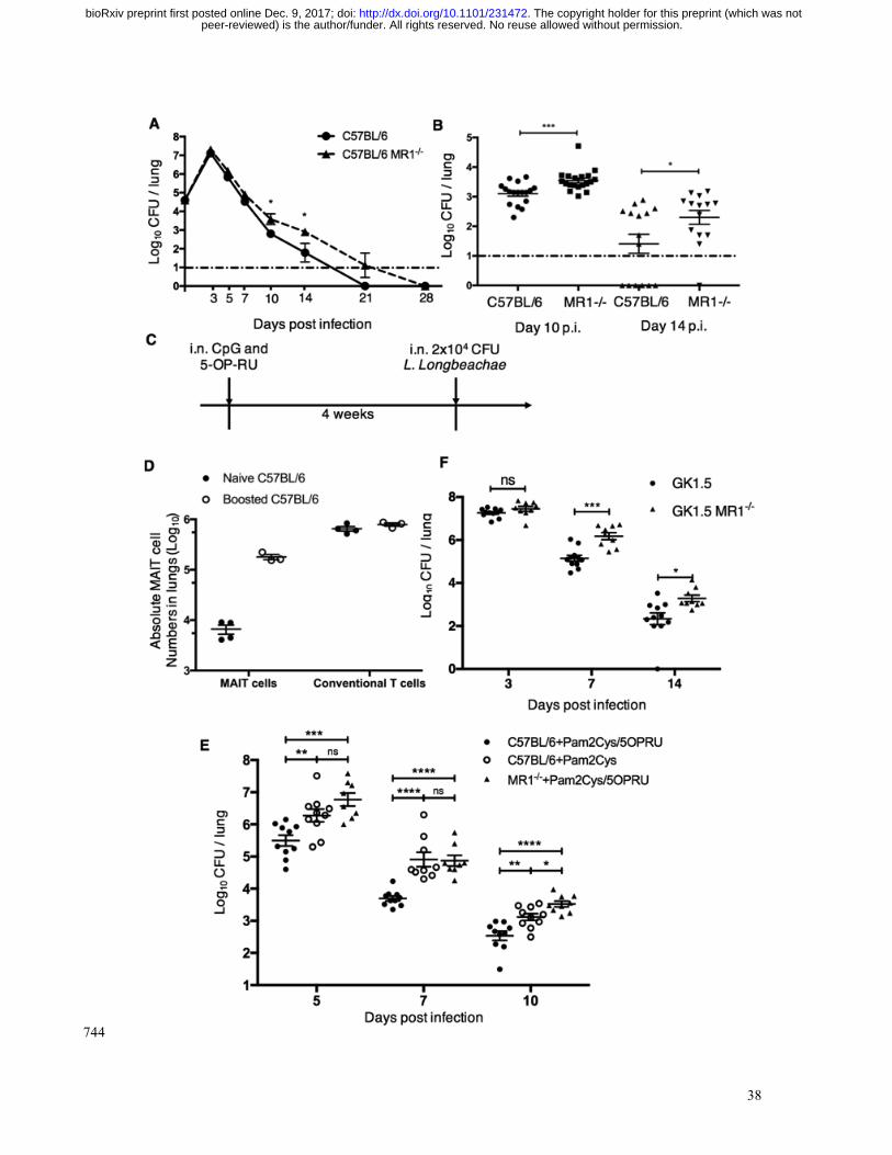
Figure 4. MAIT cells contribute to protection in murine Legionella infection in vivo, 745
which can be accelerated by prior ligand-induced MAIT cell expansion 746
(A,B). Bacterial load (CFU) in lungs C57BL/6 or MR1-/- mice following intranasal 747
infection with 2x10⁴ CFU L. longbeachae. Dashed line represents limit of detection. (A) 748
Bacterial load over the time-course of infection. (B). Bacterial load at days 10 and 14 749
post infection, from further three separate replicates. (C). Schematic for panel D and E: 750
C57BL/6 or MR1-/- mice were treated with 20nmol S-[2,3-bis(palmitoyloxy)propyl] 751
cysteine (Pam2Cys) and 76pmol 5-OP-RU (in 50µl) intranasally 1 month before 2x10⁴ 752
CFU L. longbeachae inoculation. (D) Absolute numbers of MAIT cells and conventional 753
T cells from lungs of naïve or ligand-boosted C57BL/6 mice 30 days after administration 754
of ligand. (E) Differences in bacterial load in lungs of C57BL/6 or ligand-boosted 755
C57BL/6 or MR1-/- mice apparent at 5, 7 and 10 DPI. (F) Bacterial load in lungs of mice 756
lacking CD4+ cells (GK1.5Tg) or CD4+ and MAIT cells (GK1.5Tg.MR1-/-) 3, 7 and 14 757
days after infection with 2x10⁴ CFU i.n. L. longbeachae. Pooled data (mean±SEM) from 758
two replicates with similar results using 5-6 mice per group, compared using t tests on 759
log-transformed data. *, P<0.05; **, P<0.01; ***, P<0.001; **** P<0.0001. 760
peer-reviewed) is the author/funder. All rights reserved. No reuse allowed without permission. The copyright holder for this preprint (which was not. http://dx.doi.org/10.1101/231472doi: bioRxiv preprint first posted online Dec. 9, 2017;

40
761
Figure 5. Adoptive transfer of MAIT cells rescues Rag2-/-gc-/- mice from fatal 762
pulmonary Legionella infection 763
(A) Schematic of protocol: 105 pulmonary MAIT cells from C57BL/6 mice previously 764
infected with 106 CFU S. Typhimurium BRD509 for 7 days to expand the MAIT cell 765
population were sorted and transferred intravenously into Rag2-/-gC-/- mice, followed by 766
C
A
MAITtransfer
Antibodyinjections
i.n. 103 or 104 CFUL. Longbeachae
1 week 1-2 weeks
D E.
B
WithoutMAIT transfer
WithMAIT transfer
TCR-
MR1
-5-O
P-RU
tet
RAG2-/- C-/-
mice
+ MAIT cells + MAIT cells /Anti-MR1 treatment
Log 1
0CF
U / l
ung
Days post infection
Perc
ent s
urviv
al
Days post infection
Perc
ent s
urviv
al
peer-reviewed) is the author/funder. All rights reserved. No reuse allowed without permission. The copyright holder for this preprint (which was not. http://dx.doi.org/10.1101/231472doi: bioRxiv preprint first posted online Dec. 9, 2017;

41
intraperitoneal anti-CD4 and anti-CD8 antibody injection (0.1mg each) twice within 1 767
week to deplete contaminating conventional T cells. After 2 weeks, mice were infected 768
with 103 or 104 CFU i.n. of L. longbeachae. (B) Representative plots showing live 769
(7AAD-) hematopoietic (CD45.2+) cells with percentages of MAIT cells in the lungs of 770
Rag2-/-gC-/- mice which were untreated or were recipients of adoptively-transferred 771
MAIT cells. (C) Survival of Legionella-infected untreated or MAIT cell-recipient Rag2-772
/-gC-/- mice after 103 CFU i.n. L. longbeachae infection. (D) Survival of Legionella-773
infected untreated or MAIT cell-recipient Rag2-/-gC-/- mice after 104 CFU i.n. L. 774
longbeachae, with or without MR1 blockade. One group received 0.25mg anti-MR1 775
monoclonal antibody alternate days after infection. (E) Pulmonary bacterial load in 776
surviving Rag2-/-gC-/- mice in (D) 23 DPI. Pooled data (mean±SEM) from two replicates 777
with similar results, each with 7-12 mice per group. See also Figure S5. 778
peer-reviewed) is the author/funder. All rights reserved. No reuse allowed without permission. The copyright holder for this preprint (which was not. http://dx.doi.org/10.1101/231472doi: bioRxiv preprint first posted online Dec. 9, 2017;

42
779
780
Figure 6. Protection of Rag2-/-gc-/- mice from fatal pulmonary Legionella infection 781
is dependent on IFN-g 782
(A) Survival of Rag2-/-gC-/- mice adoptively transferred with pulmonary MAIT cells 783
generated in different mouse strains after 104 CFU i.n. L. longbeachae. ****, Gehan-784
Breslow-Wilcoxon P<0.0001. (B) Pulmonary bacterial load in surviving Rag2-/-gC-/- 785
mice in (A) 23 DPI. Pooled data (mean±SEM) from two replicates with similar results, 786
B
A
Days post infection
Perc
enta
ge s
urvi
val
CFU
in lu
ng (l
og10
)
6
4
2
0
*** *
****
C57BL/6 PFP-/-IFN- -/- IL-17-/- GM-CSF-/- GranzymeA & B-/-
TNF- -/-
100
50
0
C57BL/6 n = 7
PFP-/- n=20
IFN- -/- n=21
TNF- -/- n=20
IL-17-/- n=20
GM-CSF-/- n=20GranzymeA & B-/- n=20
0 5 10 15 20 25
Genotype of transferred MAIT cells
peer-reviewed) is the author/funder. All rights reserved. No reuse allowed without permission. The copyright holder for this preprint (which was not. http://dx.doi.org/10.1101/231472doi: bioRxiv preprint first posted online Dec. 9, 2017;

43
each with 7-13 mice per group. Fig B shows mean. PFP, perforin. Bonferroni-corrected t-787
tests *, P<0.05; **, P<0.01; ***, P<0.001. 788
789
Supplementary Information 790
Supplementary Figures S1-S5 791
792
peer-reviewed) is the author/funder. All rights reserved. No reuse allowed without permission. The copyright holder for this preprint (which was not. http://dx.doi.org/10.1101/231472doi: bioRxiv preprint first posted online Dec. 9, 2017;