
Leg edema family medicine
79
Dr.Saeid Khezer Ahmed MBChB.,MSc. Family Medicine Duhok-Kurdistan of Iraq 2017.2.26 Practical Approach to Lower Extremity Edema
-
Upload
saeid-khezer -
Category
Health & Medicine
-
view
66 -
download
0
Transcript of Leg edema family medicine

Dr.Saeid Khezer AhmedMBChB.,MSc. Family Medicine
Duhok-Kurdistan of Iraq2017.2.26
Practical Approach to Lower Extremity Edema

Not Everything that Swells is Heart Failure

Overview
A common challenge for primary care physicians and cardiologists alike
Goal is to determine the cause and find an effective treatment for leg edema
Despite the prevalence, no formal existing practice guidelines at present

Definition
Edema is defined as a palpable swelling caused by an increase in interstitial fluid volume.
Edema, other than localized edema, does not become clinically apparent until the interstitial volume has increased by2.5 to 3 liters.
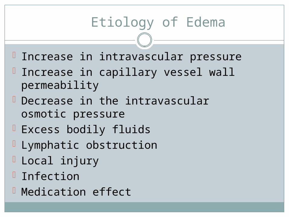
Etiology of Edema
Increase in intravascular pressure Increase in capillary vessel wall
permeability Decrease in the intravascular osmotic
pressure Excess bodily fluids Lymphatic obstruction Local injury Infection Medication effect

Anatomy and Pathophysiology
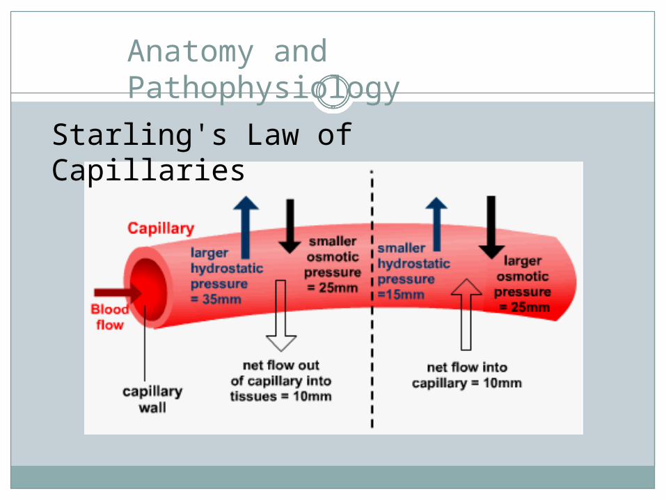
Anatomy and Pathophysiology
Starling's Law of Capillaries

Anatomy and Pathophysiology

Introduction
The most likely cause of leg edema in patients over age 50 is venous insufficiency Venous insufficiency
affects up to 30% of the population
Heart failure affects only approximately 1%

Introduction
The most likely cause of leg edema in women under age 50 is idiopathic edema (formerly known as cyclic edema).

Introduction
Most patients can be assumed to have one of these diseases unless another cause is suspected after a history and physical examination.
However, there are at least 2 exceptions to this rule: pulmonary hypertension early heart failure
Both conditions can both cause leg edema before they become clinically obvious in other ways.

Classification
There are two types of leg edema: Venous edema consists of excess low-viscosity,
protein-poor interstitial fluid resulting from increased capillary filtration that cannot be accommodated by a normal lymphatic system.
Lymphedema consists of excess protein-rich interstitial fluid within the skin and subcutaneous tissue resulting from lymphatic dysfunction.
A third type, lipidema, is more accurately considered a form of fat maldistribution rather than true edema.

History
Key elements of the history include What is the duration of the edema (acute [<72
hours] vs. chronic)? If the onset is acute, deep vein thrombosis should be
strongly considered (the 72-hour cutoff is commonly cited but arbitrary)

Unilateral
Acute (<72hours) Deep vein thrombosis
Chronic (<72 hours) Venous insufficiency
Bilateral
Acute (<72hours)
Chronic (<72 hours) Venous insufficiency Pulmonary
hypertension Heart failure Idiopathic edema Lymphedema Drugs Premenstrual edema
Common Causes of Leg Edema in the United States

Unilateral
Bilateral
Acute (<72hours) Ruptured Baker's cyst Ruptured medial
head of gastrocnemius
Compartment syndrome
Chronic (<72 hours) Secondary lymphedema (tumor,
radiation, surgery, bacterial infection)
Pelvic tumor or lymphoma causing external pressure on veins
Reflex sympathetic dystrophy
Acute (<72hours) Bilateral deep vein thrombosis Acute worsening of systemic cause
(heart failure, renal disease)
Chronic (<72 hours) Renal disease (nephrotic
syndrome, glomerulonephritis)
Liver disease Secondary lymphedema
(secondary to tumor, radiation, bacterial infection, filariasis)
Pelvic tumor or lymphoma causing external pressure
Dependent edema Diuretic-induced edema Preeclampsia Anemia
Less Common Causes of Leg Edema in the United States

Unilateral
Acute (<72hours)
Chronic (<72 hours) Primary lymphedema
(congenital lymphedema, lymphedema praecox, lymphedema tarda)
Congenital venous malformations
May-Thurner syndrome (iliac-vein compression syndrome)
Bilateral
Acute (<72hours)
Chronic (<72 hours) Primary lymphedema
(congenital lymphedema, lymphedema praecox, lymphedema tarda)
Protein losing enteropathy, malnutrition, malabsorption
Restrictive pericarditis Restrictive cardiomyopathy
Rare Causes of Leg Edema in the United States
History
Key elements of the history include What is the duration of the edema (acute [<72
hours] vs. chronic)? If the onset is acute, deep vein thrombosis should be
strongly considered (the 72-hour cutoff is commonly cited but arbitrary)
Is the edema painful? Deep vein thrombosis and reflex sympathetic
dystrophy are usually painful. Chronic venous insufficiency can cause low-grade
aching. Lymphedema is usually painless.

History
Reflex Sympathetic Dystrophy
Acute Deep Venous Thrombosis
Painful swelling?

History
Key elements of the history include: What drugs are being taken?
Calcium channel blockers, prednisone, and anti-inflammatory drugs are common causes of leg edema.

Common Causes of Leg Edema
MedicationsAntihypertensive drugsCalcium channel blockersBeta blockers Clonidine Hydralazine Minoxidil Methyldopa
Hormones Corticosteroids Estrogen Progesterone Testosterone
OtherNSAIDsMonoamine oxidase inhibitors Rosiglitazone Piogliatazone

History
Key elements of the history include: What drugs are being taken?
Calcium channel blockers, prednisone, and anti-inflammatory drugs are common causes of leg edema.
Is there a history of systemic disease (heart, liver, or kidney disease)?
Is there a history of pelvic/abdominal neoplasm or radiation?

History
Key elements of the history include: Does the edema improve overnight?
Venous edema is more likely than lymphedema to improve overnight.
Is there a history consistent with sleep apnea? Sleep apnea can cause pulmonary hypertension,
which is a common cause of leg edema. Findings that may increase suspicion of sleep
apnea include loud snoring or apnea noted by the sleep partner, daytime somnolence, or a neck circumference >17 inches.

Physical Examination
Body mass index. Obesity is associated with sleep
apnea and venous insufficiency.

Physical Examination
Distribution of edema: Unilateral leg edema is generally due to a local
cause such as deep vein thrombosis, venous insufficiency, or lymphedema.
Bilateral edema can be due to a local cause or systemic disease, such as heart failure or kidney disease.
Generalized edema is due to systemic disease.
The dorsum of the foot is spared in lipidema but prominently involved in lymphedema.
Physical Examination
Tenderness: Deep vein thrombosis and lipidema are often
tender. Lymphedema is usually nontender.
Pitting: Deep vein thrombosis, venous insufficiency,
and early lymphedema usually pit. Myxedema and the advanced fibrotic form of
lymphedema typically do not pit.

Physical Examination
Varicose veins: Leg varicosities are
often present in patients with chronic venous insufficiency, but venous insufficiency can occur without varicose veins

Physical Examination

Physical Examination
Kaposi-Stemmer sign: Inability to pinch a
fold of skin on the dorsum of the foot at the base of the second toe is a sign of lymphedema

Physical Examination
Skin changes: A warty texture (hyperkeratosis) with papillomatosis
and brawny induration are characteristic of chronic lymphedema.

Physical Examination
Skin changes: A warty texture (hyperkeratosis) with papillomatosis
and brawny induration are characteristic of chronic lymphedema.
Brown hemosiderin deposits on the lower legs and ankles are consistent with venous insufficiency.

Physical Examination
Skin changes: Venous Stasis Dermatitis

Physical Examination
Skin changes: A warty texture (hyperkeratosis) with papillomatosis
and brawny induration are characteristic of chronic lymphedema.
Brown hemosiderin deposits on the lower legs and ankles are consistent with venous insufficiency.
Reflex sympathetic dystrophy initially leads to warm tender skin with increased sweating. Later the skin is thin, shiny, and cool. In the chronic stage, the skin becomes atrophic and dry with flexion contractures.

Physical Examination
Reflex Sympathetic Dystrophy:

Physical Examination
Signs of systemic disease: findings of heart failure (especially jugular venous
distension and lung crackles) liver disease (ascites, spider hemangiomas, and
jaundice) may be helpful in detecting a systemic cause

Diagnostic Studies
Laboratory Tests
Most patients over age 50 with leg edema have venous insufficiency, but if the etiology is unclear, a short list of laboratory tests will help rule out systemic disease: complete blood count, electrolytes, creatinine urinalysis blood sugar thyroid-stimulating hormone albumin

Diagnostic Studies
Laboratory Tests
A serum albumin below 2 g/dL often leads to edema and can be caused by: liver disease nephrotic syndrome protein-losing enteropathy

Diagnostic Studies
Additional tests are indicated depending on the clinical presentation: Patients who may have a cardiac etiology should
have an electrocardiogram, echocardiogram, and chest radiograph.
Dyspneic patients should have a brain natriuretic peptide (BNP) determination to help detect heart failure.
The BNP is most helpful for ruling out (rather than ruling in) heart failure because the sensitivity is high (90%).

Diagnostic Studies
Additional tests are indicated depending on the clinical presentation: Idiopathic edema can be diagnosed in young women
without further testing if there is no reason to suspect another etiology based on history and physical examination.
However, tests to confirm idiopathic edema have been described and may be helpful in difficult cases.

Diagnostic Studies
Additional tests are indicated depending on the clinical presentation: In patients with acute edema (<72 hours)
A normal D-dimer will essentially rule out deep vein thrombosis if the clinical suspicion is low because false negative D-dimers are rare.
However, an elevated D-dimer should be followed up with a Doppler examination because false positive D-dimers are common.

Diagnostic Studies
Additional tests are indicated depending on the clinical presentation: Patients with possible nephrotic syndrome should
have serum lipids in addition to the basic laboratory studies listed above.

Imaging Studies
Patients over age 45 with edema of unclear etiology should have an echocardiogram to rule out pulmonary hypertension.

Imaging Studies
Patients over age 45 with edema of unclear etiology should have an echocardiogram to rule out pulmonary hypertension.
Lymphoscintigraphy can be helpful to distinguish lymphedema from venous edema and to determine the cause of lymphedema.
Lymphoscintigraphy is performed by injecting a radioactive tracer into the first web space and monitoring lymphatic flow with a gamma camera.

Imaging Studies
Lymphoscintigraphy

Treatment
Venous Insufficiency Chronic venous insufficiency is treated with leg
elevation and knee-high compression stockings that provide 30 to 40 mm Hg pressure at the ankle.
If arterial insufficiency is a concern, an ankle-brachial index should be performed because compression stockings are contraindicated in arterial insufficiency.
Patients who are refractory to compression stockings may improve with intermittent pneumatic compression pumps.

Treatment
Venous Insufficiency Horse chestnut seed extract (300 mg, standardized to
50 mg of escin, twice a day) has been found to be effective in several studies and can be obtained in health food stores. Horse chestnut seed extract contains escin, which
inhibits the activity of elastase and hyaluronidase. These enzymes are thought to play a role in the
pathophysiology of chronic venous insufficiency. However, the benefits are modest and the agent has
not gained widespread acceptance.

Treatment
Venous Insufficiency: Horse chestnut seed
extract

Treatment
Venous Insufficiency Diuretics (e.g., furosemide 20 to 40 mg once a
day with supplemental potassium) can be used for short periods in severely affected patients. However, venous insufficiency is not a volume overload
state, and long-term use of diuretics can lead to adverse metabolic complications.

Treatment
Idiopathic Edema Spironolactone is considered the drug of choice for
idiopathic edema because of the secondary hyperaldosteronism found in patients with this disorder.
The starting dose is 50 to 100 mg daily (maximum 100 mg, 4 times daily).
If spironolactone is not effective, low doses of a thiazide diuretic (e.g., hydrochlorothiazide, 25 mg daily) can be added with close monitoring of the serum potassium.
It is best to avoid loop diuretics.

Treatment
Idiopathic Edema The diuretic should be given in the early evening
because fluid retention is most noticeable at the end of the day.
Other measures include intermittent recumbency, avoiding environmental heat, low-salt diet, avoiding excessive fluid intake, and weight loss for obese patients.
It may be helpful to ask about depression, eating disorder, and surreptitious diuretic or laxative use.
Compression stockings are usually not helpful and not tolerated

Treatment
Idiopathic Edema Many patients with idiopathic edema are already
taking diuretics when first seen and may have "diuretic-induced edema."
Chronic use of diuretics may lead to a state of mild hypovolemia with resulting stimulation of the renin- angiotensin-aldosterone system.
When the diuretics are withdrawn, a rebound worsening of edema occurs and patients believe they must continue.

Treatment
Idiopathic Edema However, the treatment of suspected diuretic-
induced edema is to withdraw diuretics for 3 to 4 weeks after warning the patient that her edema will probably worsen initially and reassuring her that the diuretic can always be restarted.
If the edema does not improve after 4 weeks, spironolactone can be initiated at a dose of 50 to 100 mg daily and increased to a maximum of 100 mg, 4 times daily.

Treatment
Lymphedema Nonspecific treatment of lymphedema includes
exercise, elevation, compressive garments, manual lymphatic drainage, intermittent pneumatic compression, and surgery (excisional procedures, microsurgery).
Tinea pedis should be controlled, and prophylactic antibiotics may be indicated for recurrent cellulitis.
Diuretics are generally not helpful. Treatment of lymphedema is often
disappointing, and psychosocial support is important in such patients.

Treatment
Deep Vein Thrombosis An acute deep vein thrombosis is generally
treated with heparin initially. Warfarin can be initiated simultaneously with
heparin, starting with 5 to 10 mg daily for 2 days with subsequent dosage based on a target international normalized ratio range of 2.0 to 3.0.
Xarelto (rivaroxaban) has recently been approved for the treatment of DVT.
Treatment
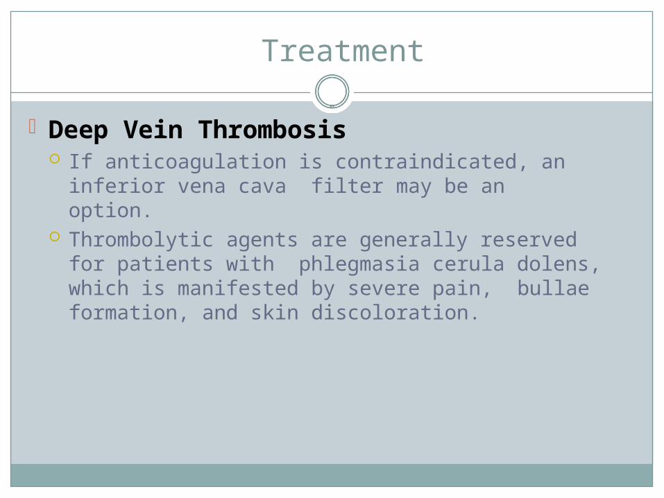
Deep Vein Thrombosis If anticoagulation is contraindicated, an inferior
vena cava filter may be an option. Thrombolytic agents are generally reserved for
patients with phlegmasia cerula dolens, which is manifested by severe pain, bullae formation, and skin discoloration.

Summary
In the approach to leg edema of unclear etiology, the physician should first rule out lipidema (fat maldistribution with sparing of feet) and lymphedema (marked foot and toe involvement, verrucous thickened skin, nonpitting when chronic) because subsequent evaluation and treatment are different for these disorders.
If systemic disease is considered unlikely, the most common causes of bilateral leg edema are idiopathic edema (in young women) and chronic venous insufficiency (in older patients).

Summary
In patients with chronic bilateral edema, the physician should consider the most common systemic causes (cardiac, renal, hepatic) and decide, based on history and physical examination, which of them need to be ruled out with further testing.
Pulmonary hypertension is a common cause and should be suspected in patients who may have sleep apnea (e.g., neck circumference >17 inches, loud snoring, or apnea noted by sleep partner).

Summary
If the patient presents with sudden onset (<72 hours) of leg swelling, a deep vein thrombosis should be ruled out using a Doppler examination.

Questions ???

Figure 1. Algorithm for leg edema

Figure 2. Common causes.

Figure 4. Unilateral edema
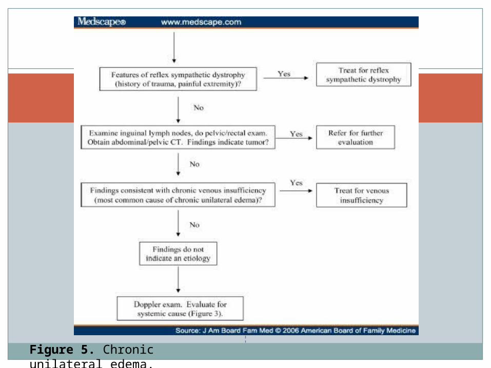
Figure 5. Chronic unilateral edema.

Figure 3. Systemic evaluation.

Common Causes of Leg Edema
Venous Insufficiency The diagnosis is usually made clinically but can be
confirmed with a Doppler study. Although chronic venous insufficiency is thought to
result from previous deep vein thrombosis, only one third of patients will give that history.
"Dependent edema" is a variant of venous insufficiency and often occurs in patients following stroke who sit in wheelchairs for long periods.

Common Causes of Leg Edema
Pregnancy Increased venous pressure resulting
from an enlarging uterus near term commonly leads to lower extremity edema and varicosities.
Edema is commonly present in patients with preeclampsia but is no longer considered a factor in making the diagnosis.

Common Causes of Leg Edema
Heart Failure Patients with congestive heart failure
complain of dyspnea, dependent edema, and fatigue.
On physical examination they may have elevated jugular venous pressure, basilar crackles on chest auscultation, gallop rhythm, and pitting edema.
BNP may be helpful in diagnosing heart failure among dyspneic patients.

Common Causes of Leg Edema
Pulmonary Hypertension Pulmonary hypertension commonly results
from sleep apnea and is under-recognized as a cause of edema.
Other causes of pulmonary hypertension include left heart failure and chronic lung disease.
Echocardiography can help in assessing pulmonary pressures.

Tests for Idiopathic Edema
Morning and Evening Weights: Patients should weigh themselves nude and with an empty bladder
before food or fluids in the morning and at bedtime. A mean weight gain >0.7 kg is consistent with idiopathic edema.
Water Load Test: After avoiding diuretics for at least 10 days, the patient drinks 20
mL/kg body weight (maximum 1500 mL) uniced water over 20 minutes, sometime between 7:30 AM and 9:00 AM.
The patient collects urine every hour, starting 1 hour before the oral fluid load and ending 4 hours after.
On the first day, the patient should be walking slowly or standing during this 4- hour period.
On the second day, the patient repeats the fluid load and urine collection, but should be recumbent during the 4-hour period.
In patients with idiopathic edema, less than 55% of water load is excreted in the upright position and more than 65% in the recumbent position.

Common Causes of Leg Edema
Pulmonary Hypertension Treating sleep apnea might improve the leg
edema that results from pulmonary hypertension, but this also is unknown.
An echocardiogram is recommended in patients who are at risk for pulmonary hypertension and in patients over age 45 with leg edema of unclear etiology.
Common Causes of Leg Edema
Idiopathic Edema Idiopathic edema occurs only in menstruating
women and is most common in the 20s and 30s. Synonyms include fluid-retention edema, orthostatic
edema, cyclical edema, and periodic edema. However, if symptoms persist throughout the
menstrual cycle, idiopathic edema should be distinguished from premenstrual edema.
Idiopathic edema leads to pathologic fluid retention in the upright position, and women typically notice a weight gain of>1.4 kg as the day progresses.

Common Causes of Leg Edema
Idiopathic Edema Patients often complain of face and hand edema in
addition to leg swelling. Several confirmatory tests are available, but the diagnosis is usually made clinically after ruling out systemic disease by history and physical examination.
The confirmatory tests in are indicated only when there is significant doubt about the diagnosis.
Obesity and depression can be associated with this syndrome, and diuretic abuse is common.

Common Causes of Leg Edema
Drugs Calcium channel blockers and nonsteroidal
anti- inflammatory drugs (NSAIDs) are most commonly implicated.
The incidence of edema in patients taking NSAIDS is approximately 5%.
Up to 50% of patients on calcium-channel blockers develop edema. Dihydropyridines (amlodipine, nifedipine) may be more
likely to induce edema than phenylalkylamines (verapamil) or benzothiazepines (diltiazem).

Common Causes of Leg Edema
Primary lymphedema is a rare disorder that is divided into 3 types according to age of presentation. Congenital lymphedema may be present at birth or
becomes manifest by age 2 years. The familial form of congenital lymphedema is an autosomal dominant disorder known as Milroy disease.
Lymphedema praecox, the most common form of primary lymphedema, has its onset between age 2 and 35 and has a female to male ratio of 10:1. Lymphedema praecox is usually unilateral and is limited to the foot and calf in most patients. The familial form of lymphedema praecox is an autosomal dominant disorder known as Meige disease.
Lymphedema tarda presents after age 35.

Common Causes of Leg Edema
Secondary lymphedema Is much more common than primary, and the
cause is generally apparent from the history. The most common causes of leg lymphedema
are: tumor (e.g., lymphoma, prostate cancer, ovarian cancer) surgery involving lymphatics radiation therapy infection (bacterial infection or filariasis)

Common Causes of Leg Edema
Venous Insufficiency Venous insufficiency is characterized by:
Chronic pitting edema, often associated with brown hemosiderin skin deposits on the lower legs.
The skin changes can progress to dermatitis and ulceration, which usually occur over the medial maleoli.
Other common findings include varicose veins and obesity. Most patients are asymptomatic but a sensation of
aching or heaviness can occur.

Common Causes of Leg Edema
Secondary lymphedema: Filariasis
Common Causes of Leg Edema
Secondary lymphedema Chronic lymphedema is usually distinguished from
venous edema based on characteristic skin changes, absence of pitting, and history of an inciting cause.
The skin becomes thickened and darkened and may develop multiple projections called lymphostatic verrucosis.
The dorsum of the foot is prominently involved and may have a squared-off appearance.
The examiner is unable to pinch a fold of skin on the dorsal aspect of the base of the second toe (Kaposi-Stemmer sign).

Common Causes of Leg Edema
Obesity Obesity itself does not cause leg edema but obesity
can lead to many other causes such as chronic venous insufficiency, lymphedema, idiopathic edema, and obstructive sleep apnea.
Premenstrual Edema Most women experience some premenstrual edema
and weight gain. The edema tends to be generalized, occurs a few days before the beginning of menses, and resolves during a diuresis that occurs with the onset of menses.
The etiology is poorly understood.

Common Causes of Leg Edema
Deep Vein Thrombosis Deep vein thrombosis classically results in an acutely
swollen, painful leg that may be discolored. However, the presentation can be more subtle
with mild, painless, asymmetric edema. The physical examination is often unreliable and
patients with acute edema usually require further evaluation, which may include a D-dimer determination and a Doppler study.
Risk factors for deep vein thrombosis include cancer, immobilization (especially following surgery or an injury), and a hypercoagulable state.


