Visit at MIT: Daniel Jackson’s Lectures on Coupling Karl Lieberherr .

1
Lectures 23-25Fall 2006
Prof. Daniel Kahne
In the last set of lectures, you learned that cell growth and division mustbe carefully coordinated. When things go wrong at key steps, cancercan occur. In this block of lectures, you are going to learn about inter-and intracellular communication -- that is, how cells receive andrespond to information. You have learned that there are specificchemical signals that control whether cells are growing or dividing. Youhave also learned that most cells are not growing or dividing at anygiven time. You will now learn about the delicate mechanisms involvedin cell signaling, and you will learn some of the ways in which cellsignaling can go wrong.

2
Anti-Viral Agents
Fuzeon (blocks HIV fusion)
AZT (blocks reverse transcription)
Dupont-Merck Inhibitor (blocks HIV protease)
FUSION WITH CELL MEMBRANEAND ENTRY INTO CELL
UNCOATING OFRNA
RTRNA
DNA
TRANSCRIPTION
RNA
ASSEMBLY OF IMMATUREVIRIONS AND EXIT FROM CELL
TRANSLATION
RNA
VIRION MATURATION
We talked a number of weeks ago about protein structure and pointed out that proteins arethe workhorses of the cell. Without proteins, information-carrying molecules (DNA andRNA) cannot be decoded by any of the organisms that live today. Without proteins, virusescan’t co-opt cells to replicate. Given the importance of proteins in mediating cellularactivities, it is not surprising that most drugs target essential functions of various proteins. Inthe case of anti-HIV agents, you learned that there are several different steps in the viral lifecycle that can be targeted, and all of them involve inhibition of protein targets. In order tounderstand how these drugs work, you needed to understand the basics of protein structureand function. Fuzeon, which Erin talked about, prevents the fusion between the HIV viralparticles and the cell. Fuzeon works by binding to glycoprotein gp41 on HIV, preventing itfrom rearranging to a conformation that brings the viral membrane and the host cellmembrane together. The conformational rearrangement that occurs involves interactionsbetween distant parts of the glycoprotein. Fuzeon mimics the C-terminal portion of theglycoprotein and thus blocks the interactions. AZT, which is a nucleotide mimic, is asubstrate for reverse transcriptase and it gets incorporated into the cellular DNA copy of theviral RNA genome. Remember that polynucleotides are synthesized from the 5’ to the 3’direction, and AZT has a normal 5’ end. Once it is incorporated, however, reversetranscription ceases because there is no 3’ OH on AZT. The Dupont-Merck HIV proteaseinhibitor works at a later step in the viral lifecycle, a step that occurs after the DNA copy ofthe viral RNA genome is synthesized, transcribed into messenger RNA, and translated tomake a polyprotein. HIV protease cleaves the inactive polyprotein into different activeproteins during the maturation of the assembled virion and unleash the active HIV particle.We tend to think of the virus of an evil monster that exists to replicate its own genome -- andthat is true -- but the RNA genome by itself has no function. Without the proteins thatmediate key steps in the lifecycle, the RNA genome wouldn’t be able to do anything. Itwould simply fall apart -- be degraded rapidly. (continues next page.)

3
Anti-Viral Agents
Fuzeon (blocks HIV fusion)
AZT (blocks reverse transcription)
Dupont-Merck Inhibitor (blocks HIV protease)
FUSION WITH CELL MEMBRANEAND ENTRY INTO CELL
UNCOATING OFRNA
RTRNA
DNA
TRANSCRIPTION
RNA
ASSEMBLY OF IMMATUREVIRIONS AND EXIT FROM CELL
TRANSLATION
RNA
VIRION MATURATION
The important point here is that only by understanding the molecular mechanisms by whichthe HIV virus infects the cell and replicates itself could we have successfully designed thesedrugs to specifically target the respective steps of the HIV life cycle. Unfortunately, it has notbeen as easy to design drugs to target cancer cells specifically over normal cells.

4
General Chemotherapeutic Agents
O
O
O
O
OH
OO
O
HO
NH
OH
OO
O
O
Taxol (blocks mitosis)
Camptosar(blocks DNA replication)
N
N
O
O
OHO
ON
O
N
G0
Until the past decade, virtually all treatments for cancer relied on the concept that tumorcells grow more rapidly than normal cells. The strategy for treating cancer was,therefore, to try to block proliferation of cells by blocking key steps in the cell divisioncycle. Andrew told you that the cell cycle is divided into distinct phases. Camptosarprevents progression through S phase, the phase in which DNA is replicated, by inhibitingtopoisomerase II (Topo II), an enzyme that facilitates DNA unwinding to enablereplication. There are many other anti-tumor drugs that block different functions requiredfor progression through S phase. Taxol functions later in the cell cycle. It stabilizesmicrotubules, which causes mitotic arrest. Blocks at any point in the cell cycle lead toapoptosis, or programmed cell death. The problem with these anti-cancer agents is thatthey are not selective for cancer cells per se. They will kill any cell that is dividing. Theyhappen to kill many more cancer cells than normal cells simply because the cancer cellsdivide more rapidly. But any normal cells that happen to be dividing will also die.Therefore, they are very toxic, and people on cancer chemotherapy get quite sick fromthe treatment itself. The reason why Andrew characterized these generalchemotherapeutic agents as blunt tools for the treatment of cancer is that these drugssimply target cellular processes that were not specific to the particular cancer treated. Inother words, these drugs are not mechanism-based drugs and they are not selective.
There are now better approaches to treat certain cancers, and it may be possible toextend these approaches to a large number of different types of cancer. Theseapproaches involve using compounds that selectively target signaling pathways in cancercells, the very pathways that go haywire when cancer develops.
5
Mechanism-specific Anti-Cancer Agents
Specific to Chronic MyelogenousLeukemia (CML)
N
N
NH
NH
N
N
N
O
Gleevec(blocks Bcr-Abl kinase
phosphorylation)
• Cell Signaling• Protein Kinases
Erin will tell you about a compound called Gleevec, the firstmechanism-based anti-cancer drug. Gleevec inhibits an enzyme calledBcr-abl, a protein kinase that is misregulated in cancer cells. Mostnormal cell types in the body don’t utilize this protein kinase and so thedrug is non-toxic. In order to understand how Gleevec does what itdoes, we need to learn about the cell signaling pathway that it affects.For this series of lectures, we will begin to talk about cell signalingpathways and go on to talk about protein kinases, enzymes that arecentral to the regulation of the cell, and of the organism.

6
OverviewRecognition
Signal transmissionand processing
Getting into the nucleusand transcription
1
2
3
Cells receive many kinds of information from their environment, andthis information influences their behavior. Extracellular “signals” carrythis information to the cell. For example, you have learned that cellsneed to control growth and regulate cell division, and to do this theyrely on growth factors and hormones produced inside other cells andsecreted into the extracellular milieu. Because growth factors andmany hormones cannot enter cells, there needs to be a way tocommunicate the presence of the signaling molecule to the proteinsthat are located on the inside of the cell. Cells have specific receptorproteins embedded in the cellular membrane, which bind to hormonesand growth factors. When a hormone or growth factor binds to areceptor, the structure of the receptor changes. We will talk about howthe receptor changes later. The structural change in the receptor issensed by proteins inside the cell, which then affect the enzymaticactivities of other proteins. The signal is relayed by a series ofproteins, which comprise a signaling pathway. The ultimatedownstream targets of these signals can vary, but three main types ofproteins can be affected: proteins with enzymatic activity, such as theenzymes involved in the metabolic pathway; regulatory proteins, suchas transcription factors which control the expression of genes; orstructural proteins, such as those involved in maintaining the shape ofthe cell.

7
Lect 23-25. Cell Signaling and Regulation of Gene Expression:1. Regulation of gene expression.
a. Review of gene expressionb. Regulation of gene expression in prokaryotesc. Regulation of gene expression in eukaryotes (lecture notes)d. Inducers: Jacob and Monod diploid analysise. Le Chatelier's Principle
2. Growth factors and cell signaling.a. Growth factors and growth factor receptorsb. Tyrosine phosphorylation: a post-translational strategy for
modulating gene expressionc. Phosphates in biologyd. Ras, GTPases, and biological timescalese. MAP kinase cascade
Alberts pp. 532-539, 557-560, 267-281, 106-109, 150-154, 67McMurry pp 192-207, 335-338, 656-658
Lecture Readings
As I said, this section is about “cell signaling”, the process by which cells detect molecules in theenvironment and transmit information about the detected molecules to other parts of the cell.Since many cellular signals elicit responses at the level of gene expression, we will start by talkingin some detail about gene expression and its regulation in prokaryotes (while the overall picture issimilar, there are differences in the regulation of gene expression between prokaryotes andeukaryotes and those differences are detailed in the notes at the end of this section of lectures).In this section we will discuss the experiments which led two scientists Jacob and Monod topropose the correct mechanism for regulation of gene expression. We will then introduce howgrowth factors function. Growth factor function involves binding events between biologicalmolecules as well as chemical reactions that alter protein structure. The chemical reactionsinvolve a process known as phosphorylation, and we will describe what phosphorylation is andthen ask why Nature uses phosphates so frequently in biological signaling pathways. Whatchemical properties make phosphates so well suited to play this role? In answering this question,we will talk about the differences between kinetic and thermodynamic stability, and how biologyrelies on molecules that are kinetically stable but thermodynamically unstable. We will also talkabout how phosphorylation can change the activity of proteins -- affecting their conformations,binding interactions, and localization in different parts of the cell. In the section on bindinginteractions, we will talk about some important quantitative aspects of biology, with an emphasison understanding the relationship between the thermodynamics of binding interactions and thekinetics of biological processes. As Andrew mentioned, there are so-called “timers” in biology thatrely on small differences in the thermodynamic and kinetic behavior of different possible substratesto achieve remarkable fidelity. Without fidelity in processes involving growth and division, wecouldn’t exist. You will see that there is very delicate balance of factors that lead to fidelity, andsmall changes in individual proteins can be disastrous. This section will set up a final lecture byErin in which an understanding of how signaling processes are regulated led to a hypothesis abouthow to develop a small molecule to cure a particular type of cancer. This work introduced a newparadigm for cancer chemotherapy.

8
Gene expression/transcription in prokaryotes
RNA polymerase binds to the -10 and -35 sequencesRNA polymerase is comprised of subunits:
Core RNA polymerase -- responsible for elongationHoloenyzme of transcript
σ factor -- guides RNA polymerase to the the start site
When we discussed HIV you heard about the important role that RNApolymerase plays in converting DNA into mRNA. There are DNA sequencescalled promoters that RNA polymerases interact with before transcriptionstarts. Promoters typically reside upstream of the coding sequence of genes.Promoters, in both bacterial and eukaryotic cells contain three important parts1) the initiation site, where transcription begins; 2) a site that is required forbinding of RNA polymerase to the promoter; and 3) sequences important forthe binding of regulatory transcription factors that influence whether the geneis turned on or off. Bacterial transcriptional systems are the simplest andbest understood and can be used to illustrate how generegulation/transcription is controlled. In bacterial systems, genes that areinvolved in common biological functions are often arranged in a group closetogether on the chromosome. These groups of genes called operons aretypically transcribed from one promoter to produce one long mRNA, which isthen transcribed to form multiple proteins. In bacterial systems, RNApolymerase is comprised of a core, which contains several subunits, and asigma factor. The core RNA polymerase is the machine that actuallycatalyzes elongation of the transcript. RNA polymerase by itself cannotinitiate transcription. It doesn’t know where to start. A Sigma factor isimportant for guiding RNA polymerase to the initiation site and it falls off onceproductive elongation has begun. Sigma factor is thus a regulatory proteinthat controls which genes get transcribed. (Walter Gilbert of HarvardUniversity discovered the subunits of the core RNA polymerase.)

9
Regulation of gene expression
• One σ factor (σ70) is responsible for transcribing 90% of genesin E. coli.
• When environmental conditions change, bacteria must turn onalternative genes to express proteins that allow them to thriveunder those new conditions.
• More specialized σ factors [(σH) and (σE)] are needed to activatedifferent genetic programs in the cell.
In bacteria, there can be many sigma factors. Some organisms containas many as 65 sigma factors. Escherichia coli contains seven sigmafactors. In E. coli, which is the major model bacterial organism, onesigma factor, sigma 70, is responsible for transcribing 90% of genes. Ittranscribes the “housekeeping” genes necessary for basic metabolicfunctions involved in cell growth and division. Bacteria also containgenes that allow them to survive and ultimately thrive even when theirenvironment changes dramatically. These genes are normally “off”, butare turned on when changes in the environment are sensed.Expression of these genes is controlled by specialized sigma factors.Escherichia coli makes its home in the intestines of animals, includinghumans, and the environment in the intestines is pretty well controlled,which may be why E. coli has so few sigma factors compared withsome other bacteria. One of the sigma factors in E. coli responds toincreases in temperature. Temperature increases lead to changes inprotein stability. Some proteins denature when the temperature risesonly a few degrees. Under conditions of “heat shock”, a set of proteinsmust be expressed that act as chaperones to refold the denaturedproteins or as proteases that clear by degradation the accumulatedmisfolded proteins. Sigma H is a heat-shock sigma factor that binds todifferent promoters from sigma 70 and activates an alternative geneticprogram. Sigma E is another important sigma factor involved inresponses to other kinds of stress, such as damage to the bacterial cellwall.

10
How does one control subsets of genesthat use the same sigma factor?
Negative regulation -- repressors
RNA pol cannot bind promoter site when repressor is bound
One way that bacteria control which genes are transcribed is throughthe agency of sigma factors. We learned, however, that one sigmafactor is responsible for transcription of 90% of genes. You mightwonder how the bacteria control which subset of these genes istranscribed at any given time. There are two ways that bacteria canindividually regulate gene transcription from promoters that use thesame sigma factor. One way is called “negative regulation” andinvolves the use of repressor proteins that bind to sites overlapping thepromoter site. The repressor binding sequence is called the operator.When a repressor is bound to an operator, RNA polymerase cannotbind to the overlapping promoter site and so it cannot transcribe thegene.

11
Some sigma factors do not bind apromoter sequence without help.
Positive regulation -- activators
Activators help recruit sigma factors to increase gene expression
Other transcriptional regulators function in the opposite manner to repressors. Thatis, rather than repressing transcription, they help to turn genes on. Proteins that dothis are called activators. The promoters of genes that are controlled by activatorstend to be poorly transcribed in the absence of the activator because sigma factordoesn’t bind well to the particular DNA sequences in these promoters. There aremany promoter sequences, and some are better than others at recruiting sigmafactor and RNA polymerase (holoenzyme). Promoters that have “consensus”sequences at -10 and -35 bind RNA holoenzyme strongly and initiate transcriptionefficiently, but other promoters initiate transcription poorly -- or sometimes not at allunless an activator is present. The activator functions by binding to DNA upstream ofthe -35 site (typically) and contacting the RNA holoenzyme. These contacts to theactivator enhance binding to the promoter so that transcription can occur.
The genes in bacteria are all contained on one continuous circular chromosome.Moreover, individual genes which code for proteins which are involved in similarprocesses are grouped together in operons. In eukaryotic systems there are 23different chromosomes, and genes with similar functions are typically not groupedtogether. Genes in eukaryotic systems respond to many more signals because theorganism is multicellular and comprised of many different cell types found in differentenvironments. Gene regulation is accordingly much more complex.Initiation of transcription in eukaryotes differs from bacteria in several ways,discussed in the following slides (These slides were not discussed in lecture).

12
Bacteria maintain one cell type;eukaryotes have many types of cells.
BacteriumBacteria
Growth
UndifferentiatedEukaryotic Cell
GrowthDifferentiation
Eukaryotic genes not grouped on chromosomes
Prokaryotic genes grouped in operons on chromosome
Note: This section not covered in lecture is for your own interest.Some is a review of Rob’s material.
In eukaryotic systems, all of the cells contain the same 23 pairs ofchromosomes. Different proteins are produced by different cell types,allowing for different form and function. (Hair follicle cells are differentform liver cells, for example.) Cells need to determine which proteinswill be produced by controlling which genes are transcribed.

13
How eukaryotic gene expression differsfrom prokaryotic gene expression
• DNA is packaged into nucleosomes in the nucleus.
• 3 RNA polymerases
• Many transcription factors
• Promoters are much more complicated
You heard about transcription in prokaryotic cells in which genes aretypically regulated by one sigma factor with perhaps an additional levelof regulation by repressors or activators. The genes are transcribed byRNA polymerase, and their promoters are simple, consisting of a DNAbinding site(s) for the sigma factor (which can be different for differentgenes) as well as a binding site for RNA polymerase.
Transcription in eukaryotic cells is different. DNA in eukaryotic cells ispackaged into nucleosomes, which prevent DNA binding proteins fromreaching their sites in the genome. Eukaryotic cells have threepolymerases rather than just one. The three polymerases areresponsible for the transcription of different sets of genes. Manydifferent proteins (transcription factors) are required for transcriptionin eukaryotic cells because binding of the RNA polymerase to itsbinding site (the TATA box) is weak. Finally, the promoters ineukaryotic cells are complex. In addition to the RNA polymeraseconsensus binding site (the TATA box), they consist of binding sites formany transcription factors. Some of these binding sites are far awayfrom the RNA polymerase binding site, so the promoter region can bevery long.

14
Eukaryotic DNA
Chromatin remodeling is important in regulationof gene transcription.
This material will not be covered on the final exam!!!!!
As you have learned, the nucleus of a typical human cell contains about 2 meters of DNA.In order to fit the DNA into the nucleus, the DNA is packaged into chromatin by specializedproteins, which bind to and compact the DNA into a series of coils and loops. The mostfundamental packing level of chromatin is called the nucleosome, which consists of acomplex of 8 histone proteins and double stranded DNA of approximately 146 base pairs.The eight histone proteins are rich in positively charged amino acid side chains and form aprotein core around which the negatively charged double stranded DNA winds. Inbetween adjacent nucleosomes is linker DNA that is exposed and not complexed with thehistone proteins. Nucleosomes in a cell are packed together to generate a compactstructure called the 30 nm fiber, which is folded upon itself to form a compactedchromosome. Packaging of DNA into nucleosomes generally inhibits binding oftranscription regulatory proteins and the transcription initiation complex.
To gain access to binding sites covered by nucleosomes, the nucleosomes must beremoved or destabilized so that transcription factors can compete for binding to theirconsensus sites, or the nucleosome must be moved. In general, nucleosomes are moved,destabilized, or removed by two kinds of protein complexes: ATP-dependent chromatinremodeling complexes that use the energy derived from ATP hydrolysis to move or removenucleosomes; and histone modifying enzymes such as histone acetyltransferases (HATS),which covalently attach an acetyl group to the amino group of lysine, neutralizing thepositive charge. This destabilizes the nucleosome (because electrostatic interactionsbetween the positively charged histones and negatively charged DNA play a role inbinding) and can also affect the binding of other proteins to the nucleosome.

15
Three RNA polymerases:
tRNA genes5S rRNA geneRNA polymerase III
All protein-coding genesRNA polymerase II
Most rRNA genesRNA polymerase I
Genes TranscribedType of Polymerase
Unlike prokaryotic cells, which use only one type of RNA polymerase,eukaryotic cells use three types of RNA polymerases. The three RNApolymerases are responsible for transcription of different sets of genes.RNA polymerase I (RNA Pol I) transcribes ribosomal RNA genes. Theribosome, the machine for protein translation, contains many ribosomalRNA molecules, which play essential roles in structure, substratebinding, and catalysis.
RNA Pol II transcribes all the regular genes that encode proteins.
RNA Pol III transcribes all the genes that encode the tRNAs required forprotein translation.
On the next slide, we are going to focus only on RNA Pol II.
16
Many proteins are required fortranscription in eukaryotes
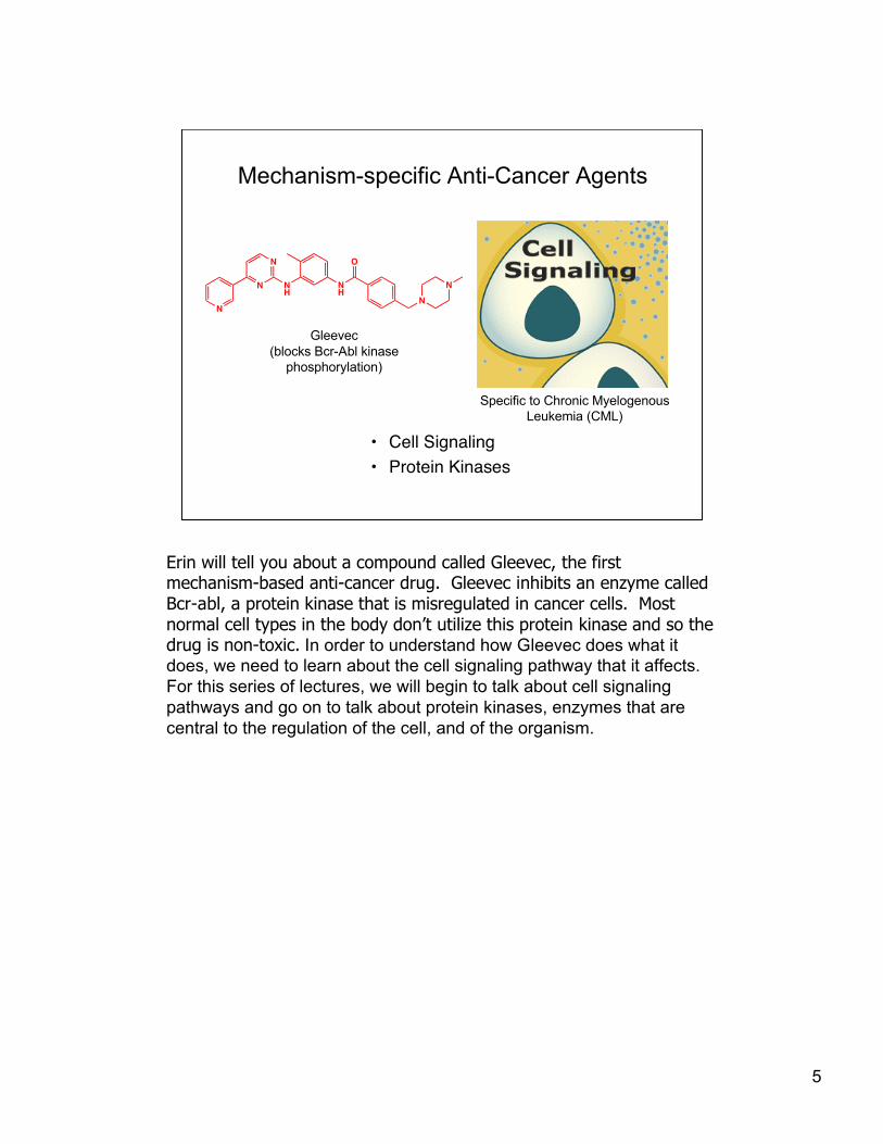
Much of what has been learned about transcription has come from biochemical studies,purifying the proteins involved in this process and studying transcription in vitro. Thesestudies have revealed that bacterial RNA pol consists of four subunits, which can transcribeDNA in the absence of any other protein. In contrast, purified RNA pol II, consists of morethan ten subunits, but is unable to transcribe DNA unless at least 20 other proteins, called“general transcription factors”, are also present. In addition, there is a TATA binding proteinthat plays an important role in bringing RNA pol II and the general transcription factors to thepromoter. The TATA binding protein binds to the TATA box that is found in the promoters ofalmost all genes transcribed by RNA pol II. The TATA box is one feature in the DNA thatallows putative protein-encoding genes to be identified from genome sequences.
17
Promoters in eukaryotic cells arecomplex
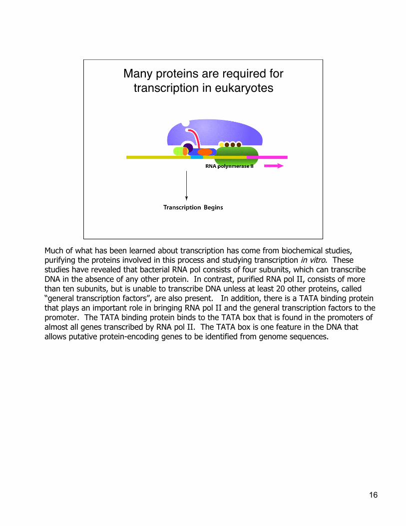
Complex promoters, numerous regulatory proteins,and chromatin structure allow for “combinatorial”control over gene transcription in eukaryotes.
You learned that prokaryotic promoters consist of two consensus sites of six base pairs each.One consensus site is found 10 base pairs upstream of the initiation site and the other isfound 35 base pairs upstream. Promoters in eukaryotic cells are far more complex. Theyconsist of binding sites for many different transcription factors as well as the TATA box. Someof the binding sites for transcription factors are located far from the RNA pol II binding site,and the intervening DNA must loop around for the proteins bound far upstream to makecontact to the RNA polymerase.
The features that make eukaryotic gene transcription different from prokaryotic genetranscription allow for complex patterns of gene regulation. Genes can be influenced bymultiple signaling pathways -- which can affect both nucleosome structure/stability (throughchanging patterns of post-translational modifications such as histone acetylation) and theavailability of particular transcription factors which can modify the efficiency of genetranscription. The integrated signals can give distinct patterns of gene expression in differentcell types.
Another way of putting it: Instead of having just one regulatory protein for transcription ofgenes, there are many regulatory proteins, which allows for greater variation of responses.This “combinatorial” control is required because the complexity of differentiated eukaryoticsystems is much higher. We need to be able to turn on different genes in different cells atdifferent times to function as a differentiated organism. (FYI: humans have only about fourtimes as many genes as many soil microorganisms -- but we, and all mammals, are clearly farmore complex.)
18
Question
But how do repressors and activatorscontrol gene expression?
Answer
The activity of the repressors andactivators must change somehow.
We just learned that proteins called repressors determine whether agene is expressed by controlling access to the promoter sites that bindRNA polymerase and that activators can cause gene expression frompromoters that don’t have the consensus sequence that is recognizedby “housekeeping” sigma factors. You might wonder what controls therepressors and activators that control gene regulation. After all, if theactivities of repressors and activators were constant, we wouldn’t needthem at all. Somehow, cells must be able to sense environmentalsignals -- changes outside or within the cell -- and respond to them toalter gene regulation in a way that is beneficial to the cell.
19
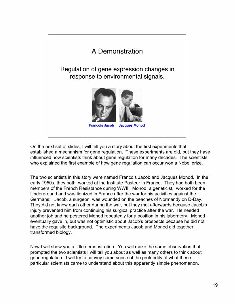
A Demonstration
Regulation of gene expression changes inresponse to environmental signals.
Francois Jacob Jacques Monod
On the next set of slides, I will tell you a story about the first experiments thatestablished a mechanism for gene regulation. These experiments are old, but they haveinfluenced how scientists think about gene regulation for many decades. The scientistswho explained the first example of how gene regulation can occur won a Nobel prize.
The two scientists in this story were named Francois Jacob and Jacques Monod. In theearly 1950s, they both worked at the Institute Pasteur in France. They had both beenmembers of the French Resistance during WWII. Monod, a geneticist, worked for theUnderground and was lionized in France after the war for his activities against theGermans. Jacob, a surgeon, was wounded on the beaches of Normandy on D-Day.They did not know each other during the war, but they met afterwards because Jacob’sinjury prevented him from continuing his surgical practice after the war. He neededanother job and he pestered Monod repeatedly for a position in his laboratory. Monodeventually gave in, but was not optimistic about Jacob’s prospects because he did nothave the requisite background. The experiments Jacob and Monod did togethertransformed biology.
Now I will show you a little demonstration. You will make the same observation thatprompted the two scientists I will tell you about as well as many others to think aboutgene regulation. I will try to convey some sense of the profundity of what theseparticular scientists came to understand about this apparently simple phenomenon.

20
All cells need energy
Glucose is the preferred carbon source.
OHOHO
OHHO
OH
Glucose
Central MetabolismCO2 + H2O + ATP (energy source)
I have to start by telling you some facts. One you already know. Cellsare not at equilibrium. (You would be dead if you reached equilibrium.)All cells need energy to drive metabolic processes. The energy comesfrom ingested nutrients. It is essential to have some carbohydratesource. (Carbohydrates contain carbon, hydrogen, and oxygen.)Oxidation of the carbons in that carbohydrate source through essentialmetabolic pathways generates ATP. ATP is the energy currency of thecell -- the molecule that is used from bacteria to humans to store energyfor future use. Glucose is the preferred carbon source in both E. coliand in humans. These facts about metabolism have been known for avery long time, although with increasing detail.

21
Other sugarscan be converted to glucose
LactoseGalactose Glucose
enzymes
β-galactosidase only “recognizes” the galactose part of the lactose so…
β-linkage
OOHO
OHHO
OH
O
HO
HO
HO
OH
OH
O
HO
HO
HO
OH
OHOHO
OHHO
OH
!-Galactosidase
H20
+
It has also been know for a long time that it is not necessary to ingestglucose itself to use glucose. Many sugars contain glucose as abuilding block or can be converted to glucose through variousenzymatic processes. Lactose, the major sugar in milk and cheese,contains one unit of glucose and one unit of galactose. Galactosediffers from glucose in having a different configuration of one hydroxylon the ring (at C4). Lactose is converted to its building blocks, glucoseand galactose, through the enzymatic hydrolysis of the glycosidiclinkage. The enzyme that performs this reaction is called beta-galactosidase. Beta-galactosidase only recognizes the galactose partof the molecule and so it will hydrolyze not only lactose, but anymolecule containing galactose coupled through a beta linkage toanother molecule. In the case of lactose, the galactose that isproduced is itself converted to glucose through the action of three otherenzymes.

22
β-galactosidase can hydrolyze otherβ-linked galactosides
O-nitrophenyl-galactoside (ONPG)
galactose O-nitrophenol
ONPG hydrolysis produces a yellow product, ONP.A yellow color formed upon addition of ONPG indicates thepresence of active β-galactosidase.
!-Galactosidase
H20
O
O
HO
HO
HO
OH
NO2
OH
O
HO
HO
HO
OH O-
NO2+
Recall that beta-galactosidase only recognizes the galactose portion ofsubstrates. Therefore, biochemists have been able to develop assaysin which chromogenic substrates (substrates containing leaving groupsthat are colored) are used to report on enzymatic activity. One beta-Galassay that is commonly used involves hydrolysis of ONPG, ortho-nitrophenyl-galactoside. The starting material is colorless but beta-Galhydrolysis generates a product, ONP (ortho-nitrophenol), which isyellow. The concentration of ONP formed as a function of timeprovides a measure of enzymatic activity. The concentration can bedetermined by measuring the absorbance and then using Beer’s law tocalculate the concentration. Beer’s law simply says that theabsorbance of a solution (how much light is absorbed at a givenwavelength) is related to the concentration by a proportionality constantcalled the extinction coefficient, which is a property of the molecule.
23
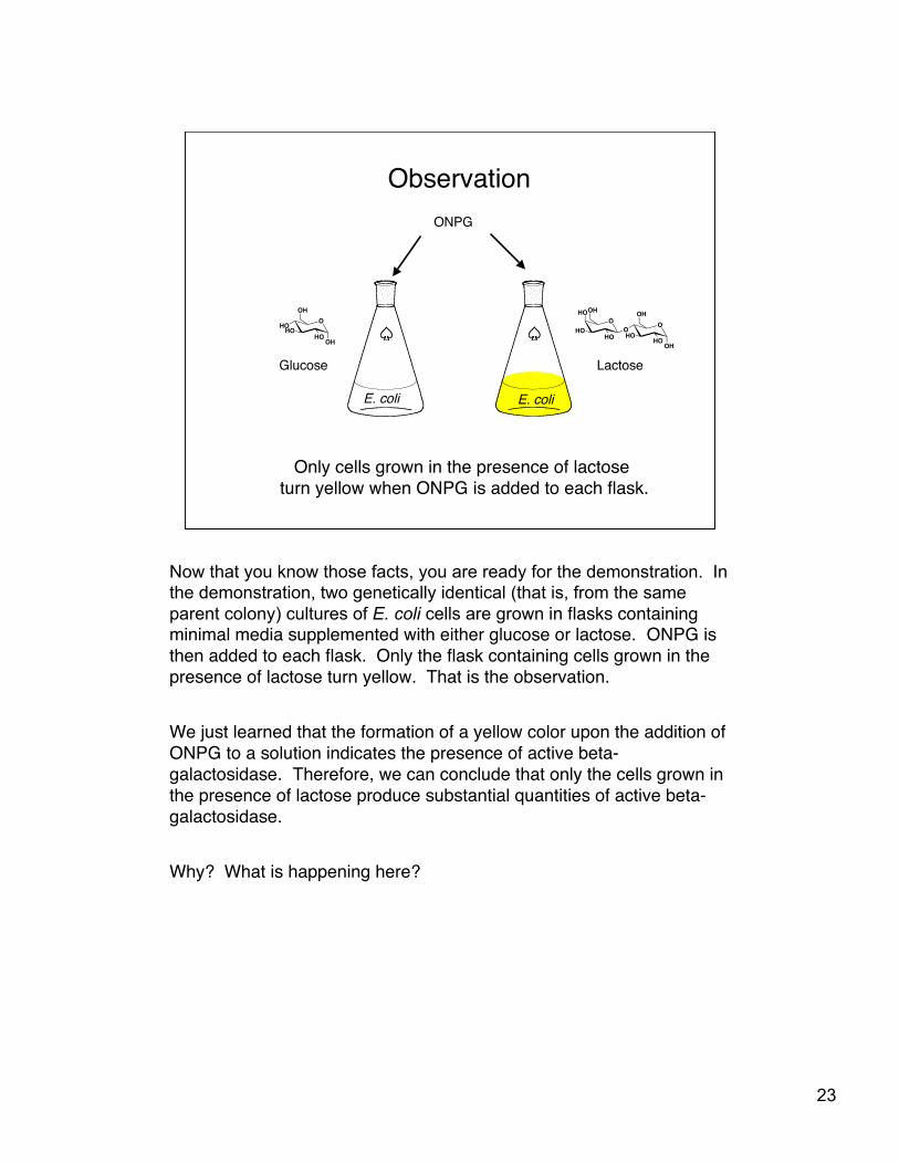
Observation
OOHO
OHHO
OH
O
HO
HO
HO
OH
OHOHO
OHHO
OH
Glucose Lactose
E. coli E. coli
Only cells grown in the presence of lactose turn yellow when ONPG is added to each flask.
ONPG
Now that you know those facts, you are ready for the demonstration. Inthe demonstration, two genetically identical (that is, from the sameparent colony) cultures of E. coli cells are grown in flasks containingminimal media supplemented with either glucose or lactose. ONPG isthen added to each flask. Only the flask containing cells grown in thepresence of lactose turn yellow. That is the observation.
We just learned that the formation of a yellow color upon the addition ofONPG to a solution indicates the presence of active beta-galactosidase. Therefore, we can conclude that only the cells grown inthe presence of lactose produce substantial quantities of active beta-galactosidase.
Why? What is happening here?
24
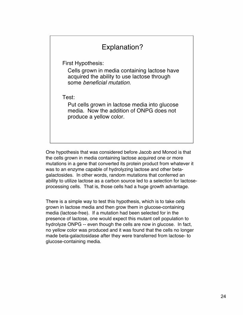
Explanation?
First Hypothesis:Cells grown in media containing lactose haveacquired the ability to use lactose throughsome beneficial mutation.
Test:Put cells grown in lactose media into glucosemedia. Now the addition of ONPG does notproduce a yellow color.
One hypothesis that was considered before Jacob and Monod is thatthe cells grown in media containing lactose acquired one or moremutations in a gene that converted its protein product from whatever itwas to an enzyme capable of hydrolyzing lactose and other beta-galactosides. In other words, random mutations that conferred anability to utilize lactose as a carbon source led to a selection for lactose-processing cells. That is, those cells had a huge growth advantage.
There is a simple way to test this hypothesis, which is to take cellsgrown in lactose media and then grow them in glucose-containingmedia (lactose-free). If a mutation had been selected for in thepresence of lactose, one would expect this mutant cell population tohydrolyze ONPG -- even though the cells are now in glucose. In fact,no yellow color was produced and it was found that the cells no longermade beta-galactosidase after they were transferred from lactose- toglucose-containing media.

25
Another explanation
Hypothesis 2:β-galactosidase is always present but inactive unlesssubstrate is present (preprotein hypothesis).
Test:Determine whether all substrates for the enzymeinduce the activity of enzyme.
Result: Some substrates do not induce!
Where does the β-galactosidase come from?
When this hypothesis was disproven, scientists began to pursue adifferent explanation: that the enzyme, beta-galactosidase, was alwayspresent but in an inactive state unless the substrate was present. Thishypothesis held that the substrate causes the enzyme to adopt itsactive conformation, perhaps by templating a folding process aroundthe substrate. Monod’s research group was exploring this hypothesisThey used radiometric experiments (pulse-chase experiments such asthe one Tim Hunt did to find cyclin) to show that the beta-galactosidaseprotein was freshly synthesized upon the addition of lactose. Thisfinding ruled out a model in which enzyme activation involves a foldingprocess.
In a key finding, Monod’s research group also identified somesubstrates that can be hydrolyzed by beta-galactosidase, but do notinduce the production of the protein in cells (for example, phenyl-galactoside).

26
Jacob and Monod…found a mutant E. coli strain that produces a yellowcolor in the absence of lactose.
Hypothesis:All E. coli cells contain a gene for β-galactosidase (lacZ).The expression of lacZ is under the control of a regulatoryprotein. The activity of the regulatory protein is controlledby lactose.
lacI lacZ
E. coli cell
Jacob entered the picture and continued to investigate the mystery ofbeta-galactosidase in Monod’s group. He showed that normal E. colicells cannot grow on plates in which the only carbon source is phenyl-galactoside, but he found a mutant that could. Mutant colonies grownon these plates and then transferred to glucose media produced ayellow color when ONPG was added to them. Therefore, these cellsproduce beta-galactosidase all the time. Jacob and Monod concludedthat there must be some gene that controls the expression of beta-galactosidase. Furthermore, that gene, whatever it is, must becontrolled by the presence of lactose.

27
Diploid analysisprovides insight into gene regulation
Hypothesis:lacI DNA makes a diffusible molecule (a protein) that turnsoff β-galactosidase expression (negative control).
lacI lacZ
E. coli cell (wild type)
lacI lacZ
E. coli cell (mutant)
lacI lacZ
Diploid cell is clear!
To try and understand the molecular basis for their observations, Jacoband Monod engineered a cell containing the DNA from wild-type cellsthat encodes the inducible expression of beta-galactosidase as well asthe DNA from mutant cells that encodes the constitutive expression ofbeta-galactosidase. (Inducible means turned on by a signal; andconstitutive means expressed all the time.) This cell has two copies(alleles) of the beta-galactosidase gene and its regulatory region (thecell is diploid with respect to these genes; you will learn more aboutMendelian genetics in LS1B). This new engineered cell did not turnyellow when ONPG was added to the media in which it was grownunless that media also contained lactose.
Jacob and Monod concluded that the DNA from the wild-type strain(containing the inducible gene) produces a diffusible molecule, aprotein, that is able to block transcription of beta-gal genes.

28
Jacob and Monod’s model
lacI lacZ lacI lacZ
OOHO
OHHO
OH
O
HO
HO
HO
OH
lactose
lactose
OOHO
OHHO
OH
O
HO
HO
HO
OH
DNA
OOHO
OHHO
OH
O
HO
HO
HO
OH
E. coli cell (wild type)
Lactose’s effect on lac repressor is an example of Le Chatelier’sPrinciple. (A system at equilibrium, subjected to a stress, will adjust to
relieve the stress and restore the equilibrium.)
lac repressor
DNA-boundlac repressor
DNA-binding conformation
Ligand-binding conformation
Ligand-boundlac repressor
Jacob and Monod proposed that the gene lacI makes a protein (shownas a red circle) that represses transcription of lacZ by binding at a sitethat overlaps the RNA polymerase binding site. Lactose, the signalingmolecule (or inducer) binds to this repressor (shown as a redrectangle), which they named the lac repressor, and causes it to fall offthe DNA. Later it was shown that lac repressor can adopt twoconformations, one that can bind DNA (circle) and one that does notbind DNA (rectangle). The two conformations are in equilibrium (theequilibrium lies very far towards the conformation that binds to DNA).Lactose stabilizes the conformation that does not bind to DNA, shiftingthe equilibrium very far in the other direction. This is an example of LeChatelier’s principle. (Le Chatelier’s principle states that a system atequilibrium subjected to a stress will adjust to relieve the stress andrestore the equilibrium.) For example, if the concentration of thereactants increases, the reaction will form more products. Equivalently,removing the products as soon as they are formed decreases theconcentration of the products and causes more products to be formed.Removing products as they form is an effective way to force all thereactants to react. In the example above, stabilization of the unboundconformation that cannot bind to DNA leads to depletion of the unboundconformation that can bind to DNA. As this concentration decreases,lac repressor falls off the DNA to reestablish an equilibrium between theDNA-bound and unbound --(but DNA-binding competent)-- forms of themolecule.)