Lecture Skin
35
Integumentary Integumentary System System
-
Upload
parveen-kumar -
Category
Documents
-
view
215 -
download
1
description
Brief description about the structure of skin in humans.
Transcript of Lecture Skin

Integumentary Integumentary SystemSystem

This system is divided into:This system is divided into:1- skin1- skin2- hair2- hair3- glands3- glands4- nails4- nails5- nerve endings5- nerve endings
I) I) Skin Skin Skin is an organ because it consists of Skin is an organ because it consists of
different tissues that are different tissues that are joined to perform a joined to perform a specific specific function.function.Largest organ of the body in surface Largest organ of the body in surface area and weight.area and weight.


Anatomy (structure)Anatomy (structure)
EpidermisEpidermis (thinner outer layer of (thinner outer layer of skin)skin)
Dermis Dermis (thicker connective tissue (thicker connective tissue layer)layer)
HypodermisHypodermis (subcutaneous layer or (subcutaneous layer or Sub-Q)Sub-Q)
Muscle and boneMuscle and bone

Epidermis: Keratinized stratified squamous epithelium Epidermis: Keratinized stratified squamous epithelium with four distinct cell types and five distinct layers. with four distinct cell types and five distinct layers.


Cells in the Epidermis:Cells in the Epidermis:
i) Keratinoytesi) Keratinoytes iii) Melanocytesiii) Melanocytesii) Merkel cellsii) Merkel cells iv) Langerhans’ cellsiv) Langerhans’ cells
1- Keratinocytes: most abundant 1- Keratinocytes: most abundant - produce keratin (fibrous protein)- produce keratin (fibrous protein)- protective; waterproofing the skin - protective; waterproofing the skin - continuous mitosis- continuous mitosis- formed in the deepest layer called the - formed in the deepest layer called the stratum basalestratum basale- cells push their way up to the surface - cells push their way up to the surface
where they are dead cells filled where they are dead cells filled with keratin; will slough off. Regenerates with keratin; will slough off. Regenerates every 25-45 days.every 25-45 days.


MelanocytesMelanocytes
- Cells produce brownish/black pigment called - Cells produce brownish/black pigment called melanin. (8% of epidermal cells) melanin. (8% of epidermal cells)
- stratum basale- stratum basale
- branching processes (dendrites)- branching processes (dendrites)
- melanin accumulates in melanosomes and - melanin accumulates in melanosomes and transported along dendrites of the transported along dendrites of the melanocytes to keratinocytes.melanocytes to keratinocytes.
- melanin accumulates on the superficial aspect - melanin accumulates on the superficial aspect of of the keratinocyte shielding its nucleus from the keratinocyte shielding its nucleus from UV rays.UV rays.
- lack of melanin: albino- lack of melanin: albino

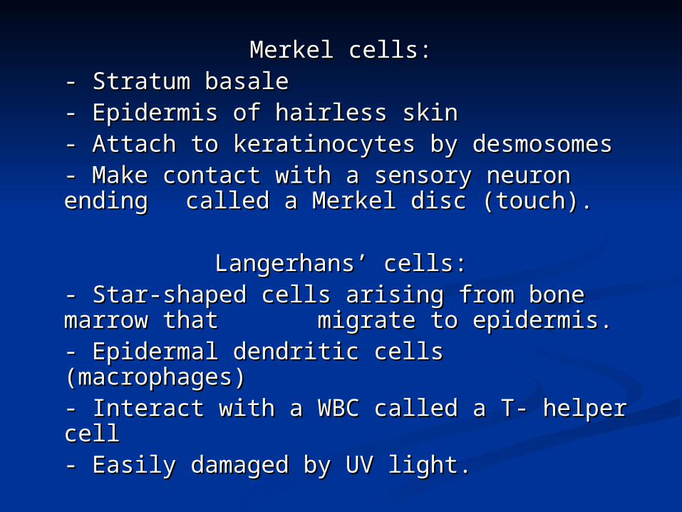
Merkel cells:Merkel cells:- Stratum basale- Stratum basale- Epidermis of hairless skin- Epidermis of hairless skin- Attach to keratinocytes by desmosomes- Attach to keratinocytes by desmosomes- Make contact with a sensory neuron - Make contact with a sensory neuron ending ending called a Merkel disc (touch).called a Merkel disc (touch).
Langerhans’ cells:Langerhans’ cells:- Star-shaped cells arising from bone - Star-shaped cells arising from bone marrow that migrate to epidermis.marrow that migrate to epidermis.- Epidermal dendritic cells (macrophages)- Epidermal dendritic cells (macrophages)- Interact with a WBC called a T- helper cell- Interact with a WBC called a T- helper cell- Easily damaged by UV light.- Easily damaged by UV light.


Stratum corneum
Stratum lucidum
Stratum spinosum
Stratum granulosum
Stratum basale

5 Layers of the epidermis5 Layers of the epidermis
1- 1- Stratum corneum (horny layer)Stratum corneum (horny layer)- - Layer has many rows of dead cells Layer has many rows of dead cells filled with filled with keratinkeratin
-- Continuously shed and replaced Continuously shed and replaced (desquamation)(desquamation) -- Effective barrier against light, heat Effective barrier against light, heat and bacteriaand bacteria- 20-30 cell layers thick- 20-30 cell layers thick

2- 2- Stratum lucidumStratum lucidum -- Seen in thick skin of the palms and Seen in thick skin of the palms and
soles of feet.soles of feet. - 3-5 rows of - 3-5 rows of clearclear flat dead cells flat dead cells
3- 3- Stratum granulosumStratum granulosum- 3-5 rows of flattened cells- 3-5 rows of flattened cells- Nuclei of cells flatten out- Nuclei of cells flatten out- Organelles disintegrate cells eventually - Organelles disintegrate cells eventually diedie
- Lamellated granules secrete - Lamellated granules secrete glycolipids into glycolipids into extracellular spaces to extracellular spaces to slow water loss in the epidermisslow water loss in the epidermis

4- 4- Stratum spinosumStratum spinosum: “spiny layer”: “spiny layer”
- 8-10 rows of polyhedral (many - 8-10 rows of polyhedral (many sided) cellssided) cells
- appearance of prickly spines- appearance of prickly spines
- melanin granules and Langerhans’ - melanin granules and Langerhans’ cell predominatecell predominate

5- 5- Stratum basaleStratum basale
-- Deepest epidermal layerDeepest epidermal layer- Attached to dermis- Attached to dermis- Single row of cells- Single row of cells
- mostly columnar keratinocytes- mostly columnar keratinocytes- with rapid mitotic division - with rapid mitotic division - stratum germinativum- stratum germinativum
- Contain merkel cells and melanocytes - Contain merkel cells and melanocytes
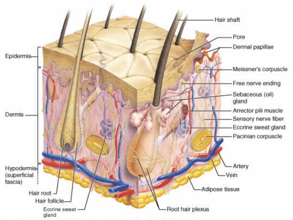

DermisDermis
- Flexible and strong connective tissue- Flexible and strong connective tissue- Elastic, reticular and collagen fibers- Elastic, reticular and collagen fibers- Cells: fibroblasts, macrophages (WBC),- Cells: fibroblasts, macrophages (WBC),
mast cells (histamine).mast cells (histamine).- Nerves, blood and lymphatic vessels- Nerves, blood and lymphatic vessels- Oil and sweat glands originate- Oil and sweat glands originate- Two layers: papillary and reticula- Two layers: papillary and reticula

1- Papillary layer:1- Papillary layer:
- Loose connective tissue with nipple like - Loose connective tissue with nipple like surface projection called dermal surface projection called dermal
papilla.papilla.- Contains capillaries- Contains capillaries- Contain pain receptors - Contain pain receptors - Contain touch receptors (Meissner’s - Contain touch receptors (Meissner’s corpusclescorpuscles- Dermal ridges- epidermal ridges- pattern - Dermal ridges- epidermal ridges- pattern
called fingerprints called fingerprints

2- Reticular layer:2- Reticular layer:
- Dense irregular.- Dense irregular.
- Collagen fibers offer strength- Collagen fibers offer strength
- Holds water- Holds water
- Dermal tearing causes stretch - Dermal tearing causes stretch marks.marks.
- striae- striae

Hair (pili)Hair (pili)

Hair anatomyHair anatomy- Composed of dead columns of keratinized - Composed of dead columns of keratinized cells.cells.
- Shaft: is the superficial portion of hair- Shaft: is the superficial portion of hair
- Root: below the surface in the dermis- Root: below the surface in the dermis
Shaft and root are composed of three layers: Shaft and root are composed of three layers: inner medulla, middle cortex and outer inner medulla, middle cortex and outer cuticle.cuticle.
Inner medulla has 2-3 rows of polyhedral Inner medulla has 2-3 rows of polyhedral cells where pigment is locatedcells where pigment is located
Cortex is major portion of shaftCortex is major portion of shaft
Cuticle is scaly and heavily keratinized.Cuticle is scaly and heavily keratinized.

Vellus hair: fine hair Vellus hair: fine hair Terminal hair : coarser hair; axillary Terminal hair : coarser hair; axillary and pubic region. Grow in response and pubic region. Grow in response to sex hormonesto sex hormonesHirsutism: excessive hairiness: incr. Hirsutism: excessive hairiness: incr. androgensandrogens

Hair follicle surrounds the root.Hair follicle surrounds the root.
Bulb is the enlargement at the end of the follicle.Bulb is the enlargement at the end of the follicle.
- Also houses the germinal layer- Also houses the germinal layer
Papilla (nipple like) is located in the bulb and is Papilla (nipple like) is located in the bulb and is where the blood supply nourishes the hair.where the blood supply nourishes the hair.

Arrector pili (pl. pilorum) is smooth muscle located in Arrector pili (pl. pilorum) is smooth muscle located in the dermis and is attached to the side of the hair the dermis and is attached to the side of the hair shaft.shaft.
- fright, cold and emotions will contract muscle and - fright, cold and emotions will contract muscle and pull hair in vertical position. “Goose bumps”.pull hair in vertical position. “Goose bumps”.

GlandsGlandsTwo types of glands exist in the integument.Two types of glands exist in the integument.
- Sebaceous glands (oil glands)- Sebaceous glands (oil glands)- Sudoriferous glands (sweat glands)- Sudoriferous glands (sweat glands)
Sebaceous glands: (holocrine glands)Sebaceous glands: (holocrine glands)- connected to hair follicle- connected to hair follicle- not found on palms and soles of feet- not found on palms and soles of feet- secretes sebum (fats, cholesterol and - secretes sebum (fats, cholesterol and
proteinsproteins- keep hair from drying out, keeps skin - keep hair from drying out, keeps skin
moistmoist- whiteheads, blackheads and acne- whiteheads, blackheads and acne


Sudoriferous glands: exocrine glandsSudoriferous glands: exocrine glands- millions located throughout the skin- millions located throughout the skin- two types:- two types:
- eccrine: more common (merocrine) - eccrine: more common (merocrine) - originate in subQ layer- originate in subQ layer- duct empties on skin - duct empties on skin
surfacesurface- palms and soles of feet- palms and soles of feet


AApocrine: axillary and pubic regionpocrine: axillary and pubic region- duct empties onto hair follicle- duct empties onto hair follicle- viscous fluid- causes body odor when bacteria break - viscous fluid- causes body odor when bacteria break
it downit down

NailsNails
- Produced by cells in the epidermis- Produced by cells in the epidermis
- Nail plate (body): visible portion- Nail plate (body): visible portion
- Nail root: located under cuticle- Nail root: located under cuticle
- Lunula: half moon crescent shaped- Lunula: half moon crescent shaped
white portion under cuticlewhite portion under cuticle
- Nail bed: located under nail plate- Nail bed: located under nail plate


HypodermisHypodermis
- Called subcutaneous, Sub-Q or - Called subcutaneous, Sub-Q or superficial fasciasuperficial fascia
- Anchors skin to underlying structures- Anchors skin to underlying structures
- Contains adipose tissue and blood vessels- Contains adipose tissue and blood vessels