
Lecture no.4.cvs,h spptx CVS
135
LECTURE №4. The syndrome affection of the heart valves. Mitral stenosis. Mitral regurgitation. Tricuspid stenosis. Tricuspid regurgitation. Aortic stenosis. Aortic regurgitation. 1
-
Upload
kumar-bharat -
Category
Education
-
view
71 -
download
0
Transcript of Lecture no.4.cvs,h spptx CVS
1
LECTURE №4. The syndrome affection of the heart valves.
Mitral stenosis. Mitral regurgitation.Tricuspid stenosis. Tricuspid
regurgitation. Aortic stenosis. Aortic regurgitation.

2
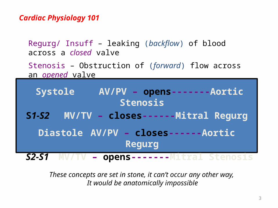
Cardiac Physiology 101
Systole AV/PV – opens S1-S2 MV/TV – closes
Diastole AV/PV – closes S2-S1 MV/TV – opens
3
Cardiac Physiology 101
Regurg/ Insuff – leaking (backflow) of blood across a closed valve
Stenosis – Obstruction of (forward) flow across an opened valve
Systole AV/PV – opens-------Aortic Stenosis
S1-S2 MV/TV – closes------Mitral Regurg
Diastole AV/PV – closes------Aortic Regurg
S2-S1 MV/TV – opens-------Mitral Stenosis
These concepts are set in stone, it can’t occur any other way, It would be anatomically impossible

4
Cardiac Anatomy 101

5
Cardiac Anatomy 101
6
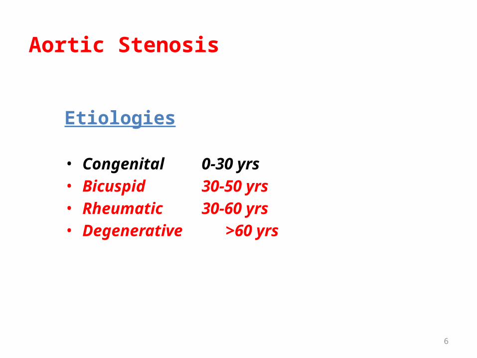
Aortic Stenosis
Etiologies
• Congenital 0-30 yrs• Bicuspid 30-50 yrs• Rheumatic 30-60 yrs• Degenerative >60 yrs

7
Aortic Stenosis

8
Aortic Stenosis – pathophysiology

9
Aortic Stenosis – pathophysiology

10
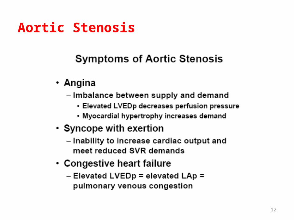
Aortic Stenosis
Symptoms
• Angina• Syncope• Congestive Heart Failure (CHF)

11
Aortic Stenosis
12
Aortic Stenosis

13
Aortic Stenosis
Diagnosis
– Ecg – LAE, LVH– Echo 2D/color doppler –test of choice– Cardiac Cath – helpful, confirmatory, needed if
the pt is older – look at the coronaries

14
Aortic Stenosis
Treatment of Symptomatic Aortic Stenosis or Decreased LV Function
Medical Therapy – treats the symptoms not the cause
Aortic Valve Replacement Bioprosthetic vs Mechanical AVR

15
Valvular Heart Disease
Aortic Valve
• Aortic Stenosis• Aortic Regurgitation

16
Aortic Regurgitation
17
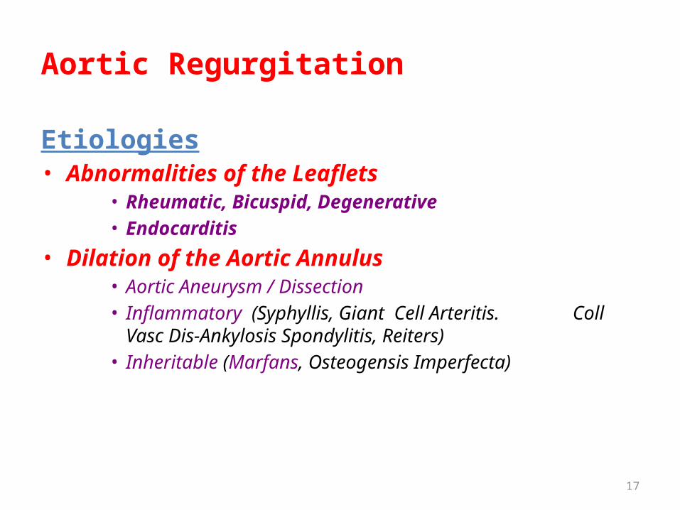
Aortic Regurgitation
Etiologies• Abnormalities of the Leaflets
• Rheumatic, Bicuspid, Degenerative• Endocarditis
• Dilation of the Aortic Annulus• Aortic Aneurysm / Dissection• Inflammatory (Syphyllis, Giant Cell Arteritis. Coll
Vasc Dis-Ankylosis Spondylitis, Reiters)• Inheritable (Marfans, Osteogensis Imperfecta)

18Plate 18
Left

19
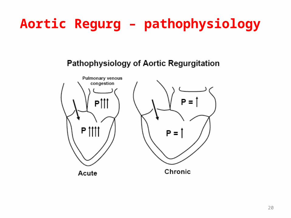
Aortic Regurg – pathophysiology
20
Aortic Regurg – pathophysiology

21
Aortic Regurg – pathophysiology
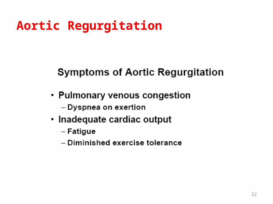
22
Aortic Regurgitation

23
Aortic Regurgitation
Physical Exam
• Diastolic Decrescendo Blowing Murmur• Hyperdynamic LV apical impulse• Bounding Pulses• S4, S3 Gallop-advanced AI• Apical Rumble – “Austin Flint Murmur”

24
Aortic Regurg – Austin Flint Murmur
Due to the vibration of the anterior leaflet of the mitral valve as it is buffetted simultaneously by the blood jets from the left atrium
and the aorta.

25
Aortic Regurgitation
Diagnosis
– Ecg – LAE, LVH– Echo 2D/color doppler –test of choice– Cardiac Cath – helpful, confirmatory, needed if
the pt is older – look at the coronaries
26
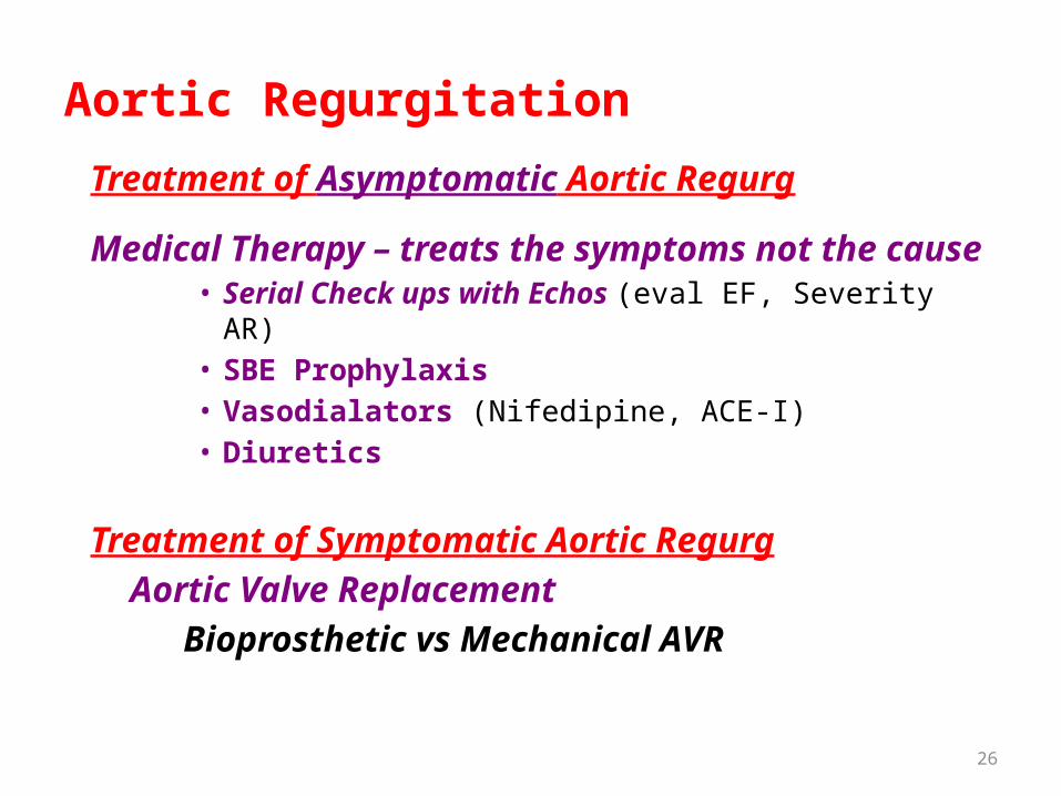
Aortic Regurgitation
Treatment of Asymptomatic Aortic Regurg
Medical Therapy – treats the symptoms not the cause• Serial Check ups with Echos (eval EF, Severity AR)• SBE Prophylaxis• Vasodialators (Nifedipine, ACE-I)• Diuretics
Treatment of Symptomatic Aortic Regurg
Aortic Valve Replacement
Bioprosthetic vs Mechanical AVR

27
If you're not confused, you're not paying
attention.Tom Peters

28
Valvular Heart Disease
Mitral Valve
• Mitral Regurgitation• Mitral Stenosis
29
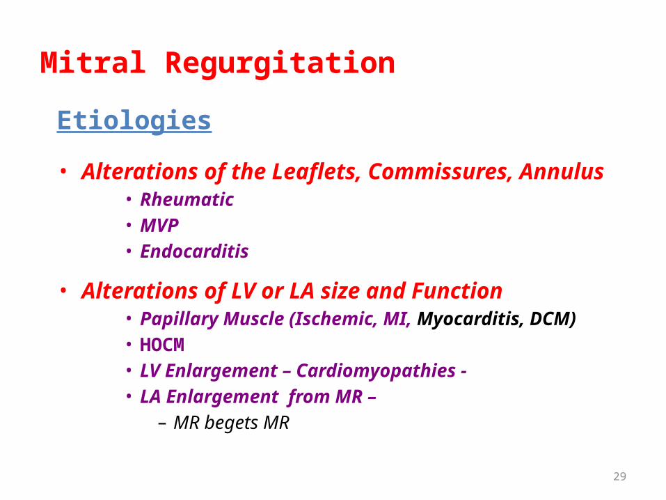
Mitral Regurgitation
Etiologies
• Alterations of the Leaflets, Commissures, Annulus• Rheumatic• MVP• Endocarditis
• Alterations of LV or LA size and Function• Papillary Muscle (Ischemic, MI, Myocarditis, DCM)• HOCM• LV Enlargement – Cardiomyopathies - • LA Enlargement from MR –
– MR begets MR

30
Mitral Regurgitation
31
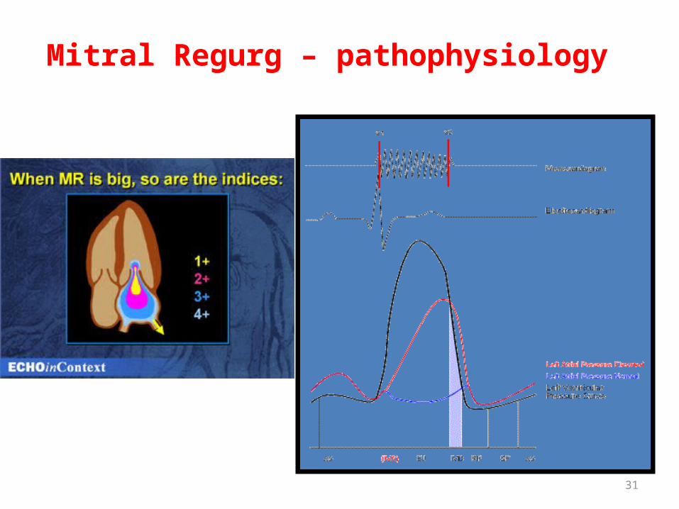
Mitral Regurg – pathophysiology

32
Mitral Regurg – pathophysiology

33
Mitral Regurg – pathophysiology

34
Mitral Regurgitation
Symptoms
• Fatigue and weakness • Dyspnea and orthopnea• Right sided HF• MVP Syndrome (if present)

35
Mitral Regurgitation
Physical Exam
• Holosystolic Apical Blowing Murmur • Laterally displaced apical impulse• Split S2 (but is obscured by the murmur)• S3 Gallop (increased volume during diastole)• Radiation depends on the etiology

36
Mitral Regurgitation
Diagnosis
– Ecg – LAE, LVH– Echo 2D/color doppler –test of choice– Cardiac Cath – helpful, confirmatory, needed if
the pt is older – look at the coronaries

37
Mitral Regurgitation
- SBE Prophylaxis

38
Mitral Regurgitation

39
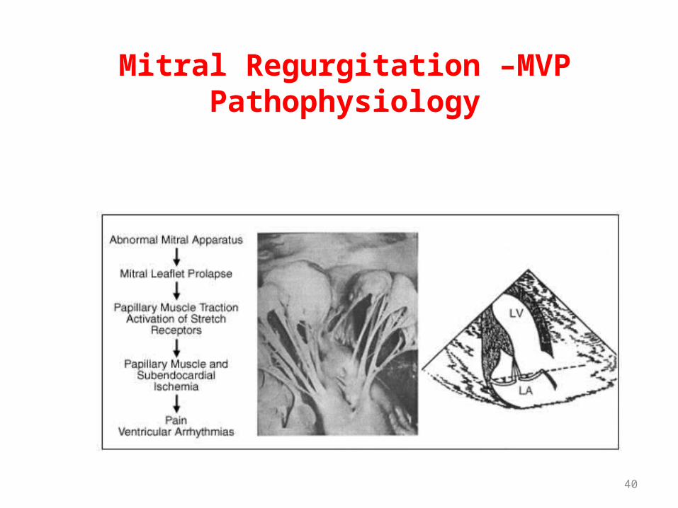
Mitral Regurgitation -MVP
40
Mitral Regurgitation –MVPPathophysiology

41
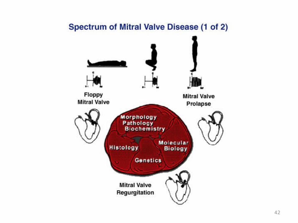
Mitral Regurgitation -MVP
42

43
Mitral Regurgitation -MVP

44
Mitral Regurgitation -MVP
Diagnosis and Treatment
• Echo 2D/Color• B-Blockers (hyperadrenergic symptoms, Palpitations)• Aspirin (TIAs without etiology)• SBE Prophylaxis (only if associated with MR)• Severe Symptomatic MR – same as chronic MR

45
Valvular Heart Disease
Mitral Valve
• Mitral Regurgitation• Mitral Stenosis

46
Mitral Stenosis
Etiologies• Rheumatic – almost all cases in adults• Mitral Annular Ca+ - massive (rare)• Congenital – rare
60% of pts don’t have a history of ARC
50% of pts who have ARC don’t develop VHD

47

48
Mitral Stenosis

49
Mitral Stenosis
50
Mitral Stenosis

51
Mitral Stenosis

52
Mitral Stenosis
Physical Exam– Loud S1– Opening Snap– Diastolic Apical Rumble (murmur)– May be associated with:
• MR or AS• Right Sided Murmurs
o PI – Graham Steel Murmuro TR

53
Mitral Stenosis
Diagnosis
– Ecg – A Fib, LAE, RAE, RVH– Echo 2D/color doppler –test of choice– Cardiac Cath – helpful, confirmatory, needed if
the pt is older – look at the coronaries

54
Mitral Stenosis
Treatment of Symptomatic Mitral Stenosis
Medical Therapy – treats the symptoms not the cause• Diuretics – for congestion• Digoxin, Beta and Ca Channel Blockers for Afib
rate control• Anticoagulation – for AFib and LA clots• SBE Prophylaxix – prevent endocarditis

55
Mitral Stenosis
Treatment of Symptomatic Mitral Stenosis
Surgical Therapy – treats the cause• Percutaneous Ballon Valvulaoplasty – Non-
calcified, pliable valve

56
Mitral Stenosis
Treatment of Symptomatic Mitral Stenosis
Surgical Therapy – treats the cause• Open Commisurotomy – valve repair• Mitral Valve Replacement
57
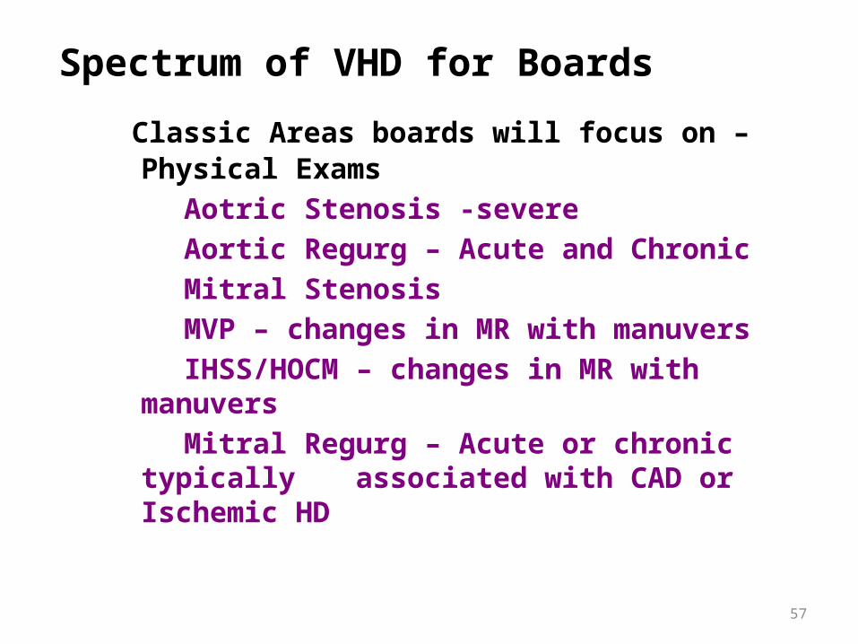
Spectrum of VHD for Boards
Classic Areas boards will focus on – Physical Exams
Aotric Stenosis -severe Aortic Regurg – Acute and ChronicMitral StenosisMVP – changes in MR with manuversIHSS/HOCM – changes in MR with manuversMitral Regurg – Acute or chronic typically
associated with CAD or Ischemic HD
58
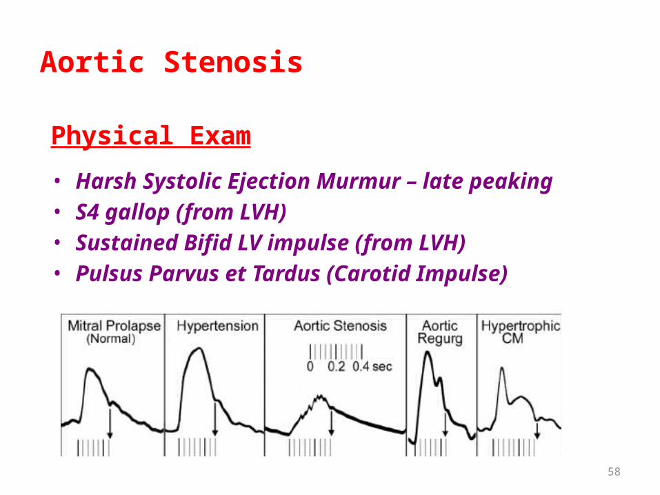
Aortic Stenosis
Physical Exam
• Harsh Systolic Ejection Murmur – late peaking• S4 gallop (from LVH)• Sustained Bifid LV impulse (from LVH)• Pulsus Parvus et Tardus (Carotid Impulse)

59
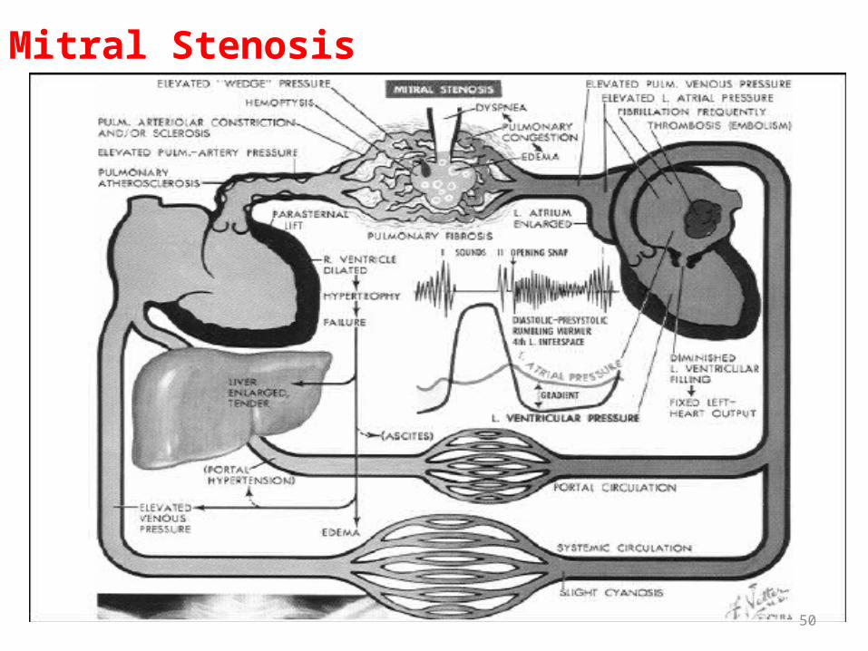
Mitral stenosis
• Obstacle to a bloodflow from the left atrium to the left ventricule, caused by narrowing left atrio-ventricular apertures.
• The causes: the causes at adults practically always is earlier transferred rheumatic fever. Children with congenital mitral stenosis seldom live more than 2th years. The causes of obstruction left АV apertures can be and micsoma the left atrium.

60Plate 17Left

61

62

63

64

65
66
67
68

69
Echocardiography

70
Echocardiography

71
Physical Exam Review:
72

73
• Complaints: at early stages the mitral stenosis usually there is a dyspnea at physical activity and fatigue. At the moderate and severe stage the dyspnea disturbs even in rest, palpitation, cough, hemoptysis, hypostases of feet. The fever, a tachycardia at physical activity or atrial fibrillation shortens time diastolic fillings of ventricules, the insufficient bloodflow through narrowed left АV aperture in a diasdolic phase promotes increase of pressure in the left atrium and to reduction of cardiac output.
• Sudden increase of pressure in the left atrium conducts to the expressed hypostasis of lungs. Hemoptysis, caused by rupture of small pulmonary vessels, and also a hypostasis of lungs, it is especially probable at pregnant women that is connected with increase in volume of blood. The dilations left atrium and the expanded pulmonary trunk can press the left returnable nerve, cause its paralysis and hoarse voices.

74
Survey:• Acrocyonosis, «mitral face» - dark-violet colouring of cheekbones
against a pale skin (is more characteristic for patients with low cardiac output and a high pulmonary hypertensia). There can be a backlog in physical development, presence of "a cardiac hump» at defect formation in the early childhood.
Palpation:• On heart apex - diastolic trembling «the cat's purring», apex beat
more often it is not changed, in left parasternal areas - the expressed pathological pulsation connected with a hypertrophy and dilatation of right ventricule. In some cases are palpated I tone on an apex of heart and click of opening of the mitral valve at a left edge of the inferior part of a breast.
Percussion:• The upper and right borders of relative dullness of heart are
displaced accordingly upwards and to the right.
75
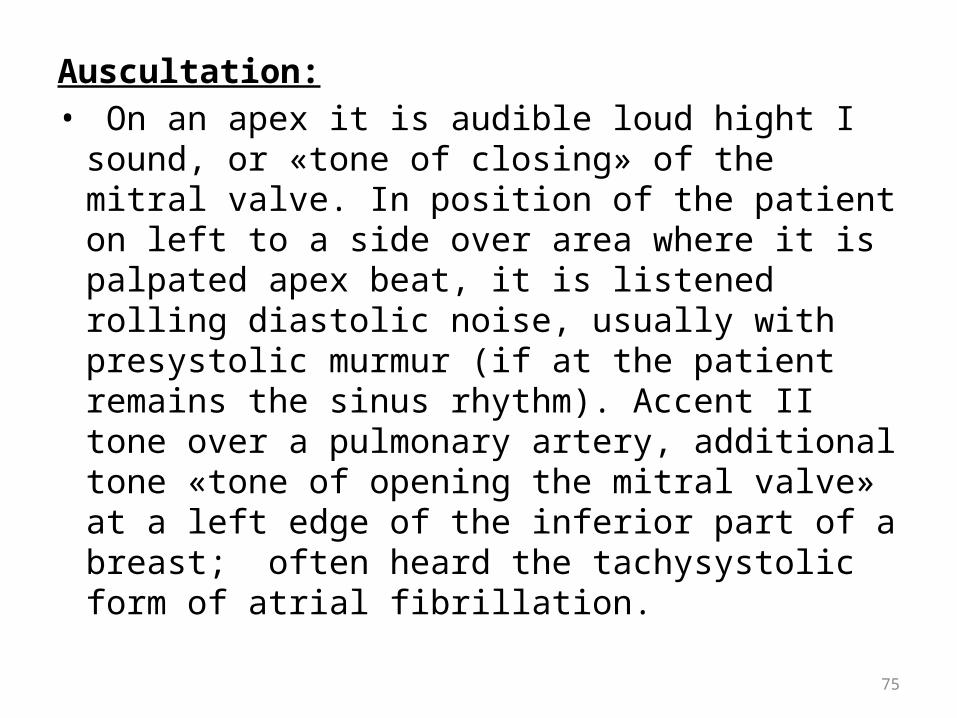
Auscultation:• On an apex it is audible loud hight I sound, or
«tone of closing» of the mitral valve. In position of the patient on left to a side over area where it is palpated apex beat, it is listened rolling diastolic noise, usually with presystolic murmur (if at the patient remains the sinus rhythm). Accent II tone over a pulmonary artery, additional tone «tone of opening the mitral valve» at a left edge of the inferior part of a breast; often heard the tachysystolic form of atrial fibrillation.

76
Laboratory and instrumental investigation:
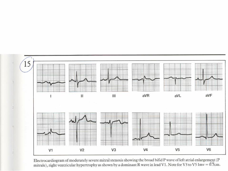
ELECTROCARDIOGRAM:• Increase of amplitude and duration of Р wave in I, II standard leads and
left chest leads (V5,6), the big area of a final negative part of Р wave in V1 – its signs of hypertrophy left atrium, infringement intraatrium conduction.
• The right type of the electrocardiogram, caused by hypertrophy RV is formed.
• Presence in V1 of high R wave, characteristic for hypertrophy RV, testifies by several pulmonary hypertensia
• It is possible atrial fibrillation: usually fibrillation waves (f) large, however at diffuse defeat of atriums of a f-wave can be small.
Phonocardiography: increase in amplitude of I tone at a heart apex, II tones over a pulmonary artery, presence III additional «tone of opening of the mitral valve» and diastolic murmur on an apex.

77
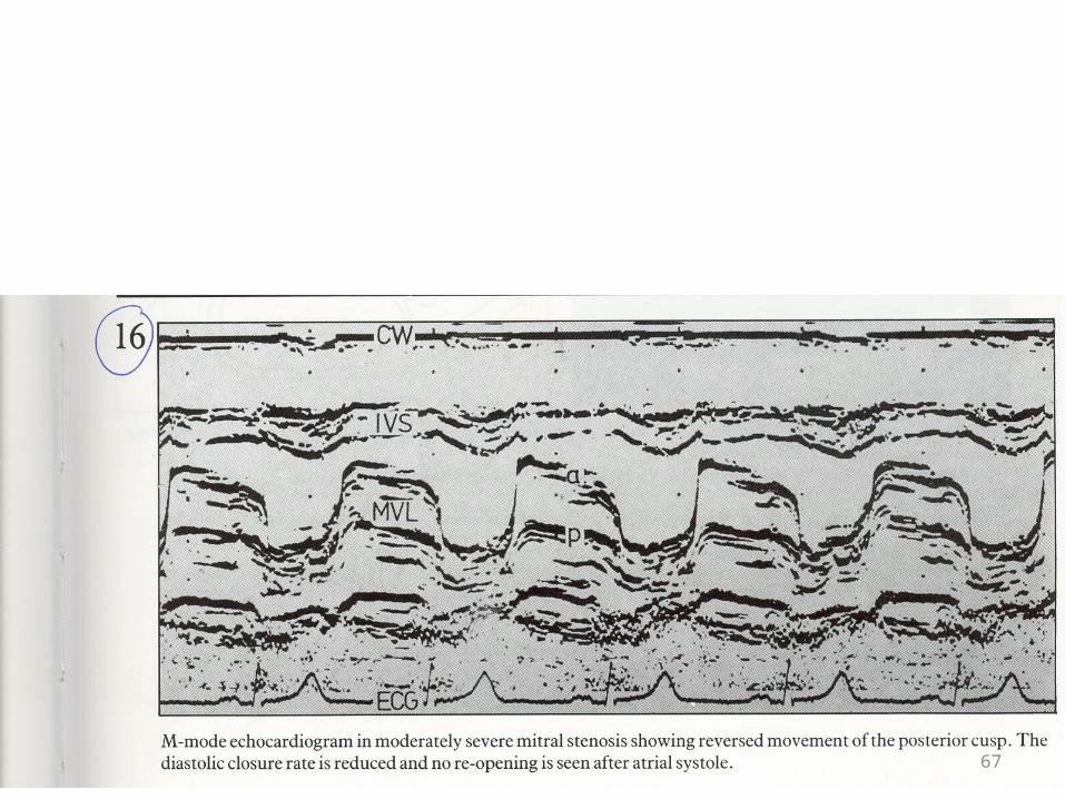
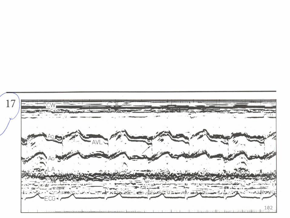
ECHO CG:• Allows to measure precisely narrowed left anАV aperture• Movement of shutters of the mitral valve changes (it gets P-shaped
configuration) - unidirectional movement of shutters of the mitral valve, expansion of cavities of the left atrium (that is important for an expediency estimation cardioversion).
X-ray: increase in the left atrium and LV - straightening left contour of heart because of dilatation of an ear of the left atrium (<< the mitral configuration »), increase of arches of a pulmonary artery, a vein of the superior areas of lungs are quite often expanded as a result of bloodflow redistribution.
Heart cathetherization - the raised pressure of jamming (pressure in the left atrium) and degree of a pulmonary hypertension, and quantitatively to estimate degree of the mitral stenosis and regurgitation.
• Insufficiency аортального the valve.• (Аортальная insufficiency; аортальная регургитация)• Ретроградный a blood-groove from an aorta in left желудочек through
leaky closed shutters аортального the valve.

78
Mitral regurgitation• Mitral regurgitation is backflow of blood from left ventricule to the
left atrium through insolvent (not closed completely) the mitral valve.Causes:• Prolapse of the mitral valve (PMV), rheumatic defeat of heart, rupture
of tendineae chords, dysfunction of papillar muscles, is more rare - mycsoma of the left atrium and expressed calcification of the mitral orifice (mainly at elderly women).
• At children the most frequent causes of mitral regurgitation- dysfunction of papillar muscles owing to abnormal of the left coronary artery from a pulmonary trunk, acute myocarditis, the micsomatous valve degeneration.
• The mitral regurgitation caused by dysfunction of papillar muscles is complication of myocardial infarction with aneurysm of LV (or without it) and fibrosis of papillar muscles (or without it).

79
Clinical featuresComplaints: long enough defect signs can be absent. At the severe mitral
regurgitation often there are attacks of palpitation to the first symptoms of insufficiency of blood circulation, is possible thanks to frequent extrasystole and to hyperdynamic effect increased LV. Complaints further prevail connected with development and progressing of cardiac insufficiency:
• Dyspnea (appears even before development of symptoms of the lowered cardiac output and it is caused by a high pressure in the left atrium,due to the big wave regurgitaton) at first at physical activity, then in rest;
• Palpitation;• The complaints caused by stagnation in small and big circles of blood
circulation (cough, hemoptysis, weight in right side due to hepatomegaly, hypostases of feet).

80
Survey:• Аcrocyonosis (lips, a tip of a nose, lobes of ears, tips of fingers,
nails).Palpation: Frequent pulse some the increased filling. The apex beat
carries poured the raising character (is increases) and displaced to the left.
• The apex beat in left parasternal areas due to increased left atriumPercussion:The upper border of relative dullness of heart is displaced
upwards, the left border - outside from left midclavicular line.Auscultation: • Easing of I tone on apex, splitting and accent II tone over a
pulmonary artery (due to a pulmonary hypertension), systolic murmar on an apex of heart (at small insufficiency - high and blowing character, with increase of degree of regurgitation, in murmur low frequencies, louder prevail, rough). Absence of an interval between I tone and systolic murmur is typicaly.

81
Laboratory and instrumental investigations
Chest X-ray: - with moderated and severe mitral regurgitation are visible the increased left atrium and LV, mitral configuration of heart.
Electrocardiogram - signs of an overload of the left atrium and left ventricule. The left type of an electrocardiogram (RI> RII> RIII SIII> and SI) with signs of hypertrophy LV is registered. At expressed atriomegaly (HLA)it is almost always registered atrial fibrillation.
ЭХОКГ - increase of cavities of the left atrium and LV, a thickening (consolidation) of shutters of the mitral valve, absence closing in a phase of systole of the ventricules. Doppler-EHOKG, especially colour, allows to estimate of mitral regurgitation is quantitatively. Many of the causes of the mitral regurgitation (for example, PMV, mycsoma of the left atrium, rupture of chords and calcification of valvular orifice) have the ECHOCG signs.
Ventriculography - allows to estimate degree of regurgitation, and pressure in RV - degree of a pulmonary hypertension which invariably accompanies a high pressure in the left atrium. The greatest pressure in the left atrium develops at rupture of chords.

82
Mitral Regurgitation
• There is acute volume overload on left ventricle with an increase in end diastolic volume. At the same time, there is new pathway for LV ejection into a low pressure system into the LA. The left ventricle initially is hypercontractile because it can eject blood back into the LA and out the aortic valve.
Forward stroke volume is actually decreased. • In acute MR, the LA cannot accommodate the increased
volume and builds up in the lungs leading to respiratory distress.

83
Mitral Regurgitation
• In chronic MR, the LA will slowly dilate, the LV will constantly be volume overloaded and eventually weaken. Both of these will eventually lead to congestive heart failure.

84
Echocardiography

85
Echocardiography

86
EKG Findings:

87
Physical Exam Review
• A spike and dome arterial pulse• PMI will be sustained with a triple apical beat secondary a
palpable a wave• There is a harsh mid systolic murmur radiating throughout the
precordium.• There is usually also a holosystolic murmur c/w MR• Maneuvers have specific affects on this murmur

88
Tricuspid stenosis
ETIOLOGY:• Rheumatic heart disease (most common). It is
frequently associated with mitral or aortic valve disease. More common in women.
PATHOPHYSIOLOGY:• Tricuspid valve stenosis results in a reduced cardiac
output which is restored towards normal when the right atrial pressure increases. The resulting systemic venous congestion produces hepatomegaly, ascites and dependent edema.

89
CLINICAL FEATURESSymptoms:• Symptoms of associated left-sided rheumatic valve lesion.• Abdominal pain (due to hepatomegaly) and swelling (due
to ascites) and peripheral edema.On examination:• JVP: giant "a" wave in JVP.• Pulsating liver: Deep palpation of liver may show
presystolic pulsation.• Murmur: Mid-diastolic murmur at tricuspid area i.e. along
lower left sternal border, loud on inspiration.ECG:• This shows "P" pulmonale due to right atria! hypertrophy.
90
TRICUSPID REGURGITATION
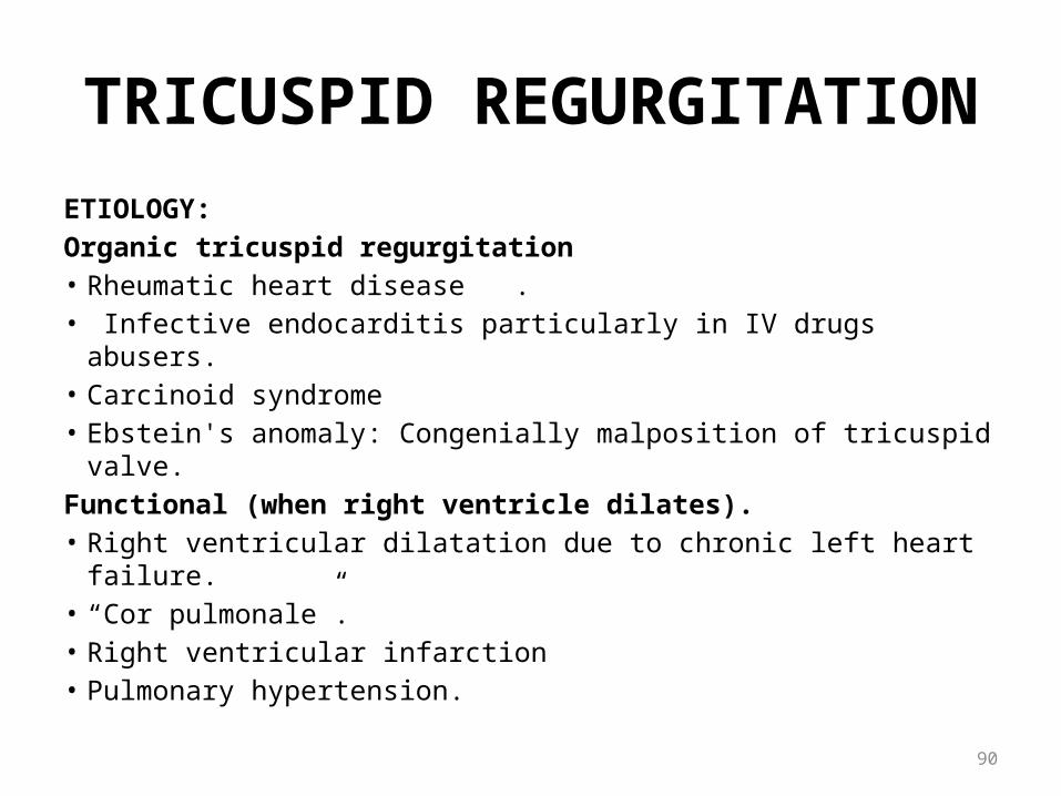
ETIOLOGY:Organic tricuspid regurgitation• Rheumatic heart disease .• Infective endocarditis particularly in IV drugs abusers.• Carcinoid syndrome• Ebstein's anomaly: Congenially malposition of tricuspid valve.Functional (when right ventricle dilates).• Right ventricular dilatation due to chronic left heart failure.• “Cor pulmonale”.• Right ventricular infarction• Pulmonary hypertension.

91
CLINICAL FEATURES:Symptoms• Features of right heart failure. Dyspnea, the feeling of a pulsation in the field of a neck
(which it is caused by high waves regurgitation in the jugular veins, reflecting of increase pressure in RV), quite often weight and pains in superior part of abdomen due to hepatomegaly.
Clinical ExaminationSurvey• Cyonosis of skin and mucous membranes with an icteric shade, swelling and a pulsation of
cervical veins, positive pulse, a pulsation of a liver and in the area of right ventricule.Palpation• The apex beat is not expressed, the pulsation hypertrophied right ventricule, pulse small
and frequent. Palpation of liver shous systolic pulsation.Percussion• Displacement of the right border of relative dullness to the right.Auscultation• At the inferior part of breast the weakened I sound, over a pulmonary artery - easing II
sound. At the inferior part of sternum listened systolic murmur amplifying at a delay of breath at height of a breath (Corvallo’ symptom). (Murmur: On auscultation, there is a pansystolic murmur along the left parasternal border. Murmur increases during inspiration (that differentiates from pansystolic murmur of mitral regurgitation that increases on expiration)).
92
Laboratory and instrumental investigations
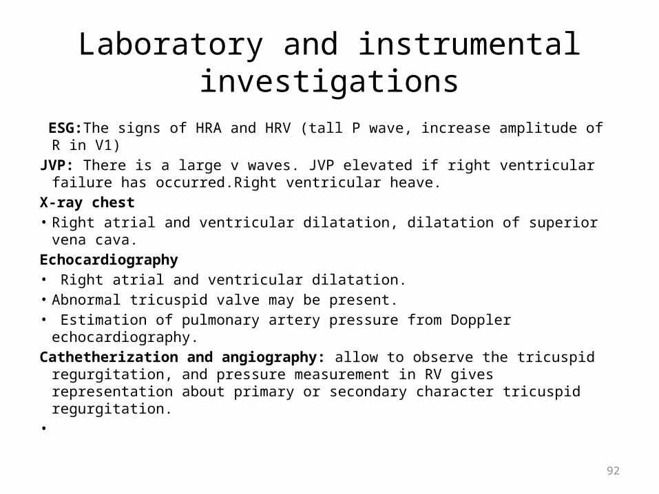
ESG:The signs of HRA and HRV (tall P wave, increase amplitude of R in V1)JVP: There is a large v waves. JVP elevated if right ventricular failure has
occurred.Right ventricular heave.X-ray chest• Right atrial and ventricular dilatation, dilatation of superior vena cava. Echocardiography• Right atrial and ventricular dilatation.• Abnormal tricuspid valve may be present. • Estimation of pulmonary artery pressure from Doppler echocardiography.Cathetherization and angiography: allow to observe the tricuspid
regurgitation, and pressure measurement in RV gives representation about primary or secondary character tricuspid regurgitation.
•

93
Aortic stenosisETIOLOGY• Rheumatic heart disease is the most, common cause resufting from
adhesions and fusions of the commissures and cusps.• Congerutally abnormal (bicuspid) aortic valve. Normal valve is tricuspid,
bicuspid valve may be stenotic with commissural fusion at birth but usually not causing serious narrowing of the aortic orifice during childhood. The abnormal architecture induces turbulent flow, which traumatizes the leaflets and leads to fibrosis, increased rigidity, calcification of leaflets and narrowing of the aortic orifice in adulthood.
• Senile AS: Age related AS degeneration and calcification of previously normal valve in elderly, usually smokers, diabetics, hypertensive and hyperlipidemic.
• SLE and severe familial hyrjerchojesterolemia occasionally cause aortic stenosis.

94
Other causes of left ventricular outlet obstruction (other than aortic valve stenosis)
• Subvalvular aortic stenosis: A congenital condition in which fibrous ridge or diaphragm is situated immediately below the aortic valve causing aortic outflow obstruction.
• Supravalvular obstruction: A congenital fibrous diaphragm above the aortic valve often associated with mental retardation and hypercalcemia (Willium's syndrome).
• Hypertrophic obstructive cardiomyopathy: Asymmetrical septal hypertrophy obstructing the left ventricular outflow.
95

96

97

98Plate 17
Right

99Plate 18
Left
100
Plate 18Right

101Plate 17Left
102

103
• Aortic valve stenosis is a heart condition caused by narrowing of the aortic valve. The aortic valve controls the direction of blood flow from the left ventricle to the aorta. When in good working order, the aortic valve does not impede the flow of blood between these two spaces. Under some circumstances, the aortic valve becomes narrower than normal, impeding the flow of blood. This is known as aortic valve stenosis, or aortic
• stenosis, often abbreviated as AS. When the aortic valve becomes • stenotic, it causes a pressure gradient between the left ventricle (LV) and
the • aorta. The more stenotic the valve, the higher the gradient between the LV
and the aorta. For instance, with a mild AS, the gradient may be 20 mmHg. This means that, at peak systole, while the LV may generate a pressure of 140 mmHg, the pressure that is transmitted to the aorta will only be 120 mmHg. So, while a blood pressure cuff may measure a normal systolic blood pressure, the actual pressure generated by the LV would be considered high.

104
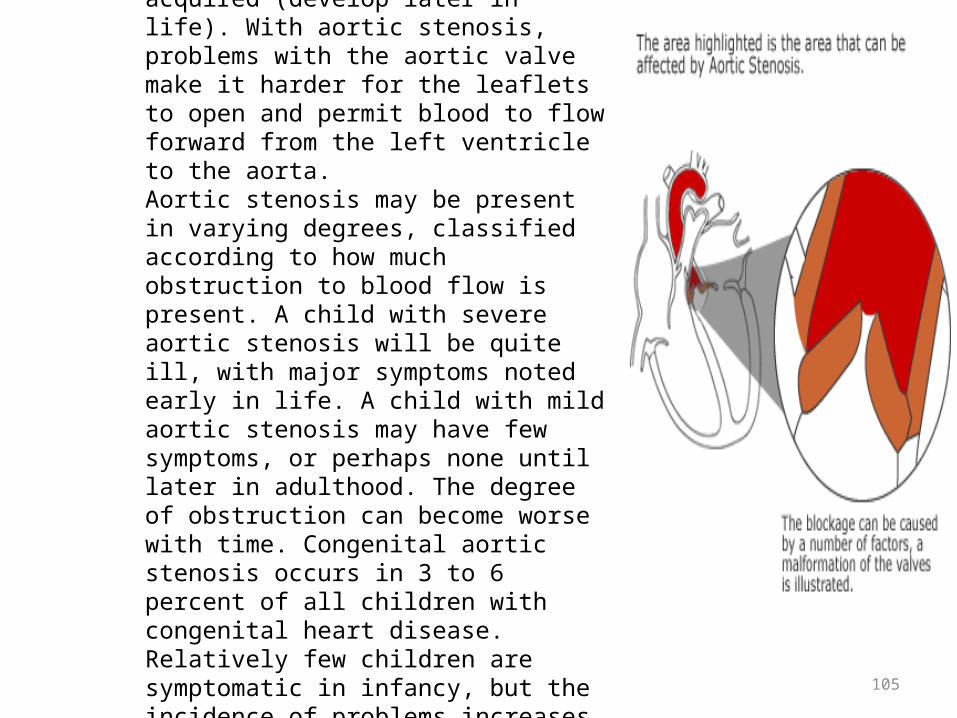
Congenital aortic stenosis occurs due to improper development of the aortic valve in the first 8 weeks of fetal growth. It can be caused by a number of factors, though, most of the time, this heart defect occurs sporadically (by chance), with no apparent reason for its development. Some congenital heart defects may have a genetic link, either occurring due to a defect in a gene, a chromosome abnormality, or environmental exposure causing heart problems to occur more often in certainfamilies. Acquired aortic stenosis may occur after a strep infection that progresses to rheumatic fever.
Age related calcification of the valve is the most common cause. Deposits of calcium build up in the valve in some older people. It is not clear why this happens. This 'calcification' can make the valve stiff and open less easily. It can be mild and cause little narrowing. But, in time it can become more severe. With aging, protein collagen of the valve leaflets is destroyed, and calcium is deposited on the leaflets. Once valve leaflet mobility is reduced by calcification, turbulence across the valve increases, causing scarring, thickening, and stenosis of the valve. Why this aging process progresses to cause significant aortic stenosis in some patients but not in others is not known.
105
Aortic stenosis is the inability of the aortic valve to open completely. Aortic stenosis is a heart defect that may be congenital (present at birth) or acquired (develop later in life). With aortic stenosis, problems with the aortic valve make it harder for the leaflets to open and permit blood to flow forward from the left ventricle to the aorta. Aortic stenosis may be present in varying degrees, classified according to how much obstruction to blood flow is present. A child with severe aortic stenosis will be quite ill, with major symptoms noted early in life. A child with mild aortic stenosis may have few symptoms, or perhaps none until later in adulthood. The degree of obstruction can become worse with time. Congenital aortic stenosis occurs in 3 to 6 percent of all children with congenital heart disease. Relatively few children are symptomatic in infancy, but the incidence of problems increases sharply in adulthood. Congenital aortic stenosis occurs four times more often in boys than in girls.
106
Most children with aortic valve stenosis have no symptoms, so it’s difficult to detect. In general, many patients will be easily fatigued, but show no other symptoms until their thirties to fifties. A small number of children may be prone to dizziness and fainting (syncope) within the first ten years of life. If the obstruction is great, infants may suffer from severe heart failure. Sudden death is uncommon, but possible. Adolescents with hypertrophic cardiomyopathy, a type of aortic stenosis where the left ventricle is noticeably enlarged, have the greatest risk of sudden heart failure.

107
Aortic stenosis is the inability of the aortic valve to open completely. Aortic stenosis is a heart defect that may be congenital (present at birth) or acquired (develop later in life). With aortic stenosis, problems with the aortic valve make it harder for the leaflets to open and permit blood to flow forward from the left ventricle to the aorta. Aortic stenosis may be present in varying degrees, classified according to how much obstruction to blood flow is present. A child with severe aortic stenosis will be quite ill, with major symptoms noted early in life. A child with mild aortic stenosis may have few symptoms, or perhaps none until later in adulthood. The degree of obstruction can become worse with time. Congenital aortic stenosis occurs in 3 to 6 percent of all children with congenital heart disease. Relatively few children are symptomatic in infancy, but the incidence of problems increases sharply in adulthood. Congenital aortic stenosis occurs four times more often in boys than in girls.

108
109

110

Serial chest radiographs of a patient with aortic stenosis taken 10 years apart. The left ventricle and aorta are prominent in both radiographs. The heart is larger and the right hilar vessels more prominent in the film at the bottom, which was taken after the development of symptoms.
111

Chest radiograph showing generalized cardiac enlargement
112
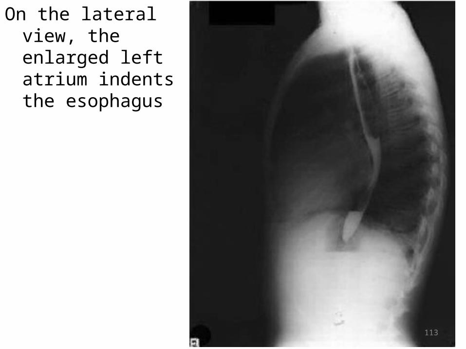
On the lateral view, the enlarged left atrium indents the esophagus
113
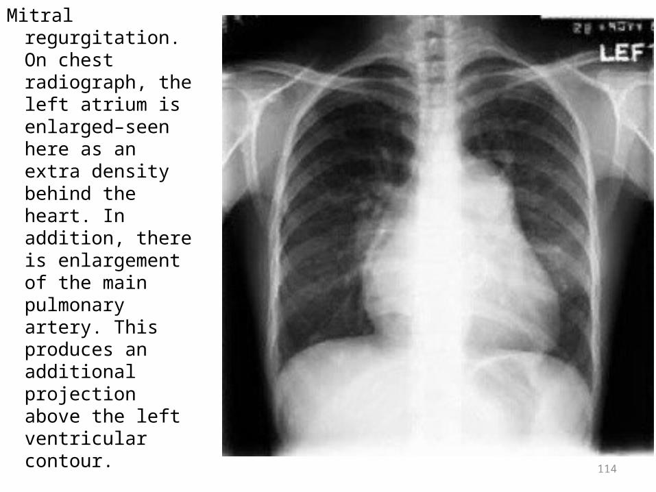
Mitral regurgitation. On chest radiograph, the left atrium is enlarged–seen here as an extra density behind the heart. In addition, there is enlargement of the main pulmonary artery. This produces an additional projection above the left ventricular contour. 114

This lateral film demonstrates a prosthetic mitral valve, which in this patient is insufficient. The left atrium is enlarged, pressing against the left main stem bronchus. In addition, the retrosternal space is filled in, typical of enlargement of the pulmonary artery and right ventricle.
115
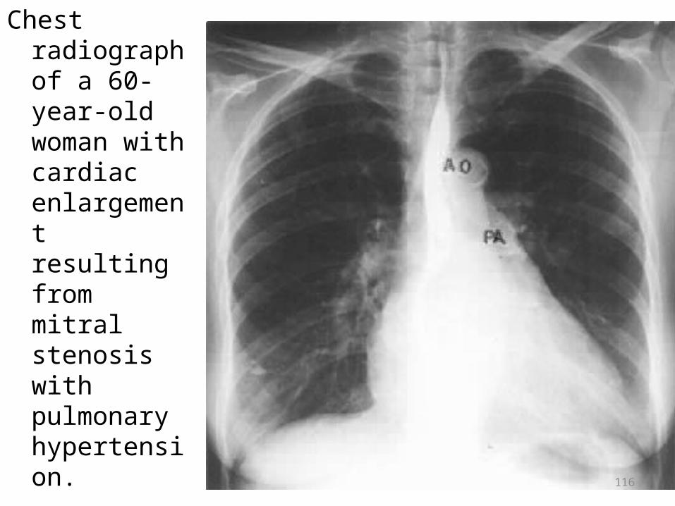
Chest radiograph of a 60-year-old woman with cardiac enlargement resulting from mitral stenosis with pulmonary hypertension.
116

117
Syncope in the setting of heart failure increases the risk of death. In patients with syncope, the 3 year mortality rate is 50%, if the aortic valve is not replaced. While it is unclear why aortic stenosis would cause syncope, the most popular theory is that severe AS produces a fixed cardiac output. When the patient exercises, their peripheral vascular resistance will decrease as the blood vesels of the skeletal muscles dilate to allow the muscles to receive more blood to allow them to do more work. This decrease in peripheral vascular resistance is normally compensated for by an increase in the cardiac output. Since patients with severe AS cannot increase their cardiac output, the blood pressure falls and the patient wil syncopize due to decreased blood persufion to the brain. A second theory as to why syncope may occur in AS is that during exercise, the high pressures generated in the hypertrophied LV causes a vasodepressor response, which causes a secondary peripheral vasodilatation that will then cause decreased perfusion to the brain.

118
Patients with aortic stenosis may also experience chest pain, pressure, or discomfort (called angina or angina pectoris), caused by an insufficient supply of oxygen to the heart. As the left ventricle thickens and works harder to expel blood through the stenotic aortic valve, its demand for oxygen increases. To compound the problem, aortic stenosis reduces blood flow to the heart itself as well as to the rest of the body (the coronary arteries and other arteries of the body originate from the aorta). Thus, while the heart's demand for oxygen increases, its supply of oxygen-rich blood decreases, causing angina.

119
Most patients with aortic stenosis develop one or more of these three classic symptoms: shortness of breath (dyspnea), passing out (syncope), and chest pain (angina pectoris). Thickening of the walls of the left ventricle causes the ventricle to become stiff and unable to relax between contractions. When this happens, the pressure in the left ventricle rises and blood can "back up" into the lungs, interfering with normal absorption of oxygen from the lungs into the bloodstream. This may cause shortness of breath, which worsens as the left ventricle becomes increasingly impaired. The aortic valve may become so constricted (stenotic) that it can open only slightly, drastically reducing the amount of blood that flows into the aorta and throughout the body. In some cases, the flow of oxygen-rich blood to the brain may not be enough to sustain normal brain function. When this occurs, patients may briefly lose consciousness, or pass out. Losing consciousness is called syncope.

120
Complaints: a classical triad of symptoms of the aortic stenosis - faints, a stenocardia and a dyspnea at physical activity. The faints arising at physical activity, are considered as a sign of the fixed cardiac output and reflect the severe aortic stenosis. The stenocardia is caused by insufficient subendocardialby arterial blood supply of hypertriphied walls of LV. Average life expectancy of patients after stenocardia occurrence - 5 years, a faint - 4 years, cardiac insufficiency - 3 years.
Survey:• Pallor of skin, apex beat of the heart it is often displaced to the left and
downwards in 5-6 intercostals space. Palpation:• The apex beat is displaced to the left and downwards, larged, strengthened and
resistant. Over an aorta in 2 intercostal space to the right of a breast it is defined systolic trembling (<<the cat's purring »), is dicrease of systolic blood pressure and increase diastolic blood pressure, i.e. low puls pressure; pulse small, slow, rare.
Percussion:• Displacement of the left border of relative dullness of heart to the left.Auscultation:• On an apex I sound is decrease, II sound over an aorta is decrease, over an aorta
rough systolic murmur.

121
Laboratory and instrumental investigations
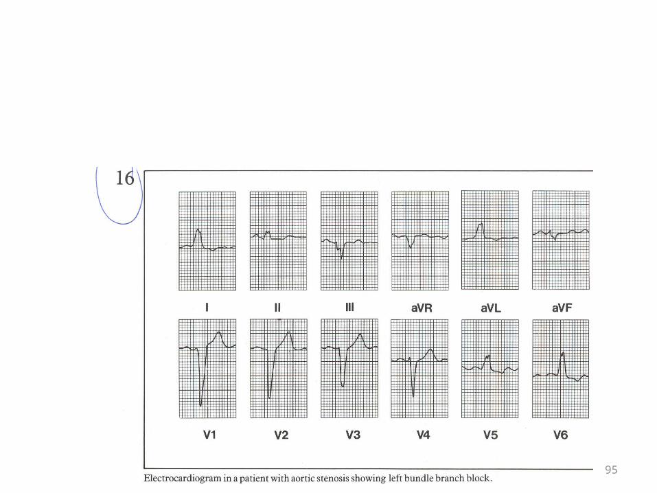
Electrocardiogram: the left type of an electrocardiogram, signs of a hypertrophy left ventricule, depression of segment ST from an isoline and inversion of Т wave in I, II standard leads and the left chest leads. In severe cases - LBBB. These changes testify to considerable overload LV and chronic coronary insufficiency.
Phocardiography: over an aorta – hight amplitude systolic murmur, beginning after 0,04 0,06 sec. after I tone and coming to an end for 0,05-0,06 sec. to II tone, amplitude of II tones it is dicrease.
ECHO CG: sharp decrease of degree of disclosing аоrtic cusps during a systole, consolidation of aortic cusps of the valve does reflexion from them by more intensive, signs of HLV. Doppler-EHOKG allows to define pressure gradients precisely.
Chest X- ray: increase of size of the LV, the heart form has of aortic configuration, contraction of left ventricule are increased.
Catheterization: allows to measure of pressure gradients, and also to reveal damage of coronary arteries.
122
Aortic regurgitation
The retrograde bloodflow from an aorta to the left ventricule through dontcomplate closed of cusps of the aortic valve.
Causes: rheumatic damage of the aortic valve, infectious endocarditis,
Mycsomatous degeneration and a trauma. At children – defect of interventricular septal with prolapse of the aortic valve. Severe or prevailing аortic regurgitation can be accompanied it calcification. The most widespread causes of easy forms аоrtic regurgitation at adults - the congenital bycuspid valve, a severe arterial hypertensia with diastolic blood pressure more than 110 mm HG. Rare аortic regurgitation find at patients with diffuse diseases of a connecting tissues. Last years the causes concern syphilitic аоrtitis, stratifying an aorta aneurysm etc.

123
• The size and strock volume of LV at aortic regurgitation are increased, as during time of diastole in it blood from pulmonary veins and blood regurgitad from an aorta also. Hypertrophy LV always develops proportionally it dilatation - for maintanance pressure. Сог bovinum - «the bull heart», characteristic for aortic regurgitation, - represents the biggest on the size and weight heart, known in a cardiac pathology.

124
• Complaints: long years high tolerance to physical activity can remain; eventually, appear a dyspnea at physical activity, orthopnea and night attacks of a cardiac asthma. Complaints to palpitation, dizziness, pains in the aries of heart (at 5 % stenocarditic character) are possible at massive regurgitation. Attacks of a stenocardia arise at night (probably because at night at decrease of heart rate increases of regurgitation) is more often.

125
Survey:• Pallor of a skin and mucous membranes, «dancing of carotids» ( the
sinocarotidic reflex low diastolic pressure is caused peripheral dilatation of vessels and backflow of blood to the cavity LV), capillary pulse or Qwinke’s symptom (pulsing palloring and reddening of a nail bed of a finger of a hand at easy pressing on a nail tip), visible on a neck a sharp pulsation carotid arteries is called corrigan’s pulse, head rocking in a step to cardiac contraction, caused by ballistic action of the big strock volume of blood, is called the Musset's symptom. An apex beat increased, displaced downwards and to the left, at survey of area of a stomach - a liver pulsation.
Palpation:• The apex beat is increased, displaced downwards and to the left,
larged, rising; the pulses waves with fast lifting and the big amplitude (<<clapping»pulse), prompt lifting and fast falling of arterial pulse - high, fast, skipping, jumping pulse.
Percussion:• Displacement of the left border of relative dullness to the left.
126
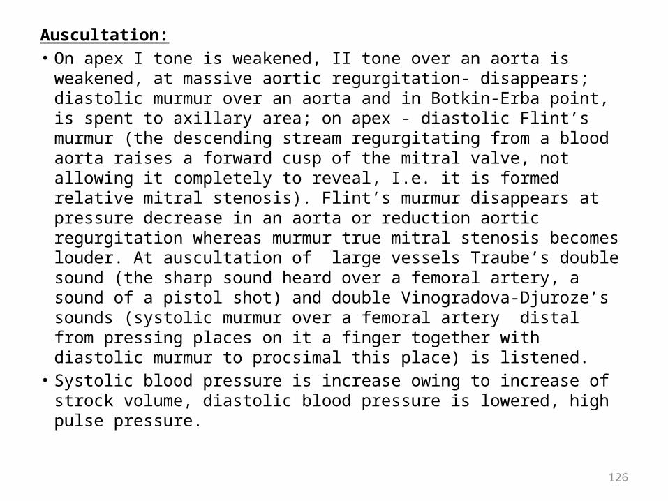
Auscultation:• On apex I tone is weakened, II tone over an aorta is weakened, at
massive aortic regurgitation- disappears; diastolic murmur over an aorta and in Botkin-Erba point, is spent to axillary area; on apex - diastolic Flint’s murmur (the descending stream regurgitating from a blood aorta raises a forward cusp of the mitral valve, not allowing it completely to reveal, I.e. it is formed relative mitral stenosis). Flint’s murmur disappears at pressure decrease in an aorta or reduction aortic regurgitation whereas murmur true mitral stenosis becomes louder. At auscultation of large vessels Traube’s double sound (the sharp sound heard over a femoral artery, a sound of a pistol shot) and double Vinogradova-Djuroze’s sounds (systolic murmur over a femoral artery distal from pressing places on it a finger together with diastolic murmur to procsimal this place) is listened.
• Systolic blood pressure is increase owing to increase of strock volume, diastolic blood pressure is lowered, high pulse pressure.

127
Laboratory and instrumental investigations
Electrocardiogram: the left type of an electrocardiogram (RI> RII> RIII is marked; SIII> SI; RV56> RV4), and also interval displacement SТ downwards and negative Т wave; LBBB is possible.
Phonocardiography: on apex - decrease amplitude of 1 tone, over an aorta is registered high-frequency the diastolic murmur, which amplitude decreases by the end of diastole, decrease amplitude of II tone.
ECHO CG: consolidation and fybrosis of cusps of the aortic valve of a rheumatic aetiology, trembling of a forward cusp of the mitral valve during time of diastole of ventricules due to of stream blow at regurgitation of blood from an aorta to the LV.
Chest X-ray: sharp increase in the sizes of LV - «the aortic configuration», a heart waist expansion and the dilated and increase pulsation of an aorta.

128
Differential diagnosis of valvular heart disease.Mitral Stenosis Mitral Regurgitation
Inspection
Malar flush, precordial bulge, and diffuse pulsation in young patients.
Usually prominent and hyperdynamic apical impulse to left of MCL.
Palpation"Tapping" sensation over area of expected PMI. Right ventricular pulsation left third to fifth ICS parasternally when pulmonary hypertension is present. P2 may be palpable.
Forceful, brisk PMI; systolic thrill over PMI. Pulse normal, small, or slightly collapsing

129
Differential diagnosis of valvular heart disease.Mitral Stenosis Mitral Regurgitation
Heart sounds, rhythm, and blood pressure
S1 loud if valve mobile. Opening snap following S2. The worse the disease, the closer the S2-opening snap interval.
S1 normal or buried in early part of murmur (exception is mitral prolapse where murmur may be late). Prominent third heart sound when severe MR. Atrial fibrillation common. Blood pressure normal. Midsystolic clicks may be present and may be multiple.

130
Differential diagnosis of valvular heart disease.Mitral Stenosis Mitral Regurgitation
Murmurs Location and transmission
Localized at or near apex. Diastolic rumble best heard in left lateral position; may be accentuated by having patient do sit-ups. Rarely, short diastolic murmur along lower left sternal border (Graham Steell) in severe pulmonary hypertension.
Loudest over PMI; posteriorly directed jets (ie, anterior mitral prolapse) transmitted to left axilla, left infrascapular area; anteriorly directed jets (ie, posterior mitral prolapse) heard over anterior precordium. Murmur unchanged after premature beat

131
Differential diagnosis of valvular heart disease.Mitral Stenosis Mitral Regurgitation
Timing Relation of opening snap to A2 important. The higher the LA pressure the earlier the opening snap. Presystolic accentuation before S1 if in sinus rhythm. Graham Steell begins with P2 (early diastole) if associated pulmonary hypertension.
Pansystolic: begins with S1 and ends at or after A2. May be late systolic in mitral valve prolapse.
Character
Low-pitched, rumbling; presystolic murmur merges with loud S1.
Blowing, high-pitched; occasionally harsh or musical.
132
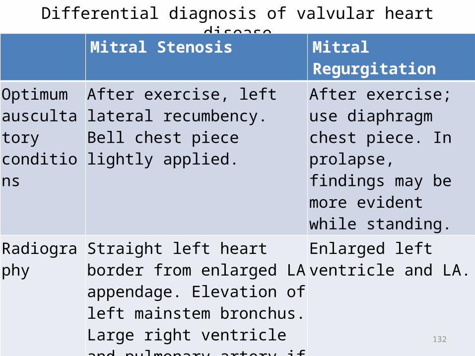
Differential diagnosis of valvular heart diseaseMitral Stenosis Mitral Regurgitation
Optimum auscultatory conditions
After exercise, left lateral recumbency. Bell chest piece lightly applied.
After exercise; use diaphragm chest piece. In prolapse, findings may be more evident while standing.
Radiography Straight left heart border from enlarged LA appendage. Elevation of left mainstem bronchus. Large right ventricle and pulmonary artery if pulmonary hypertension is present. Calcification in mitral valve in rheumatic mitral stenosis or in annulus in calcific mitral stenosis.
Enlarged left ventricle and LA.

133
Differential diagnosis of valvular heart diseaseMitral Stenosis Mitral
Regurgitation
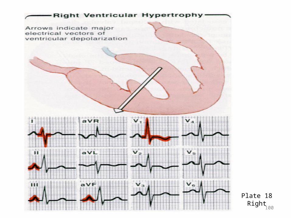
ECG Broad P waves in standard leads; broad negative phase of diphasic P in V1. If pulmonary hypertension is present, tall peaked P waves, right axis deviation, or right ventricular hypertrophy appears.
Left axis deviation or frank left ventricular hypertrophy. P waves broad, tall, or notched in standard leads. Broad negative phase of diphasic P in V1.

134
Differential diagnosis of valvular heart diseaseMitral Stenosis Mitral Regurgitation
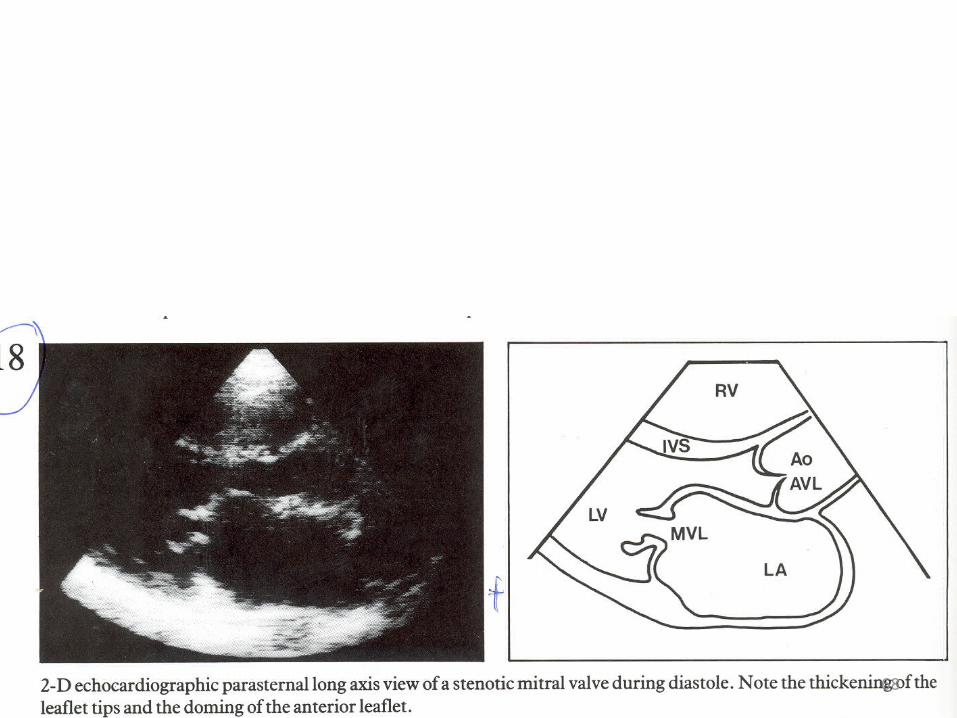
Two-dimensional echocardiography
Thickened, immobile mitral valve with anterior and posterior leaflets moving together. "Hockey stick" shape to opened anterior leaflet in rheumatic mitral stenosis. Annular calcium with thin leaflets in calcific mitral stenosis. LA enlargement, normal to small left ventricle. Orifice can be traced to approximate mitral valve orifice area.
Thickened mitral valve in rheumatic disease; mitral valve prolapse; flail leaflet or vegetations may be seen. Dilated left ventricle in volume overload. Operate for left ventricular end-systolic dimension > 4.5 cm.

135
Differential diagnosis of valvular heart diseaseMitral Stenosis Mitral Regurgitation
Continuous and color flow Doppler and TEE
Prolonged pressure half-time across mitral valve allows estimation of gradient. MVA estimated from pressure half-time. Indirect evidence of pulmonary hypertension by noting elevated right ventricular systolic pressure measured from the tricuspid regurgitation jet.
Regurgitant flow mapped into LA. Use of PISA helps assess MR severity. TEE important in prosthetic mitral valve regurgitation.