Lecture 8- Intro to Abdominal Xrays Slides
73
Introduction to Normal om na a ograp c
-
Upload
ubcradadmin -
Category
Documents
-
view
218 -
download
0
Transcript of Lecture 8- Intro to Abdominal Xrays Slides

7/31/2019 Lecture 8- Intro to Abdominal Xrays Slides
http://slidepdf.com/reader/full/lecture-8-intro-to-abdominal-xrays-slides 1/73
Introduction to Normal
om na a ograp c

7/31/2019 Lecture 8- Intro to Abdominal Xrays Slides
http://slidepdf.com/reader/full/lecture-8-intro-to-abdominal-xrays-slides 2/73
De artment of Radiolo
Vancouver General Hospital
Dr. Savvas Nicolaou
Director of ER/Trauma Radiology

7/31/2019 Lecture 8- Intro to Abdominal Xrays Slides
http://slidepdf.com/reader/full/lecture-8-intro-to-abdominal-xrays-slides 3/73
Objectives
GI Tract• Stomach• ma n es ne
• Large intestine• Liver• Spleen
GU System• Kidneys
• Bladder
Bones• Sacroiliac joint, Symphysis Pubis, Iliac crest, Femoral heads , Hip Joint• Vertebral bodies
• Pedicles, Transverse process, spinous process
Muscles• Psoas ma or
Be able to identify which abdominal organs are intraperitoneal andretroperitoneal

7/31/2019 Lecture 8- Intro to Abdominal Xrays Slides
http://slidepdf.com/reader/full/lecture-8-intro-to-abdominal-xrays-slides 4/73
Anatomy Review

7/31/2019 Lecture 8- Intro to Abdominal Xrays Slides
http://slidepdf.com/reader/full/lecture-8-intro-to-abdominal-xrays-slides 5/73
Anatomical Planes
Sagittal
Axial/Transverse

7/31/2019 Lecture 8- Intro to Abdominal Xrays Slides
http://slidepdf.com/reader/full/lecture-8-intro-to-abdominal-xrays-slides 6/73

7/31/2019 Lecture 8- Intro to Abdominal Xrays Slides
http://slidepdf.com/reader/full/lecture-8-intro-to-abdominal-xrays-slides 7/73
Basic Abdominal Anatomy
• Abdomen extends from inferior margin of
thorax to the superior margin of pelvisand lower limb
• Peritoneum lines the abdominal cavity
• Forms the mesenteries thatsuspen s v scera

7/31/2019 Lecture 8- Intro to Abdominal Xrays Slides
http://slidepdf.com/reader/full/lecture-8-intro-to-abdominal-xrays-slides 8/73
Basic Abdominal Anatomy

7/31/2019 Lecture 8- Intro to Abdominal Xrays Slides
http://slidepdf.com/reader/full/lecture-8-intro-to-abdominal-xrays-slides 9/73
BOWEL

7/31/2019 Lecture 8- Intro to Abdominal Xrays Slides
http://slidepdf.com/reader/full/lecture-8-intro-to-abdominal-xrays-slides 10/73
Basic Radiographic Terminology
• Attenuation describes the absor tion of X-ra s as the
traverse structures
Lucency = low attenuation
appear black
e.g., air
pac ty = g attenuat on appear white
. .,

7/31/2019 Lecture 8- Intro to Abdominal Xrays Slides
http://slidepdf.com/reader/full/lecture-8-intro-to-abdominal-xrays-slides 11/73
Abdomen

7/31/2019 Lecture 8- Intro to Abdominal Xrays Slides
http://slidepdf.com/reader/full/lecture-8-intro-to-abdominal-xrays-slides 12/73
Radiography
radiographs, and are
demonstrated on this ima e
Soft Tissue
Fat
Air - fat - water - bone - metal
MetalBone
Lower HigherAttenuation
• Note that THICK structures
Air
THIN structures of the same
composition

7/31/2019 Lecture 8- Intro to Abdominal Xrays Slides
http://slidepdf.com/reader/full/lecture-8-intro-to-abdominal-xrays-slides 13/73
Patient Positioning
Supine
Erect

7/31/2019 Lecture 8- Intro to Abdominal Xrays Slides
http://slidepdf.com/reader/full/lecture-8-intro-to-abdominal-xrays-slides 14/73
GASTROINTESTINAL SYSTEMInterpreting the Abdominal Radiograph
Routine “3 Views”:
Supine AP abdomen Erect AP abdomen Erect chest x-ray

7/31/2019 Lecture 8- Intro to Abdominal Xrays Slides
http://slidepdf.com/reader/full/lecture-8-intro-to-abdominal-xrays-slides 15/73
GASTROINTESTINAL SYSTEMInterpreting the Abdominal Radiograph
Routine “3 Views”:
Supine AP abdomen
• Most useful for detecting bowel
gas patterns
• Eg. dilated bowels, intra-abdominal luid

7/31/2019 Lecture 8- Intro to Abdominal Xrays Slides
http://slidepdf.com/reader/full/lecture-8-intro-to-abdominal-xrays-slides 16/73
GASTROINTESTINAL SYSTEMInterpreting the Abdominal Radiograph
Routine “3 Views”:
• Most useful for detecting free air,
Erect AP abdomen
air-fluid levels and bowel gas
patterns• Eg. Bowel obstruction

7/31/2019 Lecture 8- Intro to Abdominal Xrays Slides
http://slidepdf.com/reader/full/lecture-8-intro-to-abdominal-xrays-slides 17/73
GASTROINTESTINAL SYSTEMInterpreting the Abdominal Radiograph
Routine “3 Views”:
• Most useful in detecting intra-
Erect chest x-ray
peritoneal air (air under the
diaphragm)• May demonstrate thoracic
disease causing abdominal pain

7/31/2019 Lecture 8- Intro to Abdominal Xrays Slides
http://slidepdf.com/reader/full/lecture-8-intro-to-abdominal-xrays-slides 18/73
Normal gastric air bubble with air fluid level
Air Under the Diaphragm PathologyNormal gastric air bubble with air fluid level

7/31/2019 Lecture 8- Intro to Abdominal Xrays Slides
http://slidepdf.com/reader/full/lecture-8-intro-to-abdominal-xrays-slides 19/73
APPROACH TO EVALUATING A PLAIN ABDOMINAL FILM
3 :
SOFT TISSUES – ABDOMINALVISCERA
BOWEL GAS PATTERN
BONES- SKELETAL ELEMENTS

7/31/2019 Lecture 8- Intro to Abdominal Xrays Slides
http://slidepdf.com/reader/full/lecture-8-intro-to-abdominal-xrays-slides 20/73
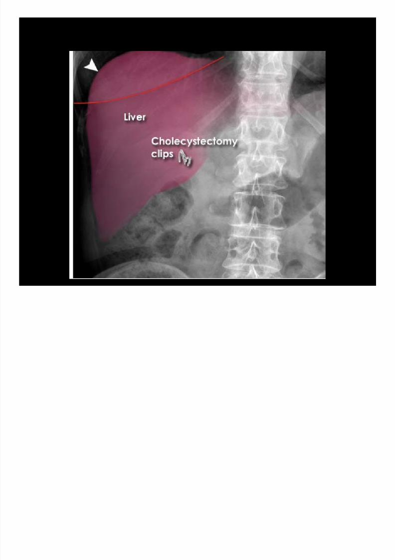
LiverSpleen
Stomach
Left kidneypedicle
Hepaticflexure
Left psoas
margin
p nous process
Left sacroiliac joint
Left hi oint
Symphisis pubis

7/31/2019 Lecture 8- Intro to Abdominal Xrays Slides
http://slidepdf.com/reader/full/lecture-8-intro-to-abdominal-xrays-slides 21/73
Soft Tissue
LiverSpleen
Left kidne
Stomach
Hepaticflexure
Left psoas

7/31/2019 Lecture 8- Intro to Abdominal Xrays Slides
http://slidepdf.com/reader/full/lecture-8-intro-to-abdominal-xrays-slides 22/73
ENLARGED SPLEEN

7/31/2019 Lecture 8- Intro to Abdominal Xrays Slides
http://slidepdf.com/reader/full/lecture-8-intro-to-abdominal-xrays-slides 23/73
Bone
Spinous
Pedicle
Right iliac
crest
Left
Right
acetabulum
sacroiliac joint
Right femoral
head

7/31/2019 Lecture 8- Intro to Abdominal Xrays Slides
http://slidepdf.com/reader/full/lecture-8-intro-to-abdominal-xrays-slides 24/73
Bowel Gas
Bowel Gas

7/31/2019 Lecture 8- Intro to Abdominal Xrays Slides
http://slidepdf.com/reader/full/lecture-8-intro-to-abdominal-xrays-slides 25/73
GAS PATTERN – Air Inside the Bowel Lumen
Property Small Bowel Large Bowel
Location Central Peripheral (Picture Frame)
Circular Folds Haustra , sacculations
Folds Uninterrupted folds cross entire width(Valvulae Conniventes)
Interrupted: folds do not crossentire width
Maximumdiameter
3cm9cm at cecum,10cm or more abnormalMax 6cm transverse colon if morethan 6 abnormal
Maximum foldthickness
3mm 5mm
Rarely contains solid fecal material Commonly contains solid fecal

7/31/2019 Lecture 8- Intro to Abdominal Xrays Slides
http://slidepdf.com/reader/full/lecture-8-intro-to-abdominal-xrays-slides 26/73
LARGE BOWEL SMALL BOWEL

7/31/2019 Lecture 8- Intro to Abdominal Xrays Slides
http://slidepdf.com/reader/full/lecture-8-intro-to-abdominal-xrays-slides 27/73
Small vs. Large Intestine
Small Intestine Large Intestine
7/31/2019 Lecture 8- Intro to Abdominal Xrays Slides
http://slidepdf.com/reader/full/lecture-8-intro-to-abdominal-xrays-slides 28/73
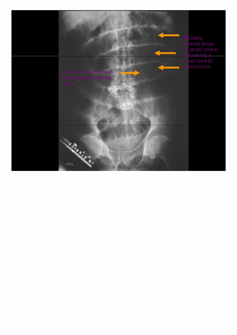
Multiple
dilated loopsof small bow el
indicat in a
Valvulae conniventes –
small bow el mucosal
small bow el
obstruction
folds

7/31/2019 Lecture 8- Intro to Abdominal Xrays Slides
http://slidepdf.com/reader/full/lecture-8-intro-to-abdominal-xrays-slides 29/73
Valvulaeconn ven es –
small bow elmucosal folds
small bow el obst ruction
numerous small bow el airfluid levels

7/31/2019 Lecture 8- Intro to Abdominal Xrays Slides
http://slidepdf.com/reader/full/lecture-8-intro-to-abdominal-xrays-slides 30/73

7/31/2019 Lecture 8- Intro to Abdominal Xrays Slides
http://slidepdf.com/reader/full/lecture-8-intro-to-abdominal-xrays-slides 31/73
Small bowel mucosal folds circumferential valvulae conniventes (plicaesemicircularis)

7/31/2019 Lecture 8- Intro to Abdominal Xrays Slides
http://slidepdf.com/reader/full/lecture-8-intro-to-abdominal-xrays-slides 32/73

7/31/2019 Lecture 8- Intro to Abdominal Xrays Slides
http://slidepdf.com/reader/full/lecture-8-intro-to-abdominal-xrays-slides 33/73
Large bowel folds - haustra

7/31/2019 Lecture 8- Intro to Abdominal Xrays Slides
http://slidepdf.com/reader/full/lecture-8-intro-to-abdominal-xrays-slides 34/73
Spleen
Left kidney
Stomach
Spinousprocess
Left
e c e
Left
joint
Rectum

7/31/2019 Lecture 8- Intro to Abdominal Xrays Slides
http://slidepdf.com/reader/full/lecture-8-intro-to-abdominal-xrays-slides 35/73
Liver
Hepatic
flexure
Right iliac
crest
Ri ht
acetabulum
head
R di h 3D R t ti

7/31/2019 Lecture 8- Intro to Abdominal Xrays Slides
http://slidepdf.com/reader/full/lecture-8-intro-to-abdominal-xrays-slides 36/73
Radiograph vs. 3D Reconstruction
3D Reconstruction

7/31/2019 Lecture 8- Intro to Abdominal Xrays Slides
http://slidepdf.com/reader/full/lecture-8-intro-to-abdominal-xrays-slides 37/73
3D Reconstruction
1. Liver
2. Spleen
3. Kidneys
4. Psoas muscle
5. Sacrum3
1
36. Rectum
7. Vertebral Body
8. Femoral Head
4
7
4
5
68 8
Radiograph Superimposition on 3D Reconstruction

7/31/2019 Lecture 8- Intro to Abdominal Xrays Slides
http://slidepdf.com/reader/full/lecture-8-intro-to-abdominal-xrays-slides 38/73
Radiograph Superimposition on 3D Reconstruction
1. Liver
2. S leen21
3
3. Kidneys
4. Psoas muscle
3
1
3
4
.
6. Rectum
7. Vertebral Body4
7
4
5
.
5
668 8
Radiograph Superimposition on 3D Reconstruction

7/31/2019 Lecture 8- Intro to Abdominal Xrays Slides
http://slidepdf.com/reader/full/lecture-8-intro-to-abdominal-xrays-slides 39/73
Radiograph Superimposition on 3D Reconstruction
1. Liver
2. S leen
3. Kidneys
4. Psoas muscle
2
3
1
3 .
6. Rectum
7. Vertebral Body4
7
4
.
9. Ilium
5
99
68 8
Radiograph Superimposition on 3D Reconstruction

7/31/2019 Lecture 8- Intro to Abdominal Xrays Slides
http://slidepdf.com/reader/full/lecture-8-intro-to-abdominal-xrays-slides 40/73
Radiograph Superimposition on 3D Reconstruction
1. Liver
2. S leen
3. Left kidney
4. Psoas muscle
34
.
6. Rectum
7. Vertebral Body
47
5
.
9. Ilium9
9
6 88
Radiograph vs 3D Reconstruction

7/31/2019 Lecture 8- Intro to Abdominal Xrays Slides
http://slidepdf.com/reader/full/lecture-8-intro-to-abdominal-xrays-slides 41/73
Radiograph vs. 3D Reconstruction
Radiograph vs. 3D Reconstruction

7/31/2019 Lecture 8- Intro to Abdominal Xrays Slides
http://slidepdf.com/reader/full/lecture-8-intro-to-abdominal-xrays-slides 42/73
Radiograph vs. 3D Reconstruction
3D Reconstruction

7/31/2019 Lecture 8- Intro to Abdominal Xrays Slides
http://slidepdf.com/reader/full/lecture-8-intro-to-abdominal-xrays-slides 43/73
3D Reconstruction
.2. Vertebral Body
3. Ilium4. Sacrum
1 1
5. Femoral Head2
33
5
Radiograph vs. 3D Reconstruction

7/31/2019 Lecture 8- Intro to Abdominal Xrays Slides
http://slidepdf.com/reader/full/lecture-8-intro-to-abdominal-xrays-slides 44/73
g p
11
.2. Vertebral Body
3. Ilium4. Sacrum
25. Femoral Head
33
5
Radiograph vs. 3D Reconstruction

7/31/2019 Lecture 8- Intro to Abdominal Xrays Slides
http://slidepdf.com/reader/full/lecture-8-intro-to-abdominal-xrays-slides 45/73
g p
11
.2. Vertebral Body
3. Ilium4. Sacrum
25. Femoral Head
33
5
Radiograph vs. 3D Reconstruction

7/31/2019 Lecture 8- Intro to Abdominal Xrays Slides
http://slidepdf.com/reader/full/lecture-8-intro-to-abdominal-xrays-slides 46/73
211. Liver
2. Spleen
33
3. Left kidney
4. Psoas muscle5. Sacrum
47
6. Rectum
7. Vertebral Body
8. Femoral Head
59
9
9. Ilium
68
8

7/31/2019 Lecture 8- Intro to Abdominal Xrays Slides
http://slidepdf.com/reader/full/lecture-8-intro-to-abdominal-xrays-slides 47/73
Coronal view
31
3
2 4
7
6
4
6
1. Liver 5. Descending colon.
3. Stomach
4. Small intestine
.7. Ascending colon

7/31/2019 Lecture 8- Intro to Abdominal Xrays Slides
http://slidepdf.com/reader/full/lecture-8-intro-to-abdominal-xrays-slides 48/73
Coronal view
11 3
2
2
. omac2. Psoas muscles
3. Spleen

7/31/2019 Lecture 8- Intro to Abdominal Xrays Slides
http://slidepdf.com/reader/full/lecture-8-intro-to-abdominal-xrays-slides 49/73
Coronal view
2
21
3
44
3
57
66
1. Liver 5. Gluteal muscles.
3. Left kidney
4. Psoas muscle
.7. Sacrum
C l i

7/31/2019 Lecture 8- Intro to Abdominal Xrays Slides
http://slidepdf.com/reader/full/lecture-8-intro-to-abdominal-xrays-slides 50/73
Coronal view
21
1 23
3
4
5
6
.2. Spleen3. Left kidney
.5. Illium6. Femoral Head
FINAL REVIEW OF PLAIN XRAY ANATOMY

7/31/2019 Lecture 8- Intro to Abdominal Xrays Slides
http://slidepdf.com/reader/full/lecture-8-intro-to-abdominal-xrays-slides 51/73

7/31/2019 Lecture 8- Intro to Abdominal Xrays Slides
http://slidepdf.com/reader/full/lecture-8-intro-to-abdominal-xrays-slides 52/73

7/31/2019 Lecture 8- Intro to Abdominal Xrays Slides
http://slidepdf.com/reader/full/lecture-8-intro-to-abdominal-xrays-slides 53/73

7/31/2019 Lecture 8- Intro to Abdominal Xrays Slides
http://slidepdf.com/reader/full/lecture-8-intro-to-abdominal-xrays-slides 54/73

7/31/2019 Lecture 8- Intro to Abdominal Xrays Slides
http://slidepdf.com/reader/full/lecture-8-intro-to-abdominal-xrays-slides 55/73

7/31/2019 Lecture 8- Intro to Abdominal Xrays Slides
http://slidepdf.com/reader/full/lecture-8-intro-to-abdominal-xrays-slides 56/73

7/31/2019 Lecture 8- Intro to Abdominal Xrays Slides
http://slidepdf.com/reader/full/lecture-8-intro-to-abdominal-xrays-slides 57/73

7/31/2019 Lecture 8- Intro to Abdominal Xrays Slides
http://slidepdf.com/reader/full/lecture-8-intro-to-abdominal-xrays-slides 58/73

7/31/2019 Lecture 8- Intro to Abdominal Xrays Slides
http://slidepdf.com/reader/full/lecture-8-intro-to-abdominal-xrays-slides 59/73
7/31/2019 Lecture 8- Intro to Abdominal Xrays Slides
http://slidepdf.com/reader/full/lecture-8-intro-to-abdominal-xrays-slides 60/73

7/31/2019 Lecture 8- Intro to Abdominal Xrays Slides
http://slidepdf.com/reader/full/lecture-8-intro-to-abdominal-xrays-slides 61/73

7/31/2019 Lecture 8- Intro to Abdominal Xrays Slides
http://slidepdf.com/reader/full/lecture-8-intro-to-abdominal-xrays-slides 62/73

7/31/2019 Lecture 8- Intro to Abdominal Xrays Slides
http://slidepdf.com/reader/full/lecture-8-intro-to-abdominal-xrays-slides 63/73

7/31/2019 Lecture 8- Intro to Abdominal Xrays Slides
http://slidepdf.com/reader/full/lecture-8-intro-to-abdominal-xrays-slides 64/73

7/31/2019 Lecture 8- Intro to Abdominal Xrays Slides
http://slidepdf.com/reader/full/lecture-8-intro-to-abdominal-xrays-slides 65/73

7/31/2019 Lecture 8- Intro to Abdominal Xrays Slides
http://slidepdf.com/reader/full/lecture-8-intro-to-abdominal-xrays-slides 66/73

7/31/2019 Lecture 8- Intro to Abdominal Xrays Slides
http://slidepdf.com/reader/full/lecture-8-intro-to-abdominal-xrays-slides 67/73

7/31/2019 Lecture 8- Intro to Abdominal Xrays Slides
http://slidepdf.com/reader/full/lecture-8-intro-to-abdominal-xrays-slides 68/73

7/31/2019 Lecture 8- Intro to Abdominal Xrays Slides
http://slidepdf.com/reader/full/lecture-8-intro-to-abdominal-xrays-slides 69/73

7/31/2019 Lecture 8- Intro to Abdominal Xrays Slides
http://slidepdf.com/reader/full/lecture-8-intro-to-abdominal-xrays-slides 70/73

7/31/2019 Lecture 8- Intro to Abdominal Xrays Slides
http://slidepdf.com/reader/full/lecture-8-intro-to-abdominal-xrays-slides 71/73

7/31/2019 Lecture 8- Intro to Abdominal Xrays Slides
http://slidepdf.com/reader/full/lecture-8-intro-to-abdominal-xrays-slides 72/73
LiverSpleen
Stomach

7/31/2019 Lecture 8- Intro to Abdominal Xrays Slides
http://slidepdf.com/reader/full/lecture-8-intro-to-abdominal-xrays-slides 73/73
Stomach
Left kidneypedicle
Hepatic
flexure
Left psoas
margin
p nous process
Left sacroiliac joint
Left hi oint
Symphisis pubis