Lecture 7: Medical imaging and -...
38
ME 328: Medical Robotics Autumn 2016 Lecture 7: Medical imaging and image-guided interventions Allison Okamura Stanford University
Transcript of Lecture 7: Medical imaging and -...
ME 328: Medical Robotics Autumn 2016
Lecture 7:Medical imaging and
image-guided interventions
Allison OkamuraStanford University

Updates
Intuitive Surgical Tour: Friday, Nov. 11
We will leave campus at 2 pm (later than originally proposed). Manufacturing tour starts at 2:45 pm and ends at 3:45 pm. Hands-on demonstrations start at 3:45 and end when the drivers need to leave. I will send a poll for attendance and drivers. 40 people max.
Assignment 3
Deadline extended until next Monday!
Note that this assignment is purposely somewhat open-ended. This and the remaining assignments will continue be like “mini projects”.

first, a brief (re-)introduction to
image-guided procedures
reference: Image-Guided Interventions, edited by Terry Peters and Kevin Cleary (link provided on course website)

idealized time-line description of image-guided procedures
Phase 1: Pre-operative planning
Phase II: Intraoperative plan execution
Phase III: Postoperative assessment

Intraoperativeupdate model update plan
real-timecomputer assistance
Postoperativecomputer- assisted
assessmentdatabasepatient
atlas
Preoperativecomputer-assisted
planning
patient-specificmodeling

image guidance enables minimally invasive procedures
previously:surgery
now:a wide variety of specialties exist
for medical interventions, and they are not all considered “surgery”(consider cardiology, radiology)

key technologies associated with image-guided procedures
medical imaging and image processing
data visualization and image segmentation
registration, tracking systems, and
human-computer interaction
replaces vision
replacesvisual reasoning
replaceshand-eye
coordination

Physicians mentally integrate their knowledge of anatomical structures with
patient-specific medical images to produce a plan and execute it.
Image-guided systems use a similar approach, where all information sources
are integrated and used to provide guidance to the physician.
Image-Guided Procedures: A Review, by Ziv Yaniv and Kevin Cleary (2006)

medical imaging

why use medical images?
intensity values are related to physical tissue characteristics which in turn relate to
(1) anatomical information and/or(2) a physiological phenomenon
physics
anatomy
physiology

what should you consider when selecting an imaging modality?
technical specifications:• spatial resolution• temporal resolution• field of view• types of biological and physiologic information
possible interaction between the imaging modality and intervention (e.g., does a metal robot cause image artifacts? does the magnet of the MRI machine cause the robot to malfunction?)

traditional imaging
functional imaging
vs.
physiologic information is interpreted
physiologic information is computed

projection imaging: • 2D cross images are generated by capturing a
“view” from a single direction
tomographic images: • 3D images are generated by stacking a set of 2D
cross sectional image slices• derived from the Greek tomos (slice) and graphein
(to write)
vs.
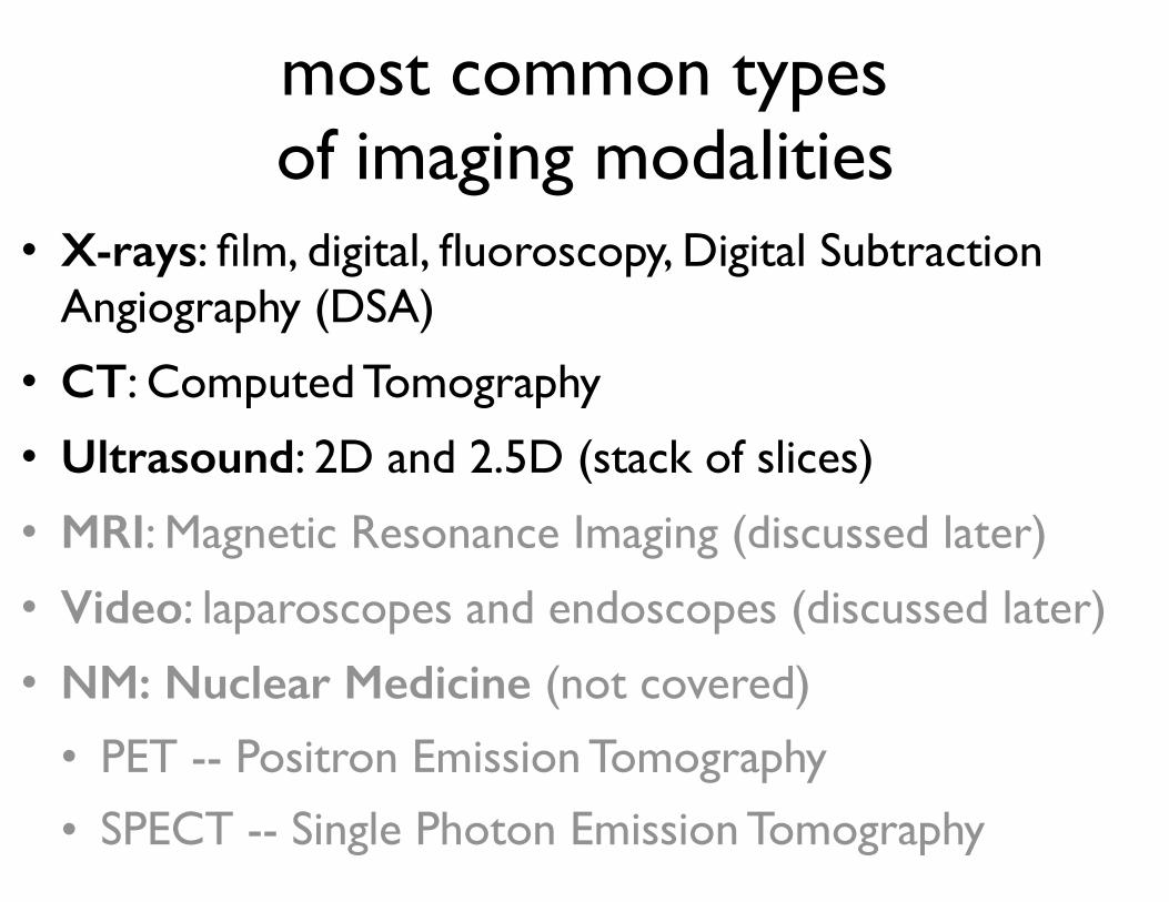
most common types of imaging modalities
• X-rays: film, digital, fluoroscopy, Digital Subtraction Angiography (DSA)
• CT: Computed Tomography
• Ultrasound: 2D and 2.5D (stack of slices)
• MRI: Magnetic Resonance Imaging (discussed later)
• Video: laparoscopes and endoscopes (discussed later)
• NM: Nuclear Medicine (not covered)
• PET -- Positron Emission Tomography
• SPECT -- Single Photon Emission Tomography

in the beginning, there was x-rayphysics: density of x-ray absorption
(x-rays are a form of ionizing radiation)
http://www.britannica.com/first “medical” x-ray, 1895
gray valueon film is
proportionalto radiation
energy

from film to digital
traditional X-ray film is replaced by solid-state detectors that convert X-rays into electrical signals (CCD camera)
Advantages:1. there is no film to process, so the images are available
immediately2. digital images can be shared or enhanced electronically3. digital images can be used for computer-assisted detection
(helps doctors confirm or draw more attention to suspicious areas on a digital image)
4. essential for real-time decision making in robot-assisted interventions

mammogram machine
uses low-energy X-rays for detection of early cancer (microcalcifications)
common screening method, lately somewhat controversial

traditional configurations of x-ray and fluoroscopy machines
Philips digital multi-functional X-ray system
early fluoroscope (Britannica Film)

c-arm fluoroscopy
Philips XperCT (CT-like imaging, more on CT later)

digital subtraction angiography (DSA)create a pre-contrast image, then subtract it from later images after a contrast medium has been introduced
iodine and barium are common types of contrast mediums for x-ray, since they attenuate x-rays (vessels become dark)

discussion
how can robots improve x-ray/fluoroscopy procedures?
how can x-ray/fluoroscopy be used in robotic interventions?

computed tomography (CT scan)
3D images are generated from a large series of 2D X-ray images taken around a single axis of rotation
(produces a volume of data for analysis)physics: same as x-ray
L. Joskowicz 2011c�Copyright L. Joskowicz, 2011
Computed Tomography: images
Single slice
Series of parallel
slices 2mm apart
single slice series of parallel slices 2mm apart
Copyright L. Joskowicz, 2011
Computed Tomography: images
Single slice
Series of parallel
slices 2mm apart

computed tomography (CT scan)
3D images are generated from a large series of 2D X-ray images taken around a single axis of rotation
(produces a volume of data for analysis)physics: same as x-ray
Copyright L. Joskowicz, 2011
CT-scan views
Size: 512 x 512 x 128
Resolution: 0.5 x 0.5 x 1 mm3
Copyright L. Joskowicz, 2011
CT-scan views
Size: 512 x 512 x 128
Resolution: 0.5 x 0.5 x 1 mm3
L. Joskowicz 2011c�

emitter/receiver configuration
http://www.youtube.com/watch?v=M-4o0DxBgZk

CT machines
two examples from Philips (Brilliance 6 and 40)differ in number of images per second, number of detectors, etc.

discussion
what challenges might exist in performing CT-guided robotic
interventions?

ultrasound imaging (diagnostic)physics: variations of acoustic impedance
1. probe sends high-frequency sound waves (1-5 MHz) into the body
2. sound waves travel into tissue and get reflected by boundaries
3. reflected waves are recorded by the probe4. time of flight gives spatial information about
the boundaries
the desired frequency of signal is chosen based on a trade-off of resolution and attenuation

ultrasoundA-mode (amplitude mode): a single transducer scans a line through the body with the echoes
plotted on screen as a function of depth.
Therapeutic ultrasound aimed at a specific tumor or calculus is also A-mode, to allow for accurate focus
of the destructive wave energy.
B-mode (brightness mode) or 2D mode: a linear array of transducers simultaneously scans a plane through the body that can be viewed as a two-
dimensional image on screen

common application: fetal ultrasound
images courtesy Nora M. Su

ultrasound characteristics• No radiation • Poor resolution (~1mm)
non-uniform, distortion, noisy
• Low penetration properties
• One 2D slice or several slices (2.5D)
• Relatively cheap and easy to use
• Preoperative and intraoperative use
L. Joskowicz 2011c�Copyright L. Joskowicz, 2011
Ultrasound imaging: characteristics
• No radiation
• Poor resolution (1mm) non-uniform, distortion, noise
• Low penetration properties
• One 2D slice or several slices (2.5D)
• Relatively cheap and easy to use
• Preoperative and intraoperative use

ultrasound machine
Ultrasonix
ultrasound transducers/probes
http://used-medicalequipmentblog.blogspot.com/

3D ultrasound
L. Joskowicz 2011c�
reconstruct 3D data from 2D slices
acquisition methods: linear, rotation, fan-like, hand
Copyright L. Joskowicz, 2011
3D ultrasound�
• Reconstruct 3D data
from 2D slices
• Acquisition methods:
linear, rotation, fan-
like, freee-hand�
Copyright L. Joskowicz, 2011
3D ultrasound�
• Reconstruct 3D data
from 2D slices
• Acquisition methods:
linear, rotation, fan-
like, freee-hand�

transrectal ultrasound
prostate brachytherapyhttp://www2.cfpc.ca
https://myhealth.alberta.ca/

Doppler ultrasoundemploys the Doppler effect to determine whether
structures (typically blood) are moving towards or away from the probe, and their relative velocity
color and pulsed Doppler of blood shunting across a muscular ventricular septal defect (in the heart)
http://www.glowm.com/

ultrasound elastography
Freehandpalpationelastograms
Boctor, Rivaz, Fleming, Foroughi, Fichtinger, Hager (2008)
width (mm)
depth (mm)
0 10 20 30
0
10
20
30-0.04
-0.03
-0.02
-0.01
0
ablated tissue
dept
h (m
m)

discussion
what challenges might exist in performing ultrasound-guided robotic
interventions?

caution!when introducing robotic (or any)
technology into the interventional suite, you should consider what imaging
modalities are already used and available
there is a conflict between the potential for improving a procedure
and the practical limitations in changing the workflow and resources required to perform the procedure

Modality Intra-operative Accessability Data
Availability Dimensionality
Computed Tomography (CT) available (not widespread) high 3D
Magnetic Resonance Imaging (MRI) available (not widespread) high 3D
X-ray available high 2D projection
functional Magnetic Resonance
Imaging (fMRI) not available moderate 3D
Positron Emission Tomography (PET) not available moderate 3D
Single Photon Emission
Computed Tomography (SPECT) not available moderate 3D
X-ray Fluoroscopy available high 2D projection
C-arm CT available low 3D
Ultrasound (US) available high 2D
optical imaging available high 2D projection
Table 1: Classification of imaging devices according to their availability for intra-operative use, their accessability to physicians around the world, the dimensionalityof the data they acquire and the type of information conveyed by the images.
The tomographic modality best suited for imaging bony structures is computed tomog-raphy (CT). This modality is considered to be geometrically accurate [138], but exhibitsintensity artifacts when metallic objects are present in the field of view. These artifactsare the result of reconstruction using corrupted projection data, which is caused by theX-rays being greatly attenuated by the metal. There are a number of approaches tometal artifact reduction, including use of higher energy X-ray beams [210], and interpo-lating the missing projection data [114, 150, 213].
Intra-operative CT is available, but it is not in widespread use. A real-time versionof this modality is CT-fluoroscopy, although most machines only provide the physicianwith a single image slice view that can be updated several times a second. The need forinexpensive intra-operative fully 3D data has led to the recent introduction of C-armCT imaging [81, 276]. This modality utilizes iso-centric motorized C-arms to acquire asmall field of view tomographic data set. Though the quality of the tomographic datais still lower than standard CT it is expected to improve with advances in C-arm imageintensifier technology.
The main drawback of CT, CT-fluoroscopy and C-arm CT is their use of ionizing radia-tion [49, 115, 202]. Previously, it was hypothesized that there may be a threshold belowwhich exposure to ionizing radiation does not increase the risk for cancer. A recentreport from the US national academies suggests that this hypothesis is not valid andthat risk of cancer proceeds in a linear manner with no lower threshold [1].
The tomographic modality best suited for imaging soft tissue is magnetic resonanceimaging (MRI). MR images exhibit both geometric and intensity distortions. Geo-metric distortions are mainly caused by inhomogeneity of the main magnetic field andnon-linearity of the magnetic field gradients [138, 237]. New MRI scanners incorporategeometric distortion schemes, so the end result may be su±ciently accurate for specificapplications [12]. A recent evaluation of geometric distortions in clinical MRI systems
5
Image-Guided Procedures: A Review, by Ziv Yaniv and Kevin Cleary (2006)