Lecture 4 - personal.psu.edupersonal.psu.edu/mxs43/472/lecture4.pdf · E d x (X 0! 1) Fractional...
43
Lecture 4 Photons transfer the energy to the matter via ionization and excitation of atoms. The process is mostly indirect - first the electrons are released. They intensively interact with the matter losing energy. Crucial for radiology. Basic electron and positron interactions. X-RAYS : DISCOVERY, EARLY HISTORY , will try to start: 1 section 4.4 intro to section 6.3
Transcript of Lecture 4 - personal.psu.edupersonal.psu.edu/mxs43/472/lecture4.pdf · E d x (X 0! 1) Fractional...

Lecture 4
Photons transfer the energy to the matter via ionization and excitation of atoms. The process is mostly indirect - first the electrons are released. They intensively interact
with the matter losing energy. Crucial for radiology.
Basic electron and positron interactions.
X-RAYS : DISCOVERY, EARLY HISTORY ,will try to start:
1
section 4.4
intro to section 6.3

• Elastic electron-electron scattering ( Möller scattering).
• Ionization of atoms
• Bresmsstrhlung
• Elastic positron-electron scattering (Bhabha scattering)
• Positron annihilation.
Interactions of electrons and positrons with matter
2

Bremsstrahlung (German)- - breaking radiation.
Physical phenomenon - electron scatters, changing direction, leading
to emission of photons mostly along initial and final electron directions.
3

The interaction of charged particles is much stronger than in the case of photon-”charged particle” interactions. The
reason long range nature of the Coulomb interaction.
Rutherford scattering of particles with charges Z and Z’
Amazing formula - valid in classical mechanics, quantum mechanics and in quantum electrodynamics. Scattering at very small angles dominates. In fact
Physical reason: even at large impact parameters there is a small deflection due to the long range Coulomb forces.
d⇥
d�(�) =
�ZZ �
4E
⇥2 1sin4(�/2)
⇥tot =�
d⇥
d�(�) d��⇥
4

Small angle scattering - happens very frequently, but leads to small - finite energy losses for the particle per unit length. Correspondingly one introduces the concept of differential energy losses which is given by Bethe- Bloch equation:
β=p/E - velocity, γ=E/m - Lorentz factor.
Ionization is a weak function of velocity for large β, and grows dramatically for small velocities ~1/β2
Here Tmax is the maximal kinetic energy which can be imparted to a
free electron is a single collisions, ze is the charge of incident particle, Z - charge of the target nucleus. A is atomic mass of the
absorber (g mol-1 ); K=.307 MeV cm2 , I is the mean excitation energy measured in eV, δ is the density effect correction to ionization energy loss.
�dE
dx= Kz2 Z
A
1�2
�12
ln2mec2�2⇥2Tmax
I2� �2 � ⇤
⇥.
5

Three dominant processes which occur when electron /positron traverses matter
Soft interactions
Interaction with atoms as a whole with atoms left in excited or sometime ionized state
Interaction with orbital electrons in which they get significant energy so they knocked out (so called delta ray electrons) - so they have tracks of their own.
Hard interactions
Bremsstrahlung - radiative interactions in which electron scatters off th Coulomb filed of the nucleus and emits a photon with energy comparable to the energy of the initial electron
★
★
★
6

32
Radiative Interactions
• Radiative interactions are inelastic scatters in which x-rays are producedwhen an electron decelerates under the influence of the electric field of thenucleus – hence, the name bremsstrahlung (German for “braking radiation”)
• Whereas ordinary collision losses (soft + hard) occur at all energies, theradiative-loss mechanism becomes relatively more and more important asthe energy increases
• EM showers are essentially the result of two high-energy processes that feedone another:
– X-rays produced by electrons (+ or –)
– Pairs produced by photons
• We can predict at which energy to expect showering to occur by means ofthe stopping power …to be discussed next
7

Stanford Linear Accelerator Center 30
• The hard collisions are also classified by the charge of theprimary electron and named after the physicist who firstdetermined the cross section for the process– Møller: electron + electron! electron + electron– Bhabha: positron + electron! positron + electron
• At low energies primary electrons basically lose energy by theexcitation and ionization of atoms – a combination of both softand hard collisions – simply referred to as collision loss
• Electrons also elastically scatter due to the Coulomb field ofthe nucleus – very many small elastic scatterings add togetherand lead to what is called multiple scattering
Bhabha scat.e+
e-8

Stanford Linear Accelerator Center 33
Stopping Powers(the average rate of energy loss)
• The average rate of energy loss along a charged particle’s track is called thestopping power and is denoted by dT/dx
• As might be expected, there are two stopping powers:– dT/dx)col (soft + hard components combined)– dT/dx)rad
• Collision loss vs. radiative loss– Collision losses occur at all energies but dominate at low energies– Radiative losses kick in at high energies and eventually overwhelm thelosses due to collisions
• There is a cross-over point called the critical energy defined bydT/dx)col = dT/dx)rad
which tells us approximately when to expect the onslaught of showering9

Stanford Linear Accelerator Center 34
Stopping Powers (cont.)
• In this figure the fractional energy loss, dT/Edt, is plotted for bothcollision and radiative processes, where t = x/Xo (r.l.)
• Cross-overs occur at 10 MeV for Pb and 100 MeV for water• Also, one easily sees how radiation completely dominates at high energy
10

11

-2Stanford Linear Accelerator Center 36
Radiation Lengths• We have seen that, starting with a single high-energy electron or photon, thenumber of particles (e–,e+ and γ) increases with depth into the medium – theso-called longitudinal development
• A special unit of length, called the radiation length, makes it significantlyeasier to plot these longitudinal shower curves
• That is, we can make showers in different materials (Al, Pb, etc.) scale so thatthey all fit on the same plot – i.e., they look more or less alike
• The key is knowing that radiative processes dominate at high energies
• We define the radiation length, Xo, as that distance in which an electron loses1/eth its energy by emitting x-rays
• Using the bremsstrahlung cross section we can show that
1-g2cm31183ln2241!
!
"
#
$
$
%
& −= ZorZoX
α
X usually measured in g cm12

13

Bremsstrahlung
Lead (Z = 82)Positrons
Electrons
Ionization
Møller (e!)
Bhabha (e+)
Positronannihilation
1.0
0.5
0.20
0.15
0.10
0.05
(cm
2g!
1 )
E (MeV)1
010 100 1000
1 E!
dE dx
(X0!
1 )
Fractional energy loss per radiation length in lead as a function of electron or positron energy. Electron(positron)scattering is considered as ionization when the energy loss per collision is below 0.255 MeV, and as Møller (Bhabha) scattering when it is above. X0(Pb)=6.37 g/cm2
Bremsstrahlung dominates at high energies, ionization dominates at low energies. The process of energy loss is statistical - large fluctuations.
14

X-RAYS : DISCOVERY, EARLY HISTORY ,
15

Wilhelm Conrad Roentgen (1845-1923) in 1896.
16

Site of the discovery, the Physical Institute of the University of Wuerzburg, taken in 1896. The Roentgens lived in apartments on the upper story, with laboratories and classrooms in the basement and first floor. He was the head of the physics department.
17

ROENTGEN’S LABORATORY
ROENTGEN’S LABORATORY
18

DISCOVERY: November 8,1895.
Tool: Cathode tube.
19

20

Roentgen switched on the tube, screened the light from the tube, and switched off the light. Waited to adjust to low light level. A cardboard screen covered by a fluorescent material - barium platinocyanide was few feet away - he noticed a glow from the screen - letter A. Presumably written by a student!!!
Could not come from cathode rays - too far away !!!
Röntgen’s Setup
21

filament
cathode
target
anode
photon flux
e-
electron kinetic energy= e (Voltage difference)x
The rough schematics of an X-ray tube
22

Electron energy is lost in the target due to two sources
1. Ionization. In diagnostic range accounts for about 99% of the energy loss and shows up as heat.
2. Bremsstrahlung. 1% Produces x-rays with energies up to the energy of the incident electron
Photon energy keV
Resulting photon spectrum is sum of bremsstrahlung and characteristic radiation
Low energy (<20keV) X-rays are filtered out by the glass envelope of the tube
(this is an exit spectrum)
59 keV
69 keV
23

Spectral distribution of X-ray calculated for a thick tungsten target
24

A bit of history leading to discovery which may explain some puzzling pieces of the story: Roentgen used the cathode tube modified by Philipp Lenard Philipp Lenard in 1888, when he was working at Heidelberg under Quincke, had done his first work with cathode rays. He investigated the view then held by Hertz that these rays were analogous to ultraviolet light and he did an experiment to find out whether cathode rays would, like ultraviolet light, pass through a quartz window in the wall of a discharge tube. He found that they would not do this; but later, in 1892, when he was working as an assistant to Hertz at the University of Bonn, Hertz called him to see the discovery he had made that a piece of uranium glass covered with aluminum foil and put inside the discharge tube became luminous beneath the aluminum foil when the cathode rays struck it. Hertz then suggested that it would be possible to separate, by means of a thin plate of aluminum, two spaces, one in which the cathode rays were produced in the ordinary way and the other in which one could observe them in a pure state, which had never been done. Hertz was too busy to do this and gave Lenard permission to do it and it was then that he made the great discovery of the "Lenard window".
25

After many experiments with aluminum foil of various thicknesses he was able to publish, in 1894, his great discovery that the plate of quartz that had, until then, been used to close the discharge tube, could be replaced by a thin plate of aluminum foil just thick enough to maintain the vacuum inside the tube, but yet thin enough to allow the cathode rays to pass out. It thus became possible to study the cathode rays, and also the fluorescence they caused, outside the discharge tube and Lenard concluded from the experiments that he then did that the cathode rays were propagated through the air for distances of the order of a decimeter and that they travel in a vacuum for several meters without being weakened. Although Lenard at first followed Hertz in believing that the cathode rays were propagated in the ether, he later abandoned this view as a result of the work of Jean Perrin in 1895, Sir J.J. Thomson in 1897 and W. Wien in 1897, which proved the corpuscular nature of the cathode rays.
26

Right after observing the effect the Roentgen started to investigate different materials - some (bone, lead...)blocked the new rays, some did not. Magnet did not deflect them as it did with cathode rays, prism did not bend them --> entirely new object ---> called them X-rays. They left black spots on the photographic plate.
December 22, brought his wife, made a photo of her hand; Shortly after Christmas send a paper to Physico-Medical Society of Wuerzburg; Published Dec. 28 !!!
27
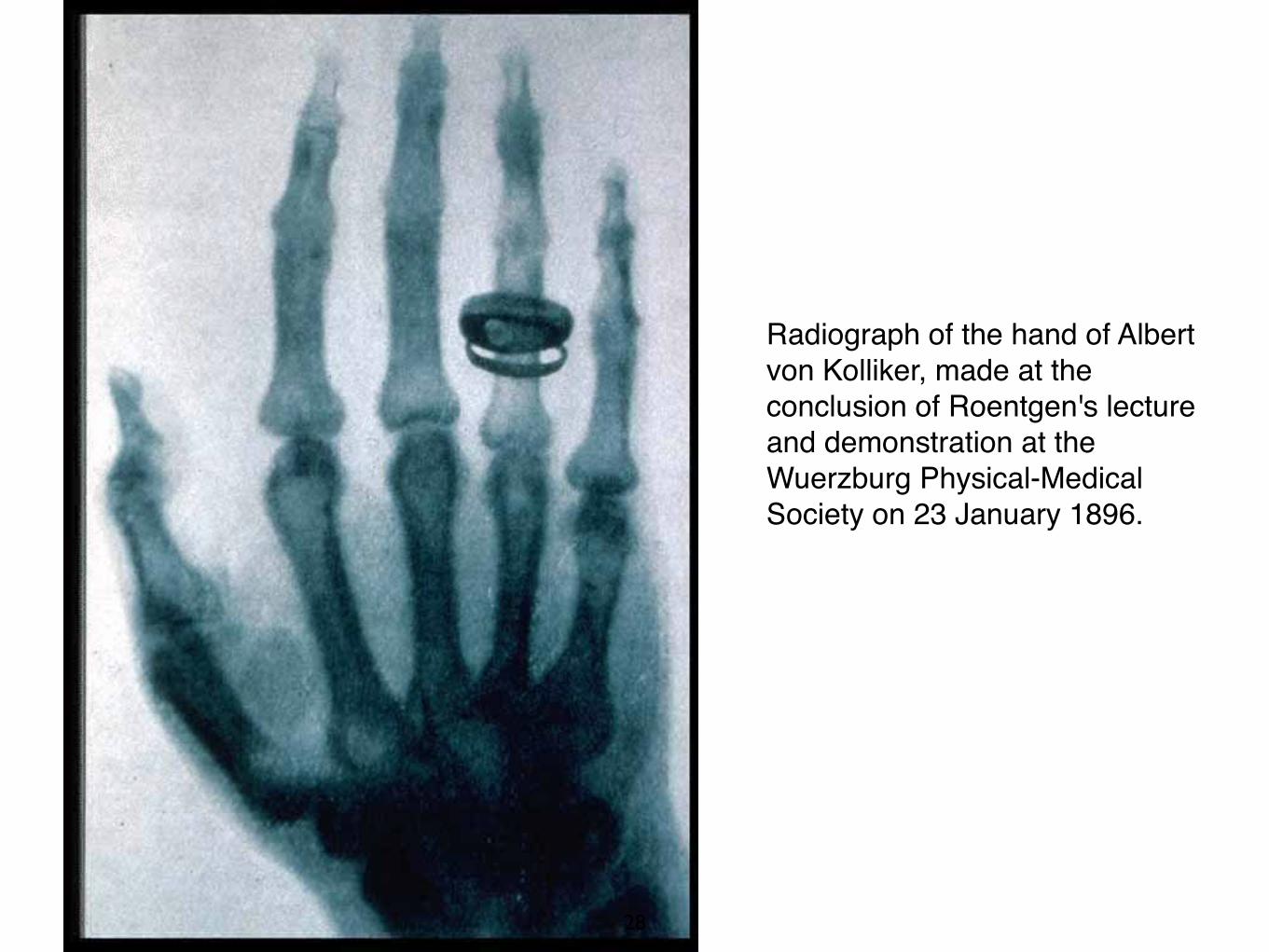
Radiograph of the hand of Albert von Kolliker, made at the conclusion of Roentgen's lecture and demonstration at the Wuerzburg Physical-Medical Society on 23 January 1896.
28

January 1 - send reprints to several physicists including Director of Physical Institute in Vienna, Exner, who at informal meeting shown it to collegues. One of them , Lechner , mentioned it to his father , editor of Neue Freie Presse - Published Sun. , Jan.5 . Next day New York Sun!!
Jan. 13, 1896 – Images needle in patient’s hand February, 1896– X-ray used presurgically
By February the first x-ray treatment for cancer was performed. Within 4 months Edison was manufacturing x-ray equipment
1901 – Receives first Nobel Prize in Physics – Given for discovery and use of X-rays
29

Radiograph of coins made by A.W. Goodspeed (1860- 1943) and William Jennings (1860-1945) in 1896, duplicating one they had made by accident in Philadelphia on 22 February 1890. Neither Goodspeed nor Jennings claimed any priority in the discovery, as the plates lay unnoticed and unremarked until Roentgen's announcement caused them to review the images.
30

31

Cunning was shot into a leg by Holder.Bullet observed using X-ray and removed. Holder
32

X-Ray Studios, like this one in New York, opened in cities large and small to take "bone portraits," often on subjects who had no physical complaints.
33

The necessary apparatus was easily acquired. An evacuated glass tube with anode and cathode, and a generator (coil or static machine), combined with photographic materials could set anyone up in business as a "skiagrapher." Greek skiagraphi, painting in light and shade
34

35

Public demonstrations, like this one by Edison in May 1896, gave the average person the opportunity to see his or her bones.
36

37

So excited was the public that each newly radiographed organ or system brought headlines. With everything about the rays so novel, it is easy to understand the frequent appearance of falsified images, such as this much-admired "first radiograph of the human brain," in reality a pan of cat intestines photographed by H.A. Falk in 1896.
38

Angiographic work began in January of 1896 with the post-mortem injection of mercury compounds. This image was made by E. Haschek and 0. Lindenthal of Vienna.
Patient Preparation amputation. Exposure
time 57 minutes.
(Reimbursement rejected by Medicare)
39

VOL. 1, NO.1, MAY,1897
40

Like this roentgen room in April 1896, early clinical settings were hot, crowded with wires and apparatus, and dangerous for patients and practitioners.
41

Early adds, though, sanitized the look of radiology practice. Here Dr. Rome Wagner and his assistant demonstrate the pleasant applications of fluoroscopy.
42

Studies of medical applications - few weeks after discovery. US first radiologist - Francis Williams - associated with MIT
650 pages, 390 illustrations many of them reproductions of radiographs
Understood dangers of X-rays, used shielding ---> lived a long life.
43