Lecture 20: DNA Delivery in Host (Part-I) - NPTel
52
NPTEL – Biotechnology – Fundamentals of Biotechnology Joint initiative of IITs and IISc – Funded by MHRD Page 1 of 52 Lecture 20: DNA Delivery in Host (Part-I) Introduction: The delivery of DNA into the host is required for generation of genetically modified organism. DNA delivery to host is a 3 stage process, DNA sticking to the host cell, internalization and release into the host cell. As a result, it depends on 2 parameters- Surface chemistry of host cell-Host cell surface charges either will attract or repell DNA as a result of opposite or similar charges. Presence of cell wall (in the case of bacteria, fungus and plant) causes additional physical barrier to the up-take and entry of DNA. Charges on DNA-Negative charge on DNA modulates interaction with the host cell especially cell surface. Modulation of these two properties is achieved in different methods to deliver DNA into the host cell and it is the topic of the discussion of today’s lecture. Transformation- it is the natural process, through which bacterial population transfer the genetic material to acquire phenotypic features. The event of transformation was first time demonstrated by Frederick Griffith in 1928. The schematic presentation of the experiment is given in Figure 20.1. Griffith has used two different Streptococcus pneumonia strains, virulent (S, causes disease and death of mice) and avirulent (R, incapable of causing disease or death of mice). In a simple experiment he injected 4 different combination of bacterial mixture, (1) live S, (2) heat killed S, (3) live R, (4) mixture of live R and heat killed S in to the mice. The observation indicates that live S has killed the mice where as mice were healthy with heat killed S or live R. Surprisingly, mice injected with mixture of live R with heat killed S were found dead, and bacteria isolated from these dead mice were virulent. Based on these observations, Griffith hypothesized the existence of a transforming agent (Protein, DNA) being transferred from heat killed virulent strain to the avirulent strain and proposed the concept of transformation. Later, Oswald has proved that the transforming factor is DNA rather than protein.
Transcript of Lecture 20: DNA Delivery in Host (Part-I) - NPTel

NPTEL – Biotechnology – Fundamentals of Biotechnology
Joint initiative of IITs and IISc – Funded by MHRD Page 1 of 52
Lecture 20: DNA Delivery in Host (Part-I)
Introduction: The delivery of DNA into the host is required for generation of genetically
modified organism. DNA delivery to host is a 3 stage process, DNA sticking to the host
cell, internalization and release into the host cell. As a result, it depends on 2 parameters-
Surface chemistry of host cell-Host cell surface charges either will attract or repell
DNA as a result of opposite or similar charges. Presence of cell wall (in the case of
bacteria, fungus and plant) causes additional physical barrier to the up-take and entry of
DNA.
Charges on DNA-Negative charge on DNA modulates interaction with the host cell
especially cell surface.
Modulation of these two properties is achieved in different methods to deliver DNA
into the host cell and it is the topic of the discussion of today’s lecture.
Transformation- it is the natural process, through which bacterial population transfer the
genetic material to acquire phenotypic features. The event of transformation was first
time demonstrated by Frederick Griffith in 1928. The schematic presentation of the
experiment is given in Figure 20.1. Griffith has used two different Streptococcus
pneumonia strains, virulent (S, causes disease and death of mice) and avirulent (R,
incapable of causing disease or death of mice). In a simple experiment he injected 4
different combination of bacterial mixture, (1) live S, (2) heat killed S, (3) live R, (4)
mixture of live R and heat killed S in to the mice. The observation indicates that live S
has killed the mice where as mice were healthy with heat killed S or live R. Surprisingly,
mice injected with mixture of live R with heat killed S were found dead, and bacteria
isolated from these dead mice were virulent. Based on these observations, Griffith
hypothesized the existence of a transforming agent (Protein, DNA) being transferred
from heat killed virulent strain to the avirulent strain and proposed the concept of
transformation. Later, Oswald has proved that the transforming factor is DNA rather than
protein.

NPTEL – Biotechnology – Fundamentals of Biotechnology
Joint initiative of IITs and IISc – Funded by MHRD Page 2 of 52
Figure 20.1: Discovery of Transformation

NPTEL – Biotechnology – Fundamentals of Biotechnology
Joint initiative of IITs and IISc – Funded by MHRD Page 3 of 52
Mechanism of Transformation- Transformation is the process by which cell free DNA
is taken up by another bacteria. The principle steps of transformation are given in Figure
20.2.The DNA from donor bacteria binds to the competent recipient cell and DNA enters
into the cell. The DNA enters into the recipient cell through a uncharacterized
mechanism. The DNA is integrated into the chromosomal DNA through a homologous
recombination. Naturally transformation is common between closely related species only.
Figure 20.2: Principle steps in transformation.

NPTEL – Biotechnology – Fundamentals of Biotechnology
Joint initiative of IITs and IISc – Funded by MHRD Page 4 of 52
Procedure: Natural ability of a bacterium to take up cell free DNA present in
extracellular environment is low and only 1% bacteria are capable to take DNA. Hence, a
number of (chemical/physical) treatments make the cell competent to take DNA through
transformation. A list of agent used for different organism is given in Table 20.1.
TABLE 20.1: LIST OF SELECTED AGENT AS POTENTIAL TO MAKE CELL
COMPETENT
Bacterial Strains Competent agents
Streptococcus
pneumoniae
mitomycin C, fluoroquinolone
In B. subtilis UV light
Helicobacter pylori ciprofloxacin
Legionella
pneumophila
mitomycin C, norfloxacin, ofloxacin, nalidixic acid,
bicyclomycin, hydroxyurea, UV light
E.Coli Calcium chloride, Rubidium Chloride
The most popular reagent for making E.coli competent cell is calcium chloride. The
complete procedure of transformation is given in Figure 20.3 and it has following steps:
1. Bacterial Culture- The growth stage of the bacteria has a significant impact for its
ability to take up foreign DNA. The bacterium at log phase is more active and efficient to
perform DNA damage and repair than stationery phase. As a result, it is preferred to use a
bacteria of log phase for making competent cells for transformation.
2. Preparation of Competent Cell-Bacteria is incubated with divalent cation (Calcium
chloride,Manganese chrloride or Rubidium chloride) for 30mins at 40C. During this
process, cell wall of treated bacteria is swell and it gather factors required for intake of
DNA docked on the plasma membrane.
3. On the day of transformation, competent cells are incubated with DNA or circular
plasmid containing appropriate resistance gene such as ampicillin resistance gene for
30mins on ice.
4. Heat Shock-Competent cells are given a brief heat shock (420C for 90 sec) to relax the
cell wall and high temperature stress causes upregulation of the factor responsible for
DNA recombination and repair.

NPTEL – Biotechnology – Fundamentals of Biotechnology
Joint initiative of IITs and IISc – Funded by MHRD Page 5 of 52
5. A chilled media is added for faster recovery of transformed cells.
6. it is plated on the solid media with appropriate antibiotics such as ampicillin and
allowed to grow for another 18-24 hrs.
7. Transformed cells with appropriate resistance will grow and give colony.
Figure 20.3: Steps in bacterial transformation by CaCl2 method.

NPTEL – Biotechnology – Fundamentals of Biotechnology
Joint initiative of IITs and IISc – Funded by MHRD Page 6 of 52
TRANSFORMATION IN YEAST
1. Lithium Acetate/ssDNA/PEG Method: In this method, yeast cells are incubated with
a transformation mixture of lithium acetate, PEG 3500, single stranded carrier DNA and
foreign plasmid at 420C for 40mins. The purpose of adding carrier DNA is to block the
non-specific sites on cell wall and made plasmid available for uptake. Post-
transformation, cells are pelleted to remove transformation mixture and re-suspended in
1ml water. It is plated on a solid media with an appropriate selection pressure such as
antibiotics.
2. Spheroplast Transformation Method: In this method, yeast cell wall is removed
partially to produce spheroplast. Spheroplasts are very fragile for osmotic shock but are
competent to takes up free DNA at high rate. In addition, polyethyl glycol (PEG) is used
to facilitate deposition of plasmid and carrier DNA on cell wall for easier uptake. The
mechanism of DNA uptake in yeast is not very clear. A schematic of spheroplast method
is given in Figure 20.4. (1) In the spheroplast method, yeast cells are incubated with
zymolyase to partially remove cell wall to produce spheroplast. (2) They are collected by
centrifugation and incubated with carrier DNA and plasmid DNA for 10mins at room
temperature. (3) It is now treated with PEG and calcium for 10mins with gentle shaking.
(4) Transformed spheroplast are plated on selective solid media and incubated on 300C
for 4 days.
Figure 20.4: Steps in yeast transformation by sphereplast method.

NPTEL – Biotechnology – Fundamentals of Biotechnology
Joint initiative of IITs and IISc – Funded by MHRD Page 7 of 52
Electroporation- Plasma membrane is composed of lipid and protein. These
macromolecules give a partial conductance to the cell membrane. When a high electric
pulse is given to the cell, the charge run across the membrane and partially disturbs the
arrangement of lipid molecule. As a result, it makes formation of pore and allow easy
passage of macromolecule especially charged molecule like DNA into the cell. After the
electroporation, cell is allowed to recover from the damage and it forms colony on the
selective solid media.

NPTEL – Biotechnology – Fundamentals of Biotechnology
Joint initiative of IITs and IISc – Funded by MHRD Page 8 of 52
Lecture 21: DNA Delivery in Host (Part-II)
DNA Delivery in mammalian cells- mammalian cell membrane surface chemistry,
intracellular comparatmentization and uptake mechanism is different from the
prokaryotic cells or yeast. Hence specialized methods have been developed to suit
mammalian cells. There are 4 major strategies to deliver the DNA in mammalian cells:
1. Chemical transfection techniques-The principle behind the chemical transfection
technique is to coat or complex the DNA with a polymeric compound to a reasonable size
precipitate (Figure 21.1). It facilitates the interaction of the precipitate with the plasma
membrane and uptake through endocytosis. There are multiple chemical compounds have
been discovered which can be able to make complex and deliver DNA into the
mammalian cell.
Figure 21.1: Mechanism of chemical method mediated DNA delivery in animal cells.

NPTEL – Biotechnology – Fundamentals of Biotechnology
Joint initiative of IITs and IISc – Funded by MHRD Page 9 of 52
Calcium Phosphate method-In this method, DNA is mixed with calcium chloride in
phosphate buffer and incubated for 20mins. Afterwards, transfection mixture is added to
the plate in dropwise fashion. DNA-calcium phosphate complex forms a precipitate and
deposit on the cells as a uniform layer. The particulate matter is taken up by endocytosis
into the internal storage of the cell. The DNA is then escapes from the precipitate and
reach to nucleus through a unknown mechanism. This method suited to the cell growing
in monolayer or in suspension but not for cells growing in clumps. But the technique is
inconsistent and the successful transfection depends on DNA-phosphate complex particle
size and which is very difficult to control.

NPTEL – Biotechnology – Fundamentals of Biotechnology
Joint initiative of IITs and IISc – Funded by MHRD Page 10 of 52
Polyplexes method- The disadvantage of calcium phosphate method is severe physical
damage to the cellular integrity due to particulate matter settling on the cell. It results in
reduced cellular viability and cyto-toxicity to the cell. An alternate method was evolved
where DNA was complexed with chemical agent to form soluble precipitate (polyplexes)
through electrostatic interaction with DNA (Figure 21.2). A number of polycationic
carbohydrate (DEAE-Dextran), positively charged cationic lipids (transfectin),
polyamines (polyethylenimines) etc are used for this purpose. The soluble aggregates of
DNA with polycationic complex is readily been taken up by the cell and it reaches to the
nucleus for expression (Figure 21.3).
Figure 21.2: Transfection of animal cell with tranfectin (polyplexes)

NPTEL – Biotechnology – Fundamentals of Biotechnology
Joint initiative of IITs and IISc – Funded by MHRD Page 11 of 52
Figure 21.3: Proposed mechanism of DNA-lipid complexes in mammalian cells
Liposome and lipoplex method-Another approach of DNA transfection in animal cell is
to pack the DNA in a lipid vesicle or liposome. In this approach, DNA containing vesicle
will be fused with the cell membrane and deliver the DNA to the target cell. Preparation
of liposome and encapsulating DNA was a crucial step to achieve good transfection
efficiency. Liposome prepared with the cationic or neutral lipid facilitates DNA binding
to form complex (lipoplex) and allow uptake of these complexes by endocytosis. The
lipoplex method was applicable to a wide variety of cells, and found to transfect large
size DNA as well. Another advantage of liposome/lipoplexes is that with the addition of
ligand in the lipid bilayer, it can be used to target specific organ in the animal or a site
within an organ.

NPTEL – Biotechnology – Fundamentals of Biotechnology
Joint initiative of IITs and IISc – Funded by MHRD Page 12 of 52
2. Bactofection-This mode of gene transfer is very popular in plant where agrobacterium
tumefaciens is used. In animal cell, bacteria is actively been taken up by the host cell
through phagocytosis and entrapped in a membraneous vesicle known as phagosome.
Then bacteria get escape from phagosome and get lysed to release the DNA into the
cytosol. In alternate mechanism, bacteria get lysed inside the phagosome and DNA is
released into the cytosol. The bacterial species used in this methods are salmonella,
shigella etc. Most of the strain used to deliver the DNA are attenuated so that they should
not harm host cell (Figure 21.4).
Figure 21.4: Proposed mechanism of transfection of mammalian cell with bacteria.
3. Transduction (Virus mediated)- Viral particle has a natural tendency to attack and
deliver the DNA into the eukaryotic cells. As discussed previously, cloning gene of
interest in to the viral vectors is a innovative way to deliver the DNA into the host cell. If
the recombination sequences are available, the delivered DNA is integrated into the host
and replicate. Virus has essential components for expression of proteins required for
DNA replication, RNA polymerase and other ligand for attachment onto the host cell. In
addition, it has additional structural components to regulate infection cycle. The virus
vector contains cassettes to perform all these functions then it is fully sufficient to
propagate independently. Few virus strains may cause disease if their propagation will be
uncontrolled. A mechanism has been devised to keep a check on the uncontrolled
propagation of virus in cell. Few crucial structural blocks are placed on another helper
plasmid, in this case virus propagate only if helper plasmid has been supplied along with

NPTEL – Biotechnology – Fundamentals of Biotechnology
Joint initiative of IITs and IISc – Funded by MHRD Page 13 of 52
the viral vector. This particular arrangement is made with the virus strains which can
cause disease after integrating into the genome such as lentivirus.
Quiz
Q1: What is the role of heat shock in the transformation of bacteria in CaCl2 method?
Q2: Why generation of spheroplast gives better transformation efficiency in yeast?
Q3: What are the draw back of calcium-phosphate method of DNA delivery in
mammalian cells?
Q4: The process through which DNA-lipid complex is taken up by the mammalian cells?
Q5: What is the significance of a helper plasmid in virus mediated DNA delivery in
mammalian cells?
NPTEL – Biotechnology – Fundamentals of Biotechnology
Joint initiative of IITs and IISc – Funded by MHRD Page 14 of 52
Lecture 22: Screening of recombinant
clone (Part-I)
Introduction- The different vectors are used in cloning techniques to produce
recombinant DNA or clone. Transformation of recombinant DNA into the suitable host
gives colonies and screening of the clone containing desired gene fragment is required for
down-stream applications.
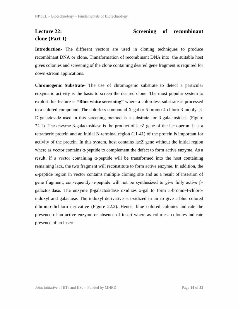
Chromogenic Substrate- The use of chromogenic substrate to detect a particular
enzymatic activity is the basis to screen the desired clone. The most popular system to
exploit this feature is “Blue white screening” where a coloroless substrate is processed
to a colored compound. The colorless compound X-gal or 5-bromo-4-chloro-3-indolyl-β-
D-galactoside used in this screening method is a substrate for β-galactosidase (Figure
22.1). The enzyme β-galactosidase is the product of lacZ gene of the lac operon. It is a
tetrameric protein and an initial N-terminal region (11-41) of the protein is important for
activity of the protein. In this system, host contains lacZ gene without the initial region
where as vector contains α-peptide to complement the defect to form active enzyme. As a
result, if a vector containing α-peptide will be transformed into the host containing
remaining lacz, the two fragment will reconstitute to form active enzyme. In addition, the
α-peptide region in vector contains multiple cloning site and as a result of insertion of
gene fragment, consequently α-peptide will not be synthesized to give fully active β-
galactosidase. The enzyme β-galactosidase oxidizes x-gal to form 5-bromo-4-chloro-
indoxyl and galactose. The indoxyl derivative is oxidized in air to give a blue colored
dibromo-dichloro derivative (Figure 22.2). Hence, blue colored colonies indicate the
presence of an active enzyme or absence of insert where as colorless colonies indicate
presence of an insert.

NPTEL – Biotechnology – Fundamentals of Biotechnology
Joint initiative of IITs and IISc – Funded by MHRD Page 15 of 52
Insertional inactivation-In this approach a foreign DNA is cloned within the coding
gene responsible for a phenotype. As a result of insertion, the gene product is not
available to modulate the phenotype of the host. This approach is known as insertional
inactivation, and it can be used with a suitable genetic system.
Figure 22.1: Molecular Mechanism of blue-white screening.
Figure 22.2: Chemical conversion of X-gal during blue-white screening.

NPTEL – Biotechnology – Fundamentals of Biotechnology
Joint initiative of IITs and IISc – Funded by MHRD Page 16 of 52
(1) Insertional Inactivation of antibiotic resistance gene- As discussed in an earlier
lecture, bacterial plasmid PBR322 has two antibiotic resistance gene, Apr and Tc
r. If a
gene fragment will be cloned in ScaI, it will disrupt the Apr gene. As a result, the clone
will be ampicillin sensitive and Tcr. where as the original plasmid will be Ap
r and Tc
r. To
select the clone, first the transformed e.coli is plated on tetracycline containing media.
Subsequently, a replica plate will be made on ampicillin containing media to identify the
clone growing on tetracycline media but not on ampicillin media. This approach is
schematically depicted in Figure 22.3.
Figure 22.3: Insertional Inactivation of antibiotic resistance gene in pBR322 to screen recombinant clone.

NPTEL – Biotechnology – Fundamentals of Biotechnology
Joint initiative of IITs and IISc – Funded by MHRD Page 17 of 52
(2) Insertional Inactivation of LacZ gene- LacZ is a part of lac operon and responsible
for synthesis of β-galactosidase. As discussed earlier, X-gal system can be used to detect
the insertional inactivation of LacZ gene to screen the cloned fragment. If the gene is
inserted into the lacz, the clone will not be able to produce a functional β-galactosidase.
Hence, blue colored colonies indicate the presence of an active enzyme or absence of
insert where as colorless colonies indicate presence of an insert. This approach is
schematically depicted in Figure 22.4.
Figure 22.4: Insertional Inactivation of LacZ gene to screen recombinant clone.

NPTEL – Biotechnology – Fundamentals of Biotechnology
Joint initiative of IITs and IISc – Funded by MHRD Page 18 of 52
(3) Insertional Inactivation of cI gene-During an infection cycle, virus undergoes a lytic
and lysogenic stages. The lytic phase is responsible for lysis of host to release the virus
particle where as lysogenic stage allow the replication of virus without lysis of host. The
cI gene encodes for cI repressor and which is responsible for the formation of lysogens.
In the presence of functional cI, the plaques contains unlysed host cells and has a turbid
appearance where as in the absence it will clear. This feature can be use to screen the
clone to detect functional cI (absence of clone) or absence of cI (presence of insert). This
approach is schematically depicted in Figure 22.5.
Figure 22.5: Insertional Inactivation of cI gene to screen recombinant clone.

NPTEL – Biotechnology – Fundamentals of Biotechnology
Joint initiative of IITs and IISc – Funded by MHRD Page 19 of 52
Antibiotic sensitivity- Vector carries a functional selection marker such as antibiotic
resistance gene to be use to select the clone. The antibiotic resistance gene product has
multiple mechanism to provide resistance in host cell (Table 22.1). In this approach, a
circular plasmid containing antibiotic resistance can be able to replicate into the host cell
plated on a antibiotic containing media. In the cloning of a fragment into this plasmid, the
plasmid is cut with restriction enzymes and a fragment in ligated to give circular plasmid
with insert. The transformation of both DNA species; cut plasmid and circularized clone
into the host and plated onto the antibiotic containing solid media. Only circularized
clone will give colonies where as cut plasmid will not grow as it has lost antibiotic
resistance gene. This approach is schematically depicted in Figure 22.6.
TABLE 22.1: ANTIBIOTICS RESISTANCE GENE AND THEIR MODE OF MECHANISM.
Antibiotic Gene product Mechanism
Ampicillin β-lactamase Degradation of ampicillin
Kanamycin Neomycin
phosphotransferase II
Covalent modification of kanamycin
Tetracycline Ribosomal protection
proteins
Efflux of tetracycline outside of the
bacteria
Chloramphenicol Chloramphenicol acetyl
transferase
Chloramphenicol to acetyl
Chloramphenicol

NPTEL – Biotechnology – Fundamentals of Biotechnology
Joint initiative of IITs and IISc – Funded by MHRD Page 20 of 52
Figure 22.6: Screening of recombinant clones using antibiotic sensitivity.
Complementation of mutation- In this approach, a mutant host strain can be used to
screen the plasmid containing the missing gene and the transformant will grow only if the
gene product from clone will complement the function. In general genes taking part in
metabolic pathway or biosynthetic pathway are routinely been used for this purpose.
There are 3 important requirement for this approach to work-
1. Host strain deficient in a particular gene. If the gene belongs to the biosynthetic
pathway, the mutant host in this case are called as auxotroph as host dependent on the
gene product or final product of the biosynthetic pathway as a supplement in media for
growth.
2. A defined media with missing nutrient.
3. A vector containing the gene to supply the gene product to complement.

NPTEL – Biotechnology – Fundamentals of Biotechnology
Joint initiative of IITs and IISc – Funded by MHRD Page 21 of 52
Lecture 23: Screening of recombinant clone (Part II)
In continuation of earlier lecture, an auxotroph yeast strain can be use to screen
recombinant clone. Yeast vector discussed in previous lecture has 4 different gene His3,
Leu2, Trp1 and Ura3 as selectable marker. Yeast host with a mutation in these gene are
available and can be use with the yeast vector to screen the recombinant clone. Ura3 and
Lys2 marker offer both positive and negative selection.
Positive selection- In the positive selection, host strain doesn’t grow on the media
lacking the functional gene but the host transformed with the recombinant clone can be
able to supply the gene product required to grow in the media.
Negative selection-In the negative selection, a chemical compound is added to the media
which will be converted to the cyto-toxic agent in the presenc of gene product, and as a
result it doesn’t allow the growth of the wild type cells. But host strain transformed with
the recombinant clone has non-functional gene product and grows in the presence of the
compound in the media. Ura3 codes for orotine-5'-monophosphate (OMP) decarboxylase
and an active enzyme process the 5-fluoro-orotic acid to the toxic fluorodeoxyuridine.
Generation of this cyto-toxic agent kills the cells carrying functional Ura3 gene product.
Figure 23.1: Negative selection of yeast cells transformed with the recombinant clone.

NPTEL – Biotechnology – Fundamentals of Biotechnology
Joint initiative of IITs and IISc – Funded by MHRD Page 22 of 52
Screening of transfected mammalian cells- mammalian cells containing a recombinant
clone can be screened using multiple approaches. In the current we will discuss few of
these approaches-
Reporter Gene Assay- In the reporter gene assay system, a chimeric construct is
produced with an enzyme gene is cloned in front of the promoter of gene of interest. A
schematic design of reporter gene construct is given in Figure 23.2. The general reporter
gene construct contains a eukaryotic promoter and a enzyme for easy read out. The
reporter gene construct is transfected to the mammalian cells with a suitable transfection
reagent as discussed in previous lecture. Afterwards, the cells are stimulated with the
agents to stimulate the production to transcription factor to binds promoter and drive the
expression of the reporter gene. A suitable substrate is added to measure the activity of
the reporter enzyme. Different enzymes used for this purpose is given in Table 23.1.
Figure 23.2: an over-view of the reporter gene assay.

NPTEL – Biotechnology – Fundamentals of Biotechnology
Joint initiative of IITs and IISc – Funded by MHRD Page 23 of 52
Table 23.1: Reporter gene construct used for screening mammalian clones.
Gene Gene Product Reaction Catalyzed
CAT Chloramphenicol acetyl
transferase
Chloramphenicol to acetyl Chloramphenicol
lacZ β-galactosidase o-nitrophenol-β-galactoside to o-nitrophenol and
galacose.
Luc Luciferase Luciferin to oxyluciferin
phoA Alkaline Phosphatase Release of inorganic phosphate
GFP Green Fluorescent Protein Fluorescence

NPTEL – Biotechnology – Fundamentals of Biotechnology
Joint initiative of IITs and IISc – Funded by MHRD Page 24 of 52
Luciferase reporter gene system-Luciferase is an enzyme present in the abdomen of
firefly photinus pyralis. The enzyme utilizes D-luciferin as a substrate to form
oxyluciferin. In the presence of ATP, Mg2+, luciferin is converted into the
luciferinadenylate involving pyrophosphate cleavage and transfer of AMP to luciferin.
Luciferin adenylate undergoes oxidative decarboxylation to oxyluciferin with
simultaneous emission of light (Figure 23.3). The assay setup is shown in Figure 23.4.
The reporter gene construct containing luciferase is transfected to the mammalian cells.
The cells are washed with PBS and lysed with a lysis buffer. Take the lysate into the
lumiometer cuvette or plate and luciferin substrate is injected to start the reaction and
measured immigately in a luminometer.
Figure 23.3: Luciferase reporter assay measures the formation of oxyluciferin formation.

NPTEL – Biotechnology – Fundamentals of Biotechnology
Joint initiative of IITs and IISc – Funded by MHRD Page 25 of 52
Figure 23.4: Different steps of luciferase reporter assay setup.

NPTEL – Biotechnology – Fundamentals of Biotechnology
Joint initiative of IITs and IISc – Funded by MHRD Page 26 of 52
Chimeric Construct with green fluorescent protein (GFP) - In the live cell, green
fluorescent protein is a good choice as reporter gene to screen cells containing
recombinant protein fluorescently tagged with the GFP at their c-terminus (Figure 23.5,
A). The cell receiving recombinant DNA will give green fluorescence and it can be
visualized with an inverted fluorescence microscope (Figure 23.5,B) and it can be
analyzed in flow cytometer to separate the GFP containing cells from the untransfected
cells. Flow cytometer analysis the cell based on its shape, size and fluorescence level. A
non-fluorescent cell is giving separate peak as compare to the fluorescently labeled cells
(Figure 23.5, C) and with the help of flow cytometer, both of these peaks can be collected
in separate tubes. Besides, GFP, red fluorescent protein, yellow fluorescent protein, cyan
fluorescent protein are also popular to use to label the protein.
Figure 23.5: GFP represents a good candidate for reporter gene construct. (A) 3-D structure of green fluorescent protein, (B)
COS-7 Cells expressing GFP and (C) Flow cytometric analysis of GFP expressing COS-7 Cells.

NPTEL – Biotechnology – Fundamentals of Biotechnology
Joint initiative of IITs and IISc – Funded by MHRD Page 27 of 52
Lecture 24 Protein Production strategies in Expression System
(Part-I)
Introduction-Heterologous expression system offers a cheap and reproducible
production of the protein in large quantities for applications. Various prokaryotic and
eukaryotic expression system are developed for this purpose with distinct features. E.coli
as a expression system is cost effective and manipulation procedures are easy but it has
limitation to express proteins with post-translation modification. Similarly, eukaryotic
expression system can be able to perform post-translational modifications but it may not
be cost effective. In the current lecture, we will discuss different expression system and
the underlying strategies to produce foreign protein in large quantities.
Criteria to choose a expression system-Choosing a host expression system is the first
step to decide the strategies to clone the gene in a suitable vector and subsequent down-
stream procedures. The number of factors need to consider to choose the host expression-
vector system suited for over-expression of a protein.
1. Quantity of the desired protein- if a protein is required in small amount, any host
expression system may be suitable for the purpose but if a large quantity of the protein is
required, a e.coli, yeast or baculo expression system might be more suitable than
mammalian expression system.
2. Size of the protein- E.Coli expression system is not preferred for large size of the
protein but an eukaryotic expression system is more suitable.
3. Compatibility between source organism and expression system- In general a close
distance between source organism and expression system is preferred as it may increases
the chances of getting the expression of cloned gene and presence of the protein in
soluable fraction.
4. Down-stream application- This is the most important criteria to choose a host-vector
system. If the protein production is for generating antibodies, any expression system may
suit well for this purpose but if the protein is required for activity or for ELISA, then a
compatible expression system is preferred.

NPTEL – Biotechnology – Fundamentals of Biotechnology
Joint initiative of IITs and IISc – Funded by MHRD Page 28 of 52
In this series of lecture, we will discuss 4 different expression system:
1. E.COLI as a Expression System-
2. Yeast as a Expression System-
3. Insect Cell line as a Expression System-
4. Mammalian cells as a Expression System
E.COLI as a Expression System
An Over-view of protein synthesis machinery in E.coli- Protein production in
prokaryotic system is a multistep process and an over-view of these steps are as follows:
1. Binding of RNA polymerase to the promoter elements to start the transcription to form
m-RNA.
2. As soon m-RNA is synthesized, a translation machinery start the synthesis of protein.
Protein synthesis start usually at the start codon AUG and ends at the stop codon (UAA,
UGA or UAG). In bacteria transcription and translation occurs simultaneously and
translation start as soon as m-RNA comes out from the RNA polymerase.
The details of these steps can be followed from any molecular cell biology text book
such as, B. Alberts, A. Johnson, J. Lewis, M. Raff, K. Roberts, P. Walters, Molecular
Biology of Cell, 5th Ed, Garland Publishing, 2007.

NPTEL – Biotechnology – Fundamentals of Biotechnology
Joint initiative of IITs and IISc – Funded by MHRD Page 29 of 52
Figure 24.1: Steps of protein synthesis in e.coli expression system.
Typical Components of an e.coli expression vector- In our previous lecture we
discussed the salient feature of cloning vector of e.coli but additional structural features
are essential for an expression vector.
1. Promoter-This is upstream sequence to the gene and provides the docking site for
RNA polymerase.
2. Ribosome binding site- Ribosome binding site including Shine-Dalgarno sequence is
the docking site for assembly of ribosome.
3. Termination site- it terminates the synthesis of m-RNA.
4. Affinity tag- The presence of affinity tag either before or after gene sequence provides
a mean to purify the protein using affinity chromatography.

NPTEL – Biotechnology – Fundamentals of Biotechnology
Joint initiative of IITs and IISc – Funded by MHRD Page 30 of 52
Promoter regulates the production of protein- The structural elements of an E.coli
promoter is given in Figure 24.2. The sequence at -10 and -35 are crucial to facilitate
RNA polymerase and consequently determine the strength of the promoter. The
nucleotide substitution in this region severaly affects the turn over of RNA polymerase
binding events or transcription initiation events. Subsquently a number of promoters are
designed for over-expression of protein in E.coli using a strong or a weak promoter to
suit the over-expression strategy.
Figure 24.2: A generalized promoter structure in E.coli
IPTG inducible promoter- IPTG is an synthetic analogue of lactose and it is an inducer
for lac operon. The lac promoter is very widely been used to construct different
expression plasmid to express protein in E.coli. The different vector contains lac
promoters or its derivatives:
1. The lac promoter- example of plasmid, pUC, pGEM etc.
2. The trp-lac (tac) promoter- it is a hybrid promoter where -10 region from lac UV5
promoter is fused with the -35 region of trp promoter. Ex. of plasmid is pKK223-3
3. The trp-lac (trc) promoter- it is similar to tac promoter except that distance
separating -10 and -35 region of promoter is different from the tac prommoter. Ex. of
plasmid pTrc 99A.
Bacteriophage λ PL promoter- This promoter keeps the tight control over the protein
production. It is regulated by the presence of repressor cIts857 to either repress the
transcription or not. cIts857 is temperature sensitive and degraded at high temperature
and consequently in a temperature depenent fasion it represses the transcription at low
temp but not at high temperature. This promoter is useful in the cases where the protein is
toxic in nature.

NPTEL – Biotechnology – Fundamentals of Biotechnology
Joint initiative of IITs and IISc – Funded by MHRD Page 31 of 52
Bacteriophage T7 Promoter- Similar to Bacteriophage λ PL promoter, T7 RNA
polymerase promoter is used to design plasmid with tight control on the protein
production. These vectors contains most of the structural blocks from pBR322 and MCS
is in front of the T7 promoter to drive the transcription of the insert. Hence, vector
contains foreign gene in front of the T7 promoter for expression. The host E.coli also
need modification to suits the T7 promoter expression system. Host E.coli is either
transformed with a plasmid which carries the T7 RNA polymerase gene or the T7 RNA
polymerase gene is integrated into the bacterial chromosome. In few host strain T7 RNA
polymerase is placed under the control of IPTG inducible lacUV5 promoter to tightly
control the production of T7 RNA polymerase. A schematic is given in Figure 24.3 to
explain the control mechanism in T7 promoter based expression system. After induction
with IPTG, the inducer binds the lac repressor and stimulate the production of T7 RNA
polymerase using E.coli RNA polymerase. The T7 RNA polymerase binds to the T7
promoter and drive the transcription of the target gene to eventually give large amount of
protein.
Figure 24.3: T7 Promoter mediated protein production control mechanism in expression host.

NPTEL – Biotechnology – Fundamentals of Biotechnology
Joint initiative of IITs and IISc – Funded by MHRD Page 32 of 52
Expression of gene using E.coli expression system- The steps in an expression of a
gene is outlined in Figure 24.4 and it has following steps:
1.Transformation- As discussed we can use multiple methods to transform the host
with a recombinant clone containing suitable selection marker.
2. Growth of the bacteria- A single colony of the transformed colony is inoculated in
the suitable media as discussed before upto the log phase (OD=0.6-0.7).
3. Induction- The bacterial culture is now induced with IPTG (0-1mM) for 3-6 hrs to
produce the protein.
4. Recovery of the bacteria and analysis of protein expression- Bacteria can be
recovered from the culture with a brief centrifugation at 8000-9000 RPM and analyzed
on a SDS-PAGE. The details of SDS-PAGE will be discussed in a future lecture. The
SDS-PAGE analysis of a particular expression study in E.coli is given in Figure 24.4 and
it indicates a prominent expression of the target protein in the induced cells as compare to
the uninduced cells.
Figure 24.4: Protein Production using E.coli as expression system.

NPTEL – Biotechnology – Fundamentals of Biotechnology
Joint initiative of IITs and IISc – Funded by MHRD Page 33 of 52
Lecture 25 Protein Production strategies in Expression System
(Part-II)
Factor govering expression of foreign protein in E.coli
Translation efficiency: Translation efficiency is goverened by composition of promoter
especially the sequence of ‘shine dalgaro sequences’ which enables the binding of
ribosome protein production machinery. In addition, the distance between shine-dalgaro
sequence and the start codon is also important for efficient translation start. More-over,
secondary structure of promoter elements also affect the efficiency of gene expression.
Codon Usage: Genetic codes are degenerate and there are 61 codes available for 20
amino acids (Figure 25.1). As a result, organism has a preference towards a set of genetic
codes. Expressing these sequences require t-RNA to recongnize the genetic code. But if
the host expression system has no t-RNA or low level of a particular t-RNA then it will
either delay the synthesis or stop the synthesis at the particular amino acid. Consequently
either it will produce less protein or a truncated protein.
Figure 25.1: Genetic Code table

NPTEL – Biotechnology – Fundamentals of Biotechnology
Joint initiative of IITs and IISc – Funded by MHRD Page 34 of 52
Growth Conditions: Growth media has dramatic effect on the gene expression. Either
the media components provide the raw material for the synthesis of amino acid or provide
amino acid for synthesis of a protein. In addition, growth media rich with carbon source
may provide high cell mass and as a result it will give more protein.
Expression of Fusion proteins-The proteins in E.coli expression system can be
expressed as a hybrid protein where reading frame of two gene (one for fusion tag and
other is for foreign gene) are in frame. The fusion tag can be placed either at N-terminus
or C-terminus. A list of commonly used fusion tag is given in Table 25.1.
The advantages of fusion proteins:
1. Easy Purification: A detail of this aspect will be discussed in the future lecture.
2. Targeting to the cellular compartment- A fusion protein can be targeted to the
different cellular compartments for various reasons. Such as periplasm targeting sequence
will allow the protein to accumulate into the periplasm and hence can help to easy
isolation.
3. Half life of protein- In many cases a fusion tag hides the potential protease sites on the
foreign protein and enhances its half-life.
4. Soluability- Keeping a tag at N-terminus direct the protein synthesis and hence help in
increasing the soluability of the foreign protein.

NPTEL – Biotechnology – Fundamentals of Biotechnology
Joint initiative of IITs and IISc – Funded by MHRD Page 35 of 52
Table 25.1: Selected List of Fusion tags and their applications.
S.No. Fusion Tag Vector Features
1 β-galactosidase pUC, pBluescript,
pGEM
Blue-white screening
and affinity
purification
2 Maltose Binding Protein
(MBP)
pMAL Affinity purification
3 Thioredoxin (trx) pTrx Affinity purification
4 Poly His6 pET series Affinity purification
5 GST pGEX Affinity purification
and reporter gene
assay
6 Alkaline phosphatase pTA1529 Reporter Gene assay

NPTEL – Biotechnology – Fundamentals of Biotechnology
Joint initiative of IITs and IISc – Funded by MHRD Page 36 of 52
Removal of fusion Tag- For many biotechnology applications, a protein is expressed as
fusion protein with a N-terminus or C-terminus tag, to easily purify the protein. But after
purification the tag needs to be removed for down-stream applications such as vaccine or
protein crystallographic studies. A list of reagent is given in Table 25.2 to cleave the
fusion protein to remove the tag. In general fusion protein junction point has either the
protease cutting site or the site is sensitive to the chemical treatment. Treating the fusion
protein with protease or chemical agent cuts the fusion protein to release the target
protein (Figure 25.2). Passing the cleavage mixture allows the binding of the tag into the
affinity column whereas target protein does not bind and comes out in the flow through.
Target protein free of fusion tag can be collected and used for down-stream applications.
Table 25.2: Selected List of reagents for cleavage of fusion protein.
S.No. Reagent Cleavage Sequence
1 Cynogen Bromide -Met ↓
2 Hydroxylamine -Asn-↓-Pro
3 Enterokinase -Asp-Asp-Asp-Asp-Lys↓
4 Factor Xa -Ile-Glu-Gly-Arg ↓
5 α-thrombin -Leu-Val-Pro-Arg↓-Gly-Ser
6 Trypsin -Arg ↓ or Lys ↓
7 Subtilisin -Gly-Ala-His-Arg ↓
Yeast as a expression system-yeast is the simpliest unicellular eukaryotic cells available
for protein production. It is easy to manipulate and the production cost is also very low in
comparison to the other eukaryotic expression system. It offers most of the advantages
available in a typical eukaryotic cells. In addition, a large amount of genetic, molecular
and cell biology aspect of yeast is known and this knowledge has help us to design better
protein production strategy and troubleshooting.

NPTEL – Biotechnology – Fundamentals of Biotechnology
Joint initiative of IITs and IISc – Funded by MHRD Page 37 of 52
Host Species Used- There are two different varities of yeast strains available for protein
production.
Non-methylotrophic-These species don’t have ability to utilize one carbon compounds
such as methanol. But it can be able to utilize other carbon sources such as glucose,
lactose, maltose, starch and alkane. The example of yeast in this class are S.cerevisiae,
K.lactis, and Y.lipolytica. These yeast strains are mostly been used for fermentation to
produce alchol etc. The major advantage of this class is better understanding of molecular
biology, biochemistry and fermentation technology aspect of these strains. But still the
technology is not eveolved to utilize this class of yeast for production of heterologous
protein.
Figure 25.2: Schematic presentation of cleavage of fusion tag from target protein.

NPTEL – Biotechnology – Fundamentals of Biotechnology
Joint initiative of IITs and IISc – Funded by MHRD Page 38 of 52
Methyltrophic yeasts- The major advantage of this class is ability to utilize one carbon
compounds such as methanol as carbon and energy source. In addition, these strains have
high level of methanol oxidizing enzyme and that allow them to be very strong and grow
in high density. The example of yeast in this class are, pichia pastories, pichia angusta,
P.methanolica and C.boidini.
Transformation- A number of transformation methods specific to yeast is discussed in
the previous lecture. Lithium acetate and electroporation is the method popular method
for transformation of yeast.
Vector and selection- The different yeast vector are already discussed in the previous
lecture. Transformant are selected either using a auxotroph marker (such as URA3,
LEU2, TRP1, HIS4) or antibiotic resistance (such as G418, hygromycin etc).
Promoters in yeast expression vector- Similar to E.Coli expression system, yeast
vectors have different promoters to drive the expression of foreign protein. A list of
promoter is given in Table 25.3. In general, yeast expression vector offers two types of
promoters:
1. Constitutive Promoter- These promoter belongs to the house keeping gene and as a
result the expression is non-inducible. The protein production starts with the growth of
the yeast and as a result it is proportional to the cell mass. Example of these promoters
are GAPDH, GAM1 etc.
2. Inducible Promoter- Pichia pastoris expresses two different alchol oxidases, AOX1
and AOX2 where as pichia angusta expresses methanol oxidase (MOX). The promoter of
AOX1 and MOX are present on yeast vector and it has been used to drive the expression
of foreign protein. The protein production is controlled by a balance of repression and
induction. Presence of other carbon source such as glucose represses the transcription of
AOX1 gene but in the presence of trace amount of methanol, it induces the AOX1
promoter mediated protein production.

NPTEL – Biotechnology – Fundamentals of Biotechnology
Joint initiative of IITs and IISc – Funded by MHRD Page 39 of 52
Table 25.3: Different promoter(s) in Yeast expression system.
Strain Type Species Constitutive Inducible
Non-Methyltrophic S. Cerevisiae GAPDH UAS, ADH 1
K.Lactis PGK LAC 4, ADH 4
Y.lipolytica TEF, RPS 7
S.Occidentalis GAM1 AMY 1
Z. rouxii GAPDH
Methyltrophic P.Pastoris GAP AOX 1, FLD 1
H. polymorpha MOX
P. methanolica AUG 1
Production of protein in yeast- The protein production in yeast can be done in such as
way to either express the protein in cytosol or secreted into the media supernatant.
Cytoplasmic targeted protein- The expression of protein targeted to the cytoplasm is
very high but the recovery is very difficult. Yeast cell wall is very hard and high pressure
homogenization is used to disrupt the cell wall. The recovery is very less and a fraction of
total soluable protein comes out.

NPTEL – Biotechnology – Fundamentals of Biotechnology
Joint initiative of IITs and IISc – Funded by MHRD Page 40 of 52
Secreted protein- Protein tagged with secretory signal such as S.cerevisiae α-mating
factor signal target the protein into the secretory pathway. The signal peptide is processed
in ER/Golgi vesicular transport system and appear in culture media.
It is difficult to say which pathway will be useful for over-expression of protein in yeast
expression system. Irrespective of the pathway choosen the protein expression protocol in
yeast expression system is given in Figure 25.3 and it has multiple steps:
(1) Transfer the transformed yeast into 5 ml medium with suitable selection marker and
incubate for 2 days at 280C with shaking at 180rpm.
(2) Allow the culture to reach the OD600 to 5-7 and now resuspend the cells in a new
media without carbon source.
(3) Induce the culture with a methanol of 1% (v/v) twice daily.
(4) harvest the cells and analyze the expression on SDS-PAGE.
Figure 25.3: Schematic presentation of different steps in protein production in Yeast.

NPTEL – Biotechnology – Fundamentals of Biotechnology
Joint initiative of IITs and IISc – Funded by MHRD Page 41 of 52
Lecture 26 Protein Production strategies in expression system
(Part-III)
Insect cell lines as expression system: In the previous lecture we discussed the
prokaryotic and unicellular eukaryotic expression system. As a eukaryote baculovirus
expression system offers protein modification, processing and transport system. Compare
to yeast where the down-stream processing and recovering of cytosolic protein is much
easier in baculovirus system. In addition, molecular biology manipulation, transfection
and the down-stream processing. The different steps needed to produce protein are as
follows:
1. Cloning of foreign gene in transfer vector.
2. Generation of recombinant baculovirus vector
3. Screening of recombinant baculovirus
4. Culturing of recombinant insect cell lines
5. Protein production
1. Cloning of foreign DNA into the transfer vector-The structural components of
transfer vector (Figure 26.1) is discussed in a previous lecture. It has 3 distinct structural
units, (1) A polyhedrin promoter and a unstream sequence from the the virus genome. (2)
A cloning site for foreign DNA, (3) polyhedrin termination site and down-stream region
of virus genome. The up-stream and down-stream sequences from virus genome helps in
homologous recombination. The foreign DNA is cloned in the cloning site and the
recombinant transfer-vector can be propogated in E.coli.
Figure 26.1: Organization of structural elements of transfer-vector AcMNPV.

NPTEL – Biotechnology – Fundamentals of Biotechnology
Joint initiative of IITs and IISc – Funded by MHRD Page 42 of 52
2. Generation of Recombinant baculovirus: There are two approaches to construct the
recombinant baculovirus:
Approach 1: In approach1, insect cell lines are first transfected with baculovirus to
produce transformed insect cells (Figure 26.2). It is subsequently transfected with transfer
vector containing foreign DNA and allowed to grow. In one or two divisons, a double
cross over event occurs between viral genome and the transfer-vector with the help of
flanking virus genome sequences. As a result, the viral genome losses the polyhedrin
gene and receives the DNA stretch from transfer vector containing polyhedrin promoter,
foreign DNA and termination signal.
Figure 26.2: Generation of recombinant baculovirus with the double cross over event (Approach-I)

NPTEL – Biotechnology – Fundamentals of Biotechnology
Joint initiative of IITs and IISc – Funded by MHRD Page 43 of 52
Approach 2: In approach2, baculovirus genome is engineered to introduce two unique
restriction site Bsu36I introduced into the polyhedrin gene within viral genome (Figure
26.3). When modified viral genome is treated with restriction enzyme and transfected
into the insect cell, no viral particle found as it is missing the function of a crucial gene
ORF1629 which is required for viral replication. The linearized truncated viral genome
(with missing polyhedrin gene, ORF1629 and gene 603) is transfected into the insect
cells, followed by transfection of transfer vector containing foreign DNA along with the
gene 603 and essential gene. In one or two divisons, a double cross over event occurs
between truncated viral genome and the transfer vector with the help of flanking gene
603 and essential gene sequences. As a result, the viral genome recives the lost portion of
gene 603 and ORF1629 from transfer vector and foreign DNA is incorporated into the
viral genome.
Figure 26.3: Generation of recombinant baculovirus with the modified baculo-virus (Approach-II)

NPTEL – Biotechnology – Fundamentals of Biotechnology
Joint initiative of IITs and IISc – Funded by MHRD Page 44 of 52
Screening of recombinant baculovirus- The recombinant baculovirus can be screened
by a plaque assay. It involves multiple steps as depicted in the Figure 26.4. The major
steps are as follows:
1. Dilute a culture of insect cell line Sf9 to a density of 105/ml.
2. Make serial dilution of baculovirus stock in serum containing media.
3. Add 1ml of viral sample to each well and incubate for 1h at 270C.
4. Remove the viral diluted suspension and then detect the presence of plaque. There are
three popular method to detect the plaque:
(A) Over-lay a agarose and allowed it to harden. Incubate this plate for 6-8 days at 270C
to plaque form and these can be visualized.
(B) If the recombinant virus contains a lacZ gene, then plaque can be identified by adding
b-galactosidase substrate, x-gal, plaque containing cells will appear blue.
(C) In third method, cells can be stain with trypan blue, plaque containing cells will
takeup the dye and appear blue where as other cells will remain colorless.
Figure 26.4: Screening of recombinant baculovirus in a plaque assay.

NPTEL – Biotechnology – Fundamentals of Biotechnology
Joint initiative of IITs and IISc – Funded by MHRD Page 45 of 52
Culture media for growth: The culture media for growing insect cell lines is discussed
in a previous lecture.
Maintenance and culture of insect cell line: Sf9 cell lines is derived from the ovaries of
the armyworm (Spodotera frugiperda). It is maintained in TNH-FH insect media
containing 10% foetal bovine serum (FBS) and gentamycin.
Culture media for protein production: BaculoGold or other serum free, low protein
media is suitable for the secreted proteins as it facilitates easy purification.
Protein Production in Baculovirus expression system: The major steps to produce the
protein are summarized in Figure 26.5. The whole process is as follows-
1. Seed 106 Sf9 cells in a 60mm cell culture dish and allow the cells to adhere to the dish.
2. Add 0.1 ml high titer baculovirus stock at an MOI of 1:10. Incubate the cell for 3 days
at 270C.
3. Collect the cells and media. Centrifuge at 1000xg for 10min at 40C.
If the protein is secreted:
4a. Transfer the culture supernanat to new tube and determine the protein concentration
with Bradford reagent.
If the protein is cytosolic:
4b. discard the supernatant and wash the cell pellet with PBS.
4c. Lysed the cells and analyzed the protein on SDS-PAGE.

NPTEL – Biotechnology – Fundamentals of Biotechnology
Joint initiative of IITs and IISc – Funded by MHRD Page 46 of 52
Figure 26.5: Schematic Presentation of different steps in protein production in insect cells.

NPTEL – Biotechnology – Fundamentals of Biotechnology
Joint initiative of IITs and IISc – Funded by MHRD Page 47 of 52
Lecture 27 Protein Production strategies in Expression System
(Part-IV)
Mammalian expression system: Similar to all other expression system, protein
production in mammalian cell system can be achieved with either from a vector present
as extrachromosomal DNA (transient ) or the sequence is integrated into the genome
through homologous recombination to estabilish the permamnent cell line. The
expression from transient or permanent cell lines can be from a constituted or inducible
promoter. Irrespective of the expression mode in mammalian system, the different basic
steps needed to produce protein are as follows:
1. Cloning of foreign gene in mammalian expression vector.
2. Transfection of a cell line with recombinant construct.
3. Screening and selection of transfected cells.
4. Culturing of transfected cells.
5. Protein production.
Different Cell line for protein production: The most popular cell lines used for
mammalian expression system are given in Table 27.1. The cell lines are derived from
the different origin (organ of the body/cell type/animal) and consequently they are
preferred to produce foreign protein with similar origin.
Table 27.1: Popular Cell lines for protein production
S. No. Cell lines Origin
1 CV1 Kidney
2 COS-7 Fibroblast
3 3T3 Fibroblast
4 CHO Ovary
5 J774A.1 Macrophage
6 HeLa Cervical
7 BHK 21 Kidney
8 HEK 293 Kidney

NPTEL – Biotechnology – Fundamentals of Biotechnology
Joint initiative of IITs and IISc – Funded by MHRD Page 48 of 52
Growth media for mammalian cell line: The growth media for different mammalian
cells is discussed earlier in a previous lecture.
Figure 27.1: Different mammalian cell lines used for protein production.
Protein Production in mammalian expression system:
Transient expression: The expression is high but for short time period. The cells
transfected with DNA express proein until 72h post-transfection. Transient expression
system is used to screen cDNA library, isolation of a particular cDNA clone expressing
surface antigen and to test the applicability of the recombinant construct going to use for
permanent expression. There are multiple steps required to transiently express a protein
in COS-7 cell line. Although we are giving the details for COS-7 but these procedures
can be applied to other cell lines with slight modifications (Figure 27.2). The steps are as
follows:
1. Clone the foreign DNA into the appropriate mammalian expression vector to obtain
recombinant DNA. Transfection efficiency is maximum for a supercoiled circular DNA.
Purify the recombinant DNA by a miniprep kit to prepare high quality supercoiled DNA.
2. Seed COS-7 cells in DMEM media supplemeted with 10% foetal bovine serum at 20%
confluence in 100mm dish.
3. Transfect the cells with recombinant DNA using a transfection reagent. [Several
transfection reagent and methods are discussed in a previous lecture].

NPTEL – Biotechnology – Fundamentals of Biotechnology
Joint initiative of IITs and IISc – Funded by MHRD Page 49 of 52
If expressing secreted proteins-wash the transfected cells with PBS and add serum free
media. It allows to secrete the protein for next 72hr. Harvest the medium, remove dead
cells and debris by centrifugation and filter the media from 0.45µm syringe filter before
storage. Detect the presence of protein in media either by activity assay or by western
blotting.
If expressing cell surface or intracellular protein-Transfected cells are allowed to
express the protein for another 72 hrs. Remove media, wash the cells with PBS and detect
the presence of protein on cell surface or in cell lysate by activity assay or by western
blotting.
Figure 27.2: Different Steps in transient expression system.

NPTEL – Biotechnology – Fundamentals of Biotechnology
Joint initiative of IITs and IISc – Funded by MHRD Page 50 of 52
Permanent expression: The permanent expression of a gene is possible, if it will be
integrated into the chromosomal DNA. The most crucial step to estabilish a permanent
expression system for a gene is the frequency of integration events rather than number of
DNA uptake. In simpler word, permanent transfection depends on recombination
frequency instead of transfection efficiency. Stable transformant are selected by a
selection marker (such as antibiotics or auxotrophic factor or negative selection with an
inhibitor) for a prolong period to ensure the integration of recombinant DNA into the
genome. The steps follow to generate a stable cell line expressing foreign protein is given
in Figure 27.3. The different steps are as follows-
Step 1-3 are indentical as discussed for the transient expression of a foreign gene.
4. Selection of trasnfected cells
4.1. Fourty eight hrs after transfection, split the transfected cells in a selection media
containing antibiotic and allowed to grow for another 4 days.
4.2. Gently wash the cells with PBS and observe the descrete colonies.
4.3. Delineate the boundry of each colony with a marker from the back side of the plate.
4.4. Remove the media and put cloning ring to each colony. Wash the colony with PBS
and add 100µl trypsin-EDTA to remove the colony.
4.5. wash the colony with PBS and transfer into one well of 24 well dish. Allow it to
grow and become 80% confluent.
4.6. Transfer these cells to the 6 well dish in the presence of selection media and allow it
to reach 80% confluency.
5. Take a small aliquot of the cell and test the expression of foreign protein with western
blotting. In addition, integration of gene into the genome can be checked by performing a
southern blotting with radioactive probe derived from the gene of interest.

NPTEL – Biotechnology – Fundamentals of Biotechnology
Joint initiative of IITs and IISc – Funded by MHRD Page 51 of 52
Figure 27.3: Different Steps in permanent expression system.

NPTEL – Biotechnology – Fundamentals of Biotechnology
Joint initiative of IITs and IISc – Funded by MHRD Page 52 of 52
Inducible expression system. Inducible expression system is useful for the expression of
a toxic protein or proteins with pleiotropic or non-specific effects. The tetracycline-
controlled inducible system is given in Figure 27.4. In this system, seven tandenly
arranged tet operator [(Tet-op)7] are placed upstream to the minimum CMV promoter and
transcriptional activator (tTA) gene. In another set, target gene is replaced with tTA gene.
In the presence of tetracycline, the binding of tTA is blocked to the tetracycline operator.
Consequently, it causes low level of expression of tTA and target gene. In the absence of
tetracycline, low level of tTA binds to the operator and drive the enhanced expression of
tTA which in-turn stimulates further amplification of initial signal. Transcription
activator (tTA) produced in the absence of tetracycline eventually stimulates the
expression of target gene.
Figure 27.4: Tet inducible system of mammalian expression system. Molecular events in the presence (A) or absence (B) of
tetracycline.