Lecture 2 Physiology of the heart Objectives – Describe each of the components of the cardiac...
20
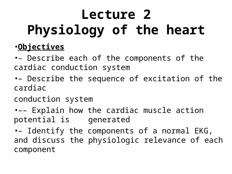
Lecture 2 Physiology of the heart • Objectives •– Describe each of the components of the cardiac conduction system •– Describe the sequence of excitation of the cardiac conduction system •–– Explain how the cardiac muscle action potential is generated •– Identify the components of a normal EKG, and discuss the physiologic relevance of each component
-
Upload
oswin-riley -
Category
Documents
-
view
215 -
download
1
Transcript of Lecture 2 Physiology of the heart Objectives – Describe each of the components of the cardiac...
Lecture 2Physiology of the heart
•Objectives•– Describe each of the components of the cardiac conduction system•– Describe the sequence of excitation of the cardiac
conduction system•–– Explain how the cardiac muscle action potential is generated•– Identify the components of a normal EKG, and discuss the physiologic relevance of each component
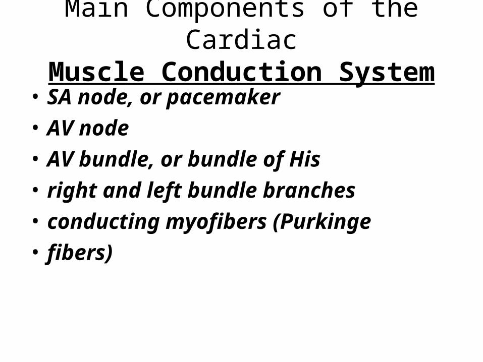
Main Components of the CardiacMuscle Conduction System
• SA node, or pacemaker
• AV node
• AV bundle, or bundle of His
• right and left bundle branches
• conducting myofibers (Purkinge
• fibers)
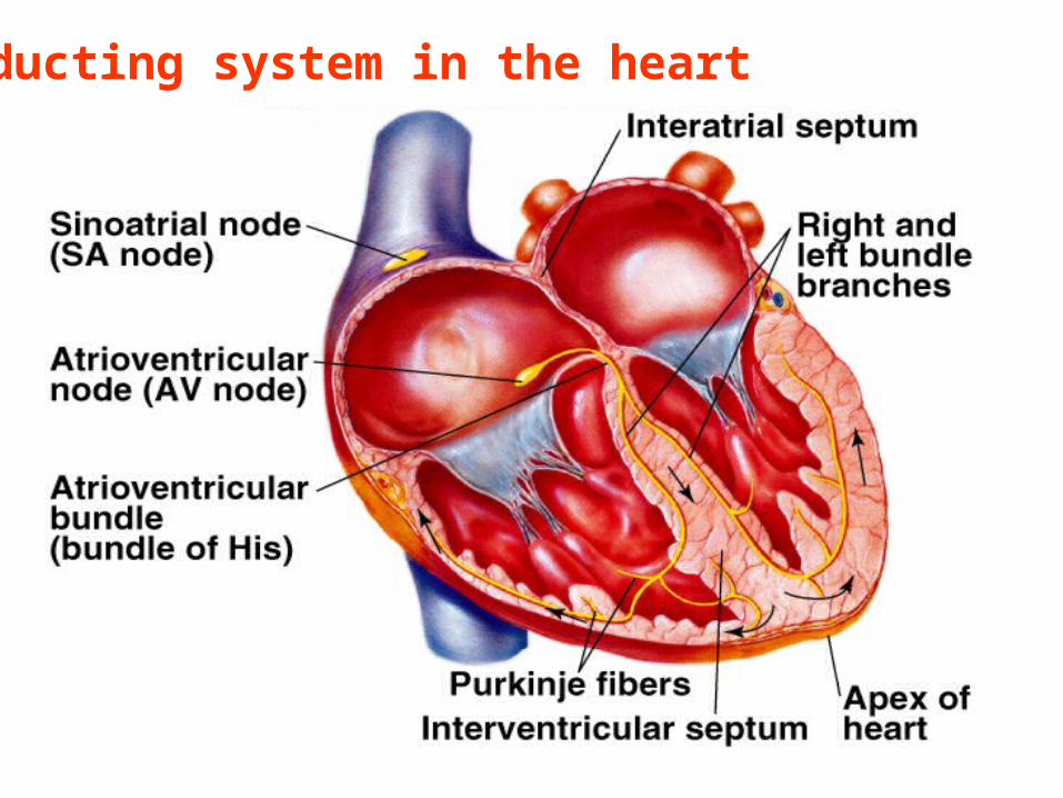
Conducting system in the heart
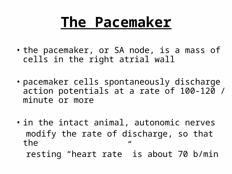
The Pacemaker
• the pacemaker, or SA node, is a mass of cells in the right atrial wall
• pacemaker cells spontaneously discharge action potentials at a rate of 100-120 / minute or more
• in the intact animal, autonomic nerves modify the rate of discharge, so that the resting “heart rate” is about 70 b/min

Sequence of Excitation of Heart Muscle
• the atria and ventricles must contract in a coordinated fashion
• the sequence of cardiac muscle excitation is such that the first event is
• 1- depolarization of the SA node
• 2- the impulses then travel down and across both atria, causing atrial muscle fiber contraction

Sequence of excitation of heartmuscle, Con’t.
• at the AV border there is band of non-conducting tissue
the impulse must pass through the AV node
and through this tissue mass in order to reach the ventricles
• the presence of the non-conducting tissue slows the impulse by about 0.1 sec
• this gives the atria time to fully empty before
the ventricles begin to contract.
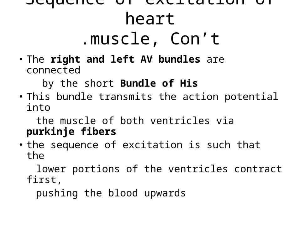
Sequence of excitation of heartmuscle, Con’t.
• The right and left AV bundles are connected by the short Bundle of His• This bundle transmits the action potential into the muscle of both ventricles via purkinje
fibers• the sequence of excitation is such that the lower portions of the ventricles contract first, pushing the blood upwards

Conduction pathway

Cardiac Muscle Action Potential
• an action potential is the result of depolarization of a muscle or nerve cell
• the action potential propagates along nerves and into the muscle fibers, causing muscle
contraction• the cardiac muscle action potential has three distinct phases; rapid depolarization; the• plateau; and repolarization

Action potential

Electrical and Mechanical Correlations
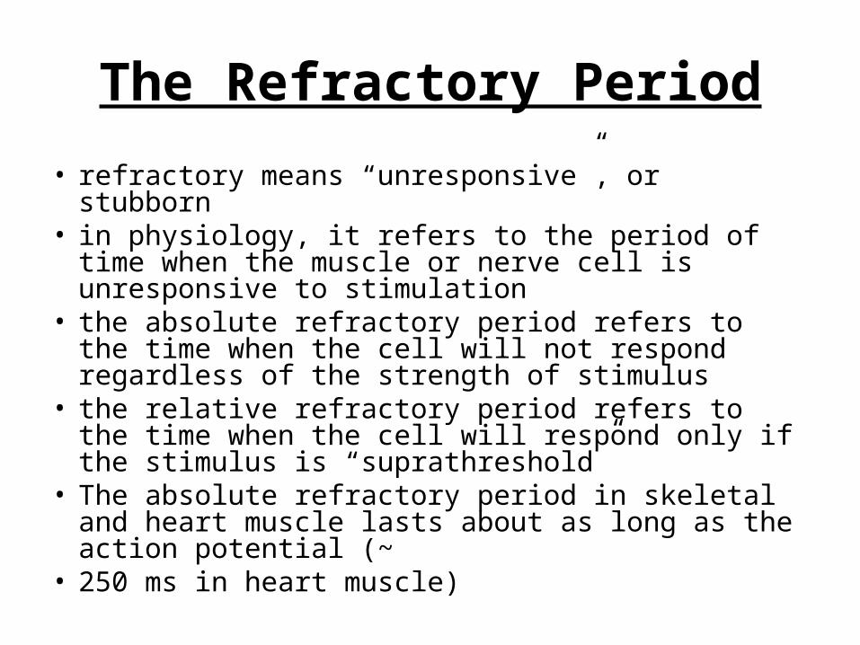
The Refractory Period
• refractory means “unresponsive”, or stubborn• in physiology, it refers to the period of time when the
muscle or nerve cell is unresponsive to stimulation• the absolute refractory period refers to the time when
the cell will not respond regardless of the strength of stimulus
• the relative refractory period refers to the time when the cell will respond only if the stimulus is “suprathreshold”
• The absolute refractory period in skeletal and heart muscle lasts about as long as the action potential (~
• 250 ms in heart muscle)

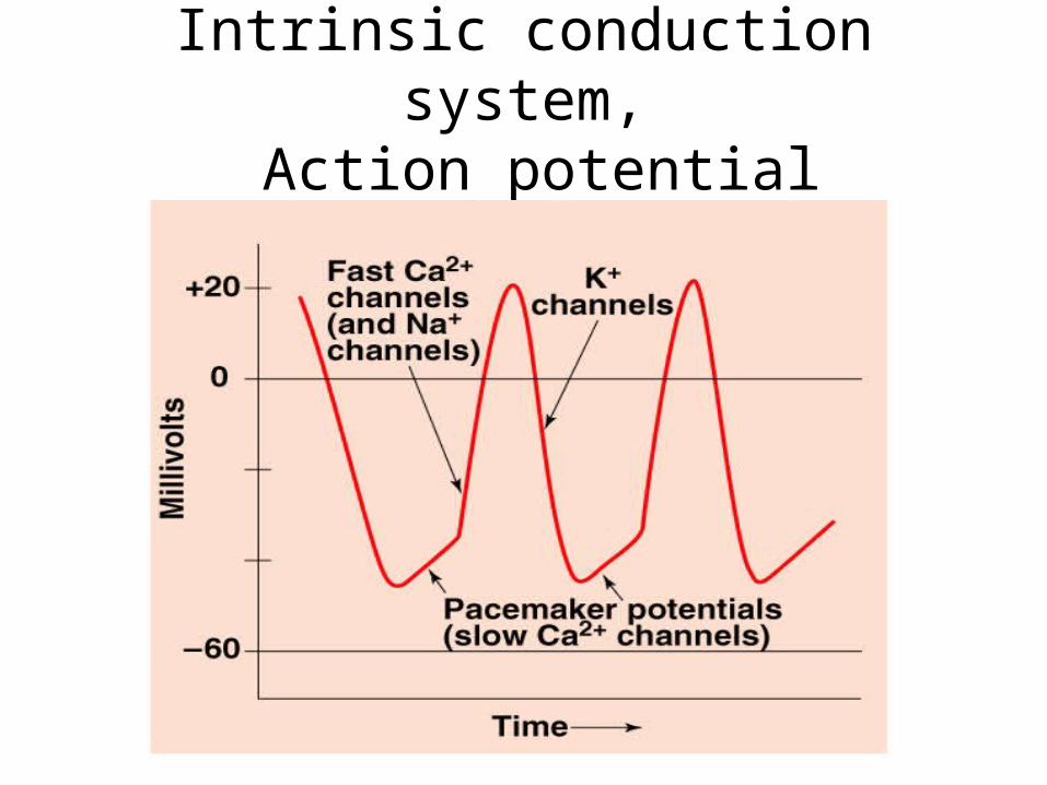
Heart Physiology: IntrinsicConduction System
• Autorhythmic cells:
– Initiate action potentials
• – Have unstable resting potentials called
pacemaker potentials
• – Use calcium influx (rather than sodium)
for rising phase of the action potential

Pacemaker potential and action potential
Intrinsic conduction system,Action potential

Electrical activity of the heart
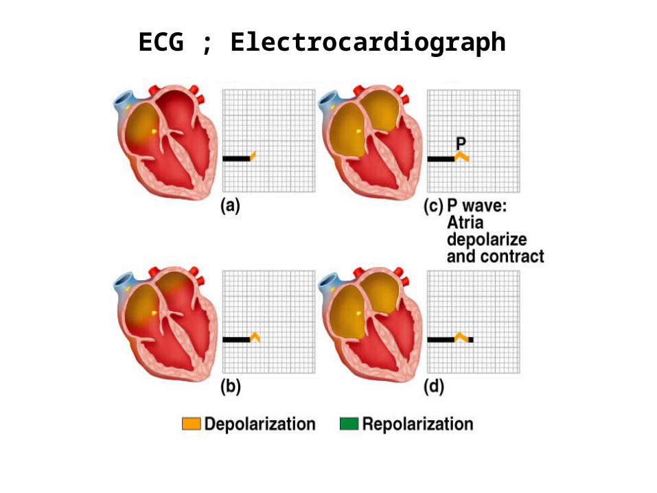
ECG ; Electrocardiograph
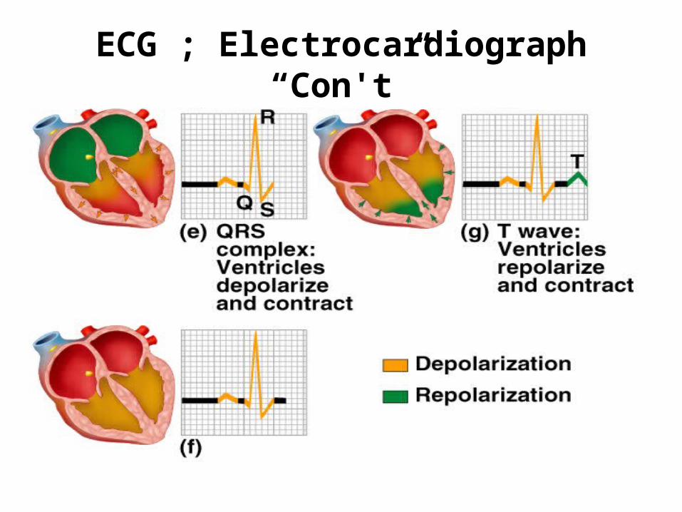
ECG ; Electrocardiograph “Con't”

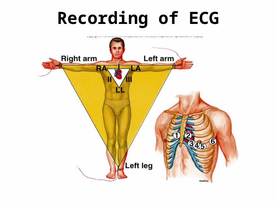
Recording of ECG