An Introduction to SIMS and the MiniSIMS ToF © Millbrook Instruments Limited Blackburn, UK .
Upload
basil-morganCategory
view
218download
0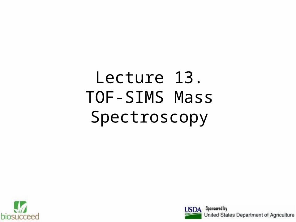
Lecture 13.TOF-SIMS Mass Spectroscopy
• Routine analytical technique
• Detailed chemical structure information
• High sensitivity
• New primary ion sources (Au, Bi, & buckministerfullerene

Principles of ToF-SIMS
• Pulsed primary ion beam
• Emission of particles – SECONDARY IONS
• Ions are mass analyzed by FLIGHT TIMES
• Two modes of analysis: static & spectroscopic

TOF-SIMS
Bombard surface with gallium and run through mass spectrometer
Gives both chemical composition of surface and “SEM-like” image of where chemicals are located
Lightly used on lignocellulosic materials
Find concentrations as low as 10 ppm
http://www.phi.com/genf.asp?ID=83

Static Mode
• Delicate organics (biomaterials)• Undamaged (opposed to X-ray
fluorescence microscopy)• Surface sensitive (outermost couple of
nm)

Spectroscopic Mode
• ONLY mass spectral (MS) data provided• Chemical imaging is POSSIBLE• Raster a micro-focused ion beam (sound
familiar???) over surface• Collect MS• Map distribution of species
Organic imaging technique
• Previously, limted by most significant signals – polyatomic clusters
• For example, most biomaterials dominated by fragments (CxHy
+/-) at low mass (< m/z 100) – MORE than one species
• NOT DIAGNOSTIC

Higher-order chemical imaging
• Larger masses (m/z > 200) more structurally assignable and unique
• Chemical mapping possible• Ga+ bombardment doesn’t allow for sufficient
sensitivity for good imaging• Polyatomic primary ion sources overcomes
deficiency (Aun+, Bin+, C60
+) 100x increases in secondary ion yields
Chemical imaging of pharmaceuticals
• Drug-loaded particles can be visualized with Bi3+, whereas with Ga+ they cannot
• Due to low intensity of molecular ion peak• Tablet formulations can be studied – distribution
of drug, excipient(s), lubricant(s) on surface and in bulk
• Thickness & uniformity can be assessed

ToF-SIMS Images
Nylon mesh – 10 micron depth Plasma cleaned scalpel blade
Distribution of materials
10 micron diameter hair fibers

Lignocellulosic biomaterials
• The work of Thompson with superoxide (Potassium superoxide) in DMSO found attack in amorphous regions first
• Hemicellulose and lignin removed more rapidly than cellulose
• The work of Kim with periodate oxidation suggests that the attack on crystalline cellulose proceeds highly heterogeneously
• Once an area is damaged, however, the area becomes more susceptible to damage due to loss of crystalline order
Thompson, N.S., Corbett, H.M, “The effect of potassium superoxide on cellulose”, TAPPI, 68:12, pp. 68-72, 1985.
Kim, U., Kuga, S., Wada, M., Okano, T., Kondo, T. “Periodate oxidation of crystalline cellulose”, Biomacromolecules, 1:488-492, 2000.

TOF-SIMS hypotheses• Lind studied the ability of hydroxyl radicals to induce viscosity
loss in cellulose fibers• In their work they found that the decrease in viscosity was
proportional to the imparted irradiation dose • This can be read to mean that as the number of hydroxyl
radicals increases, so does the cellulose degradation• The work of Lind studied the role of hydroxyl radicals in
viscosity loss using ionizing radiation • In their work they found that almost no amount of radical
scavenger could protect against depolymerization of the cellulose
• This means that hydroxyl radicals produced outside the cellulose surfaces have a minimal effect on degradation and are more likely produced very near to their consumption point
Lind, J., Merényi, G., “Hydroxyl radical induced viscosity loss in cellulose fibers”, J. Wood Chem. Technol., 17,(1,2), pp. 111-117 (1997).

TOF-SIMS hypotheses, continued
• Metal-induced peroxide cellulose degradation causes the creation of carboxylic acid content
• Work of Lind shows that hydroxyl radicals are formed and react very near to the carbohydrate surface
• Work of Kleen has been used to measure metals on fiber surface as compared to bulk during bleaching, Found majority of metals, 5 to 55 times bulk, on surface
• Not likely to be precipitates due to the fact that the sheets were made at a pH of 5
M. Kleen, Sixth European Workshop on Lignocellulosics and Pulp, 41-44 (2000)

Central hypothesis of work
Hypothesis: We are seeing metals bound to carboxylic acid groups caused by radical degradation of cellulose
Metal distribution begins rather homogeneous in the unbleached case, but becomes heterogeneous in the bleached cases
Attack appears heterogeneous concentrated and surface orientated due to the fact it does not appear “deep” enough to be seen by SEM under comparable resolutions
Total Ion Image Unbleached Mg Bleached Mg Bleached + 50ppm iron Fe

ESCA
• Used to identify carbon and oxygen and the oxidation level
• Has been utilized for the detection of carboxyl content (mostly fiber modification work) in literature and is comparable to other methods
• We appear to have a difference between the bleached and unbleached samples in COOH content
ip0212_101.spe: sample 2124* Evans PHI
02 Feb 12 Al mono 350.0 W 0.0 45.0° 23.50 eV 3.5325e+004 max 30.00 sC1s/Full/1 (SG5 Shft)
2802822842862882902922942962983000
0.5
1
1.5
2
2.5
3
3.5
4x 10⁴ ip0212_101.spe
Binding Energy (eV)
c/s
C-C
/C-H
C-O
C=O
/O-C
-O
O-C
=O

Viscosity and physical testing
• As the load bearing structure of a fiber, the cellulose chains, are being cleaved or “peeled” the mechanical strength of a fiber should decrease
• This change should manifest itself in test to include the zero-span tensile and the standard tensile test

Viscosity and physical testing• Chain scission count increases as
degradation conditions become more favorable
• Zero-span tensile tests show strength decrease as well. Perhaps we also see chemical refining
1.5% H2O2, 70C
3.0% H2O2, 90C3.0% H2O2, 90C, 50 ppm Fe
0
0.05
0.1
0.15
0.2
0.25
Cha
in S
ciss
ion
Cou
nt
Dry Zero-Span Breaking Length
0
5
10
15
20
25
Mature Bleached Bleached + iron
Sample
km Series1

Summary
• TOF-SIMS appears to visualize degradation through the indirect measurement of COOH groups
• The analysis of this degradation can be coupled with other techniques including ESCA, viscosity, and zero-span tensile
• Degradation appears to be a heterogeneous surface phenonemon

Other work
Phytic Acid Chelation• Relatively unstudied chelant that is a product of
unwanted by-products of corn• Current data shows performance on par with
DTPA/EDTA, but effectiveness is very pulp dependent
• Agriculture literature says excellent chelant for iron• Studying it as a bleaching additive and chelant on
many different pulp samples and at differing pH
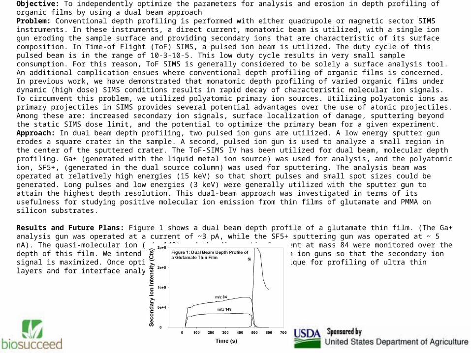
Depth Profiling of Organic Films using the Time-of-Flight SIMS S.V. RobersonObjective: To independently optimize the parameters for analysis and erosion in depth profiling of organic films by using a dual beam approach Problem: Conventional depth profiling is performed with either quadrupole or magnetic sector SIMS instruments. In these instruments, a direct current, monatomic beam is utilized, with a single ion gun eroding the sample surface and providing secondary ions that are characteristic of its surface composition. In Time-of Flight (ToF) SIMS, a pulsed ion beam is utilized. The duty cycle of this pulsed beam is in the range of 10-3-10-5. This low duty cycle results in very small sample consumption. For this reason, ToF SIMS is generally considered to be solely a surface analysis tool. An additional complication ensues where conventional depth profiling of organic films is concerned. In previous work, we have demonstrated that monatomic depth profiling of varied organic films under dynamic (high dose) SIMS conditions results in rapid decay of characteristic molecular ion signals. To circumvent this problem, we utilized polyatomic primary ion sources. Utilizing polyatomic ions as primary projectiles in SIMS provides several potential advantages over the use of atomic projectiles. Among these are: increased secondary ion signals, surface localization of damage, sputtering beyond the static SIMS dose limit, and the potential to optimize the primary beam for a given experiment. Approach: In dual beam depth profiling, two pulsed ion guns are utilized. A low energy sputter gun erodes a square crater in the sample. A second, pulsed ion gun is used to analyze a small region in the center of the sputtered crater. The ToF-SIMS IV has been utilized for dual beam, molecular depth profiling. Ga+ (generated with the liquid metal ion source) was used for analysis, and the polyatomic ion, SF5+, (generated in the dual source column) was used for sputtering. The analysis beam was operated at relatively high energies (15 keV) so that short pulses and small spot sizes could be generated. Long pulses and low energies (3 keV) were generally utilized with the sputter gun to attain the highest depth resolution. This dual-beam approach was investigated in terms of its usefulness for studying positive molecular ion emission from thin films of glutamate and PMMA on silicon substrates. Results and Future Plans: Figure 1 shows a dual beam depth profile of a glutamate thin film. (The Ga+ analysis gun was operated at a current of ~3 pA, while the SF5+ sputtering gun was operated at ~ 5 nA). The quasi-molecular ion (m/z 148) and the diagnostic fragment at mass 84 were monitored over the depth of this film. We intend to optimize the parameters of both ion guns so that the secondary ion signal is maximized. Once optimized, we will utilize this technique for profiling of ultra thin layers and for interface analysis.

What direction next?