Basic Histology and Histological Techniques (MLS-HIST 222 ...

Lecture 11 Basic histology Dr.Gaida kadhim
)1)
It is a specialized connective tissue which is characterized by its rigidity
and hardness. In adult, its constitute most of the skeleton of the body.
General functions of bone
1- Support fleshy structures (as ribs)
2-To permit locomotion (as long bones)
3- Provide protection to vital organs such as brain, heart, and lungs (as skull).
4- Red blood cells are manufactured in the red bone marrow, which is
situated in the spongy tissue at the ends of long bones.
5-It play a vital role in the mineral metabolism of the body. It considered as a
reservoir for several minerals of the body such as calcium, and phosphate.
Bone like other types of connective tissue consist of cells and
extracellular matrix but distinguish from other connective tissue is the matrix
is mineralized. This matrix is calcified and acidophilic. The matrix is
composed of approximately 35% organic and 65% inorganic components. The
organic components include ground substance and type I collagen fibers. The
ground substance is rich in proteoglycans with chondroitin sulfate and
keratin sulfate, also found glycoprotein. The inorganic components include
mineral salts which is responsible for the hardness and rigidity of bone, and
these include calcium phosphate (85%), calcium carbonate (10%), and other
salts (5%). All bones are lined on both internal and external surfaces by layers
of tissue include endosteum on the internal surfaces and periosteum on the
external surface.
The bone
Lecture 11 Basic histology Dr.Gaida kadhim
)2)
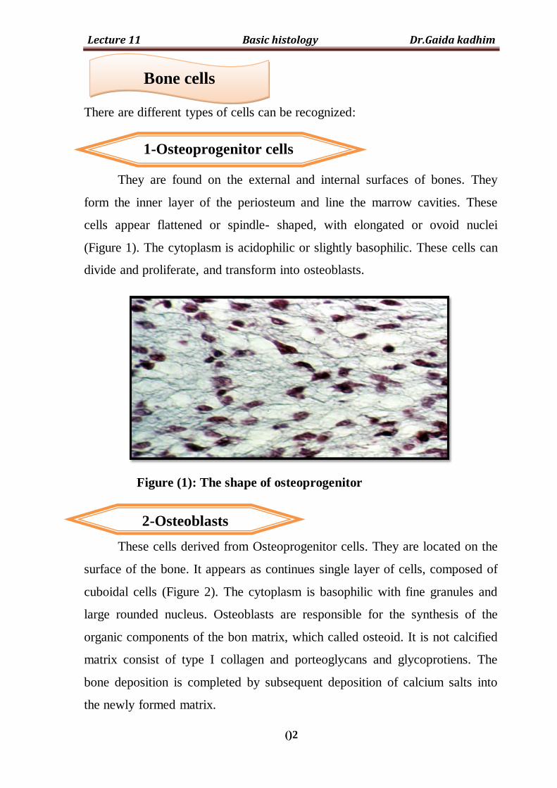
There are different types of cells can be recognized:
They are found on the external and internal surfaces of bones. They
form the inner layer of the periosteum and line the marrow cavities. These
cells appear flattened or spindle- shaped, with elongated or ovoid nuclei
(Figure 1). The cytoplasm is acidophilic or slightly basophilic. These cells can
divide and proliferate, and transform into osteoblasts.
Figure (1): The shape of osteoprogenitor
These cells derived from Osteoprogenitor cells. They are located on the
surface of the bone. It appears as continues single layer of cells, composed of
cuboidal cells (Figure 2). The cytoplasm is basophilic with fine granules and
large rounded nucleus. Osteoblasts are responsible for the synthesis of the
organic components of the bon matrix, which called osteoid. It is not calcified
matrix consist of type I collagen and porteoglycans and glycoprotiens. The
bone deposition is completed by subsequent deposition of calcium salts into
the newly formed matrix.
Bone cells
1-Osteoprogenitor cells
2-Osteoblasts

Lecture 11 Basic histology Dr.Gaida kadhim
)3)
Figure (2): The shape of osteoblasts
These are mature bone cells derived from osteoblasts that become lie
within a space or lacunae situated between lamellae of calcified bone matrix.
The osteocytes have flat, or almond-shape with faintly basophilic cytoplasm
and flattened darkly staining nucleus (Figure 3). These cells have numerous
slender cytoplasm processes called filopoidal processes extend into the
canaliculi which radiate out from the lacuna in the matrix (Figure 4). These
processes make contact with similar process of neighboring osteocytes via gap
junction through which ions and small molecules can move between the cells.
This provide mechanism for nutrients and exchange of metabolites between
blood stream and osteocytes. These cells are actively involved in the
maintenance of the bone matrix.
Figure (3): The shape of osteocytes.
3- Osteocytes

Lecture 11 Basic histology Dr.Gaida kadhim
)4)
Figure (4): The canaliculi of lacunae of osteocytes.
These cells originate from bone marrow. They are very large cells
which called giant cells and multinucleated, where they contain 5-50 nuclei in
each cell (Figure 5). The cytoplasm is acidophilic, granular, and vacuolated in
appearance, some of these vesicles are lysosomal in nature. The osteoclasts
secrete collagenase and other enzymes promoting the localized digestion of
collagen and dissolving calcium salts. Osteoclasts are found attached to the
bone surface, often in shallow depressions called Howships lacunae. These
cells play an important role in resorption and remodeling of bone tissue.
Figure (5): The shape of osteoclasts.
4-Osteoclasts

Lecture 11 Basic histology Dr.Gaida kadhim
)5)
It is fibrous sheath covered the external surfaces of bone. It is consist of
two layer, the outer layer is dense irregular connective tissue with fibroblasts
and containing a network of blood vessels and nerves. The inner layer, more
cellular layer of the periosteum is composed of loose connective tissue which
is less vascular and highly cellular contain flattened cells called
osteoprogenitor cells. These cells have potential to divide and differentiate
into osteoblasts. Osteoprogenitor cells ( osteogenic cells) play important role
in bone growth and repair.
It is thin sheath that lines the of internal cavities within the bone. It is
composed of a single layer of flattened osteoprogenitor cells and very small
amount of connective tissue. The endosteum is thinner than the periosteum.
The principle function of periosteum and endosteum are nutrition of bone
tissue and for repair and growth of bone.
Bones are classified according to their shape into: long bones, short
bones, flat bones and irregular bones. Also bones tissue is classified into
compact or spongy bone (Figure 6).
A long bone such as the femur, consists of a center piece, the shaft
called diaphysis, and a thickened head each called epiphysis at each end. The
articular surface of the epiphysis is covered with a thin layer of hyaline
cartilage. The remainder of the bone is covered membrane, the periosteum
which is richly supplied with blood vessels. Beneath the periosteum is a layer
of compact bone which is thicker in the shaft than in the two heads. The shaft
Periosteum
Endosteum
Types of bone

Lecture 11 Basic histology Dr.Gaida kadhim
)6)
encloses a hollow, the marrow cavity, which is lined with a thin soft
membrane known as the endosteum The marrow cavity contains a soft tissue
richly supplied with fat cells and blood corpuscles, the yellow marrow. The
epiphysis of a long bone consists of spongy (or cancellous) bone covered with
a thin layer of compact bone (Figure 7). If a bone is cut, the cross section
shows the epiphysis composed of spongy bone covered by a thin layer of
compact bone. The diaphysis is composed of compact bone, with small
amount of spongy bone on its inner surface around the marrow cavity.
Figure (6): Type of bones Figure (7): The structure of long bone
Under the microscope, the bone consist of two distinct structural
arrangement of bone tissue can be recognized:
Consist of slender, irregular trabeculae or bars which branch and unite
with one another to form network (Figure 8). These trabeculae consist of
several lamellae in which the lacunae containing osteocytes. These trabeculae
surround spaces called marrow cavities which filled with bone marrow
(Figure 9). There are two types of bone marrow, red bone marrow which
1- Spongy bone (cancellous)

Lecture 11 Basic histology Dr.Gaida kadhim
)7)
contains primitive stem cells that responsible of forming blood cells and
yellow bone marrow which composed mainly of fat cells.
Figure (8): The shape of spongy bone.
Figure (9): The structure of spongy bone.
It is composed of structural units called osteon or Haversion system,
where the calcified bone matrix organized into lamellae. The osteon consists
of concentric lamellae of bone matrix surrounding a central canal called
Haversian canal (osteonal canal), which contains the blood vessels, nerves and
loose connective tissue. The function of Haversian system is to bring nutrients
to compact bone.
2- Compact bone (lamellar bone)

Lecture 11 Basic histology Dr.Gaida kadhim
)8)
In Haversian system, the Haversian canal is surrounded by varying
number of concentric lamellae. Each Haversian canal lined by osteoblasts,
osteoprogenitor cells and loose connective tissue in addition to blood vessels
and nerves. In each lamella, collagen fibers are parallel to each other
(Figure 10).Within the lamellae there are small cavities called lacunae that
have numerous narrow channels called canaliculi containing the processes of
osteocytes which penetrate adjacent lamellae to join with canaliculi of
neighboring lacunae, which serves for the passage of substances between the
osteocytes and blood vessels. The Havresian canals communicate with each
other and with marrow cavity and periosteum through transverse or oblique
canals called Volkmanns canals (Figure 11).
The intervals between Haversian system are filled with interstitial or
intermediate lamellae, which act as packing between adjacent Haversian
system. Also there are inner circumferential lamellae, located around the
marrow cavity. Also outer circumferential lamellae located beneath the
periosteum (Figure 12). These structures are lamellae left by previous
Haversian system destroyed during growth and remodeling of bone.
Microscopic examination of bone shows two types:
1-Primary bone, immature bone, or woven bone
It is the first bone tissue to appear in embryonic development and in
fracture repair and other repair processes. It is temporary and is replaced in
adults by secondary bone, except in a very few places in the body, e.g. near
the sutures of the flat bones of the skull and in tooth sockets.
The type of bone is characterized by irregular array of collagen fibers in
the matrix therefore this bone nonlamellar. Smaller content of minerals and
contains higher proportion of osteocytes than in secondary bone. These cells
tend to be randomly arranged.

Lecture 11 Basic histology Dr.Gaida kadhim
)9)
1-Secondary bone, mature bone, or lamellar bone
It found in adults and its characterized by collagen fibers arranged in
lamellae that are parallel to each other or concentrically organized around a
vascular canal. The whole complex of concentric lamellae of bone called
Haversian system. The features of this type of bone as mention in compact
bone.
Figure (10): The structure of compact bone.
Figure (11): The structure of Volkmann canal.
Figure (12): The arrangement of lamellae within bone.


![Histology Slides - mediconotes.commediconotes.com/freenotes/basic/histology_laboratory_slides.pdf[Histology] Histology Slides MedicoNotes provides real laboratory Histological slides](https://static.fdocuments.in/doc/165x107/5ae110e87f8b9a5a668e6aa3/histology-slides-histology-histology-slides-mediconotes-provides-real-laboratory.jpg)