LECTURE: 10 IMMUNOLOGICAL MECHANISM IN TISSUE DAMAGE TYPE … · mediated damage that follows is...
22
LECTURE : 10 Title: IMMUNOLOGICAL MECHANISM IN TISSUE DAMAGE TYPE-III IMMUNE-COMPLEX MEDIATED HYPERSENSITIVITY LEARNING OBJECTIVES : The student should be able to: • Define type III hypersensitivity reactions. • Identify the role of the immune complex in type III reactions, and indicate which cause more damage small or large immune complexes, and locate the possible deposition of each complex. • Compare type III and type I reactions. • List some of the immune complex diseases: - Autoimmune diseases: ♦ Systemic lupus erythematosus (SLE). ♦ Rheumatoid arthritis (RA). ♦ Hyperthyroidism. - Infectious agents: ♦ Infectious mononucleosis. ♦ Meningitis. ♦ Viral hepatitis (Chronic stage). ♦ Poststreptococcal glomerulonephritis. ♦ Streptokinase (bacterial enzymes) in cardiac patients. - Drugs: ♦ Sulfonamides + penicillin. • Determine either type III reactions act locally or systematically. • Explain the mechanism of local immune complex deposition and give one example. • Explain the mechanism of generalized immune complex deposition, and give one example. • Explain the main target organ (s) for type III reactions. • Describe the mechanism of activation of type III reaction. • List a diagnostic method, and some examples of clinical features of type III such as:
Transcript of LECTURE: 10 IMMUNOLOGICAL MECHANISM IN TISSUE DAMAGE TYPE … · mediated damage that follows is...

LECTURE: 10
Title: IMMUNOLOGICAL MECHANISM IN TISSUE DAMAGETYPE-III IMMUNE-COMPLEX MEDIATED HYPERSENSITIVITY
LEARNING OBJECTIVES:
The student should be able to:
• Define type III hypersensitivity reactions. • Identify the role of the immune complex in type III reactions,
and indicate which cause more damage small or large immune complexes, and locate the possible deposition of each complex.
• Compare type III and type I reactions. • List some of the immune complex diseases:
- Autoimmune diseases:
♦ Systemic lupus erythematosus (SLE). ♦ Rheumatoid arthritis (RA). ♦ Hyperthyroidism.
- Infectious agents:
♦ Infectious mononucleosis. ♦ Meningitis. ♦ Viral hepatitis (Chronic stage). ♦ Poststreptococcal glomerulonephritis. ♦ Streptokinase (bacterial enzymes) in cardiac patients.
- Drugs:
♦ Sulfonamides + penicillin.
• Determine either type III reactions act locally or systematically.
• Explain the mechanism of local immune complex deposition and give one example.
• Explain the mechanism of generalized immune complex deposition, and give one example.
• Explain the main target organ (s) for type III reactions. • Describe the mechanism of activation of type III reaction. • List a diagnostic method, and some examples of clinical
features of type III such as:
- Arthus reactions (Local immune complex-deposition). - Serum sickness (Systemic immune complex-deposition).
LECTURE REFRENCE:
1. TEXTBOOK: ROITT, BROSTOFF, MALE IMMUNOLOGY. 6th edition. Chapter 23. pp. 357-367. 2. HANDOUT.
Hypersensitivity – Type III
Immune complexes are formed every time antibody meets antigen and are removed by the mononuclear phagocyte system following complement activation.
Persistence of antigen from continued infection or in autoimmune disease can lead to immune-complex disease.
Immune complexes can form both in the circulation, leading to systemic disease, and at local sites such as the lung.
Complement helps to disrupt antigen-antibody bonds and keeps immune complexes soluble.
Primate erythrocytes bear a receptor for C3b and are important for transporting complement-containing immune complexes to the spleen for removal.
Complement deficiencies lead to formation of large, relatively insoluble complexes which deposit in tissues.
Charged cationic antigens have tissue-binding properties, particularly for the glomerulus, and help to localize complexes to the kidney.
Factors that tend to increase blood vessel permeability enhance the deposition of immune complexes in tissues.
Immune complexes are formed every time antibody meets antigen, and generally they are removed effectively by the mononuclear phagocyte system, but occasionally they persist and eventually deposit in a range of tissues and organs. The complement and effector-cell mediated damage that follows is unknown as a Type III hypersensitivity reaction, or immune-complete disease. The sites of immune complex deposition are partly determined by the locialization of the antigen in the tissues and partly by how circulating complexes become deposited.
TYPES OF IMMUNE-COMPEX DISEASE
Disease resulting from immune-complex formation can be divided broadly into three groups: those due to persistent infection, those due to autoimmune disease, and those caused by inhalation of antigenic material (Figure-1).
Persistent infection – The combined effects of a low-grade persistent infection and a weak antibody response lead to chronic immune-complex formation, and eventual deposition of complexes in the tissue (Figure-2). Diseases with this aetiology include leprosy, malaria, dengue haemorrhagic fever, viral hepatitis and staphylococcal infective endocarditis. Autoimmune disease – Immune-complex disease is a frequent complication of autoimmune disease, where the continued production of autoimmune disease, where the continued production of autoantibody to a self-antigen leads to prolonged immune complex formation. As the number of complexes in the blood increases, athe systems that are responsible for the removal of complexes (mononuclear phagocyte, erythrocyte and complement) become overloaded, and complexes are deposited in the tissues (Figure-3). Diseases with this aetiology include rheumatoid arthritis, systemic lupus erythematous (SLE) and polymyositis. Inhalation of antigenic material – Immune complexes may be formed at body surfaces following exposure to extrinsic antigens. Such reactions are seen in the lungs following repeated inhalation of antigenic materials from moulds, plants or animals. This is exemplified in Farmer's lung and pigeon fancier's lung, where there are circulating antibodies to actinomycete fungi (found in mouldy hay) or to pigeon antigens. Bothe diseases are forms of extrinsic allergens are primarily IgG, rather than the IgE seen in Type I hypersensitivity reactions.) When antgen again enters the body by inhalation, local immune complexes are formed in the alveoli leading to inflammation and fibrosis (Figure-4). Precipitating antibodies to actinomycete antigens are found in the sera of 90% of patients with Farmer's lung. However, they are also found in some people with no disease, and are absent from some sufferers, so it seems that other factors are also involved in the disease process, including Type IV hypersensitivity reactions. MECHANISMS IN TYPE III HYPERSENSITIVITY Immune complexes are capable of triggering a wide variety of inflammatory processes:
• Complexes interact directly with basophils and platelets (via Fc receptors) to induce the release of vasoactive amines (Figure-5).
• Macrophages are stimulated to release cytokines, particularly TNFα and IL-1, that are very important during inflammation.
• They interact with the complement system to generate C3a and C5a (anaphylatoxins). These complement fragments stimulate the release of vasoactive amines (including histamine and 5 hydroxytryptamine) and
chemotactic factors from mast cells and basophils. C5a is also chemotactic for basophils, cosinophils and neutrophils.
Recent work with knockout mice indicates that complement has a less pro-inflammatory role than previously thought, wheras cell bearing Fc receptors for IgG and IgE appear to be critical for developing inflammation, with complement having a protective effect. The vasoactive amines released by platelets, basophils and mast cells cause endothelial cell retraction and thus increase vascular permeability, allowing the deposition of immune complexes on the blood vessel wall (Figure-6). The deposited complexes continue to generate C3a and C5a. Platelets also aggregate on the exposed collagen of the vessel basement membrane, assisted by interactions with the Fc regions of deposited immune complexes, to form microthrombi. The aggregate platelets continue to produce vasoactive amines and to stimulate the production of C3a and C5a. (Platelets are also a rich source of growth factors – these may be involved in the cellular proliferation seen in immune-complex diseases such as glomerulo-nephritis and rheumatoid arthritis). Polymorphs are chemotactically attracted to the site by C5a. They attempt to engulf the deposited immune complexes, but are unable to do so because the complexes are bound to the vessel wall. They therefore exocytose their lysosomal enzymes onto the site of deposition (Figure-6). If simply released into the blood or tissue fluids these lysosomal enzymes are unlikely to cause much inflammation, because they are rapidly neutralized by serum enzyme inhibitors. But if the phagocyte applies itself closely to the tissue-trapped complexes through Fc binding, then serum inhibitors are excluded and the enzymes may damage the underlying tissue. EXPERIMENTAL MODELS OF IMMUNE-COMPLEX DISEASE Experimental models are available for each of the three main types of immune-complex disease described above:
• Serum sickness, induced by injections of foreign antigen, mimics the effect of a persistent infection.
• The NZB/NZW mouse demonstrates autoimmunity.
• The Arthus reaction is an example of local damage by extrinsic antigen.
Care must be taken when interpreting animal experiments, as the erythrocytes of rodents and rabbits lack the receptor for C3b (known as CR1) which readily binds immune complexes that have fixed complement. This receptor is present on primate erythrocytes. Serum sickness can be induced with large injections of foreign antigen
In serum sickness, circulating immune complexes deposit in the blood vessel walls and tissues, leading to increased vascular permeability and thus to inflammatory diseases such as glomerulonephritis and arthritis. In the pre-antibiotic era, serum sickness was a complication of serum therapy, in which massive doses of antibody were given for diseases such as diphtheria. Horse anti-diphtheria serum was usually used, and some individuals made antibodies against the horse proteins. Serum sickness is now commonly studied in rabbits by giving them an intravenous injection of a foreign soluble protein such as bovine serum albumin (BSA). After about one week antibodies are formed which enter the circulation and complex with antigen. Because the reaction occurs in antigen excess, the immune complexes are small (Figure-7). These small complexes are only removed slowly by the mononuclear phagocyte system and therefore persist in the circulation. The formation of complexes is followed by an abrupt fall in total haemolytic complement; the clinical signs of serum sickness that develop are due to granular deposits of antigen-antibody and C3 forming along the glomerular basement membrane (GBM) and in small vessels celsewhere. As more antibody is formed and the reaction moves into antibody excess, the size of the complexes increases and they are cleared more efficiently, so the animals recover. Chronic disease is induced by daily administration of antigen. Autoimmunity causes immune-complex disease in the NZB/NZW mouse The F1 hybrid NZB/NZW mouse produces a range of autoantibodies (including anti-erythrocyte, anti-nuclear, anti-DNA and anti-Sm) and suffers from an immune complex disease similar in many ways to SLE in humans. An NZB/NZW mouse is born clinically normal, but within 2-3 months shows sign of haemolytic anaemia. Tests for anti-erythrocyte anibody (the Coombs' test), anti-nuclear antibodies, lupus cells and circulating immune complexes are all positive, and there are deposits in the glomeruli and choroid plexus of the brain. The disease is much more marked in the female, who die within a few months of developing symptoms (Figure-8). Injection of antigen into the skin of presensitized animals produces the Arthus reaction The Arthus reaction takes place at a local site in and around the walls of small blood vessels; it is most frequently demonstrated in the skin. An animal is immunized repeatedly until it has appreciable levels of serum antibody (mainly IgG). Following subcutaneous or intradermal injection of the antigen a reaction develops at the injection site, sometimes with marked oedema and haemorrhage, depending on the amount of antigen injected. The raction reaches a peak after 4-10 hours, then wanes and is usually minimal by 48 hours (Figure-9). Immunofluoescence studies have shown that initial deposition of antigen, antibody and complement in the vessel wall is followed by neutrophil infiltration and intravascular clumping of platelets (Figure-10).
This platelet reaction can lead to vascular occlusion and necrosis in severe cases. After 24-48 hours the neurophils are replaced by mononuclear cells and eventually some plasma cells appear. Complement activation via either the classical or alternative pathways was thought to be essential for the Arthus reaction to develop. But C3, C4 or C5 deficient mice were able to mount a normal Arthus reaction. However, when mice were made deficient in FcγRI or FcγRIII they were unable to produce the reaction. Furthermore when recombinant soluble FcγRII receptors were given they inhibited the development of the Arthus reaction. TNFα enhances cell-mediated immune responses in various ways. Treatment with antibodies to TNF can reduce severity in the Arthus reaction and, interestingly, anti-TNF is useful in treating rheumatoid arthritis. The ratio of antibody to antigen is directly related to the severity of the ensuing reaction. Complexes formed in either antigen or antibody excess is much less toxic than those formed at equivalence. PERSISTENCE OF COMPLEXES Immune complexes are normally removed by the mononuclear phagocyte system Immune complexes are opsonized with C3b following complement activation, and removed by the mononuclear phagocyte system, particularly in the liver and spleen. Removal is mediated by the complement C3b receptor, CR1. In primates, the bulk of CR1 in blood is found on erythrocytes. (Non-primates do not have erythrocyte CR1, and must therefore rely on platelet CR1.) There are about 700 receptors per erythrocyte, and their effectiveness is enhanced by the grouping of receptors in patches, allowing high-avidity binding to the large complexes. CR1 readily binds immune complexes that have fixes complement as has been shown by experiments with animals lacking complement (Figure-11). In normal primates the erythrocytes provide a buffer mechanism, binding complexes which have fixed complement and effectively removing them from the plasma. In small blood vessels 'streamline flow' allows the erythrocyte to travel in the centre of the vessel surrounded by the following plasma. Thus it is only the plasma that makes contact with the vessel wall. Only in the sinusoids of the liver and spleen, or at sites of turbulence, do erythrocytes make contact with the lining of the vessels. The complexes are transported to the liver and spleen, where they are removed by fixed tissue macrophages (Figure-12). Most of the CR1 is also removed in the process so, in situations of continuous immune-complex formation, the number of active receptors falls rapidly impairing the efficiency of immune complex handing. In patients with SLE, for example, the number of receptors may well be halved. With less complement receptors the complexes are cleared rapidly to the liver, but these complexes which have arrived

directly rather than on red cells are later released onto the circulation again and may then deposit in the tissues elsewhere and lead to inflammation. Complexes can also be released from erythrocytes in the circulation by the enzymatic action of Factor I, which cleaves C3b leaving a small fragment (C3dg) attached to the CR1 on the cell membrane. These soluble complexes are then removed by phagocytic cells, particularly those in the liver, bearing receptors for IgG Fc (Figure-13). Complement solubilization of immune complexes It has been known since Heidelberger's work on the precipitin curve in the 1930s that complement delays precipitation of immune complexes, although this information was forgotten for a long time. The ability to keep immune complexes soluble is a function of the classical complement pathway. The complement components reduce the number of antigen epitopes that the antibodies can bind (i.e. they reduce the valency of the antigen) by intercalating into the lattice of the complex, resulting in smaller, soluble complexes. In primates these complement-bearing complexes are readily bound by the C3b receptor (CR1) on erythrocytes. Complement can rapidly resolubilize precipitated complexes through the alternative pathway (Figure-14). The solubilization appears to occur by the insertion of complement C3b and C3d fragments into the complexes. It may be that complexes are continually being deposited in normal individuals, but are removed by solubilization. If this is the case, then the process will be inadequate in hypocomplementaemic patients and lead to prolonged complex deposition. Solubilization defects have indeed been observed in sera from patients with systemic immune-complex disease, but whether the defect is primary or secondary is not known. Complement deficiency impairs clearance of complexes In patients with low levels of classical pathway components there is poor binding of immune complexes to erythrocytes. The component deficiency may be due to depletion, caused by immune-complex disease, or could be due to a hereditary disorder, as is the case in C2 deficiency. This might be expected to result in persistent immune complexes in the circulation but in fact the reverse occurs, with the complexes disappearing rapidly from the circulation. These non-erythrocyte-bound complexes are taken up rapidly by the liver (but not the spleen) and are then released to be deposited in tissues such as skin, kidney and muscle, where they can set up inflammatory reactions (Figure-15). Infusion of fresh plasma, containing complement, restores the clearance patterns to normal, illustrating the importance of complement in clearance of immune complexes. Failure to localize in the spleen not only results in immune-complex disease, but may also have important implications for the development of appropriate immune responses. This is because the spleen plays a vital role in antigen processing and induction of immune responses. The size of immune complexes affects their deposition
In general, larger immune complexes are rapidly removed by the liver within a few minutes, whereas smaller complexes circulate for longer periods (Figure-16). This is because larger complexes are more effective at binding to Fc receptor and at fixing complement so binding better to erythrocytes. Also larger complexes are released more slowly from the erythrocytes by the action of Factor I. Anything that affects the size of complexes is therefore likely to influence clearance. It has been suggested that a genetic defect which favours production of low-affinity antibody could well lead to formation of smaller complexes, and so to immune-complex disease. Affinity maturation is dependent on efficient somatic mutation and selection of B cells within germinal centres following binding of antigen. This process is far more effective when B cells are stimulated b antigen or immune complexes coated with complement. Patients with complement deficiencies are particularly prone to develop immune complex disease and recent evidence indicates that one of the ways this is brought about is through poor targeting of antigen complexes to germial centres so preventing affinity maturation. Antibodies to self antigens may have low affinity and recognize only a few epitopes. This results in small complexes and long clearance times, because the formation of large, cross-linked lattices is restricted. Immunoglobulin classes affect the rate of immune-complex clearance Striking differences have been observed in the clearance of complexes with different immunoglobulin classes. IgG complexes are bound by etythrocytes and are gradually removed from the circulation, whereas IgA complexes bind poorly to erythrocytes but disappear rapidly from the circulation, with increased deposition in the kidney, lung and brain. Phagocyte defects allow complexes to persist Opsonized immune complexes are normally removed by mononuclear phagocytes system, mainly in the liver and spleen. However, when large amounts of complex are present, the mononuclear phagocyte system may become overloaded, leading to a rise in the level of circulating complex ad increased deposition in the glomerulus and elsewhere. Defective mononuclear phagocytes have been observed in human immune complex disease, but this may well be the result of overload rather than a primary defect. Carbohydrate on antibodies effects complex clearance Carbohydrate groups on immunoglobulin molecules have been shown to be important for the efficient removal of immune complexes of phagocytic cells. Abnormalities of these carbohydrates occur in immune-complex diseases such as rheumatoid arthritis, thus aggravating the disease process. IgGFc oligosaccharides lack the normally terminating galactose residue, enhancing rheumatoid factor binding. Recently, manna binding protein has been shown to bind agalactosyl Ig G and subsequently activate complement. DEPOSTION OF COMPLEXES IN TISSUES Immune complexes may persist in the circulation for prolonged periods of time. However, simple persistence is not usually harmful in itself; the problems only start when complexes are deposited in the tissues.
Two questions are relevant to tissue deposition:
• Why are complexes deposited? • Why do complexes show affinity for particular tissues in different diseases?
The most important trigger for tissue deposition of immune complexes is probably an increase in vascular permeability Animal experiments have shown that inert substances such as colloidal carbon will be deposited in vessel walls following administration of vasoactive substances, such as histamine or serotonin. Circulating immune complexes are deposited in a similar way following the infusion of agents that cause liberation of mast cell vasoactive amines (including histamine). Pretreatment with antihistamies blocks this affect. In studies of experimental immune-complex disease in rabbits, long-term administration of vasoactive amine antigents, such as chlorpheniramine and methysergide, has been shown considerably to reduce immune complex deposition (Figure-17). More importantly (from the point of view of disease prevention), young NZB/NZW mice treated with methysergide show less renal pathology than controls (Figure-18). Increase in vascular permeability can be initiated by a range of mechanisms which vary in important, depending on the diseases and species concerned. This variability makes interpretation of some of the animal models difficult. In general however, complement, mast cells,basophils and platelets must all be considered as potential producers of vasoactive amines. Immune-complex deposition is most likely where there is high blood pressure and turbulence Many macromolecules deposit in the glomerular capillaries, where the blood pressure is approximately four times that of most other capillaries (Figure-19). If the glomerular blood pressure of rabbit is reduced by partially constricting the renal artery or by ligating the ureter, deposition is also reduced. If the glomerular blood pressure is increased by experimentally induced hypersensitivity, immune complex deposition is also enhanced as shown by the development of serum sickness. Elsewhere, the most severe lesions also occur at sites of turbulence. They occur at turns of bifurcations of arteries, and in vascular filters such as the choroids plexus, and the ciliary body of the eye. Affinity of antigens for specific tissues can direct complexes to particular sites Local high blood pressure explains the tendency for deposits to form in certain organs, but does not explain why complexes are deposited on specific organs in certain diseases. In SLE, the kidney is a particular target, whereas in rheumatoid arthritis, although circulating complexes are present, the kidney is usually spared and the joints are the principal target.

It is possible that the antigen in the complex provides the organ specificity, and a convincing model has been established to support this hypothesis. In the model, mice are given endotoxin causing cell damage and release of DNA, which then binds to healthy glomerular basement membrane. Anti-DNA is then produced by polyclonal membrane. Anti-DNA is then produced by polyclonal activation of B cells, and is bound by the fixed DNA leading to local immune complex formation (Figure-20). The production of rheumatoid factor IgM anti-IgG allows further immune-complex formation to occur in situ. It is possible that in other diseases antigens will be identified with affinity for particular organs. The charge of the antigen and antibody may be important in some systems. For example, positively charged antigens and antibodies are more likely to be deposited in the negatively charged glomerular basement membrane. The degree of glycosylation also affects the fate of complexes containing glycoprotein antigens because certain clearance mechanisms are activated by recognition of sugar molecules, e.g. mannan binding protein. In certain diseases the antibodies and antigens are both produced within the target organ. The extreme of this is reached in rheumatoid arthritis, where IgG anti-IgG rheumatoid factor is produced by plasma cells within the synovium; these antibodes then combine with each other (self-association), so settling up an inflammatory reactions. The site of immune-complex deposition depends partly on the size of the complex This is exemplified in the kidney: small immune complexes can pass through the glomerular basement membrane, and end up on the epithelial side of the membrane; large complexes are unable to cross the membrane and generally accumulate between the endothelium and the basement membrane or the mesangium (Figure-21). The size of immune complexes depends on the valency of the antigen, and on the titre and affinity of the antibody. The class of immunoglobulin in an immune complex can also influence its deposition There are marked age- and sex-related variations in the class and subclass of anti-DNA antibodies seen in SLE. Similarly, as NZB/NZW mice grow older there is a class switch, from predominantly IgM to IgG2a. This occurs earlier in females than in males and coincides with the onset of renal disease, indicating the importance of antibody class in the tissue deposition of complexes (Figure-22). DETECTION OF IMMUNE COMPLEXES Deposited immune complexes can be visualized using immunofluorescence The ideal place of look for complexes is in the affected organ. Tissue samples may be examined by immunofluorescence for the presence of immunoglobulin and complement. The composition, pattern and particular area of tissue affected all provide useful
information on the severity and prognosis of the disease. For example, patients with the continuous, granular, subepithelial deposits of IgG found in membranous glomerulonephritis have a poor prognosis. In contrast, those whose complexes are localized in the mesangium have a good prognosis. Not all tissue-bound complexes give rise to an inflammatory response; for example in SLE, complexes are frequently found in skin biopsies from normal looking skin, as well as from inflamed skin. Assays for circulating immune complexes Circulating complexes are found in two separate compartments: bound to erythrocytes and free in plasma. Erythrocyte-bound complexes are less likely to be damaging, so it is of more interest to determine the level of free complexes. Care is required when collecting the sample: bound complexes can easily be released during clotting by the action of Factor I. To obtain accurate assays of free complexes, the erythrocytes should be rapidly separated from the plasma to prevent the release of bound complexes.
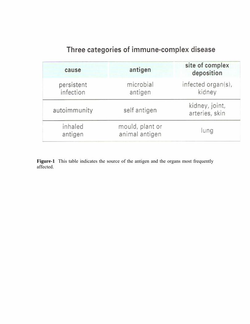
Figure-1 This table indicates the source of the antigen and the organs most frequently affected.
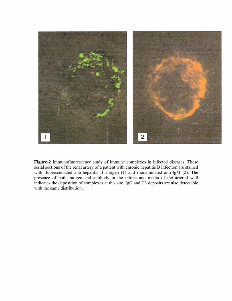
Figure-2 Immunofluorescence study of immune complexes in infected diseases. These serial sections of the renal artery of a patient with chronic hepatitis B infection are stained with fluoresceinated anti-hepatitis B antigen (1) and rhodaminated anti-IgM (2). The presence of both antigen and antibody in the intima and media of the arterial wall indicates the deposition of complexes at this site. IgG and C3 deposits are also detectable with the same distribution.
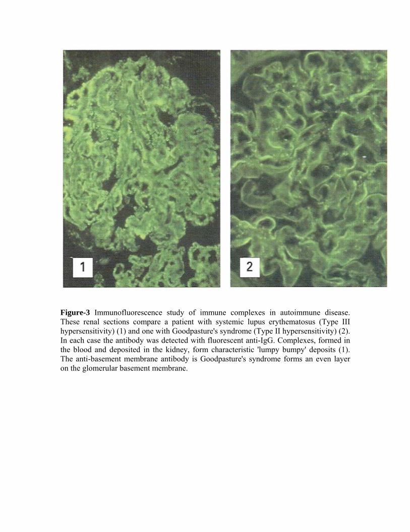
Figure-3 Immunofluorescence study of immune complexes in autoimmune disease. These renal sections compare a patient with systemic lupus erythematosus (Type III hypersensitivity) (1) and one with Goodpasture's syndrome (Type II hypersensitivity) (2). In each case the antibody was detected with fluorescent anti-IgG. Complexes, formed in the blood and deposited in the kidney, form characteristic 'lumpy bumpy' deposits (1). The anti-basement membrane antibody is Goodpasture's syndrome forms an even layer on the glomerular basement membrane.

Figure-4 When fungal antigen is inhaled into the lung of a sensitized individual, immune complexes are formed in the alveoli (2). Complement fixation leads to cell accumulation, inflammation and fibrosis. The histological appearance of the lung in extrinsic allergic alveolitis (1) shows consolidated areas due to cell accumulation. Precipitin antibody present in the serum of a patient with pigeon fancier's lung: P (3) is directed against the fungal antigen Micropolyspora faeni. A normal serum (N) lacks antibodies to this fungus.
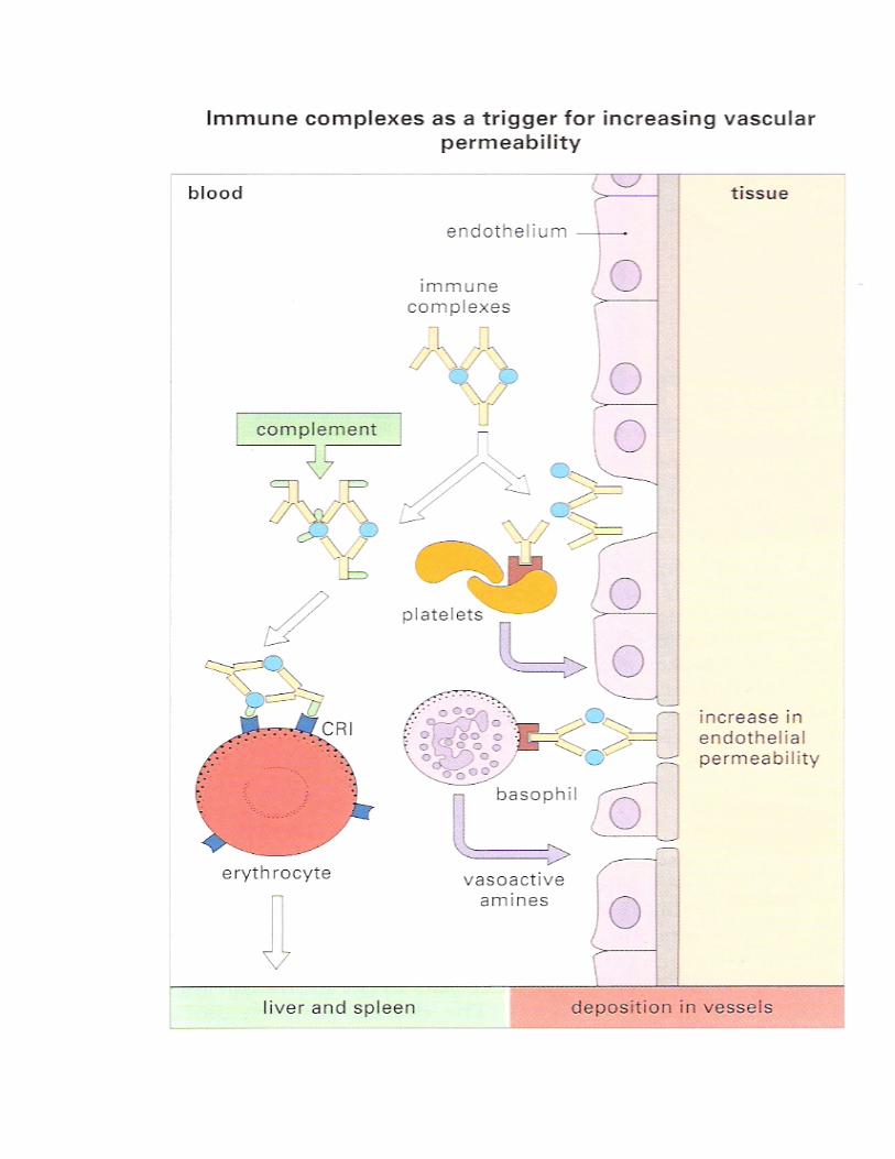
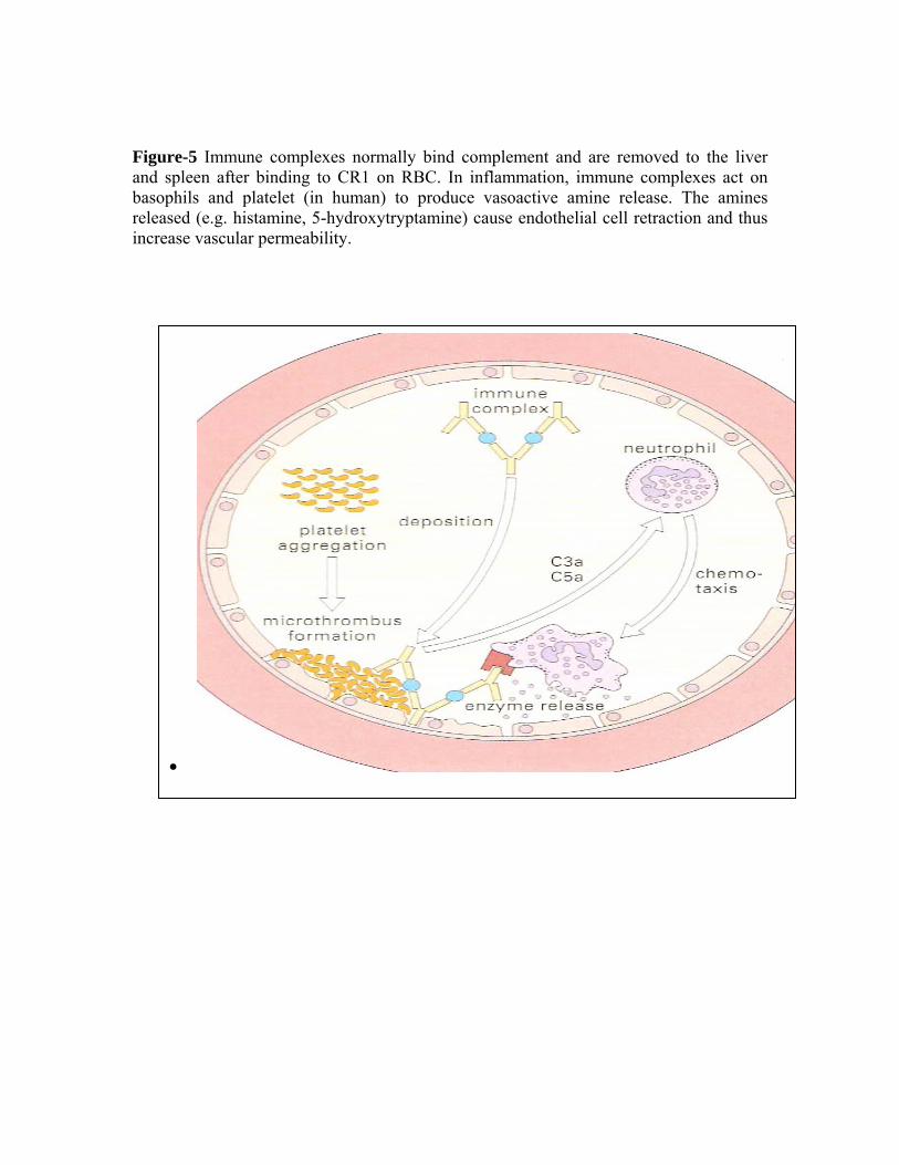
Figure-5 Immune complexes normally bind complement and are removed to the liver and spleen after binding to CR1 on RBC. In inflammation, immune complexes act on basophils and platelet (in human) to produce vasoactive amine release. The amines released (e.g. histamine, 5-hydroxytryptamine) cause endothelial cell retraction and thus increase vascular permeability.
•

Figure-6 Increased vascular permeability allows immune complexes to be deposited in the blood vessel wall. This induces platelet aggregation and complement activation. The aggregated platelets form microthrombi on the exposed collagen of the basement membrane of the endothelium. Neutrophils are attracted to the site by complement products, but cannot ingest the complexes. They therefore exocytose their lysosomal enzymes, causing further damage to the vessel wall.
Figure-7 Following an injection of xenogeneic serum there is a lag period of approximately 5 days, in which only free antigen is detectable in serum. After this time, antibodies are produced to the foreign proteins and immune complexes are formed in serum; it is during this period that the symptoms of nephritis and arteritis appear. To begin with, small soluble complexes are found in antigen excess with increasing antibody titres, larger complexes are formed which are deposited and subsequently cleared. At this stage the symptoms disappear.
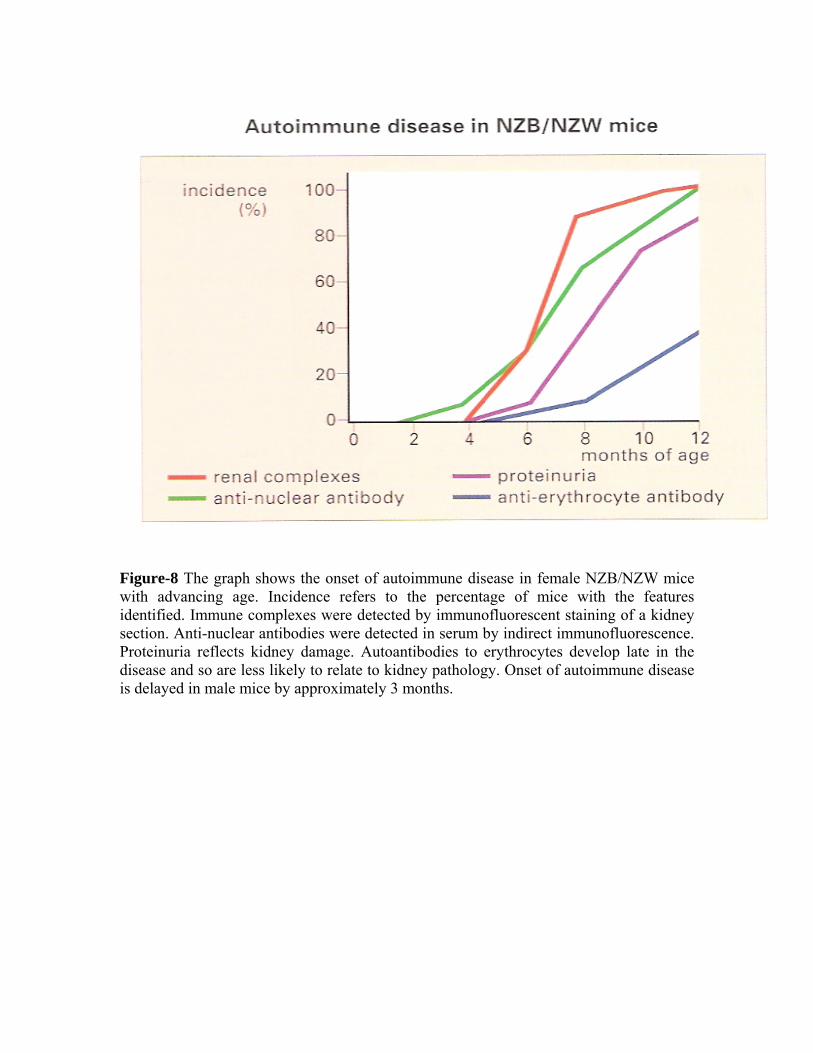
Figure-8 The graph shows the onset of autoimmune disease in female NZB/NZW mice with advancing age. Incidence refers to the percentage of mice with the features identified. Immune complexes were detected by immunofluorescent staining of a kidney section. Anti-nuclear antibodies were detected in serum by indirect immunofluorescence. Proteinuria reflects kidney damage. Autoantibodies to erythrocytes develop late in the disease and so are less likely to relate to kidney pathology. Onset of autoimmune disease is delayed in male mice by approximately 3 months.
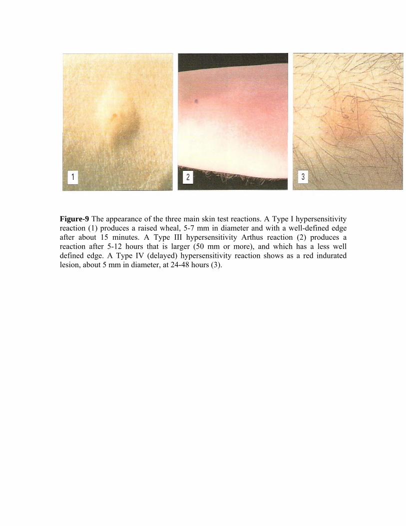
Figure-9 The appearance of the three main skin test reactions. A Type I hypersensitivity reaction (1) produces a raised wheal, 5-7 mm in diameter and with a well-defined edge after about 15 minutes. A Type III hypersensitivity Arthus reaction (2) produces a reaction after 5-12 hours that is larger (50 mm or more), and which has a less well defined edge. A Type IV (delayed) hypersensitivity reaction shows as a red indurated lesion, about 5 mm in diameter, at 24-48 hours (3).
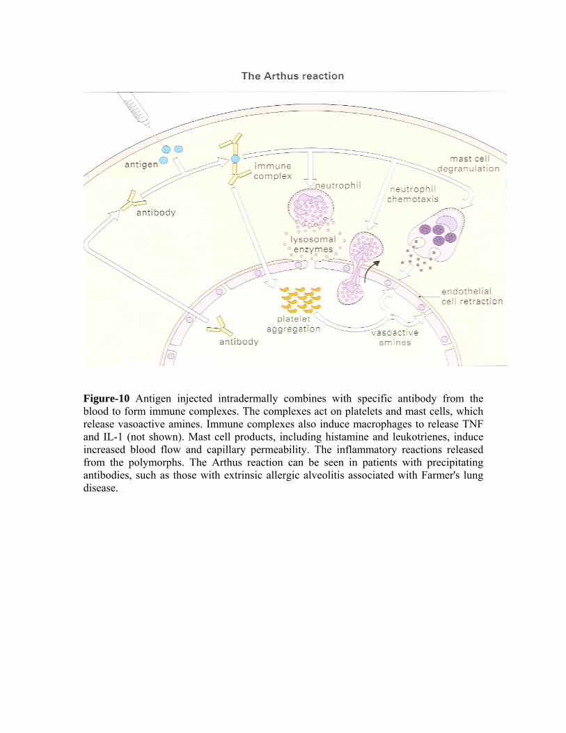
Figure-10 Antigen injected intradermally combines with specific antibody from the blood to form immune complexes. The complexes act on platelets and mast cells, which release vasoactive amines. Immune complexes also induce macrophages to release TNF and IL-1 (not shown). Mast cell products, including histamine and leukotrienes, induce increased blood flow and capillary permeability. The inflammatory reactions released from the polymorphs. The Arthus reaction can be seen in patients with precipitating antibodies, such as those with extrinsic allergic alveolitis associated with Farmer's lung disease.
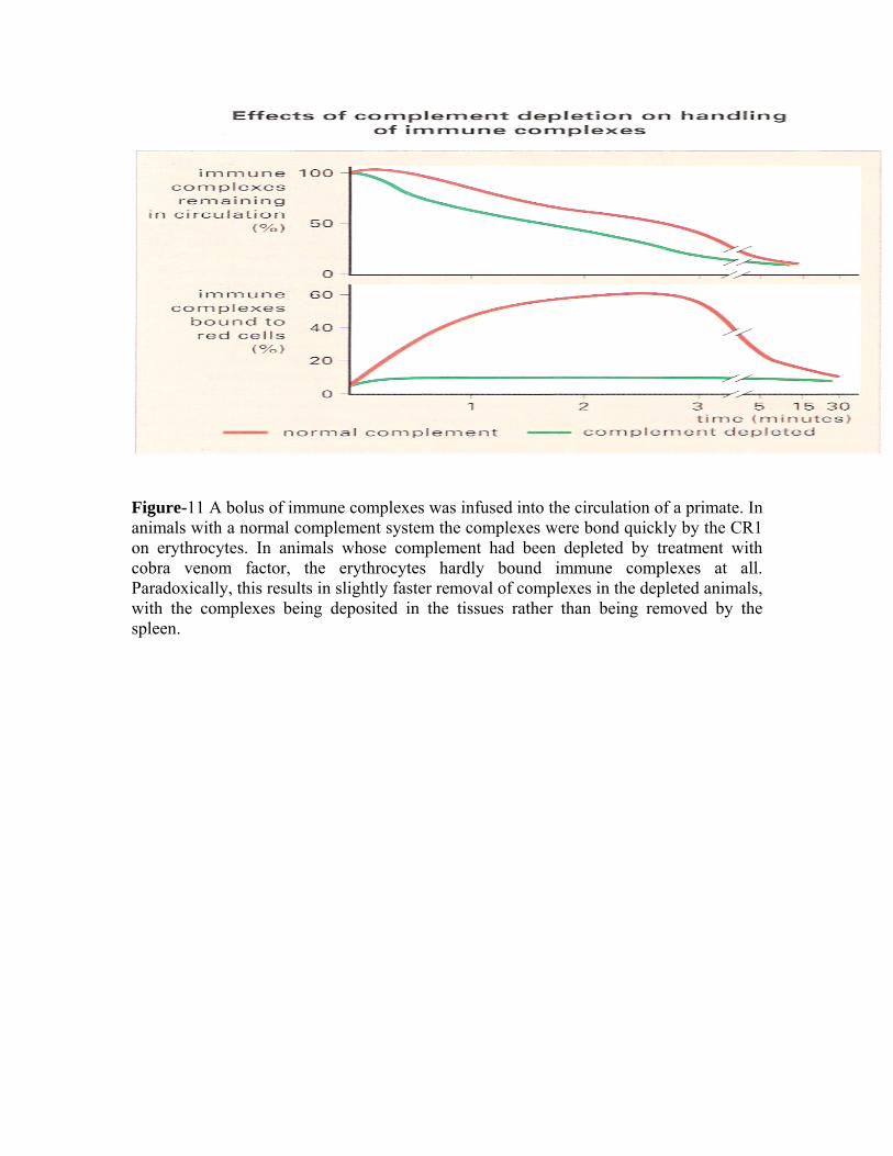
Figure-11 A bolus of immune complexes was infused into the circulation of a primate. In animals with a normal complement system the complexes were bond quickly by the CR1 on erythrocytes. In animals whose complement had been depleted by treatment with cobra venom factor, the erythrocytes hardly bound immune complexes at all. Paradoxically, this results in slightly faster removal of complexes in the depleted animals, with the complexes being deposited in the tissues rather than being removed by the spleen.