Lecture 1. Alteration. Dystrophy...
18
1 Introduction To Pathological Anatomy Lecture 1. Alteration. Dystrophy Contents • Introduction • Parenchymal Dystrophies. Classification, Ethiology, Mechanisms, Morphology • Stromally-vascular dystrophies Classification, Ethiology, Mechanisms, Morphology Introduction • - There are different morphological manifestations of a lesion in the cells and tissues. • - Lesion (Alteration) js a change in the cell structure, intercellular substance, tissues, and organs accompanied by disturbances in their vital activity. • Subcellular alteration, which largely occurs as a response to more of less constant stimuli and intracellular accumulation of a number of substances, due to the disturbances in cellular metabolism or excessive storage, as well as cell death (necrosis and apoptosis) have been described in the cells. • - Two types or stages of lesions in the tissues have been described: degeneration and necrosis. Dystrophies. Classification • Dystrophy is a complicated pathologic process based on tissue (or cellular) metabolism impairment that causes structural changes. Classification by the kind of metabolism disturbance prevail: • a) protein, • b) fat, • c) hydrocarbon, • d) mineral Dystrophies. Classification By pathologic process localization: • a) parenchymatous (modifications in the organs parenchymatous cells - cardiomyocytes, hepatocytes, ganglionic cells of cerebrum, etc.); • b) stromal-vascular (modifications in organs stroma); • c) mixed (changes in parenchyma and stroma). Depending on genetic factors influence: • a) congenital, • b) acquired. By process spread: • a) general, • b) local. Mechanisms of metabolism products abnormal uptake. Infiltration is excessive penetration of metabolism products from blood into cells and intercellular substance with their subsequent uptake due to ferment system, providing their metabolism, insufficiency. Substances metabolism products abnormal uptake by way of infiltration is observed in liver, kidneys, aorta wall. Mechanisms of metabolism products abnormal uptake. Decomposition (phanerosis) occurs under cell and intercellular substance ultrastructures destruction due to intoxication, hypoxia or other reasons. Ultrastructures membranes are made of proteins, fats and hydrocarbons, so under their destruction these substances are accumulated stored in cells.
-
Upload
duongkhanh -
Category
Documents
-
view
215 -
download
0
Transcript of Lecture 1. Alteration. Dystrophy...

1
Introduction To Pathological Anatomy
Lecture 1. Alteration. Dystrophy
Contents
• Introduction
• Parenchymal Dystrophies. Classification, Ethiology, Mechanisms, Morphology
• Stromally-vascular dystrophies Classification, Ethiology, Mechanisms, Morphology
Introduction
• - There are different morphological manifestations of a lesion in the cells and tissues.
• - Lesion (Alteration) js a change in the cell structure, intercellular substance, tissues, and
organs accompanied by disturbances in their vital activity.
• Subcellular alteration, which largely occurs as a response to more of less constant stimuli and
intracellular accumulation of a number of substances, due to the disturbances in cellular
metabolism or excessive storage, as well as cell death (necrosis and apoptosis) have been
described in the cells.
• - Two types or stages of lesions in the tissues have been described: degeneration and necrosis.
Dystrophies. Classification
• Dystrophy is a complicated pathologic process based on tissue (or cellular) metabolism
impairment that causes structural changes.
Classification by the kind of metabolism disturbance prevail:
• a) protein,
• b) fat,
• c) hydrocarbon,
• d) mineral
Dystrophies. Classification
By pathologic process localization:
• a) parenchymatous (modifications in the organs parenchymatous cells - cardiomyocytes,
hepatocytes, ganglionic cells of cerebrum, etc.);
• b) stromal-vascular (modifications in organs stroma);
• c) mixed (changes in parenchyma and stroma).
Depending on genetic factors influence:
• a) congenital,
• b) acquired.
By process spread:
• a) general,
• b) local.
Mechanisms of metabolism products abnormal uptake.
Infiltration is excessive penetration of metabolism products from blood into cells and intercellular
substance with their subsequent uptake due to ferment system, providing their metabolism,
insufficiency.
Substances metabolism products abnormal uptake by way of infiltration is observed in liver,
kidneys, aorta wall.
Mechanisms of metabolism products abnormal uptake.
Decomposition (phanerosis) occurs under cell and intercellular substance ultrastructures destruction
due to intoxication, hypoxia or other reasons. Ultrastructures membranes are made of proteins, fats
and hydrocarbons, so under their destruction these substances are accumulated stored in cells.

2
Mechanisms of metabolism products abnormal uptake.
• Distored synthesis is synthesis of those substances in cells and tissues which are not observed
in them as a norm.
• As an example, it’s glycogen synthesis in nephron tubules epithelium under diabetes mellitus,
alcohol hyaline synthesis in hepatocytes.
Mechanisms of metabolism products abnormal uptake.
• Transformation is the creation of one kind of metabolism products from intermediate
disintegration products, which should be utilized for proteins, fats and hydrocarbons synthesis.
For example, it’s fats and hydrocarbons components transformation into proteins under
starvation, fats and hydrocarbons components transformation into glycogen under diabetes
mellitus.
Morphology of proteins abnormal uptake (proteinosis) • Occurs under proteins metabolism disturbance. Tissues proteins form cells as plastic materials
(capsule, nucleus, cytoplasm, intracellular organelles) as well as intercellular stroma –
collagen, elastic, reticulin fibers, basic intercellular substance, vessels, nerves.
• By proteins metabolism disturbance development location proteinosises are divided into
parenchymatous, stromal-vascular and mixed.
parenchymatous proteinosis
• Under parenchymatous proteinosis physical-chemical features of intracellular proteins are
violated.
• At the beginning grain effect occurs in cytoplasm at the cost of protein inclusions, which is
manifestation of cell ultrastructures overstrain (hyper function).
• This process is reversible. Quite often proteins disbolism is combined with Na-K-pump
operation faults, which is accompanied with natrium ions uptake and cells hydration.
• In case intoxication, hypoxia, inflammation or other reasons of proteinosis increase this cause
cells destructive changes intensification.
kinds of parenchymatous proteins degenerations (proteinosis)
• hydropic (vacuolar),
• hyaline-drop,
• keratinization.
Hydropic or dropsy proteinosis
• is characterized by intracellular fluid increase, in which cytoplasm proteins are dissolved due
to hydrolytic pigments action. Vacuoles full of cytoplasm fluid occur in cells. Further on cells
cytoplasm transforms into blisters full with fluid, intracellular organelles destroy, cell dies off
and coliquation necrosis develops.
• Organs also didn’t change macroscopically.
• Hydropic proteinosis often develops in liver under viral hepatitis, in kidneys under
glomerulonephritis, etc.

3
Hydropic degeneration of nephrothelium.
Keratinization proteinosis
• is characterized with excessive keratin generation on the surface of plane keratinized
epithelium – hyperkeratosis, ichthyosis.
• The causes of keratinization development is chronic inflammation, avitaminosis, skin
development abnormalities.
• Leukoplakia which is mucous tunics epithelium pathologic keratinization, also belongs to
this process and can become a source of malignant growth.
hyaline-drop proteinosis
• proteins compacts and become similar to hyaline cartilage. Big hyalinoid drops of protein
occur in cells cytoplasm.
• Sometimes coagulation necrosis develops and cells die,
• organ function decreases,
• but macroscopic changes are not found.
• This kind dystrophy is often observed in hepatocytes under alcoholic hepatitis (Mellori
bodies),
• in renal tubules epithelium under nephrotic syndrome, etc.
Extracellular proteinosis • occur in the result of metabolism disturbance in organs stroma or in vessels walls, so they are
named stromal-vascular or mesenchymal proteinosis.
• Important attention is paid to proteinosis developing in the result of proteins metabolism in
conjunctive tissue and are found in stroma and vessels walls

4
Extracellular proteinosis • Primary pathologic changes are developed on histion level, consisting of microcirculation
channel: basic substance,
• fibers (collagen, reticulum, elastic),
• cells (fibroblasts, fibrocytes, lymphocytes, labrocytes, histiocytes),
• nerves.
• Basic substance is connecting, cementing, fiber and cells are situated in it.
• By chemical composition it is polymer of composite protein-hydrocarbon molecules –
mucopolysaccharides (glycosamineglycanes).
The types of stromal-vascular proteinosis
• The following relates to stromal-vascular proteinosis:
• mucoid swelling,
• fibrinoid swelling (fibrinoid),
• hyalinosis, which are considered to be consequent stages of conjunctive tissue destruction.
Mucoid swelling • It is primary disorganization of conjunctive tissue.
• Causes: hypoxia,
• allergy,
• endocrine pathologies.
• It often occurs under rheumatic and infection diseases, atherosclerosis,
• it is found in artery walls, cardiac valves, endocardium, heart.
Mucoid swelling • Basic substance depolymerization underlies its development.
• As a consequence it becomes hydrophilic, attracts liquid, vessel wall penetrability increases.
• Basic substance hydration, collagen fibers swelling occurs.
• With vascular-tissue penetrability growth conjunctive tissue saturates with blood plasm
proteins, in first turn with albumines and globulins.
• Macroscopically organ or tissue mostly doesn’t change.
Mucoid swelling
Microscopically phenomenon of metachromasia is observed: glycosamineglycanes are painted with
toluidine blue in red color. Described changes in conjunctive tissue provided that the reason was
eliminated are reversible and tissue structure is rehabilitated

5
Fibrinoid swelling • It is following stage of conjunctive tissue disorganization. Under substantial growth of
vascular-tissue penetrability fibrinogen sweats in stroma from vessels, which rather quickly
precipitates in strings of fibrin, collagen fibers are destroyed (broken, fragment), conjunctive
tissue basic substance chemical composition is changed.
• Under fibrinoid swelling deep and irreversible disorganization of conjunctive tissue is
observed, which is accompanied with basic substance and fibers destruction against the
background of considerable increase of balls vascular permeability.
Fibrinoid swelling • Macroscopically organ doesn’t change, Consequence Fibrinoid necrosis is developed in the
final of the process.
• Significance – organ function disturbance under heart disease formation, joints immobility,
luminal narrowing and vessel wall elasticity decrease,
• organ function termination under renal insufficiency at malignant hypertension,
• when fibrinoid changes as well as arterioles and capillars necrosis develops.
Fibrinoid swelling
microscopically collagen fibers become homogenous, eosinophilic, becomes yelow when painted
with picrofuchsin, pyroninophil and argyrophil.

6
Hyalinosis It is the final stage of tissue disorganization and is characterized with uptake of collagen destruction
products, plasm proteins, polysaccharides, which merge into homogenous mass which consolidates
as time passes, becomes semi-transparent similar to hyaline cartilage, so it is called hyaline.
This is complex fibrillar protein.
Hyalinosis occurs as a consequence of fibrinoid swelling, plasmorrhagia, sclerosis, necrosis.
It develops as the result of peculiar completion of sclerosis in scarring, cardiac valves under
rheumatism (local conjunctive tissue hyalinosis).
Hyalinosis Macroscopically fibrous conjunctive tissue becomes dense, cartilaginous, whitish, semi-transparent.
Microscopically collagen fibers loss fibrillarity and merge into homogenous dense cartilaginous
mass, cells squeeze and atrophy.
Hyalinosis
Heart in such cases is enlarged,
ventricular cavities are dilated,
mitral valve flappers are dense, whitish color,
considerably deformed.
This kind of hyalinosis is peculiarly expressed in rough vicious cicatrix after burns (keloid).
Consequences are unfavorable because of considerable deterioration of organ or injured tissue
function.

7
Systemic hyalinosis
develops in vessels walls under
hypertension disease,
diabetes mellitus (vascular hyalinosis) and
is mostly expressed in kidneys, cerebrum, eye retina, pancreas.
Systemic hyalinosis
Considering occurrence pathogenesis three kinds of vascular hyaline are recognized:
simple is observed under hypertension disease, atherosclerosis;
lipohyaline is developed under diabetes mellitus;
complex hyaline occurs in the result of immunopathologic disturbances and vessel wall fibrinoid
disorganization at collagenosis.
Hyalinosis of splens
Cardiac valves hyalinosis
mitral valves are thickened, deformed, glassy.

8
Hyalinosis of endocard
Morphology of lipids pathological uptake (lipidosis) – Occurs as the result of fats metabolism disturbance.
– Lipidosis are divided into parenchymatous and stromal-vascular (mesenchymal)
fatty (adipose) degenerations. To reveal fats frozen sections are colored with sudan ІІІ
or ІV.
9
Parenchymatous lipidosis
– are manifested with neutral lipids (triglycerides) drops uptake in cells cytoplasm and
are the results of cytoplasm fats metabolism disturbance.
– Mostly they are found in
– myocardium,
– liver,
– kidneys.
Myocardium lipidosis • It is characterized with lipoproteids drops uptake in cardiac hystiocytes.
• Aetiology: As a rule it is observed under intoxications
• diphtherial,
• alcohol,
• with phosphoric and arsenic compounds,
• diseases of liver, kidneys, thyrotoxicosis, etc.),
• long time general hypoxia (anemia, chronic pulmonary and cardiovascular insufficiency),
• Under oxygen deficiency process of oxidative phosphorylation and ATP synthesis in
cardiomyocytes decreases, fatty acids beta-oxidation violates. So fats coming into cell are not
completely utilized as plastic and power material and they accumulates in cytoplasm.
• Besides that under hypoxia membrane lipoprotein complexes destruction is observed
(decomposition or phanerosis).
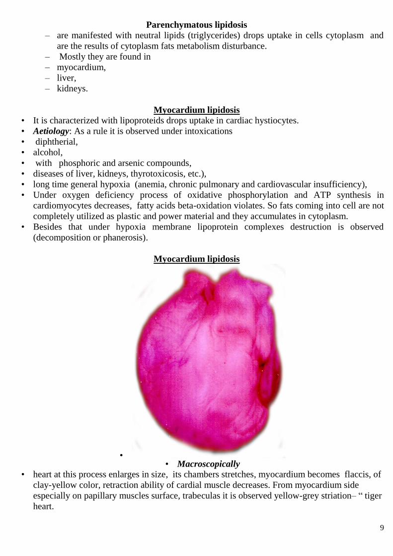
Myocardium lipidosis
• • Macroscopically
• heart at this process enlarges in size, its chambers stretches, myocardium becomes flaccis, of
clay-yellow color, retraction ability of cardial muscle decreases. From myocardium side
especially on papillary muscles surface, trabeculas it is observed yellow-grey striation– “ tiger
heart.

10
Myocardium lipidosis
• Microscopically
• fat uptakes in muscular cells groups,
• situated downstream capillars and small veins where hypoxia factor is mostly expressed.
Liver lipidosis • It is characterized with fat content increase in hepatocytes.
• Quite often it is the result of imbalance between increased fats supply under hyper lipidemia
• (alcoholism,
• diabetes mellitus,
• general obesity), their decreased
• assimilation (fatty acids oxidation in mitochondrions under hypoxia or toxic influences) and
lipids excretion decrease by liver cells under apoprotein production decrease which transports
fats in the form of lipoproteins. This is observed in case protein insufficiency in food or under
gastrointestinal disturbances, poisoning with ethanol, phosphor, etc., congenital defects of
ferments metabolizing fats.
Liver lipidosis • Microscopically
• first occurs saw type,
• then small drop and
• large drop degeneration.
• Macroscopically
• liver is enlarged,
• loose (of pastry consistency),
• yellow or clay color.
Liver lipidosis
• Three stages of liver lipidosis are distinguished:
• 1- fat uptake in hepatocytes,
• 2- fat uptake with mesenchymal reaction development,
• 3- fat uptake with liver fibrosis and cirrhosis development.

11
• Fat fills all cytoplasm and gradually pushes nucleus aside to periphery and modified
hepatocytes becomes similar to adipocytes.
• Fatty degeneration prevalence in peripheral portions of liver part confirms infiltration
mechanism of its development, which is observed under hyperlipidemia.
• Fatty degeneration development prevalence in central portions of liver part is connected with
decompensation mechanism and is observed under hypoxia or intoxication.
Liver lipidosis (“goose liver”).
• Liver is enlarged, of flaccid consistency, of ochre-yellow color.

12
Liver lipidosis-incision
Liver lipidosis.

13
Kidneys lipidosis • It is often observed under
• nephrotic syndrome,
• chronic renal insufficiency when hyperlipidemia and lipiduria occur.
• Fat excess is excreted from organism with kidneys and constipates them.
Kidneys lipidosis • Microscopically fat occurs in proximal, distal or convoluted renal tubules epithelium in cells
basal portions. Nephrocytes lipidosis often joins hyaline-drop degeneration and hydropic
proteinosis.
• Macroscopically kidney is enlarged,
• Flaccid
• , cortical layer is dilated with signs of swelling, of grey color with yellow specks.
Kidneys lipidosis
Congenital lipidosis
• metabolism of lipid disturbances are manifested with systemic lipidosis and pertain to
fermentopathies (diseases of storage or uptake).
• The following diseases are marked out: cerebrosine lipidosis (Gaucher's disease),
sphingomyelin lipidosis (Niemann-Pick disease), gangliosidosis (Tay-Sachs disease),
generalized gangliosidosis (Norman-Landig disease), which are accompanied with liver,
spleen, marrow, nervous system and other organs and tissues damage.
14
Stromal-vascular lipidosis
• Stromal-vascular lipidosis include neutral fat metabolism disturbance in adipose tissue and
adipose depot as well as cholesterol and its ethers in arteries walls under atherosclerosis.
Stromal-vascular lipidosis
– General disturbance of neutral fats metabolism is manifested with neutral fat stocks increase
or decrease in hypodermic fat tissue, mesentery, pericardium, marrow, etc.
– General uptake of neutral fat in fat depots is called obesity.
obesity. – The following is recognized:
– primary or idiopathic obesity the cause of which is unknown and
– secondary obesity which occurs under
– endocrine,
– cerebral and
– hereditary diseases.
obesity. – By external signs obesity kinds are as follows:
– upper,
– mid,
– lower and
– universal symmetric.
– By morphologic signs hyper plastic type is marked out characterized with fat cells
(adipocytes) quantity increase in organism as well as hypertrophic (malignant) type the basis of
which is adipose cells size increase several times and triglycerides content increase in cytoplasm
several times.
obesity. – Under general obesity the important clinical attention is paid to heart injury. In this case
adipose tissue grows under pericardium, surrounding organ like case. Lipocytes uptake in
myocardium stroma between cardiac hystiocytes, squeezing the latter ones which causes their
atrophy. Right portion of the heart is the most injured one. Sometimes the whole thickness of right
ventricle myocardium is changed with adipose tissue, that can cause cardiac rupture or accelerate
decompensation process.

15
Excessive growth of subcutaneous fatty tissue of ventral abdominal wall under general
obesity
Neutral fat local uptake • It is observed under Madelung's syndrome,
• Dercum's disease and
• Weber-Krischen’s desease,
• as well as vacant obesity when organ atrophied portion is substituted.
• The essence of Dercum's disease is in painful lipomas occurrence in subcutaneous adipose
tissue of extremities and trunk. Weber-Krischen’s disease is characterized with recurrent
nonpurulent cellulites with productive granulomatous inflammation development around
sphacelous adipose tissue.
General decrease of adipose tissue
• occurs under emaciation (cachexia). Tissue becomes loose,
• flabby is saturated with liquid, sliming.
Cholesterol and its ethers metabolism imbalance It is a basis of atherosclerosis development. Uptake of cholesterol fractions, lipoproteins of various
density, proteins in arteries’ walls causes formation of fat detritus (atheroma) and conjunctive tissue
enlargement (sclerosis).
Cholesterol and its ethers metabolism imbalance • Hereditary cholesterol metabolism disturbance is observed under family
hypercholesterolemic xanthomatosis, manifested with xanthalasms formation (cholesterol
deposition in skin, big vessels’ walls, heart valves and other organs).

16
Carbohydrates pathologic uptake (glycogenosis) morphology • The most valuable in carbohydrates metabolism disturbance is
• glycogen,
• glycosamineglycanes and
• glycoproteins.
• The most important in this pathology is glycogen metabolism disturbance occurring under
diabetes mellitus.
diabetes mellitus.
• In case insulin deficiency in blood the tissues utilize sugar insufficiently causing sugar level
increase in blood (hyperglycemia), and glycogen quantity in tissues decreases.
• Kidneys remove sugar excess with urine (glucosuria).
• n the result of glucose polymerization under its resorption from plasma ultrafiltrate glycogen
is accumulated in tubules epithelium, mesangium and membranes of glomerule vessels.
• The most of it is in epithelial cells and in Henle’s loop lumens (narrow segment). Epithelium
in these sections of nephron becomes high, with light and foamy cytoplasm.
• Changes in kidneys under diabetes mellitus are finalized with sclerosis development called
diabetic glomerulosclerosis.

17
Kidney tubules epithelium glycogene infiltration.
Hereditary (glycogenosis
• It occurs under deficiency of ferment which splits glycogen and the latter accumulates in cells.
• These includes hepatorenal glygenosis,
• Pompe disease,
• MacArdles and Gerce’s
• under which glycogen structure is not damaged, as well as Forbes-Cori (type 3 glycogenosis)
and Anderson’s disease (type 4 glycogenosis), under which this structure is changed.
Acquired glycogenosis
• (glycogenosis) occurs by next diseases:
• Insuloma
• Hypothyrios
• Addison`s disease
• Hypophisar`s cachexia
glycoproteins metabolism disturbance
• Under glycoproteins metabolism disturbance (mucins and mucoids which are the base of
mucus) mucus degeneration develops.
• The typical manifestation of it is mucoviscidosis which is systemic disease, charactritic of
which is high viscosity of mucus, causing development of retention cysts and sclerosis in
pancreas, bronchi, digestive and other glands.
• Besides that this degeneration is often observed under catarrhal inflammation of nose
mucous tunic (rhinitis), mucous tunic of larynx (laryngitis), bronchi (bronchitis), stomach
(gastritis), etc.
18
glycoproteins metabolism disturbance
• Macroscopically excess of mucus is seen on mucous tunic, and this mucous trickles down
from the surface.
• Microscopically wine glass like cells appear in mucous tunic and release mucus. Also
desquamation or cells necrosis is observed, glands’ excretory ducts are clogged with mucus
which is accompanied with cysts formation.
Glycoproteins and glycosamineglycanes disturbance
• Glycoproteins and glycosamineglycanes uptake in organs’ stroma is accompanied with
collagen fiber as well as cartilage and adipose tissue substitute with mucus-like mass.
Damaged tissues cells have star-like shape. This process is called tissue sliming and it is
observed under cachexias and myxedema. Carbohydrates uptake consequence can be
reversible and under process progress they become semi-transparent, looks like mucus,
colliquative necrosis develops.