Lec 1 biomechanics of the spine
33
Biomechanics of the Spine PREPARED BY, DR. MADIHA ANEES LECTURER RMI-CRS MS-PT *, T-DPT, BS-PT 0 3 / 1 6 / 2 0 2 2 1
-
Upload
madiha123anees -
Category
Health & Medicine
-
view
624 -
download
1
Transcript of Lec 1 biomechanics of the spine
05/02/2023
1
Biomechanics of the SpinePREPARED BY,DR. MADIHA ANEESLECTURER RMI-CRSMS-PT *, T-DPT, BS-PT
2Objectives
Identify the main structures of the spine. Identify normal curvatures of the spine, including the cervical, thoracic,
and lumbar regions. Identify mobile segment. Ligaments of spine. To know about different spine movements.

3Introduction
“ The vertebral column also known as back bone is a complex structure which meet the demands of mobility and stability of the trunk and
extremities, also protect the spinal cord”
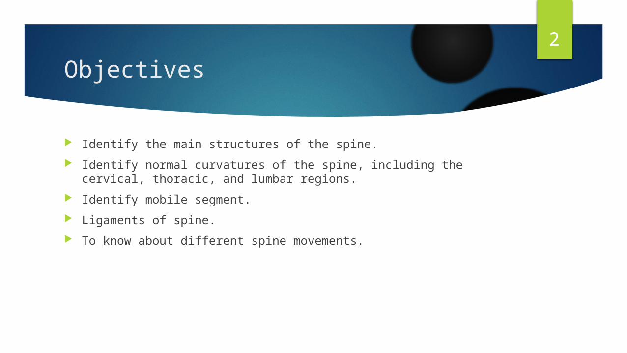
4Vertebrae total 33 vertebras are divided as
Cervical (neck) - 7 vertebrae (C1-C7) Thoracic (chest) - 12 vertebrae (T1-T12) Lumbar (lower back) - 5 vertebrae (L1-L5) Sacral (pelvis) - 5 fused vertebrae (S1-S5) Coccygeal (tail) - 4 vertebrae (Co1-Co4)

5Cervical Spine
Seven vertebrae More flexible Supports the head Wide range of motion Rotation to left and right Flexion Up and down Lateral bending Peripheral nerves• Shoulder, Chest and diaphragm• Arms
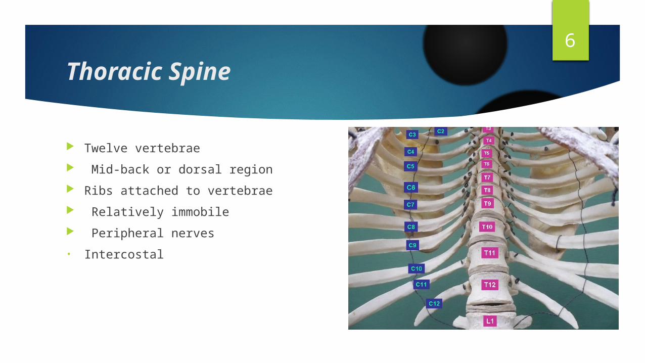
6Thoracic Spine
Twelve vertebrae Mid-back or dorsal region Ribs attached to vertebrae Relatively immobile Peripheral nerves• Intercostal
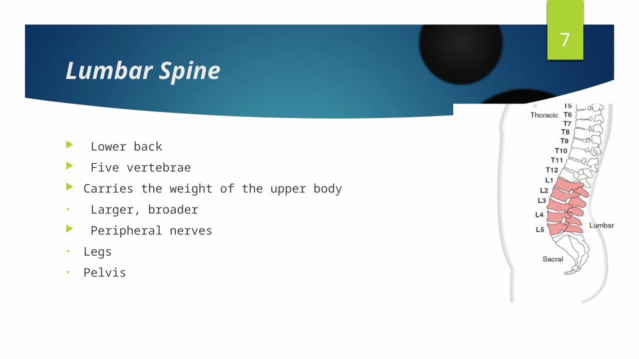
7Lumbar Spine
Lower back Five vertebrae Carries the weight of the upper body• Larger, broader Peripheral nerves• Legs• Pelvis
8Sacral and Coccygeal region
Sacrum • Triangular structure• Base of the spine• Connects spine to pelvis• Nerves to pelvic organs Coccyx• Few small bones• Remnant of tail
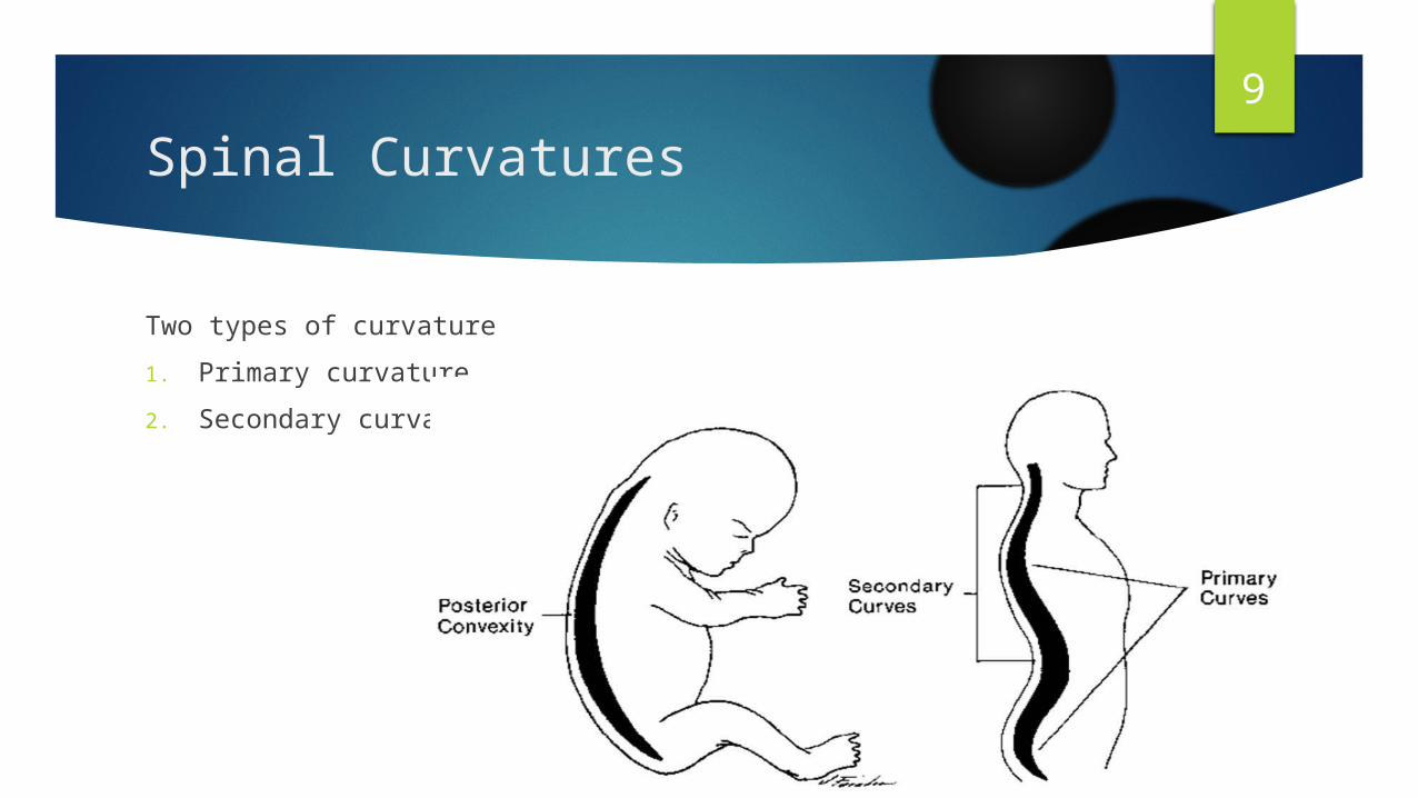
9Spinal Curvatures
Two types of curvature1. Primary curvature 2. Secondary curvature
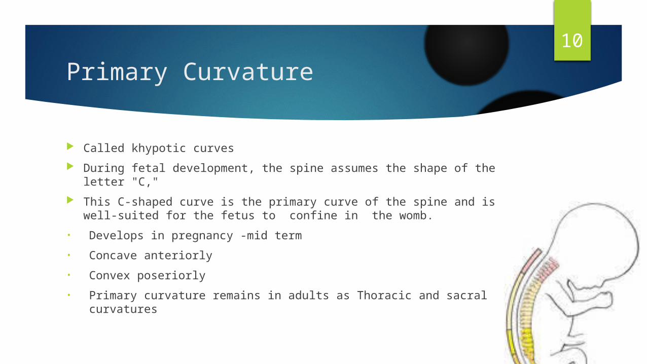
10Primary Curvature
Called khypotic curves During fetal development, the spine assumes the shape of the letter
"C," This C-shaped curve is the primary curve of the spine and is well-suited
for the fetus to confine in the womb.• Develops in pregnancy -mid term• Concave anteriorly• Convex poseriorly• Primary curvature remains in adults as Thoracic and sacral curvatures
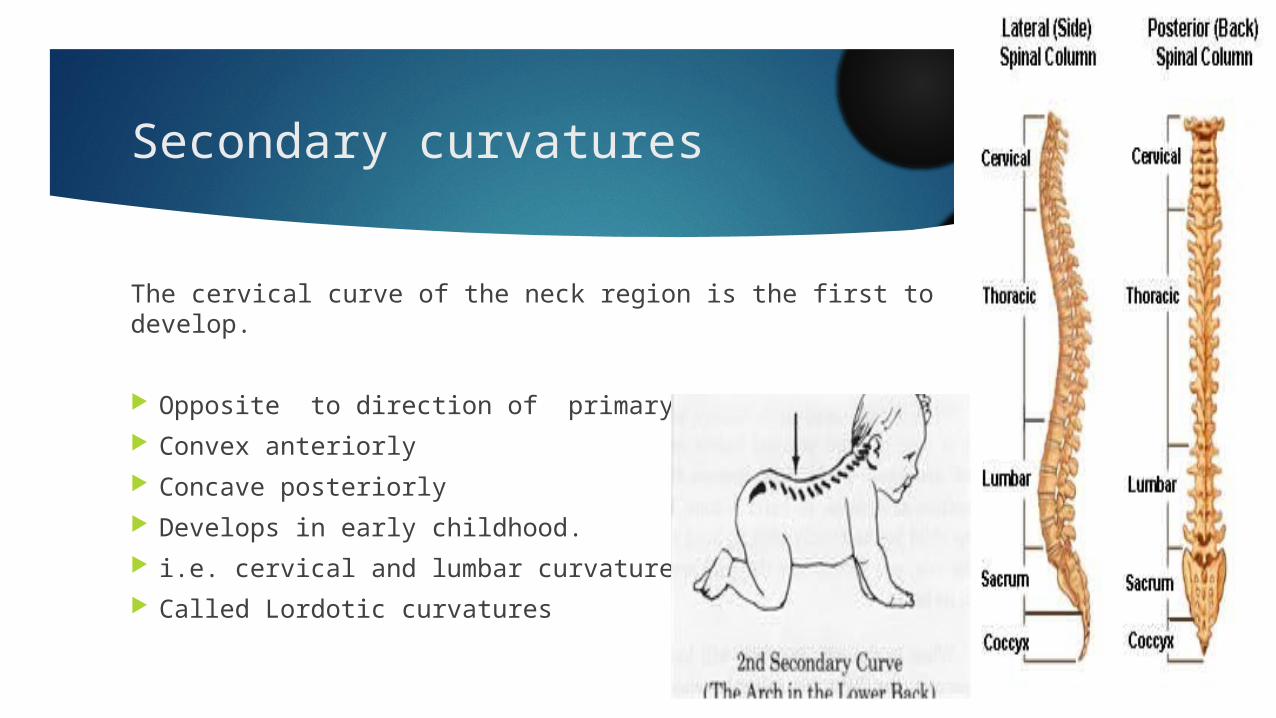
11Secondary curvatures
The cervical curve of the neck region is the first to develop.
Opposite to direction of primary curves. Convex anteriorly Concave posteriorly Develops in early childhood. i.e. cervical and lumbar curvatures Called Lordotic curvatures

12Mobile Segment
“ The smallest functional unit of the spine is called as mobile segment”Parts: Two adjacent vertebras Intervertebral disc Soft tissues which secure them, i.e. Ligaments and muscles.

13Mobile Segment
The anterior portion of the segment is composed of Two superimposed intervertebral bodies The intervertebral disc The longitudinal ligaments
The posterior portion of the segment is composed of Vertebral arches, The intervertebral joints formed by the facets, The transverse and spinous processes, Ligaments

14

15Anterior PortionThe Vertebral Bodies
are designed to bear mainly compressive loads progressively larger caudally as the superimposed weight of the upper
body increases. lumbar region are thicker and wider ; their greater size allows them to
sustain the larger loads to which the lumbar spine is subjected.
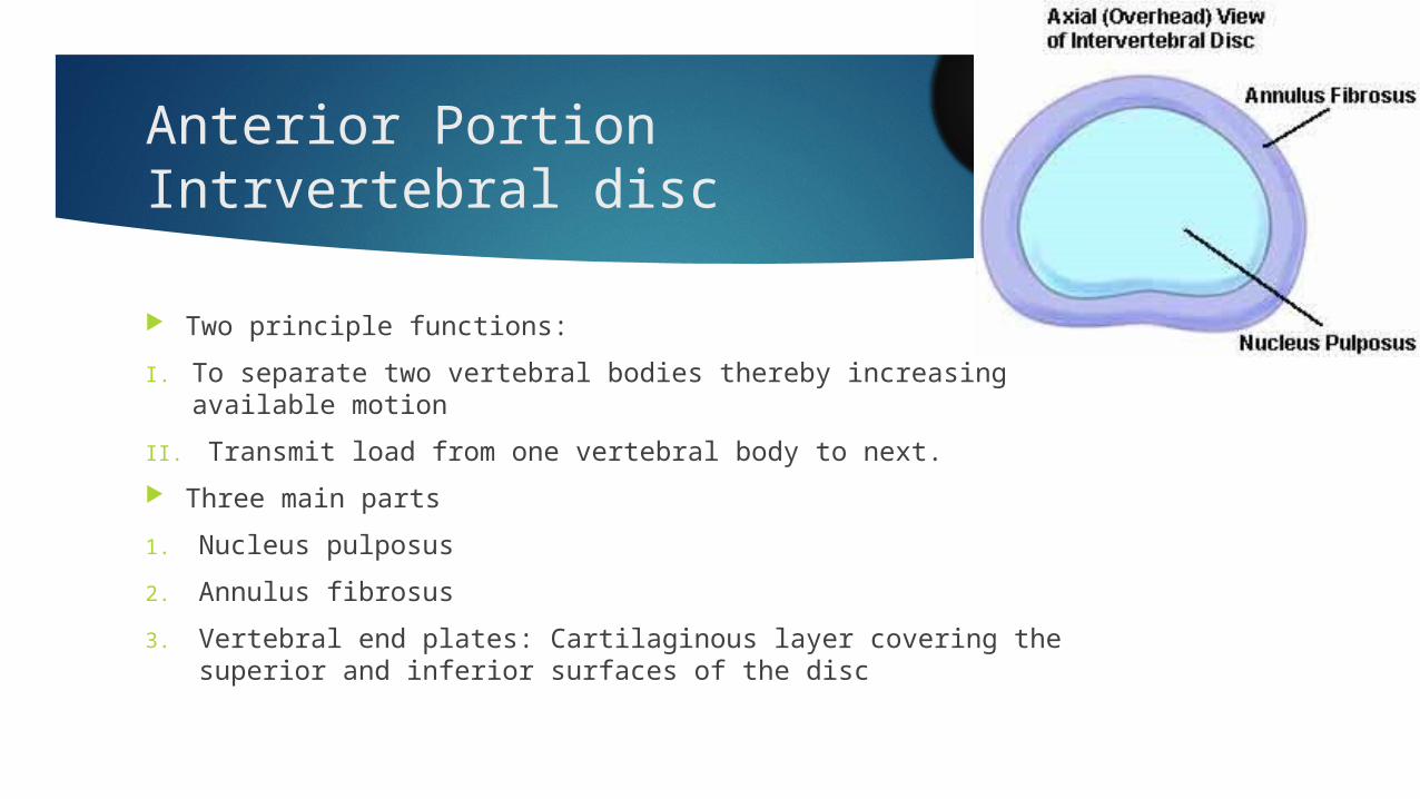
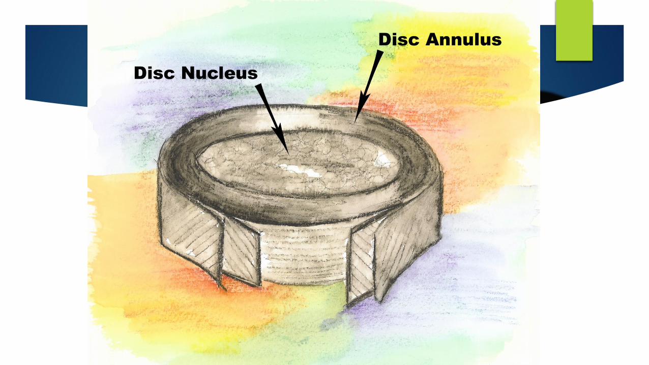
16Anterior PortionIntrvertebral disc
Two principle functions: I. To separate two vertebral bodies thereby increasing available motionII. Transmit load from one vertebral body to next. Three main parts1. Nucleus pulposus2. Annulus fibrosus3. Vertebral end plates: Cartilaginous layer covering the superior and
inferior surfaces of the disc
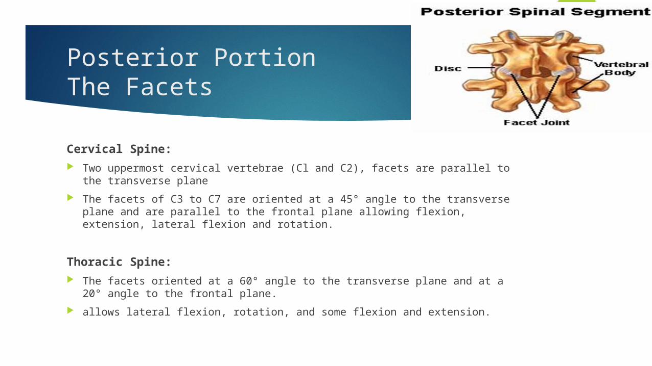
18Posterior PortionThe Facets
Cervical Spine: Two uppermost cervical vertebrae (Cl and C2), facets are parallel to the transverse
plane The facets of C3 to C7 are oriented at a 45° angle to the transverse plane and are
parallel to the frontal plane allowing flexion, extension, lateral flexion and rotation.
Thoracic Spine: The facets oriented at a 60° angle to the transverse plane and at a 20° angle to
the frontal plane. allows lateral flexion, rotation, and some flexion and extension.

19Posterior PortionThe Facets
Lumbar Spine: The facets are oriented at right angles to the transverse plane and at a
45° angle to the frontal plane. This alignment allows flexion, extension, and lateral flexion, but
almost no rotation. The lumbosacral joints differ from the other lumbar intervertebral joints
in that the oblique orientation of the facets allows appreciable rotation.


21Posterior PortionThe Facets
The facets guide movement of the motion segment and have a load-bearing function, and may have some role in the lateral stability of the motion segment
Load sharing between the facets and the disc varies with the position and the health of the spine.
The loads on the facets are greatest with axial rotation of the spine.
22Clinical Condition
With disc degeneration, a greater amount of force is transferred to the facet joints, thereby redistributing the load through the motion segment .
Because the facets are not the primary support structure in extension, if total compromise of these joints occurs, an alternate path of loading is established.
This path involves the transfer of axial loads to the annulus and anterior longitudinal ligament as a way of supporting the spine.
High loading of the facets is also present during forward bending, coupled with rotation.

23Posterior Portion vertebral arches and intervertebral joints
They play an important role in resisting shear forces. For Example: This function is demonstrated by the fact that patients with deranged
arches or defective joints (e.g., from spondylolysis and spondylolisthesis) are at increased risk for forward displacement of the vertebral body.

24Posterior PortionTransverse and Spinous Processes
Serve as sites of attachment for the spinal muscles that, when activated Initiate spine motion Provide extrinsic stability.
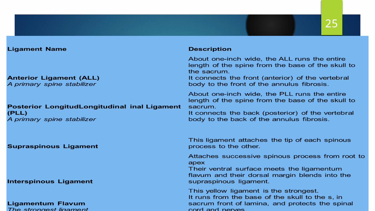
25THE LIGAMENTS OF THE SPINE

26
27Kinematics
The movements available in the vertebral column as a whole area. Flexion and extensionb. lateral flexionc. rotation Agonistic muscle initiate and carry out motion antagonistic muscles control and modify the motion co-contraction of both groups stabilizes the spine.
28Kinematics
The ROM differs at various levels of the spine and depends on the orientation of the facets.
Motion between two vertebrae is small and does not occur independently.
All spine movements involve the combined action of several motion segments.
The skeletal structures that influence motion of the trunk are The rib cage, which limits thoracic motion The pelvis, which augments trunk movements by tilting.

29SEGMENTAL MOTION OF THE SPINE
The vertebrae have six degrees of freedom: rotation about and translation along a transverse, a sagittal, and a longitudinal axis.
The motion produced during flexion, extension, lateral flexion, and axial rotation of the spine is a complex combined motion resulting from simultaneous rotation and translation which is called as coupling.
30Kinematics
Coupling is defined as the association of one movement about an axis with another movement around a different axis.
The most predominant movements that exhibit coupled behaviors are lateral flexion and rotation.
Pure lateral flexion and pure rotation do not occur in any region of the spine.

31THE MUSCLES
The spinal muscles can be divided into flexors and extensors. The trunk muscles play an important role in the mechanical behavior of
the spine, including spine stability and intradiscal pressure. The main flexors are the abdominal muscles and the psoas muscles. The main extensors are the erector spinae, the multifidus, and the
intertransversarii attached to the posterior elements.
32THE MUSCLES
When extensor muscles contract symmetrically, extension is produced. When right and left side flexors and extensor muscles contract
asymmetrically, lateral bending or twisting of the spine is produced.

33
THAT’S ALL