LDBIO TOXO II IgG - ldbiodiagnostics.com · CONTROLE POSITIF (Prêt à l'emploi ... 6. Lavage :...
24
1 WB‐TOXO II G vs09 fr‐en 161012 LDBIO TOXO II IgG Technique d'immunoblot pour CONFIRMATION usage diagnostique in vitro #TOXO II-24G : 24 tests #TOXO II-12G : 12 tests #TOXO II-96G : 96 tests ENGLISH VERSION PAGE 13 NOTICE D'UTILISATION Indication du test LDBIO-TOXO II IgG est un test qualitatif de diagnostic sérologique IgG par immunoblot de la toxoplasmose proposé comme test de confirmation d'un résultat positif ou équivoque obtenu par les tests classiques de dépistage. Il peut être pratiqué sur le sérum, sur le liquide céphalo-rachidien (LCR) ou sur l’humeur aqueuse. Principe du test Technique de Western Blot : Les antigènes Toxoplasma gondii, après séparation électrophorétique ont été fixés par électro-transfert à la surface d’une feuille de nitrocellulose (appelée le transfert) découpée en 24 bandelettes identifiées de 1 à 24. Déroulement du test : Chaque échantillon sérique (ou LCR/humeur aqueuse) à tester est incubé séparément avec une bandelette. Les anticorps spécifiques éventuellement présents dans le prélèvement se fixent sélectivement sur les antigènes. A l'étape suivante, le conjugué Phosphatase Alcaline-anti-IgG humaines se lie aux anticorps fixés. Enfin, les immun- complexes réagissent avec le substrat. Les antigènes reconnus par les anticorps spécifiques de classe IgG présents dans les échantillons sont ainsi révélés sous forme de bandes transversales violettes.
Transcript of LDBIO TOXO II IgG - ldbiodiagnostics.com · CONTROLE POSITIF (Prêt à l'emploi ... 6. Lavage :...

1
WB‐TOXO II G vs09 fr‐en 161012
LDBIO TOXO II IgG Technique d'immunoblot pour
CONFIRMATION usage diagnostique in vitro
#TOXO II-24G : 24 tests #TOXO II-12G : 12 tests #TOXO II-96G : 96 tests
ENGLISH VERSION PAGE 13
NOTICE D'UTILISATION
Indication du test LDBIO-TOXO II IgG est un test qualitatif de diagnostic sérologique IgG par immunoblot de la toxoplasmose proposé comme test de confirmation d'un résultat positif ou équivoque obtenu par les tests classiques de dépistage. Il peut être pratiqué sur le sérum, sur le liquide céphalo-rachidien (LCR) ou sur l’humeur aqueuse. Principe du test Technique de Western Blot : Les antigènes Toxoplasma gondii, après séparation électrophorétique ont été fixés par électro-transfert à la surface d’une feuille de nitrocellulose (appelée le transfert) découpée en 24 bandelettes identifiées de 1 à 24. Déroulement du test : Chaque échantillon sérique (ou LCR/humeur aqueuse) à tester est incubé séparément avec une bandelette. Les anticorps spécifiques éventuellement présents dans le prélèvement se fixent sélectivement sur les antigènes. A l'étape suivante, le conjugué Phosphatase Alcaline-anti-IgG humaines se lie aux anticorps fixés. Enfin, les immun-complexes réagissent avec le substrat. Les antigènes reconnus par les anticorps spécifiques de classe IgG présents dans les échantillons sont ainsi révélés sous forme de bandes transversales violettes.

2
WB‐TOXO II G vs09 fr‐en 161012
Réactifs fournis avec la trousse
italique : conditionnement 12 tests (#TOXO II-12G) - gras : Conditionnement 96 tests (#TOXO II-96G).
ID Qté Description Composition
R1 1
Pochette(s) de 24 (12, 4x24) BANDELETTES prédécoupées + Standards colorés. (Chaque pochette et chaque transfert est identifié par un numéro de série unique)
Nitrocellulose sensibilisée. Poids Moléculaires Colorés (kDa) : Bleu : 250, Bleu : 150, Bleu : 100, Rose : 75, Bleu : 50, Vert : 37, Rose : 25, bleu : 20, bleu : 15.
R2 1 Flacon de 30 (30, 125) ml de DILUANT ECHANTILLON (Prêt à l'emploi - solution rose).
Tampon + surfactant + NaN3 (inf. 0.1%).
R3 1 Flacon(s) de 30 (30, 2x60) ml de CONJUGUE ANTI-IgG (Prêt à l'emploi - solution bleue).
Tampon + sérum polyclonal de chèvre anti-IgG humaines conjugué à la phosphatase alcaline + NaN3 (inf. 0.1%) + stabilisants.
R5 1 Flacon de 30 (30, 125) ml de SUBSTRAT (Prêt à l'emploi - flacon opaque marron).
Tampon + NBT + BCIP + stabilisants.
R6 1
Flacon de 60 (60, 250) ml de TAMPON DE LAVAGE CONCENTRE 10X (A diluer 10 fois dans de l'eau distillée - solution incolore).
Tampon + surfactant + NaN3 (inf. 0.1%).
R10 1 Tube de 100 (100, 2x100) µl de SERUM DE CONTROLE POSITIF (Prêt à l'emploi - bouchon rouge).
Tampon + pool de sérums humains positif en sérologie toxoplasmose + NaN3 (inf. 0.1%) + stabilisants.
R2, R3, R5 et R6 sont communs à tous les kits et présentent un numéro de lot unique qui ne dépend que de la date de leur production. Il est recommandé de réaliser des séries multiparamétriques (Cf. la gamme immunoblots LDBIO) pour limiter le nombre de flacons entamés et pour assurer un meilleur contrôle de qualité.
Matériel nécessaire mais non fourni
Cuves d'incubation multicanaux en polypropylène adaptées aux miniblots (# WBPP- 08 ou équivalent). Agitateur oscillant pour immunoblots, système d'aspiration pour les liquides (les cuves # WBPP- 08 que nous fournissons peuvent être vidées par simple retournement). Tubes et matériel pour le prélèvement des échantillons, éprouvettes graduées, récipients adaptés. Pipettes automatiques, micropipettes et pointes à usage unique (volumes de 10µl, 25µl, 1.2ml et 2ml). Eau distillée ou désionisée. Papier absorbant (ex : papier filtre Whatman), ruban adhésif transparent. Gants en latex, pincette pour manipuler les bandelettes, cutter ou scalpel, règle plate transparente. Remarque : Nos réactifs peuvent être utilisés sur automate pour immunoblots. Attention aux possibles contaminations chimiques de nos réactifs si l’automate est partagé avec des réactifs d’un autre fabricant (exemple connu : contamination par le TWEEN 20) et aux contaminations bactériologiques. Dédier des flacons à l’automate. Ne pas rempoter les réactifs en fin de manipulation.

3
WB‐TOXO II G vs09 fr‐en 161012
Conditions de conservation et péremption
Conservation entre 2 et 8°C. Les réactifs du coffret sont valables jusqu'à la date de péremption indiquée sur le couvercle de la boîte et les étiquettes des flacons. Le tampon de lavage dilué 1/10 est stable 2 mois entre +2 et +8°C et une semaine à température ambiante.
Précautions d'emploi Sécurité Pour usage in vitro exclusivement. Manipuler selon les Bonnes Pratiques de Laboratoire et
considérer tout réactif et tout échantillon comme potentiellement toxique et/ou infectieux. Porter une blouse, des gants et lunettes, ne pas boire, manger ou fumer dans le laboratoire. Ne pas
pipeter avec la bouche. Le contrôle positif est un sérum d’origine humaine qui a subi un dépistage négatif concernant les
anticorps anti-VIH 1 et 2, les anticorps anti-VHC et l’antigène HBs. Il doit cependant être manipulé comme un produit potentiellement infectieux.
Le substrat contient un mélange NBT et BCIP, toxique par contact (peau et muqueuses) et inhalation.
Les réactifs contiennent de l'azide de sodium susceptible de former des sels métalliques explosifs avec le plomb ou le cuivre. Rincer à l'eau tout rejet à l'évier.
Éliminer les déchets (prélèvements, pointes, tubes, liquides de lavage, réactifs usagés...) conformément aux bonnes pratiques en usage dans la profession et aux règlements en vigueur dans le Pays.
Précautions Ne pas utiliser ensemble des réactifs liquides de lots différents. Utiliser les bandelettes dans leur ordre numérique. Ne pas mélanger des bandelettes de plusieurs
numéros de série mais utiliser les transferts successivement. Etablir un plan de distribution précis avant de commencer la manipulation.
Ne pas toucher les bandelettes avec les doigts, utiliser une pincette. Les réactifs doivent être bien mélangés avant usage, en particulier le tampon de lavage concentré. Refermer les flacons après usage, ne pas utiliser en cas de pénétration accidentelle de substance
dans les réactifs. Ne pas utiliser de réactif provenant d’un flacon présentant des signes de fuite. Ne pas utiliser de solution trouble ou précipitée.
N'utiliser que des cônes de pipette à usage unique. Eviter toute contamination inter-puits. Attention à la formation d'aérosols.
Ne nettoyer les cuves d’incubation qu’à l’eau claire puis distillée (ne jamais utiliser de détergeant ou de javel).
L’omission de distribution d’un échantillon ou la distribution d’un volume inapproprié peut faire considérer comme négatif le résultat du test quel que soit son statut réel.
Prélèvement des échantillons Prélever de manière aseptique les échantillons sur tube sec. Un minimum de 10µl de sérum, d’humeur aqueuse ou de LCR sont nécessaires. Dans les cas particuliers de l’humeur aqueuse ou du LCR, l’utilisation de 25 µl permettront d’augmenter la sensibilité du test. Conserver les échantillons à 2-8°C. S’ils doivent être conservés, les congeler à -20 ± 5°C. Ne pas utiliser d’échantillon contaminé. Éviter de congeler et décongeler les échantillons plusieurs fois.

4
WB‐TOXO II G vs09 fr‐en 161012
Préparation des réactifs Tampon de lavage : Pour 4 tests, diluer dans un flacon propre 10ml de tampon de lavage concentré 10x (R6) dans 90ml d'eau distillée ou désionisée .
Mode opératoire Nota Bene : Il est recommandé de réaliser des séries multiparamétriques (Cf. la gamme immunoblots LDBIO) pour limiter le nombre de flacons entamés et pour assurer un meilleur contrôle de qualité. 1. Préparer le plan de distribution des échantillons et du contrôle positif C+ (R10).
Seule l'utilisation de ce contrôle permet de valider techniquement la manipulation et d’identifier pour un numéro de série donné, les bandes spécifiques révélées. On ne peut pas utiliser une bandelette C+ pour interpréter les résultats de bandelettes issues d’un transfert de numéro de série différent.
2. Découper le nombre de bandelettes (R1) nécessaires, à l'aide d'un scalpel et d'une règle plate
transparente, propre et sèche, en conservant le trait bleu de positionnement sur les bandelettes : les maintenir fermement plaquées par la règle et les découper du côté de la souche (les numéros étant visibles au travers de la règle par transparence).
3. Distribuer 1.2ml de tampon échantillon (R2) dans chacun des puits selon le plan établi. 4. Déposer dans leur ordre numérique les bandelettes numérotées dans les puits : Laisser les
bandelettes se réhydrater pendant environ 1 minute, numéro visible vers le haut en agitant doucement la cuve pour les immerger totalement dans le tampon.
5. Distribuer échantillons et contrôle(s) positif(s) : selon le plan de distribution, à raison de 10 µl par
puits (de préférence 25 µl pour l’humeur aqueuse ou le LCR). Agiter doucement la cuve après chaque dépôt. La placer sur un agitateur oscillant. Incubation 90mn ± 5mn à 18-25°C.
6. Lavage : Vider le contenu des puits à l'aide d’une pipette pasteur ou par retournement de la cuve
d’incubation et répartir 2 à 3 ml de tampon de lavage dilué dans chacun d'eux et incuber 3 mn sur l'agitateur. Répéter 2 fois, puis vider le contenu des puits. Faire attention à ce que les bandelettes ne se retournent pas pendant ces opérations.
7. Distribuer 1.2 ml de conjugué anti-IgG (R3) dans chacun des puits. Placer la cuve sur l’agitateur
oscillant. Incubation 60mn ± 5mn à 18-25°C 8. Lavage : procéder comme pour l'étape 6. 9. Distribuer 1.2 ml de substrat NBT/BCIP (R5) dans chacun des puits et placer sur l’agitateur
oscillant, à l'abri de la lumière directe. Incubation 60mn ± 5mn à 18-25°C.
Quelque soit le paramètre, surveiller le développement de la coloration. La révélation peut être interrompue si la couleur du fond de la bandelette s'assombrit au point de rendre la lecture difficile (La qualité des lavages a une influence fondamentale sur cette coloration). Noter que les bandelettes s’éclairciront en séchant.

5
WB‐TOXO II G vs09 fr‐en 161012
10. Arrêter la réaction par aspiration du substrat avec une pipette pasteur ou par retournement de la cuve d’incubation puis par la distribution de 2ml d'eau distillée dans le puits. Répéter une fois ce dernier lavage.
11. Séchage des bandelettes : Les puits toujours remplis d’eau, saisir les bandelettes par leur extrémité
numérotée à l'aide de la pincette et les déposer, numéro visible, sur un papier absorbant de type Whatman. Les laisser sécher à l'air. La couleur des bandelettes s'éclaircit naturellement en séchant. La lecture ne doit s'effectuer qu'après séchage complet.
12. Stockage : Transférer les bandelettes sur la feuille de papier qui servira à les archiver. Aligner les
traits de positionnement. Maintenir les bandelettes avec la règle plate et les coller par le haut à l'aide du ruban adhésif transparent.
Pour une bonne interprétation, les bandelettes doivent être ordonnées par transfert et dans leur ordre numérique, espacées d’au maximum quelques millimètres. Il est aléatoire de vouloir comparer des bandelettes très espacées (ex : n°2 avec n°15). Il est dangereux (faux résultats) de vouloir comparer des bandelettes de kits différents (N° de série de bandelettes différent).
Contrôle qualité et interprétation Le sérum de contrôle (R10) fourni avec le coffret doit systématiquement être inclus dans toute série d'immunoblots. Il présente le profil type et permet de valider techniquement le bon déroulement du test (les bandes doivent apparaître très nettement sur la bandelette) et d'étalonner précisément la position et l’aspect des bandes spécifiques pour permettre l'interprétation des résultats de bandelettes issues d’un même transfert (même numéro de série).
Description des bandes : Un échantillon positif peut présenter de nombreuses bandes situées entre 15 et 200k Kilo-Daltons (KDa). Rechercher la présence des bandes spécifiques dans la zone 30-45kDa pour chacun des échantillons testés à l'aide des outils d'étalonnage décrits ci-dessus. Ces bandes, groupées et bien isolées, sont caractéristiques et généralement très facilement repérables.

6
WB‐TOXO II G vs09 fr‐en 161012
Fig. 1 : Exemples de résultats positifs et négatifs A gauche et à droite : représentation du contrôle positif du kit (R10) qui permet d’identifier les bandes spécifiques dans la zone 30 - 45 kDa : 30, 31, 33, 40 et 45.
Interprétation : La présence sur la bandelette d'un minimum de 3 bandes parmi les bandes spécifiques 30, 31, 33, 40, 45, et incluant la bande à 30kDa, permet d'interpréter le test comme positif et de conclure à la présence d'anticorps IgG anti-T. gondii dans l'échantillon testé. A : exemple d’une séroconversion. Les sérums 2 et 3, positifs par LDBIO-TOXO II IgG, étaient négatifs en technique de dépistage (ci-après dénommée ELISA 2 IgG dans l'étude des performances). B : exemple sur un suivi néonatal. Les sérums 5 et 6, positifs par LDBIO-TOXO II IgG, était négatifs en technique de dépistage ELISA 2 IgG . Nota Bene : d’autres bandes peuvent être observées. Elles ne sont pas prise en compte dans la lecture du test.
30 33
3 1
45 40
Contrôle positif PM en kDa
A B
Contrôle positif PM en kDa
30 3 3
3 1
45 40

7
WB‐TOXO II G vs09 fr‐en 161012
Pour la validation des résultats, toujours comparer le profil de l’immuno-blot de chaque échantillon avec celui du contrôle positif R10. L’aspect des bandes est important dans l’interprétation du test.
Limites du test Les résultats sérologiques doivent être interprétés en fonction des renseignements disponibles (épidé-miologie, clinique, imagerie, biologie) afin d'établir le diagnostic.
Performances L’évaluation a été réalisée dans un laboratoire de référence, spécialiste du diagnostic de la toxoplasmose. Le principe de l’évaluation a consisté à comparer sur 529 sérums les résultats obtenus par la technique LDBIO-TOXO II IgG, les résultats du DYE TEST de Sabin et Feldman, les résultats de deux techniques de dépistage commercialisées, « ELISA 1 IgG » et « ELISA 2 IgG », ainsi que les données cliniques et biologiques des patients. Seuil des techniques utilisées :
NEGATIF EQUIVOQUE POSITIFDYE TEST (UI/ml) <2 - ≥ 2 ELISA 1 (UI/ml) <4 4 - 8 ≥ 8 ELISA 2 (UI/ml) <6 - ≥ 6 LDBIO TOXO II IgG 0 - ≥ 1
Analyse statistique des résultats : Nous avons établi les valeurs de sensibilité, spécificité lorsque cela était possible. La corrélation des résultats trouvés par différentes techniques a été évaluée par le test CHI-2 de Mac Nemar sur séries appariées.
PATIENTS :
Toutes les analyses ont été effectuées sur des sérums conservés congelés à –20°C. Les échantillons proviennent de 5 groupes de patients différents.
o Groupe I – DYE TEST
Etude sur 200 sérums obtenus lors du dépistage de la toxoplasmose chez des femmes enceintes, et testés par le DYE TEST. Le sous-groupe « positif » correspond à 98 sérums positifs en DYE TEST provenant de femmes immunisées contre T. gondii. Ce sous-groupe comprenait des sérums présentant des titres d’IgG modérés en DYE TEST (de 2 à 32 UI/ml) afin de tester la sensibilité de LDBIO-TOXO II IgG versus les autres techniques. Le sous-groupe « négatif » correspondait à 102 sérums négatifs en DYE TEST, provenant de femmes enceintes non immunisées contre la toxoplasmose. Ces 200 sérums ont été testés en parallèle avec les techniques LDBIO-TOXO II IgG, ELISA 1 IgG et ELISA 2 IgG.

8
WB‐TOXO II G vs09 fr‐en 161012
o Groupe II - Séroconversions
Il s’agit de l’analyse rétrospective de 17 séquences de sérums (101 échantillons) provenant de patientes ayant présenté une séroconversion toxoplasmique pendant leur grossesse.
Chaque série séquentielle comprend le dernier sérum négatif puis une série de 3 à 5 sérums montrant l’apparition des IgM spécifiques et la synthèse d’ IgG spécifiques (ELISA 2 IgG).
o Groupe III – suivi enfants non-infectés
Il s’agit de l’analyse rétrospective de 74 échantillons correspondant à 20 séquences du suivi post-natal d’enfants nés de mères ayant présenté une séroconversion toxoplasmique en cours de grossesse. Chaque séquence de 2 à 6 sérums montre la décroissance du titre des IgG maternelles transmises jusqu’à négativation de la sérologie par la technique ELISA 2 IgG (entre 5 et 13 mois).
o Groupe IV – suivi enfants infectés
Il s’agit de l’analyse rétrospective de 85 échantillons provenant du suivi post natal de 30 enfants ayant une infection congénitale. Le suivi sérologique était réalisé par ELISA 2 IgG.
o Groupe V – sensibilité - spécificité (infections virales et paludisme)
Etude sur 69 sérums de patients atteints d’infections virales ou de paludisme (tableau 1). Ces échantillons ont été testés par ELISA 2 IgG. (Tous étaient négatifs en recherche d'IgM). Tous les négatifs ainsi que les discordants ont été testés en DYE TEST.
Agent infectieux (n=69)
ELISA 2 IgG POSITIF (n=44)
ELISA 2 IgG NEGATIF (n=25)
EBV (n=5) 0 5 VZV (n=3) 2 1 CMV (n=5) 2 3 HBV (n=9) 8 1 HAV (n=2) 0 2 HCV (n=10) 8 2 HIV (n=10) 6 4
PALU (n=25) 18 7 Tableau 1 : différentes infections testées dans l’étude

9
WB‐TOXO II G vs09 fr‐en 161012
RESULTATS :
o Groupe I : DYE TEST
DYE TEST LDBIO TOXO II IgG ELISA 1 IgG ELISA 2 IgG POSITIF 98 97 61 93 NEGATIF 102 103 114 107 EQUIVOQUE - - 25 - SPECIFICITE - 100% 100% 100% SENSIBILITE - 99% 85% 95% Tableau 2 : Corrélation du DYE TEST avec les 3 techniques.(La technique ELISA 1 IgG présente une zone équivoque). - 4 sérums ELISA 2 IgG négatifs sont positifs en LDBIO-TOXO II IgG et Dye test - 11 sérums ELISA 1 IgG négatifs sont positifs en LDBIO-TOXO II IgG et Dye test - 25 sérums ELISA 1 IgG sont équivoques : 24 sont positifs en LDBIO-TOXO II IgG et Dye test, 1 sérum est négatif en LDBIO-TOXO II IgG et Dye test
o Groupe II : Séroconversions
ELISA 2 IgG POSITIF NEGATIF
LDBIO TOXO II POSITIF 70 10
NEGATIF 0 21 Tableau 3 : Corrélation LDBIO-TOXO II IgG / ELISA 2 IgG sur 101 sérums de séroconversion. p=0.0016 Pour 8 / 17 séroconversion (47%) les IgG sont dépistées plus précocement par LDBIO-TOXO II IgG.
o Groupes III et IV : Suivis de nouveaux nés
ELISA 2 IgG POSITIF NEGATIF
LDBIO TOXO II POSITIF 130 18
NEGATIF 0 11 Tableau 4 : Corrélation LDBIO-TOXO II IgG / TEST 2 IgG sur 159 sérums de suivi post natal. p<0.0001
Enfants indemnes : 13 sérums, correspondant à 10 / 20 suivis postnataux (50%) négatifs en ELISA 2 IgG, restent positifs en LDBIO-TOXO II IgG qui met en évidence des anticorps maternels transmis alors que la technique ELISA 2 IgG ne les détecte plus.
Enfants contaminés : 5 sérums correspondant à 3 enfants sont discordants. L’un d’entre eux montre une négativation transitoire de sa sérologie par ELISA 2 IgG. Le test LDBIO-TOXO II IgG demeure positif, confirmant sa contamination. Pour les 2 autres enfants, le test LDBIO TOXO II IgG montre une positivité plus précoce que ELISA 2 IgG.
Il est cependant impossible d’affirmer une néosynthèse d’IgG, ce test ne différenciant pas les anticorps maternels transmis des anticorps néo-synthétisés.

10
WB‐TOXO II G vs09 fr‐en 161012
o Groupe V : Sensibilité et Spécificité (infections virales et paludisme)
ELISA 2 IgG POSITIF NEGATIF
LDBIO TOXO II + DYE TEST
POSITIF 42 2 NEGATIF 2 23
Tableau 5 : Corrélation LDBIO-TOXO II IgG / DYE TEST / ELISA 2 IgG sur 69 sérums d’infections virales ou de paludisme.
Sur cette population, la concordance de LDBIO-TOXO II IgG IgG avec le DYE TEST est de 100% : ces résultats confirment la spécificité et la sensibilité du test LDBIO-TOXO II IgG.
L’étude met en évidence quatre résultats discordants par la technique ELISA 2 IgG, 2 faux négatifs (un HIV et un P.falciparum) et 2 faux positifs (deux P.falciparum) soulignant l’intérêt d’une technique de confirmation pour tous les résultats proches du seuil.
CONCLUSION :
o Groupe I (DYE TEST) :
La corrélation LDBIO-TOXO II IgG / DYE TEST est excellente (sensibilité 99%, spécificité 100%). LDBIO-TOXO II IgG permettrait de confirmer le statut immunitaire des patients présentant au dépistage un résultat équivoque ou un titre faible d’anticorps.
o Groupe II (séroconversions) : La sensibilité de LDBIO-TOXO II IgG est supérieure à ELISA 2 IgG (p=0.0016). LDBIO-TOXO II IgG permettrait de confirmer une séroconversion plus précocement que ELISA 2 IgG.
o Groupes III et IV (suivis de nouveaux nés) : la sensibilité de LDBIO-TOXO II IgG est très supérieure à ELISA 2 IgG (p<0.0001). Lors du suivi de l’enfant, LDBIO-TOXO II IgG pourrait être utilisé pour confirmer ou infirmer la négativation de la sérologie. Cependant LDBIO-TOXO II IgG ne permet pas de différencier les anticorps maternels transmis des anticorps néo-synthétisés par l’enfant.
o Groupe V (infect. virales et paludisme) : La corrélation LDBIO-TOXO II IgG / DYE TEST est excellente (sensibilité 100%, spécificité 100%). Ces résultats mettent en évidence la nécessité d’utiliser un test de confirmation pour contrôler les échantillons présentant au dépistage un résultat proche du seuil.

11
WB‐TOXO II G vs09 fr‐en 161012
Les excellentes performances de la trousse LDBIO-TOXO II IgG justifient son utilisation en confirmation des résultats obtenus par les techniques IgG de dépistage (résultats équivoques, faiblement positifs ou posant des problèmes d’interprétation).
Reproductibilité : Reproductibilités inter-séries et inter-lots ont été testées. Dans les deux cas, la corrélation sérum à sérum vis-à-vis des bandes spécifiques est excellente.
Interférences : Bien qu’aucune interférence particulière n’ait été relevée avec des sérums hémolysés, ictériques ou lipidiques, il est conseillé d’interpréter les résultats provenant de l’utilisation de tels échantillons avec prudence.
Problèmes rencontrés "Les bandes sont pâles et peu contrastées" : Certains sérums très peu chargés en anticorps peuvent donner de tels résultats. "Des zones d'ombre se voient, plus ou moins colorées, légèrement diffuses" : La bandelette n'était pas totalement immergée dans l'un des réactifs et n'a pas incubé correctement sur toute sa longueur. Des taches peuvent être également présentes à l’endroit du dépôt de l’échantillon si la cuve n’a pas été agitée après la distribution. "Le bruit de fond est important, rendant la lecture très difficile" : Les lavages ont été insuffisants ou la dernière incubation a été trop longue. S'assurer du bon déroulement technique du test, du respect des temps de lavage, de la qualité de l'eau. Diminuer le temps de la dernière incubation. Exceptionnellement, certains sérums peuvent réagir ainsi de façon non spécifique. Le résultat de l'immunoblot ne peut alors être rendu. Ce bruit de fond non spécifique peut ne concerner qu'une partie de la bandelette, rendant les résultats ininterprétables sur cette partie seulement. "Un précipité apparaît dans la solution lors de la dernière étape de révélation" : le substrat peut effectivement partiellement précipiter (flocons noirs) dans le tampon en fin de révélation. Ce phénomène n'altère pas la qualité de la révélation qui doit être poursuivie normalement. Le lavage final à l'eau distillée élimine les particules solides éventuellement présentes.

12
WB‐TOXO II G vs09 fr‐en 161012
Bibliographie Franck, Jacqueline, Yves Jean-François Garin, et Henri Dumon. « LDBio-Toxo II immunoglobulin G
Western blot confirmatory test for anti-toxoplasma antibody detection ». Journal of clinical microbiology 46, no 7 (juillet 2008): 2334‑38. doi:10.1128/JCM.00182-08.
Jost, C, F Touafek, A Fekkar, R Courtin, M Ribeiro, D Mazier, et L Paris. « Utility of immunoblotting for early diagnosis of toxoplasmosis seroconversion in pregnant women ». Clinical and vaccine immunology: CVI 18, no 11 (novembre 2011): 1908‑12. doi:10.1128/CVI.05303-11.
Khammari, Imen, Fatma Saghrouni, Sami Lakhal, Aida Bouratbine, Moncef Ben Said, et Jalel Boukadida. « A New IgG Immunoblot Kit for Diagnosis of Toxoplasmosis in Pregnant Women ». The Korean Journal of Parasitology 52, no 5 (22 octobre 2014): 493‑99. doi:10.3347/kjp.2014.52.5.493.
Khammari, Imen, Fatma Saghrouni, Alia Yaacoub, Sondoss Gaied Meksi, Hinda Ach, Lamia Garma, Akila Fathallah, et Moncef Ben Saïd. « IgG Western Blot for Confirmatory Diagnosis of Equivocal Cases of Toxoplasmosis by EIA-IgG and Fluorescent Antibody Test ». The Korean Journal of Parasitology 51, no 4 (août 2013): 485‑88. doi:10.3347/kjp.2013.51.4.485.
Leslé, F, F Touafek, A Fekkar, D Mazier, et L Paris. « Discrepancies between a new highly sensitive Toxoplasma gondii ELISA assay and other reagents: interest of Toxo IgG Western blot ». European journal of clinical microbiology & infectious diseases: official publication of the European Society of Clinical Microbiology 30, no 10 (octobre 2011): 1207‑12. doi:10.1007/s10096-011-1214-1.
Maudry, A., G. Chene, R. Chatelain, B. Bellete, H. Patural, J. Hafid, H. Raberin, R. Tran Manh Sung, et P. Flori. « Expertise du nouveau test Access® TOXO-IgGII et comparaison avec trois autres techniques automatisées et la technique Western Blot LDBIO TOXO II IgG® ». Immuno-analyse & Biologie Spécialisée 24, no 1 (février 2009): 42‑49. doi:10.1016/j.immbio.2008.11.004.
Maudry, Arnaud, Gautier Chene, Rémi Chatelain, Hugues Patural, Bahrie Bellete, Bernard Tisseur, Jamal Hafid, et al. « Bicentric evaluation of six anti-toxoplasma immunoglobulin G (IgG) automated immunoassays and comparison to the Toxo II IgG Western blot ». Clinical and vaccine immunology: CVI 16, no 9 (septembre 2009): 1322‑26. doi:10.1128/CVI.00128-09.
Robert-Gangneux, F., et M.-L. Darde. « Epidemiology of and Diagnostic Strategies for Toxoplasmosis ». Clinical Microbiology Reviews 25, no 2 (1 avril 2012): 264‑96. doi:10.1128/CMR.05013-11.
Villard, O., B. Cimon, C. L’Ollivier, H. Fricker-Hidalgo, N. Godineau, S. Houze, L. Paris, H. Pelloux, I. Villena, et E. Candolfi. « Serological Diagnosis of Toxoplasma Gondii Infection: Recommendations from the French National Reference Center for Toxoplasmosis ». Diagnostic Microbiology and Infectious Disease, 18 septembre 2015. doi:10.1016/j.diagmicrobio.2015.09.009.
19A rue Louis Loucheur - 69009 LYON - FRANCE TEL : +33(0)4 7883 3487 - FAX : +33(0)4 7883 3430 www.ldbiodiagnostics.com - email : contac t@ldbiodiagnostics .com
NF EN ISO 13485 - ISO 9001

13
WB‐TOXO II G vs09 fr‐en 161012
LDBIO TOXO II IgG Immunoblot assay for
CONFIRMATION in vitro diagnostic use
#TOXO II-24G : 24 tests #TOXO II-12G : 12 tests #TOXO II-96G : 96 tests
INSTRUCTIONS FOR USE
Intended use
LDBIO TOXO II IgG is a qualitative test of serological IgG diagnosis by Immunoblot Assay of toxoplasmosis intended for confirmatory testing of a positive or equivocal result obtained through classic screening tests. It can be performed on sera, cerebrospinal fluid (CSF) or aqueous humour.
Principle of the test
Western Blot technique: The antigens of Toxoplasma gondii, once separated by electrophoresis, are bound by electroblotting to the surface of a nitrocellulose membrane (called the transfer) cut into 24 strips numbered from 1 to 24.
Conduct of the test: Each sera (CSF or aqueous humour) specimen to be tested is separately incubated with a strip. The anti-Toxoplasma antibodies potentially present in the sample selectively bind themselves onto the antigens of T. gondii. The alkaline phosphatase-anti human IgG conjugate then binds itself to the bound anti-Toxoplasmaa antibodies. Finally, the immunocomplexes react with the substrate. The antigens recognized by the anti-Toxoplasma antibodies of type IgG present in the samples are revealed as purple transversal bands.

14
WB‐TOXO II G vs09 fr‐en 161012
Reagents supplied with the kit
italic: package of 12 tests (#TOXO II 12G) - bold: Package of 96 tests (#TOXO II 96G).
ID Qty Description Composition
R1 1
Folder(s) of 24 (12, 4x24) STRIPS: precut + coloured Standards. (Each folder and each transfer is identified by a unique serial number)
Sensitized nitrocellulose. Coloured Molecular Weight (kDa): Blue: 250, Blue: 150, Blue: 100, Pink: 75, Blue: 50, Green: 37, Pink: 25, Blue: 20, Blue: 15.
R2 1 Vial of 30 (30, 125) ml of SAMPLE BUFFER (Ready to use - pink solution).
Buffer + surfactant + NaN3 (<0.1%).
R3 1 Vial(s) of 30 (30, 2x60) ml of ANTI IgG CONJUGATE (Ready to use - blue solution).
Buffer + anti-human IgG polyclonal goat sera conjugated with Alkaline Phosphatase + NaN3 (<0.1%) + stabilisers.
R5 1 Vial of 30 (30, 125) ml of SUBSTRATE (Ready to use - opaque brown vial).
Buffer + NBT + BCIP + stabilisers.
R6 1
Vial of 60 (60, 250) ml of WASH CONCENTRATE 10X BUFFER (To be diluted 10 times in distilled water - colourless solution).
Buffer + surfactant + NaN3 (<0.1%).
R10 1 Tube of 100 (100, 2x100) µl of POSITIVE CONTROL SERUM (Ready to use - red cap).
Buffer + pool of human sera positive in Toxoplasma serology + NaN3 (<0.1%) + stabilisers.
R2, R3, R5 and R6 are common to all kits and have a unique lot number depending only on the date of their production. It is recommended to perform multiparameter testing (see the LDBIO immunoblot range) to limit the number of vials opened and to ensure better quality control.
Additional material required but not provided
Multi-channel polypropylene incubation trays for mini-blots (# WBPP- 08 or equivalent). Rocking platform for immunoblots, vacuum system for liquids (the # WBPP- 08 tubs that we
supply can be emptied by simply turning them over). Tubes and material for drawing the samples, graduated cylinders, adapted containers.
Automatic pipettes, micropipettes and disposable tips (volumes of 10 µl, 25µl, 1.2 ml and 2 ml). Distilled or deionised water. Absorbent paper (e.g., Whatman filter paper), transparent adhesive
tape. Latex gloves, tweezers to handle the strips, cutter or scalpel, flat transparent ruler.
Note: Our reagents can be used in an automated immunoblot processor. Care should be taken with possible chemical contaminations of our reagents if the processor is shared with reagents from another manufacturer (known example: contamination by the TWEEN 20), and bacterial contaminations. Reserve vials for the processor. After processing, do not place the remaining used reagents back into the original vials.

15
WB‐TOXO II G vs09 fr‐en 161012
Storage and stability
Store between 2 and 8°C. The reagents from the kit are stable until the expiry date indicated on the outer box and the vial labels. Wash buffer diluted to 1/10 is stable for 2 months at +2 to +8 °C and one week at room temperature.
Precautions for use
Safety
For in vitro use only. Handle according to Good Laboratory Practices and consider any reagent and any sample as potentially toxic and/or infectious.
Wear a lab coat, gloves and glasses; do not drink, eat or smoke in the laboratory. Do not mouth the pipettes.
Positive control is a serum of human origin that has been screened and found negative for to HIV 1 and 2 antibodies and HCV antibodies, and HB antigen. However, it must be handled like a potentially infectious product.
The substrate contains a mixture of NBT and BCIP, toxic on contact (skin and mucous membranes) and inhalation.
The reagents contain sodium azide which can form explosive metallic salts with lead and copper. Rinse any spill with water.
Dispose of waste (samples, tips, tubes, wash liquid, used reagent...) according to good practices used in the industry and current regulations in the country.
Precautions Do not use liquid reagents from different lots together. Use the strips in numerical order. Do not mix strips from different serial numbers; use the
transfers in succession. Establish a specific distribution plan before starting the test. Do not touch the strips with your fingers; use tweezers. The reagents must be mixed well before use, particularly the concentrated wash buffer. Close the vials after use; do not use if a substance was accidentally introduced in the reagents. Do
not use reagent from a vial that presents signs of leakage. Do not use cloudy or precipitated solution.
Use only disposable pipette tips. Avoid any inter-channel contamination. Watch for the formation of foam or bubbles in the pipette tips (bacterial contamination of reagent vials).
Clean incubation trays only with clear water followed by distilled water (never use detergent or bleach).
The omission of a sample or the distribution of an inadequate volume may render the test result negative, regardless of its actual status.
Specimen collection
Aseptically collect the samples in dry tubes. A minimum of 10 µl of serum, aqueous humour or CSF are required. In cases of aqueous humour or CSF, using 25 µl will increase the sensitivity of the test. Keep the samples at 2-8 °C until they are processed. If they need to be stored, freeze the samples at -20 ± 5 °C. Do not use a contaminated sample. Avoid freezing and thawing the samples repeatedly.
16
WB‐TOXO II G vs09 fr‐en 161012
Preparation of reagents
Wash buffer: For 4 tests, in a clean bottle, dilute 10 ml of Wash Concentrate 10X (R6) in 90 ml of distilled or deionised water.
Test procedure
Nota Bene: It is recommended to perform multiparameter testing (see the LDBIO immunoblot range) to limit the number of vials opened and to ensure better quality control.
1. Prepare a distribution plan for the samples and C+ positive control (R10). Only by using this control can the test be technically validated and identification made, for a given serial number, of the specific bands developed. A C+ strip cannot be used to interpret the results of strips from a blot of a different serial number.
2. Cut the required number of strips (R1) using a scalpel and a clean and dry flat transparent ruler,
keeping the blue positioning line on the strips: hold the strips firmly in place with the ruler and cut them on the side of the strain (the numbers are visible through the ruler).
3. Distribute 1.2 ml of sample buffer (R2) in each channel according to the established plan.
4. Deposit, in their numerical order, the numbered strips in the channels: Let the strips rehydrate themselves for approximately 1 minute, with the number visible at the top, by gently shaking the tray to totally immerse them in the buffer.
5. Distribute the samples and positive control(s): according to the distribution plan, at a rate of 10 µl per channel (preferably 25 µl for aqueous humour or CSP). Gently shake the tray after each dispense. Place the tray on a rocking platform. Incubate for 90 min ± 5 min at 18-25 °C.
6. Wash step: Empty the contents of the channels with a Pasteur pipette or by turning the incubation tray over. Dispense 2 to 3 ml of diluted Wash Buffer in each channel. Incubate on the rocking platform for 3 min. Repeat 2 times, then empty the contents of the channels. Ensure that the strips don’t turn during these steps.
7. Dispense 1.2 ml of anti IgG conjugate (R3) into each channel. Place the tray on the rocking platform. Incubate for 60 min ± 5 min at 18-25 °C
8. Wash step: repeat step 6.
9. Distribute 1.2 ml of NBT/BCIP substrate (R5) into each of the channels. Place on the rocking platform and protect from direct light. Incubate for 60 min ± 5 min at 18-25 °C.
Regardless of the parameter, monitor the development of the colour. The development can be stopped if the background colour of the strip darkens to the point where reading is difficult (the quality of the wash steps has a fundamental influence on the background coloration). Note that the strips will lighten as they dry.
10. Stop the reaction by aspirating substrate with a Pasteur pipette or by turning the incubation tub over and dispensing 2 ml of distilled water in the channels. Repeat this last washing step one more time.
11. Drying the strips: With the channels still water-filled, take the strips by the numbered end using the tweezers and deposit them, with the number visible, onto a Whatman absorbent paper. Let air dry.
17
WB‐TOXO II G vs09 fr‐en 161012
The colour of the strips will naturally lighten while drying. Interpretation must only be performed after drying is complete.
12. Storage: Transfer the strips onto a sheet of paper, which will be used to archive them. Align the positioning lines. Keeping them in place with the flat ruler, stick the top of the strips with transparent adhesive tape.
For a good interpretation, the strips must be ordered by transfer and in their numerical order, spaced at a maximum of a few millimetres apart. It is unreliable to compare strips that are spaced far apart (e.g., no.2 with no.15). It is dangerous (false results) to compare strips from different kits (strips with different serial numbers).
Quality control and interpretation
The serum control (R10) provided with the kit must be systematically included in any immunoblot series. It shows the typical profile and allows for technical validation of the good conduct of the test (the bands must appear very clearly on the strip) and to calibrate precisely the position and aspect of the specific bands to allow interpretation of the results of the strips from the same transfer (same serial number).
Description of the bands: A positive sample can present numerous bands located between 15 and 200k kilodaltons (kDa). Search for the presence of specific bands in the 30-45 kDa area for each of the tested samples with the calibration tools described above. These bands, grouped and well isolated, are typical and can generally be found very easily.

18
WB‐TOXO II G vs09 fr‐en 161012
Fig. 1: Examples of positive and negative results
Left and right: picture of the positive control of the kit (R10), which identifies the specific bands in the 30-45 kDa area: 30, 31, 33, 40 and 45.
Interpretation:
The presence on the strip of a minimum of 3 bands out of specific bands 30, 31, 33, 40 and 45, and the inclusion of the band at 30 kDa, allows the assay to be interpreted as positive and to conclude that anti-T. gondii IgG antibodies are present in the tested sample.
A: seroconversion example. Sera 2 and 3, positive with LDBIO-TOXO II IgG, were negative with the screening technique (named ELISA 2 IgG below in the performance study).
B: example in neo-natal monitoring. Sera 5 and 6, positive with LDBIO-TOXO II IgG, were negative with the ELISA 2 IgG screening technique. NB: other bands may be observed. They are not taken into account in the reading of the assay.
30 33
3 1
45 40
Positive control MW (kDa)
A B Positive control
MW (kDa)
30 3 3
3 1
45 40

19
WB‐TOXO II G vs09 fr‐en 161012
To validate the results, always compare the profile of the immunoblot of each sample with that of the R10 positive control. The aspect of the bands is important when interpreting the test.
Limitations of use
These serological results must be interpreted according to available information (epidemiological, clinical, imaging, biological) in order to establish a diagnosis.
Performances
The evaluation was done in a reference laboratory specialising in the diagnosis of toxoplasmosis. The principle of the evaluation consisted of comparing, the results of 529 sera obtained with the LDBIO-TOXO II IgG technique, the results obtained with Sabin and Feldman’s DYE TEST, the results of two marketed screening techniques, “ELISA 1 IgG” and “ELISA 2 IgG”, as well as the patients’ clinical and biological data. Threshold of the techniques used:
NEGATIVE EQUIVOCAL POSITIVEDYE TEST (IU/ml) < 2 - ≥ 2 ELISA 1 (IU/ml) < 4 4 - 8 ≥ 8 ELISA 2 (IU/ml) < 6 - ≥ 6 LDBIO TOXO II IgG 0 - ≥ 1
Statistical analysis of the results: We established the sensitivity and specificity values when possible. The correlation between the results found with the various techniques was evaluated with McNemar’s CHI-2 test on matched series.
PATIENTS:
All the analyses were done on sera stored frozen at -20°C. The samples come from 5 different groups of patients.
o Group I – DYE TEST
Study on 200 sera obtained during toxoplasmosis screening in pregnant women and tested by the DYE TEST. The “positive” sub-group corresponds to 98 sera, positive with the DYE TEST, from women immunised against T. gondii. This sub-group included sera with moderate titres of IgG with the DYE TEST (from 2 to 32 IU/ml) in order to test the sensitivity of LDBIO-TOXO II IgG versus the other techniques. The “negative” sub-group corresponded to 102 sera, negative with the DYE TEST, from pregnant women not immunised against toxoplasmosis. These 200 sera were tested in parallel with the LBDIO-TOXO II IgG, ELISA 1 IgG and ELISA 2 IgG techniques.

20
WB‐TOXO II G vs09 fr‐en 161012
o Group II - Seroconversions
This is a retrospective analysis of 17 serum sequences (101 samples) from patients having presented toxoplasmosis seroconversion during their pregnancy.
Each sequential series includes the last negative serum and then a series of 3 to 5 sera showing the appearance of specific IgMs and the synthesis of specific IgGs (ELISA 2 IgG).
o Group III – Monitoring of uninfected children
This is a retrospective analysis of 74 samples corresponding to 20 sequences of post-natal monitoring of children born to mothers who presented toxoplasmosis seroconversion during pregnancy. Each sequence of 2 to 6 sera shows the decrease in the titre of transmitted maternal IgGs until the serology became negative with the ELISA 2 IgG technique (between 5 and 13 months).
o Group IV – Monitoring of infected children
This is a retrospective analysis of 85 samples from post-natal monitoring of 30 children with a congenital infection. Serological monitoring was done by ELISA 2 IgG.
o Group V – Sensitivity - Specificity (malaria and viral infections)
Study of 69 sera from patients suffering from malaria or viral infections (Table 1). These samples were tested by ELISA 2 IgG. (They were all negative in their IgM screening). All the negative as well as discordant results were tested with the DYE TEST.
Infectious agent (n = 69)POSITIVE ELISA 2 IgG
(n = 44) NEGATIVE ELISA 2 IgG
(n = 25) EBV ( n = 5) 0 5 VZV (n = 3) 2 1 CMV (n = 5) 2 3 HBV (n = 9) 8 1 HAV (n = 2) 0 2 HCV (n = 10) 8 2 HIV (n = 10) 6 4
MALARIA (n = 25) 18 7 Table 1: Different infections tested in the study
21
WB‐TOXO II G vs09 fr‐en 161012
RESULTS:
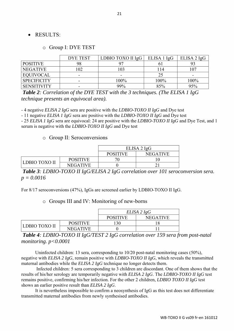
o Group I: DYE TEST
DYE TEST LDBIO TOXO II IgG ELISA 1 IgG ELISA 2 IgG POSITIVE 98 97 61 93 NEGATIVE 102 103 114 107 EQUIVOCAL - - 25 - SPECIFICITY - 100% 100% 100% SENSITIVITY - 99% 85% 95% Table 2: Correlation of the DYE TEST with the 3 techniques. (The ELISA 1 IgG technique presents an equivocal area). - 4 negative ELISA 2 IgG sera are positive with the LDBIO-TOXO II IgG and Dye test - 11 negative ELISA 1 IgG sera are positive with the LDBIO-TOXO II IgG and Dye test - 25 ELISA 1 IgG sera are equivocal: 24 are positive with the LDBIO-TOXO II IgG and Dye Test, and 1 serum is negative with the LDBIO-TOXO II IgG and Dye test
o Group II: Seroconversions
ELISA 2 IgG POSITIVE NEGATIVE
LDBIO TOXO II POSITIVE 70 10
NEGATIVE 0 21 Table 3: LDBIO-TOXO II IgG/ELISA 2 IgG correlation over 101 seroconversion sera. p = 0.0016 For 8/17 seroconversions (47%), IgGs are screened earlier by LDBIO-TOXO II IgG.
o Groups III and IV: Monitoring of new-borns
ELISA 2 IgG POSITIVE NEGATIVE
LDBIO TOXO II POSITIVE 130 18
NEGATIVE 0 11 Table 4: LDBIO-TOXO II IgG/TEST 2 IgG correlation over 159 sera from post-natal monitoring. p<0.0001
Uninfected children: 13 sera, corresponding to 10/20 post-natal monitoring cases (50%), negative with ELISA 2 IgG, remain positive with LDBIO-TOXO II IgG, which reveals the transmitted maternal antibodies while the ELISA 2 IgG technique no longer detects them.
Infected children: 5 sera corresponding to 3 children are discordant. One of them shows that the results of his/her serology are temporarily negative with ELISA 2 IgG. The LDBIO-TOXO II IgG test remains positive, confirming his/her infection. For the other 2 children, LDBIO TOXO II IgG test shows an earlier positive result than ELISA 2 IgG.
It is nevertheless impossible to confirm a neosynthesis of IgG as this test does not differentiate transmitted maternal antibodies from newly synthesised antibodies.

22
WB‐TOXO II G vs09 fr‐en 161012
o Group V: Sensitivity and Specificity (malaria and viral infections)
ELISA 2 IgG POSITIVE NEGATIVE
LDBIO TOXO II + DYE TEST
POSITIVE 42 2 NEGATIVE 2 23
Table 5: LDBIO-TOXO II IgG/DYE TEST/ELISA 2 IgG correlation over 69 malaria or viral infection sera.
In this population, there is 100% agreement between LDBIO-TOXO II IgG IgG and DYE TEST: these results confirm the specificity and sensitivity of the LDBIO-TOXO II IgG test.
The study reveals 4 discordant results with the ELISA 2 IgG technique, 2 false negatives (1 HIV and 1 P. falciparum) and 2 false positives (2 P. falciparum), emphasising the utility of a confirmation technique for all the results that are close to the threshold.
CONCLUSION:
o Group I (DYE TEST):
The LDBIO-TOXO II IgG/DYE TEST correlation is excellent (99% sensitivity, 100% specificity). LDBIO-TOXO II IgG could confirm the immune status of patients presenting an equivocal result or a low antibody titre in the screening.
o Group II (seroconversions):
The sensitivity of LDBIO-TOXO II IgG is greater than that of ELISA 2 IgG (p = 0.0016). LDBIO-TOXO II IgG could confirm a seroconversion earlier than ELISA 2 IgG.
o Group III and IV (monitoring of new-borns):
the sensitivity of LDBIO-TOXO IgG is much greater than that of ELISA 2 IgG (p < 0.0001). When monitoring children, LDBIO-TOXO II IgG could be used to confirm or set aside negative serology results. Nevertheless, LDBIO-TOXO II IgG does not differentiate transmitted maternal antibodies from antibodies that are newly synthesised by the child.
o Group V (malaria and viral infect.):
The LDBIO-TOXO II IgG/DYE TEST correlation is excellent (100% sensitivity and 100% specificity). These results demonstrate the need to use a confirmation assay to test the samples presenting a result in the screening that is close to the threshold.

23
WB‐TOXO II G vs09 fr‐en 161012
The excellent performance of the LDBIO-TOXO II IgG kit justifies its use in confirming the results obtained by the IgG screening techniques (results that are equivocal, slightly positive or that pose problems of interpretation).
Reproducibility:
Inter-series and inter-lot reproducibility were tested. In both cases, the serum to serum correlation with respect to specific bands is excellent.
Interferences:
Even though no particular cross-reaction has been observed with haemolysed, icteric or lipidic sera, it is recommended to interpret the results from the use of such samples with care.
Trouble shooting
"The bands are pale with little contrast": Certain sera with low concentrations of antibodies may give such results.
"Shaded areas can be seen, more or less coloured, slightly diffuse": The strip was not totally submerged in one of the reagents and did not incubate correctly along its entire length. Stains can also be present where the sample was deposited if the tray was not shaken after dispensing. "The background noise is significant, making reading very difficult": The washes were insufficient or the last incubation was too long. Ensure good test performance techniques, respect wash times and ensure water quality. Reduce the time of the last incubation. Exceptionally, certain sera may react in a non-specific manner. Then, the result of the immunoblot cannot be used. This non-specific background noise may involve only part of the strip, making the results uninterpretable for that part only. "A precipitate appears in the solution during the last step of development": the substrate may in fact partially precipitate (black flakes) in the buffer at the end of development. This phenomenon does not alter the quality of the development which must be continued normally. The last wash with distilled water eliminates the possible solid particles present.

24
WB‐TOXO II G vs09 fr‐en 161012
Bibliography
Franck, Jacqueline, Yves Jean-François Garin, et Henri Dumon. « LDBio-Toxo II immunoglobulin G Western blot confirmatory test for anti-toxoplasma antibody detection ». Journal of clinical microbiology 46, no 7 (juillet 2008): 2334‑38. doi:10.1128/JCM.00182-08.
Jost, C, F Touafek, A Fekkar, R Courtin, M Ribeiro, D Mazier, et L Paris. « Utility of immunoblotting for early diagnosis of toxoplasmosis seroconversion in pregnant women ». Clinical and vaccine immunology: CVI 18, no 11 (novembre 2011): 1908‑12. doi:10.1128/CVI.05303-11.
Khammari, Imen, Fatma Saghrouni, Sami Lakhal, Aida Bouratbine, Moncef Ben Said, et Jalel Boukadida. « A New IgG Immunoblot Kit for Diagnosis of Toxoplasmosis in Pregnant Women ». The Korean Journal of Parasitology 52, no 5 (22 octobre 2014): 493‑99. doi:10.3347/kjp.2014.52.5.493.
Khammari, Imen, Fatma Saghrouni, Alia Yaacoub, Sondoss Gaied Meksi, Hinda Ach, Lamia Garma, Akila Fathallah, et Moncef Ben Saïd. « IgG Western Blot for Confirmatory Diagnosis of Equivocal Cases of Toxoplasmosis by EIA-IgG and Fluorescent Antibody Test ». The Korean Journal of Parasitology 51, no 4 (août 2013): 485‑88. doi:10.3347/kjp.2013.51.4.485.
Leslé, F, F Touafek, A Fekkar, D Mazier, et L Paris. « Discrepancies between a new highly sensitive Toxoplasma gondii ELISA assay and other reagents: interest of Toxo IgG Western blot ». European journal of clinical microbiology & infectious diseases: official publication of the European Society of Clinical Microbiology 30, no 10 (octobre 2011): 1207‑12. doi:10.1007/s10096-011-1214-1.
Maudry, A., G. Chene, R. Chatelain, B. Bellete, H. Patural, J. Hafid, H. Raberin, R. Tran Manh Sung, et P. Flori. « Expertise du nouveau test Access® TOXO-IgGII et comparaison avec trois autres techniques automatisées et la technique Western Blot LDBIO TOXO II IgG® ». Immuno-analyse & Biologie Spécialisée 24, no 1 (février 2009): 42‑49. doi:10.1016/j.immbio.2008.11.004.
Maudry, Arnaud, Gautier Chene, Rémi Chatelain, Hugues Patural, Bahrie Bellete, Bernard Tisseur, Jamal Hafid, et al. « Bicentric evaluation of six anti-toxoplasma immunoglobulin G (IgG) automated immunoassays and comparison to the Toxo II IgG Western blot ». Clinical and vaccine immunology: CVI 16, no 9 (septembre 2009): 1322‑26. doi:10.1128/CVI.00128-09.
Robert-Gangneux, F., et M.-L. Darde. « Epidemiology of and Diagnostic Strategies for Toxoplasmosis ». Clinical Microbiology Reviews 25, no 2 (1 avril 2012): 264‑96. doi:10.1128/CMR.05013-11.
Villard, O., B. Cimon, C. L’Ollivier, H. Fricker-Hidalgo, N. Godineau, S. Houze, L. Paris, H. Pelloux, I. Villena, et E. Candolfi. « Serological Diagnosis of Toxoplasma Gondii Infection: Recommendations from the French National Reference Center for Toxoplasmosis ». Diagnostic Microbiology and Infectious Disease, 18 septembre 2015. doi:10.1016/j.diagmicrobio.2015.09.009.
19A rue Louis Loucheur - 69009 LYON - FRANCE TEL : +33(0)4 7883 3487 - FAX : +33(0)4 7883 3430 www.ldbiodiagnostics.com - email : contac t@ldbiodiagnostics .com
NF EN ISO 13485 - ISO 9001


![NetApp OpenVMS Guide to Best Practices...à . ,à ˙ ˘ ˝ à! à˝ ˜'à à à ˝ $ % à˝ ! 'à à!à, à˜ 'à à à ˚ ˜ ˚ ˘˘ ˚˝ ) ; ]ˇ ˘ ˚ ! à](https://static.fdocuments.in/doc/165x107/5f05acf27e708231d414244f/netapp-openvms-guide-to-best-practices-oe-.jpg)