Lasting modulation of in-vitro oscillatory activity with weak direct...
20
Lasting modulation of in-vitro oscillatory activity with weak direct current stimulation Davide Reato, Marom Bikson, Lucas C. Parra Department of Biomedical Engineering The City College of the City University of New York, New York, USA December 3, 2014 Abstract Transcranial Direct Current Stimulation (tDCS) is emerging as a versatile tool to affect brain function. While acute neurophysiological effects of stimulation are well understood, little is know about the long term effects. One hypothesis is that stimulation modulates ongoing neural activity which then translates into lasting effects via physiological plasticity. Here we used carbachol-induced gamma oscillations in hippocampal rat slices to establish whether prolonged constant current stimulation has a lasting effect on endogenous neural activity. During 10 minutes of stimulation, power and frequency of gamma oscillations, as well as multi-unit activity were modulated in a polarity specific manner. Remarkably, the effects on power and multi-unit activity persisted for more than 10 minutes after stimulation terminated. Using a computational model we propose that altered synaptic efficacy in excitatory and inhibitory pathways could be the source of these lasting effects. Future experimental studies using this novel in-vitro preparation may be able to confirm or refute the proposed hypothesis. Introduction The number of studies on transcranial direct current stimulation (tDCS) has rapidly increased in recent years (Brunoni et al., 2012). Human studies have shown improvements in behavioral and cognitive performances after transcranial stimulation (Foerster et al., 2012; Javadi et al., 2011). Pharmacological interventions in human studies point to possible synaptic changes as well as changes in neuromodulator release as the cause for the after-effects of the stimulation (Nitsche and Paulus, 2000; Nitsche et al., 2003) (for a review, Stagg and Nitsche, 2011). A few animal studies have also shown effects that outlast the stimulation period using evoked response (M´ arquez-Ruiz et al., 2012; Cambiaghi et al., 2010; 1
Transcript of Lasting modulation of in-vitro oscillatory activity with weak direct...
Lasting modulation of in-vitro oscillatory activity with weak direct current
stimulation
Davide Reato, Marom Bikson, Lucas C. Parra
Department of Biomedical Engineering
The City College of the City University of New York, New York, USA
December 3, 2014
Abstract
Transcranial Direct Current Stimulation (tDCS) is emerging as a versatile tool to affect brain function. While acute
neurophysiological effects of stimulation are well understood, little is know about the long term effects. One hypothesis is
that stimulation modulates ongoing neural activity which then translates into lasting effects via physiological plasticity.
Here we used carbachol-induced gamma oscillations in hippocampal rat slices to establish whether prolonged constant
current stimulation has a lasting effect on endogenous neural activity. During 10 minutes of stimulation, power and
frequency of gamma oscillations, as well as multi-unit activity were modulated in a polarity specific manner. Remarkably,
the effects on power and multi-unit activity persisted for more than 10 minutes after stimulation terminated. Using a
computational model we propose that altered synaptic efficacy in excitatory and inhibitory pathways could be the source
of these lasting effects. Future experimental studies using this novel in-vitro preparation may be able to confirm or refute
the proposed hypothesis.
Introduction
The number of studies on transcranial direct current stimulation (tDCS) has rapidly increased in recent years (Brunoni
et al., 2012). Human studies have shown improvements in behavioral and cognitive performances after transcranial
stimulation (Foerster et al., 2012; Javadi et al., 2011). Pharmacological interventions in human studies point to possible
synaptic changes as well as changes in neuromodulator release as the cause for the after-effects of the stimulation (Nitsche
and Paulus, 2000; Nitsche et al., 2003) (for a review, Stagg and Nitsche, 2011). A few animal studies have also shown
effects that outlast the stimulation period using evoked response (Marquez-Ruiz et al., 2012; Cambiaghi et al., 2010;
1

Fritsch et al., 2010; Ranieri et al., 2012; Gartside, 1968). However, how these results may relate to the effects measured
in human studies is not fully understood.
In recent years, brain oscillations – rhythmic neuronal activity reflecting coherent spiking – have become a target for
transcranial electrical stimulation (Herrmann et al., 2013). Gamma oscillations in the range of 25-100 Hz, for example, are
ubiquitous in the brain (Buzsaki and Draguhn, 2004), play a role in neuronal coding (Fries et al., 2007; Wang, 2010) and
have been associated with attention and memory in humans (Jensen et al., 2007). Gamma rhythms in the 30 Hz range
synchronize hippocampal activity during memory replay (Carr et al., 2012) and reduced gamma power leads to impaired
spatial working memory and exploratory behavior (Fuchs et al., 2007). Therefore, modulation of gamma rhythms with
weak currents could potentially affect brain function. While many human studies used transcranial alternating current
stimulation (tACS), only few considered how DC stimulation could affect brain oscillations (Antal et al., 2004; Polanıa et
al., 2011).
The acute effects of DC stimulation on single neurons and networks of neurons have been extensively characterized in
animals (Bikson et al., 2004; Frohlich and McCormick, 2010; Reato et al., 2010). However, no animal studies have shown
lasting effects of DC stimulation on brain oscillations. Here, we present an in-vitro model of gamma oscillations and DC
stimulation that shows lasting effects of stimulation.
Oscillations in the low-gamma frequency range can be reliably induced in hippocampal slices using carbachol, a
cholinergic agonist (Fisahn et al., 1998). We have shown previously that weak electric fields can modulate the magnitude
of these in-vitro oscillations acutely in an amplitude and frequency specific manner (Reato et al., 2010). Here we use
the same slice model and report that modulation of gamma oscillations with weak constant electric fields applied for
a prolonged time (10 minutes) outlasts the period of stimulation. Based on simulations with a previously validated
computational model we propose that the after-effects of stimulation are the result of balanced changes in excitatory and
inhibitory synaptic strength.
Materials and Methods
Hippocampal slice recordings: Slice preparation and recordings from male 3-5 weeks old Wistar rats (CCNY-IACUC,
protocol 0846) followed the procedures of (Reato et al., 2010). Extracellular recordings were performed using 3 electrodes
per slice placed in the CA3c stratum pyramidale area of the hippocampus at the distance of ∼ 250 µm. Perfusion with
carbachol (20 µM) started 5 minutes after placing the glass pipettes and the recordings lasted for the following 2 hours
without moving the electrodes. Recordings were aligned in time across slices using the beginning of carbachol perfusion.
Electrical field stimulation: Spatially-uniform electric fields were applied to slices with varying amplitudes by
passing current between two parallel Ag-AgCl wires placed in the ACSF across the slice (Gluckman et al., 1996; Bikson
et al., 2004). All experimental results are reported as a function of this electric field magnitude. Slices were aligned
2
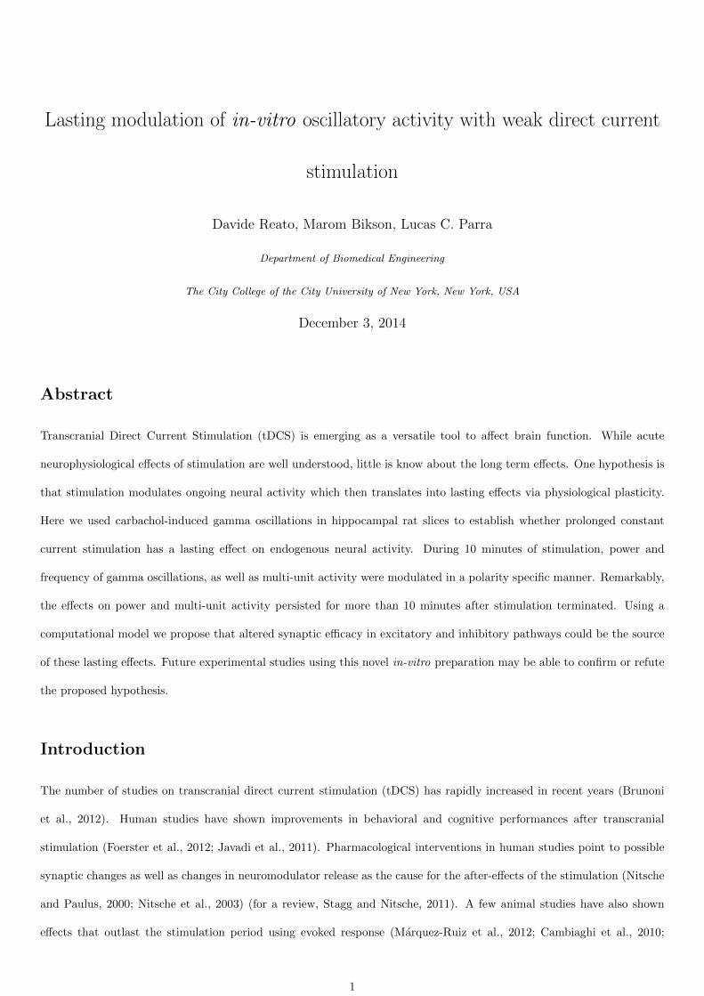
in the chamber such that the induced uniform electric field was parallel to CA3c pyramidal neurons (Fig. 1A). Before
every recording, electric fields were calibrated by passing current through the field wires and measuring the corresponding
voltages between them (representative voltage measurements in Fig. 1A). Note that a linear voltage indicates a uniform
value of the electric field. “Positive” (anodal) field polarity was defined as the positive electrode on the CA1 alveus side,
and “negative” (cathodal) field polarity was defined as negative on the CA1 alveus side (Bikson et al., 2004). Weak positive
field stimulation is thus typically expected to depolarize CA3 pyramidal cell somata, while weak negative field stimulation
should hyperpolarize CA3 somata (Deans et al., 2007). DC stimulation commenced 60 minutes after the recordings started
(55 minutes after application of carbachol). During this time, the oscillations largely stabilized in power and frequency,
although a continued drift in power is often evident (Fig. 1C). Stimulation was applied for 10 minutes with amplitudes of
-20 V/m (n = 6), -10 V/m (n = 24), 0 V/m (control, n = 26), +10 V/m (n = 23) and +20 V/m (n = 8). Each slice was
stimulated with a single stimulation intensity.
Power and frequency analysis: Gamma power and frequency were estimated using multitaper spectral analysis.
Power was computed in 1 minute segments using the Chronux toolbox (http://chronux.org/, Mitra and Bokil, 2008) with
a time-bandwidth product of WT = 2 and using 3 tapers. The frequency range of carbachol-induced activity was then
detected semi-automatically for each slice (location of the peak ±2σ but manually reduced to exclude, if present, electric
noise contaminating that frequency range) and mean-power was calculated for that frequency range. Gamma power was
averaged across electrodes provided it was at least 5 dB above noise as compared to the first 5 minutes of recording.
To compare across slices, these mean values were normalized by the pre-stimulation gamma power (50-60 min). Gamma
peak-frequency was estimated from the multitaper analysis for each 1 minute segment by considering the location of the
peak-power in the gamma band.
Multi-unit activity detection: Multi-unit activity (MUA) was detected by thresholding the extracellular recordings
after high-pass filtering (300 Hz cut-off frequency). The value of the threshold for automatic units detection was set to
7 ·median(|x|
0.6745
)(Quiroga et al., 2004) where x was the high-pass filtered extracellular signal during the first 5 minutes
of recording (before carbachol perfusion). A 1 ms dead time for detection was used. Our main results do not depend
strongly on the specific threshold for MUA detection. A high threshold for detection, as we used here, was chosen to
decrease false positive detections possibly due to electric artifacts (note that in this study we used conventional electrodes
for LFP recordings). This simple method allowed to easily estimate MUA changes in our different stimulation conditions.
Coherence between the candidate units and the extracellular local field potentials (LFP) was estimated using Chronux
(Mitra and Bokil, 2008) (http://chronux.org/). Only electrodes that showed a strong unit-to-field-potential coherence
(> 0.3) in our frequencies of interest were considered for further analysis. When multiple electrodes detected MUA,
the frequency of events was averaged across these electrodes in 1 minute temporal windows. To compare across slices,
estimated average rates (events per second) before the stimulation (50-60 min) where subtracted from each trace. Note
3

that since the recordings started before the emergence of coherent activity in the slice and the electrodes were not moved
throughout the experiment, a good MUA signal (high coherence with the LFP) was not always detected. Therefore, the
number of slices for LFP and MUA analysis differ in the Results section.
Sensitivity of power and multi-unit activity to electrical stimulation: We measured the sensitivity of the
network oscillatory power to the applied field. In equations, ∆P = gpE, where gp represents how much power changes
(in dB) per V/m electric field applied. MUA modulation follows a similar equation, ∆R = grE, where gr indicates how
many Hz the estimated rate changes with stimulation intensity. These sensitivities were estimated with a linear fit as a
function of the stimulation intensities using all available slices. Non-parametric statistics were obtained by randomizing
the stimulation amplitudes and performing the linear fits to this random data. p-values were then computed using these
shuffled statistics. Statistical tests were performed for the 10 minutes of stimulation and the subsequent 10 minutes.
This was based on previous literature showing that excitability changes in motor cortex outlast the stimulation period for
durations comparable to the stimulation period (5 minutes in (Nitsche and Paulus, 2000), and 20 minutes in (Nitsche and
Paulus, 2001).
Computational model: Modeling of gamma oscillations induced by carbachol and its response to electric stimulation
follows the methods of (Reato et al., 2010). Briefly, the voltage behavior of single neurons is captured by Izhikevich’s single-
compartment neuron model (Izhikevich, 2003). Here the network consists of 800 excitatory and 200 inhibitory neurons
synaptically connected with all-to-all connections and 40% sparseness. Gamma oscillations in the model are generated by
the interplay of increased excitation (simulating the effect of carbachol, Fisahn et al., 2002) and fast inhibitory feedback
(Bartos et al., 2007). Weak electrical stimulation was implemented as a low-pass filtered current that polarizes all pyramidal
neurons, according to experimental data (Deans et al., 2007). The parameters of the models are set such that 1 V/m
electric field induces a polarization of about 0.1 mV, consistent with previous studies (Radman et al., 2007; Deans et al.,
2007; Bikson et al., 2004; Frohlich and McCormick, 2010).
Here we tested how changes in the strength of synaptic connections affect power, frequency and firing rate of excitatory
neurons during simulated gamma oscillations. The form of synaptic connections in our model is wxx = wxx + [0, kxxwxx]
if the connection is excitatory or wxx = wxx + [kxxwxx, 0] if inhibitory, and where xx = {ee,ei,ie,ii} indicates the type
of connection (excitatory to excitatory, excitatory to inhibitory, inhibitory to excitatory, inhibitory to inhibitory). wxx
represents the baseline value of the connection, wxx the maximum value of the uniform distribution (from 0 to wxx) and
kxx is a parameter that has been changed here to simulate changes in synaptic connections. As in Reato et al., 2010, we
used here wee = wei = 0, wie = −0.8, wii = −0.3 and wee = 0.65, wei = 2, wie = −0.9, wii = −0.8. Throughout the
text, the expression “balanced excitation/inhibition” indicate an equal level of excitatory and inhibitory inputs on single
neurons during the gamma cycle. This definition is based on previous literature indicating per-neuron balanced excitatory
and inhibitory current during gamma oscillations (Atallah and Scanziani, 2009) and slow-waves (Haider et al., 2006; Shu
4
et al., 2003).
Results
Extracellular recordings were performed with multiple electrodes located in the CA3c region of rat hippocampal slices (n
= 87, Fig. 1A). Carbachol was perfused continuously beginning 5 minutes after the start of recording. Carbachol induced
strong gamma oscillations (25-35 Hz, 5-25 dB over noise) that emerge ∼20 minutes after starting the perfusion, consistent
with other studies (Colgin et al., 2003). In the average across slices, the oscillations became relatively stable in power and
frequency after ∼60 minutes (average spectrogram in Fig. 1B, n = 26). Gamma power and multi-unit activity (MUA)
were measured over the whole duration of the recordings (2 hours) and were normalized by their average value before
the stimulation. Gamma power and MUA were not statistically different across the five stimulation conditions before
the stimulation (ANOVA, n = 87, p = 0.61 for power and n = 39, p = 0.77 for MUA). 55 minutes after the start of
carbachol perfusion, slices were electrically stimulated with constant electric fields for 10 minutes. For each slice, only one
stimulation intensity was used: -20 V/m (n = 6), -10 V/m (n = 24), 0 V/m (n = 26), +10 V/m (n = 23), +20 V/m (n
= 8). Figs. 1C-D show average traces of gamma power and MUA in the different stimulation conditions (the stimulation
starts at 0 minutes). The significant variability observed over the recording period for individual traces required recording
of a large number of slices (n=87).
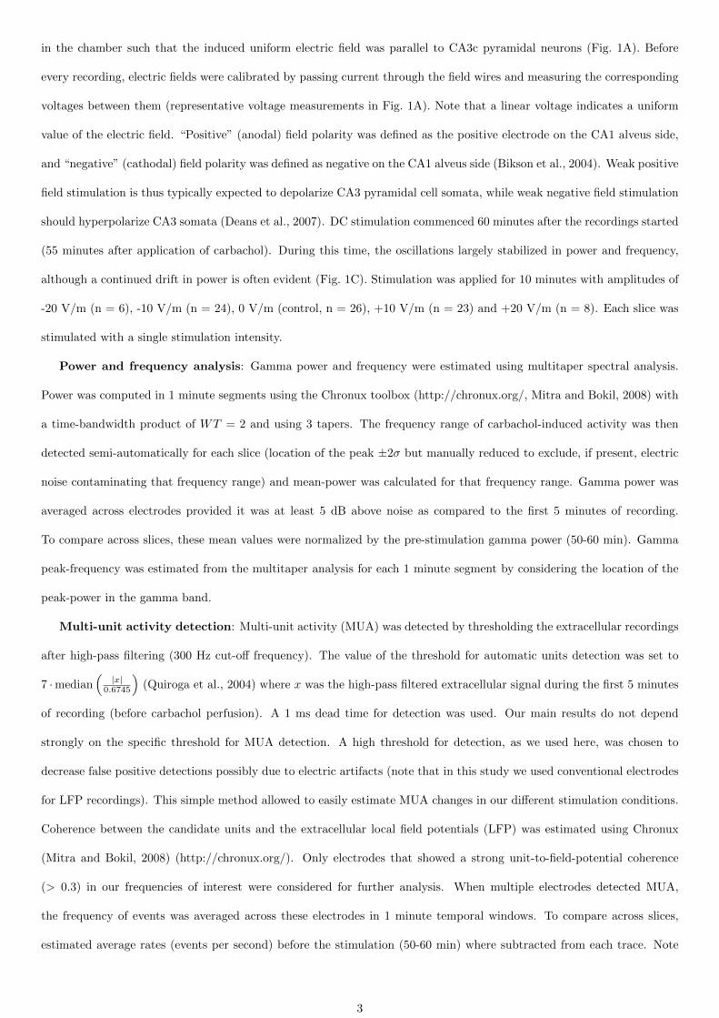
We tested whether electric fields modulated gamma oscillations and MUA in an intensity-dependent manner. We
grouped the data from all the slices (n = 87 for power, and n = 39 for MUA) and performed a linear regression as a
function of stimulation intensity for the 10 minute interval during the stimulation (acute effects) and after stimulation
(persisting effects). Combining data from all stimulation conditions was necessary in order to average out the strong
fluctuation observed over time and across slices. The first minute in both intervals was excluded from the analysis to avoid
possible transients or artifacts resulting from turning the stimulator on/off. Gamma power was significantly modulated
during the stimulation (n = 87, estimated slope of the linear fit, gp = (0.03± 0.01) dB/(V/m), p = 0.001, Fig. 2A left)
and a similar effect was also measured for MUA (n=39, estimated slope of the linear fit, gr = (0.11± 0.03) Hz/(V/m),
p=3·10−6, Fig. 2B left). The positive offset of the regression lines reflect the continuous strengthening of gamma oscillations
even 55 minute after carbachol perfusion. A positive slope implies that both gamma power and MUA are higher when
positive (anodal) electric fields are applied and lower for negative (cathodal) fields. Importantly, the effects outlasted the
stimulation in the subsequent 10 minutes for both power (n=87, estimated slope of the linear fit, gp = (0.02± 0.01) dB /
(V/m), p = 0.02, Fig. 2A right) and MUA (n = 39, estimated slope of the linear fit, gr = (0.10± 0.03) Hz/(V/m), p=0.001,
Fig. 2B right). The same analysis performed on frequency changes did not reveal any significant effects of fields (p=0.1
during and 0.3 after stimulation). To determine the exact progression of power and MUA changes, we then estimated
power and MUA sensitivity to the electric field (considering data from all the slices, as previously done for Fig. 2A-B)
5
resolved in 1 minute segments (Fig. 2C-D). Both power and MUA sensitivity continuously increased during stimulation
(dark gray shading) and then decayed after the end of the stimulation (light gray shading). The increasing confidence
intervals reflect the substantial variability of the gamma oscillations (light gray area, 5% and 95% estimated by shuffle
statistics). The time courses of power and MUA are strongly correlated (r2 = 0.65). Taken together, these results show
that weak electrical stimulation can affect gamma oscillations and MUA and that the effects outlast the stimulation for
at least 10 min.
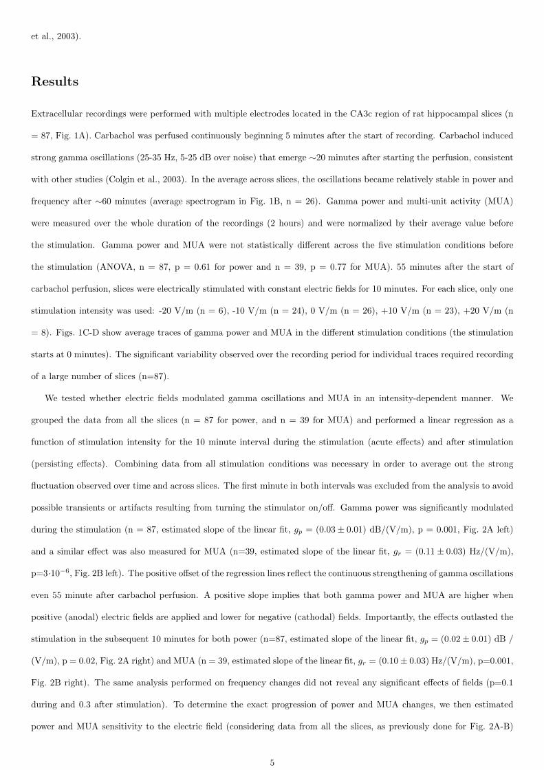
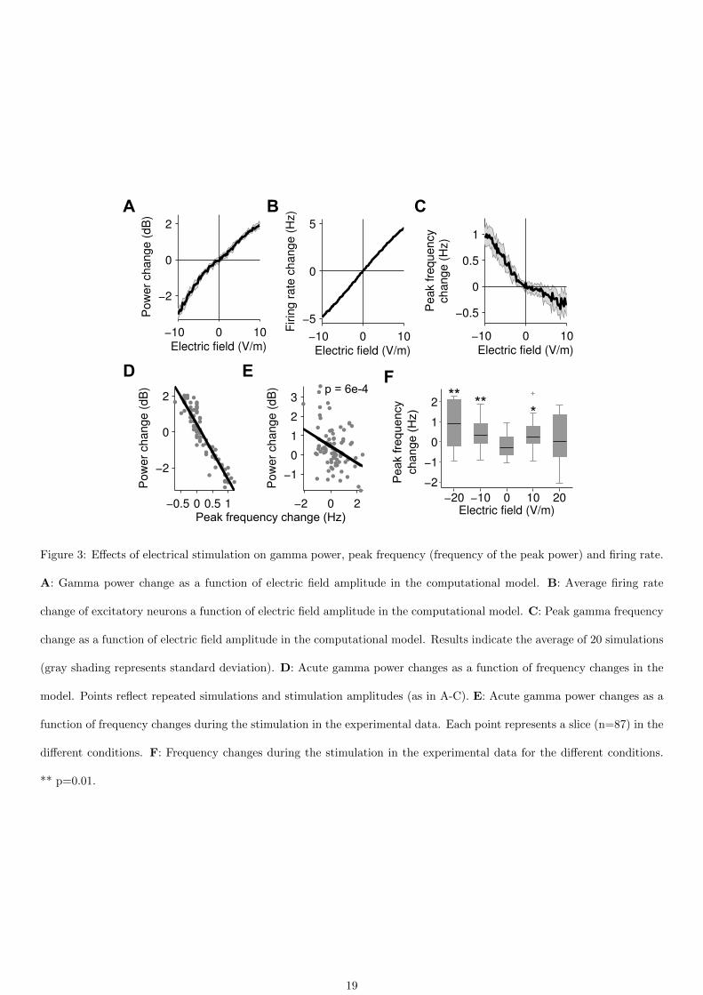
Next, we tried to determine possible causes for these lasting effects using an experimentally-validated computational
model for carbachol-induced gamma oscillations and their response to electric field stimulation (Reato et al., 2010). In the
model, increasing field magnitudes leads to a monotonic increase of gamma power and firing rate (Fig. 3A-B). Interestingly,
increasing field intensity also leads to a monotonic decrease in gamma frequency (Fig. 3C) that correlates with changes in
gamma-power (r2 = 0.85, Fig. 3D). This relationship was confirmed with our present in-vitro data during the stimulation
(Fig. 3E, n=87, p=6 ·10−4) and is consistent with previous data showing that a balance between excitation and inhibition
is the cause of this linear relationship (Atallah and Scanziani, 2009). In the computational model the relationship between
electric field magnitude and gamma frequency is non-linear (Fig. 3C). Thus, we analyzed the changes in gamma peak-
frequency in the in-vitro data separately for the different stimulation conditions and find significant effects in particular
for negative field stimulation (Fig. 3F). To summarize, our experimental results showed acute effects for gamma power,
frequency and MUA, pointing to a balanced modulation of excitatory and inhibitory activity, yet persistent effects were
only evident for oscillatory power and MUA.
We then used the computational model to investigate whether the observed lasting changes could be explained by
changes in synaptic strength, notably a change in gamma power and MUA but not frequency. We focused on synaptic
changes, as they are often assumed to underlie the persistent effects observed in human studies (see Discussion). We
modulated the strength of excitatory-to-excitatory (e→e) synaptic connections, as well as the strength of the inhibitory
feedback (excitatory-to-inhibitory, e→i, or inhibitory-to-excitatory, i→e, synaptic strength). Modulation of inhibitory
synaptic strength was motivated by recent evidence for plasticity in inhibitory pathways (Kullmann et al., 2012). Mod-
ulating the strength of e→e synapses in the model did not strongly modulate the power of the oscillations (Fig. 4A-D),
while changing e→i or i→e synapses strongly modulated gamma power (Fig. 4A,D). Both phenomena have previously
been observed experimentally for endogenous neocortical gamma activity (Morita et al., 2008; Sohal et al., 2009). This
provides further confidence in the present computational model. Here gamma frequency depends on both e→e and e→i
connections but less on i→e (Fig. 4B-E). Firing rate is more sensitive to changes in e→e connections (Fig. 4C-F). Points
in this parameter space that are consistent with the present experimental observation are indicated with an “0” for sham
stimulation and “+”, “-” for positive and negative field stimulation. Along these diagonals, power and firing-rate change,
but not oscillation frequency. Therefore, the computational results suggest that the observed lasting changes may be
6

explained by a lasting modulation of excitation matched by a corresponding change in inhibitory feedback.
Discussion
Transcranial electrical stimulation is a versatile tool to modulate brain activity (Nitsche and Paulus, 2000; Fregni et al.,
2006; Fecteau et al., 2007; Fridriksson et al., 2011). In-vivo and in-vitro studies have demonstrated that electric fields,
whose amplitude is comparable to the one expected in tDCS, can modulate firing rate (Chan and Nicholson, 1986), spike
timing (Radman et al., 2007) and the magnitude of synaptic responses (Kabakov et al., 2012; Rahman et al., 2013). We
have previously shown that acute effects of weak electrical stimulation can be amplified during endogenous oscillatory
activity (Reato et al., 2010, 2013). These results suggest that brain oscillations may be a sensitive target for transcranial
electrical stimulation with constant currents. Previous in-vitro and in-vivo studies have only shown acute effects of
stimulation on oscillatory activity (Reato et al., 2010; Frohlich and McCormick, 2010; Ali et al., 2013; Ozen et al., 2010)
and there are few reports on longer term effects in human studies (see Antal et al., 2004; Polanıa et al., 2011). The majority
of studies on oscillatory activity have used alternating current stimulation with the goal of enhancing brain oscillations
(Marshall et al., 2006; Pogosyan et al., 2009; Kirov et al., 2009; Zaehle et al., 2010; Santarnecchi et al., 2013; Helfrich et
al., 2014).
Here we found that weak constant current electrical stimulation applied for a longer period of time can induce lasting
effects, measurable as altered gamma power and multi-unit activity. These lasting effects cannot be explained as persistent
network activity in the absence of some adaptive process since in our previous work gamma power returned to baseline
activity within 100 ms after short-lasting DC field stimulation (Reato et al., 2010). Importantly, the after-stimulation effect
was consistent with the acute effect, reminiscent of Hebbian or activity-dependent plasticity and contrary to homeostatic
plasticity (Fricke et al., 2011; Reato et al., 2013).
The field intensities used in this study are above those predicted to occur during tDCS, estimated to be maximum
1 V/m using conventional electrode montages (Datta et al., 2009; Ozen et al., 2010). These currents only induce a small
polarization of the membrane (maximum 0.2 mV per V/m), which cannot lead to action potentials in quiescent neurons
(Bikson et al., 2004). Previous in-vitro studies have shown that for such low intensity fields (subthreshold), most of the
acute effects scale linearly with the change in field amplitude (Bikson et al., 2004; Deans et al., 2007; Reato et al., 2010),
including changes in synaptic response (∼1% per V/m applied; Rahman et al., 2013). Therefore, the sensitivities observed
here may also scale linearly with the field intensities. In this context we note that several factors may make the human
brain more susceptible to electric fields, including larger sensitivity of individual neurons (due to size; Radman et al.,
2009) and higher number of synaptic connections compared to our in-vitro preparation (sensitivity to fields may increase
with the number of synaptic inputs a neuron receives; Reato et al., 2013). Either way, our field amplitudes are still much
below those generated with transcranial magnetic stimulation (TMS; Pascual-Leone et al., 2002) or deep brain stimulation
7
(DBS; Perlmutter and Mink, 2006), estimated in the order of 100 V/m (Salinas et al., 2009).
To generate a hypothesis for the possible cause of the experimental results we turned to computational modeling.
The model we used matches key features of weak-field stimulation on carbachol-induced gamma oscillations (Reato et
al., 2010). Specifically, the model matches the firing properties of excitatory and inhibitory neurons and their timing
within the gamma cycle (Hajos et al., 2004; Oren et al., 2006). The model successfully predicts firing rate and spike
timing changes during AC or DC field stimulation in-vitro. Without further modifications, this model reproduced the
correlation observed in the present experiment between power and frequency changes due to DC stimulation. A variant of
this model also successfully predicted the effects of weak transcranial stimulation on slow-waves oscillations in-vivo (Ali et
al., 2013). More complex models that capture physiological details such as gap-junctions or the role of different neuronal
compartments (Tiesinga et al., 2001; Traub et al., 2000) were not necessary to replicated the relevant experimental findings.
Using the model, we focused on modulation of synaptic connections because tDCS is thought to modulate concentrations
of neurotransmitters and neuromodulators (Stagg and Nitsche, 2011; Nitsche et al., 2012), which in turn are known to
affect synaptic efficacy. Based on the computational model we hypothesize that in gamma-networks, weakly depolarizing
electric fields lead to a balanced increase of excitatory and inhibitory synaptic currents.
The lasting effects we measured experimentally could be mediated by a number of cellular mechanisms, which we
discuss below.
Brain-derived neurotrophic factor (BDNF): It has been shown in humans (Antal et al., 2010) and in-vitro
(Fritsch et al., 2010) have shown that the lasting effects of weak electrical stimulation can be mediated by BDNF release.
BDNF release is activity-dependent (Park and Poo, 2013) with a self-reinforcing feedback-loop involving acetylcholine
(Knipper et al., 1994). Thus, an acute increase in gamma activity due to electrical stimulation could be further enhanced
by increased BDNF release, and this enhancement should outlast stimulation because of the longer time scale of BDNF
release (Aicardi et al., 2004). Interestingly, BDNF affects both excitatory and inhibitory neurons (Park and Poo, 2013)
via the TrkB receptor, which has also been implicated in gamma activity (Zheng et al., 2011). Thus, any enhancing effect
resulting from increased BDNF release may strengthen both excitation and inhibitory feedback, as we have hypothesized
here.
Acetylcholine: Carbachol activates acetylcholine receptors leading to increased neuronal activity in hippocampus. It
is well established that acetylcholine can induce hippocampal plasticity (Drever et al., 2011; Galey et al., 1994; Markevich et
al., 1997; Fernandez de Sevilla et al., 2008). Indeed, carbachol alone can induce lasting effects on the acetylcholine receptors
(Auerbach and Segal, 1994) and can facilitate hippocampal LTP (Auerbach and Segal, 1996). Moreover, carbachol increases
network responsiveness to external stimuli in-vivo (Rodriguez et al., 2004; Rasmusson, 2000) and can induce lasting effects
on evoked responses (Rodriguez et al., 2004; Brocher et al., 1992), presumably by increasing precision of spike timing in the
network. Finally, long lasting effects on cortical activity can be induced when sensory stimulation is paired with activation
8

of cholinergic inputs from the basal forebrain in-vivo (Froemke et al., 2013). It is thus possible that the increased activity
due to electric fields in the presence of carbachol is translated also into increased carbachol-induced plasticity. Interestingly,
a recent in-vivo study reported reported that acetylcholine mediated-learning induces strengthening at both excitatory
and inhibitory synapses (Mitsushima et al., 2013), supporting our hypothesis that weak electrical stimulation may affect
both types of synapses.
Spike-timing dependent plasticity (STDP): The altered gamma activity with firing periods in the order of 10-30
ms may induce NMDA-mediated STDP (Wespatat et al., 2004). Thus, increased firing due to field stimulation could lead
to altered synaptic efficacies via STDP which outlast the period of stimulation.
Membrane excitability: Finally, stimulation may affect membrane excitability (non-synaptic, Ardolino et al., 2005).
For example, stimulation-induced slow changes in neuromodulator release could lead to slow changes in neuronal mem-
brane properties giving rise to changes in the population dynamics and the studied after-effects (Augustin et al., 2013).
Considering the nature of gamma oscillations in hippocampus, increase/decrease in excitability of excitatory neurons (the
most affected by electrical stimulation, Radman et al., 2009) could also lead to a balanced increase/decrease of inhibitory
feedback.
In all instances, we propose that stimulation acutely affects ongoing activity, which then leads to lasting effects via
endogenous plasticity mechanisms. We argue that using our slice preparation we will be able to test our specific hypothesis
that balanced synaptic changes mediate the effects of weak electric fields on gamma oscillations.
References
Aicardi G, Argilli E, Cappello S, Santi S, Riccio M, Thoenen H, Canossa M (2004) Induction of long-term potentiation
and depression is reflected by corresponding changes in secretion of endogenous brain-derived neurotrophic factor.
Proceedings of the National Academy of Sciences of the United States of America 101:15788–15792.
Ali MM, Sellers KK, Frohlich F (2013) Transcranial alternating current stimulation modulates large-scale cortical
network activity by network resonance. The Journal of neuroscience: the official journal of the Society for Neuro-
science 33:11262–11275.
Antal A, Chaieb L, Moliadze V, Monte-Silva K, Poreisz C, Thirugnanasambandam N, Nitsche MA, Shoukier M, Ludwig
H, Paulus W (2010) Brain-derived neurotrophic factor (BDNF) gene polymorphisms shape cortical plasticity in humans.
Brain stimulation 3:230–237.
Antal A, Varga ET, Kincses TZ, Nitsche MA, Paulus W (2004) Oscillatory brain activity and transcranial direct current
stimulation in humans. Neuroreport 15:1307–1310.
9
Ardolino G, Bossi B, Barbieri S, Priori A (2005) Non-synaptic mechanisms underlie the after-effects of cathodal transcu-
taneous direct current stimulation of the human brain. The Journal of Physiology 568:653–663.
Atallah BV, Scanziani M (2009) Instantaneous modulation of gamma oscillation frequency by balancing excitation with
inhibition. Neuron 62:566–577.
Auerbach JM, Segal M (1994) A novel cholinergic induction of long-term potentiation in rat hippocampus. Journal of
Neurophysiology 72:2034–2040.
Auerbach JM, Segal M (1996) Muscarinic receptors mediating depression and long-term potentiation in rat hippocampus.
The Journal of Physiology 492 ( Pt 2):479–493.
Augustin M, Ladenbauer J, Obermayer K (2013) How adaptation shapes spike rate oscillations in recurrent neuronal
networks. Frontiers in Computational Neuroscience 7:9.
Bartos M, Vida I, Jonas P (2007) Synaptic mechanisms of synchronized gamma oscillations in inhibitory interneuron
networks. Nature Reviews. Neuroscience 8:45–56.
Bikson M, Inoue M, Akiyama H, Deans JK, Fox JE, Miyakawa H, Jefferys JGR (2004) Effects of uniform extracellular
DC electric fields on excitability in rat hippocampal slices in vitro. The Journal of Physiology 557:175–190.
Brunoni AR, Nitsche MA, Bolognini N, Bikson M, Wagner T, Merabet L, Edwards DJ, Valero-Cabre A, Rotenberg A,
Pascual-Leone A, Ferrucci R, Priori A, Boggio PS, Fregni F (2012) Clinical research with transcranial direct current
stimulation (tDCS): challenges and future directions. Brain stimulation 5:175–195.
Brocher S, Artola A, Singer W (1992) Agonists of cholinergic and noradrenergic receptors facilitate synergistically the
induction of long-term potentiation in slices of rat visual cortex. Brain Research 573:27–36.
Buzsaki G, Draguhn A (2004) Neuronal oscillations in cortical networks. Science (New York, N.Y.) 304:1926–1929.
Cambiaghi M, Velikova S, Gonzalez-Rosa JJ, Cursi M, Comi G, Leocani L (2010) Brain transcranial direct current
stimulation modulates motor excitability in mice. The European journal of neuroscience 31:704–709.
Carr MF, Karlsson MP, Frank LM (2012) Transient slow gamma synchrony underlies hippocampal memory replay.
Neuron 75:700–713.
Chan CY, Nicholson C (1986) Modulation by applied electric fields of purkinje and stellate cell activity in the isolated
turtle cerebellum. The Journal of physiology 371:89–114.
Colgin LL, Kubota D, Lynch G (2003) Cholinergic plasticity in the hippocampus. Proceedings of the National Academy
of Sciences of the United States of America 100:2872–2877.
10

Datta A, Bansal V, Diaz J, Patel J, Reato D, Bikson M (2009) Gyri -precise head model of transcranial DC stimulation:
Improved spatial focality using a ring electrode versus conventional rectangular pad. Brain Stimulation 2:201–207.
Deans JK, Powell AD, Jefferys JGR (2007) Sensitivity of coherent oscillations in rat hippocampus to AC electric fields.
The Journal of Physiology 583:555–565.
Drever BD, Riedel G, Platt B (2011) The cholinergic system and hippocampal plasticity. Behavioural brain re-
search 221:505–514.
Fecteau S, Knoch D, Fregni F, Sultani N, Boggio P, Pascual-Leone A (2007) Diminishing risk-taking behavior by mod-
ulating activity in the prefrontal cortex: a direct current stimulation study. The Journal of neuroscience: the official
journal of the Society for Neuroscience 27:12500–12505.
Fernandez de Sevilla D, Nunez A, Borde M, Malinow R, Buno W (2008) Cholinergic-mediated IP3-receptor activation
induces long-lasting synaptic enhancement in CA1 pyramidal neurons. The Journal of Neuroscience: The Official
Journal of the Society for Neuroscience 28:1469–1478.
Fisahn A, Pike FG, Buhl EH, Paulsen O (1998) Cholinergic induction of network oscillations at 40 hz in the hippocampus
in vitro. Nature 394:186–189.
Fisahn A, Yamada M, Duttaroy A, Gan JW, Deng CX, McBain CJ, Wess J (2002) Muscarinic induction of hippocampal
gamma oscillations requires coupling of the m1 receptor to two mixed cation currents. Neuron 33:615–624.
Foerster A, Rocha S, Wiesiolek C, Chagas AP, Machado G, Silva E, Fregni F, Monte-Silva K (2012) Site-specific effects
of mental practice combined with transcranial direct current stimulation on motor learning. The European journal of
neuroscience .
Fregni F, Boggio PS, Lima MC, Ferreira MJL, Wagner T, Rigonatti SP, Castro AW, Souza DR, Riberto M, Freedman
SD, Nitsche MA, Pascual-Leone A (2006) A sham-controlled, phase II trial of transcranial direct current stimulation
for the treatment of central pain in traumatic spinal cord injury. Pain 122:197–209.
Fricke K, Seeber AA, Thirugnanasambandam N, Paulus W, Nitsche MA, Rothwell JC (2011) Time course of the induction
of homeostatic plasticity generated by repeated transcranial direct current stimulation of the human motor cortex.
Journal of neurophysiology 105:1141–1149.
Fridriksson J, Richardson JD, Baker JM, Rorden C (2011) Transcranial direct current stimulation improves naming reac-
tion time in fluent aphasia: a double-blind, sham-controlled study. Stroke; a journal of cerebral circulation 42:819–821.
Fries P, Nikolic D, Singer W (2007) The gamma cycle. Trends in neurosciences 30:309–316.
11

Fritsch B, Reis J, Martinowich K, Schambra HM, Ji Y, Cohen LG, Lu B (2010) Direct current stimulation promotes
BDNF-dependent synaptic plasticity: potential implications for motor learning. Neuron 66:198–204.
Froemke RC, Carcea I, Barker AJ, Yuan K, Seybold BA, Martins ARO, Zaika N, Bernstein H, Wachs M, Levis PA, Polley
DB, Merzenich MM, Schreiner CE (2013) Long-term modification of cortical synapses improves sensory perception.
Nature Neuroscience 16:79–88.
Frohlich F, McCormick DA (2010) Endogenous electric fields may guide neocortical network activity. Neuron 67:129–143.
Fuchs EC, Zivkovic AR, Cunningham MO, Middleton S, Lebeau FEN, Bannerman DM, Rozov A, Whittington MA,
Traub RD, Rawlins JNP, Monyer H (2007) Recruitment of parvalbumin-positive interneurons determines hippocampal
function and associated behavior. Neuron 53:591–604.
Galey D, Destrade C, Jaffard R (1994) Relationships between septo-hippocampal cholinergic activation and the improve-
ment of long-term retention produced by medial septal electrical stimulation in two inbred strains of mice. Behavioural
Brain Research 60:183–189.
Gartside IB (1968) Mechanisms of sustained increases of firing rate of neurones in the rat cerebral cortex after polarization:
reverberating circuits or modification of synaptic conductance? Nature 220:382–3.
Gluckman BJ, Neel EJ, Netoff TI, Ditto WL, Spano ML, Schiff SJ (1996) Electric field suppression of epileptiform activity
in hippocampal slices. Journal of Neurophysiology 76:4202–4205.
Haider B, Duque A, Hasenstaub AR, McCormick DA (2006) Neocortical network activity in vivo is generated through
a dynamic balance of excitation and inhibition. The Journal of Neuroscience: The Official Journal of the Society for
Neuroscience 26:4535–4545.
Helfrich RF, Schneider TR, Rach S, Trautmann-Lengsfeld SA, Engel AK, Herrmann CS (2014) Entrainment of brain
oscillations by transcranial alternating current stimulation. Current biology: CB 24:333–339.
Herrmann CS, Rach S, Neuling T, Struber D (2013) Transcranial alternating current stimulation: a review of the
underlying mechanisms and modulation of cognitive processes. Frontiers in human neuroscience 7:279.
Hajos N, Palhalmi J, Mann EO, Nemeth B, Paulsen O, Freund TF (2004) Spike timing of distinct types of GABAergic
interneuron during hippocampal gamma oscillations in vitro. The Journal of Neuroscience: The Official Journal of the
Society for Neuroscience 24:9127–9137.
Izhikevich EM (2003) Simple model of spiking neurons. IEEE Transactions on Neural Networks / a Publication of the
IEEE Neural Networks Council 14:1569–1572.
12

Javadi AH, Cheng P, Walsh V (2011) Short duration transcranial direct current stimulation (tDCS) modulates verbal
memory. Brain stimulation .
Jensen O, Kaiser J, Lachaux JP (2007) Human gamma-frequency oscillations associated with attention and memory.
Trends in neurosciences 30:317–324.
Kabakov AY, Muller PA, Pascual-Leone A, Jensen FE, Rotenberg A (2012) Contribution of axonal orientation to pathway-
dependent modulation of excitatory transmission by direct current stimulation in isolated rat hippocampus. Journal of
neurophysiology 107:1881–1889.
Kirov R, Weiss C, Siebner HR, Born J, Marshall L (2009) Slow oscillation electrical brain stimulation during waking
promotes EEG theta activity and memory encoding. Proceedings of the National Academy of Sciences of the United
States of America 106:15460–15465.
Knipper M, da Penha Berzaghi M, Blochl A, Breer H, Thoenen H, Lindholm D (1994) Positive feedback between
acetylcholine and the neurotrophins nerve growth factor and brain-derived neurotrophic factor in the rat hippocampus.
The European journal of neuroscience 6:668–671.
Kullmann DM, Moreau AW, Bakiri Y, Nicholson E (2012) Plasticity of inhibition. Neuron 75:951–962.
Markevich V, Scorsa AM, Dawe GS, Stephenson JD (1997) Cholinergic facilitation and inhibition of long-term potentiation
of CA1 in the urethane-anaesthetized rats. Brain Research 754:95–102.
Marshall L, Helgadottir H, Molle M, Born J (2006) Boosting slow oscillations during sleep potentiates memory. Na-
ture 444:610–3.
Mitra P, Bokil H (2008) Observed brain dynamics Oxford University Press, Oxford; New York.
Mitsushima D, Sano A, Takahashi T (2013) A cholinergic trigger drives learning-induced plasticity at hippocampal
synapses. Nature Communications 4:2760.
Morita K, Kalra R, Aihara K, Robinson HPC (2008) Recurrent synaptic input and the timing of gamma-frequency-
modulated firing of pyramidal cells during neocortical ”UP” states. The Journal of neuroscience: the official journal of
the Society for Neuroscience 28:1871–1881.
Marquez-Ruiz J, Leal-Campanario R, Sanchez-Campusano R, Molaee-Ardekani B, Wendling F, Miranda PC, Ruffini G,
Gruart A, Delgado-Garcıa JM (2012) Transcranial direct-current stimulation modulates synaptic mechanisms involved
in associative learning in behaving rabbits. Proceedings of the National Academy of Sciences of the United States of
America 109:6710–6715.
13

Nitsche MA, Fricke K, Henschke U, Schlitterlau A, Liebetanz D, Lang N, Henning S, Tergau F, Paulus W (2003) Phar-
macological modulation of cortical excitability shifts induced by transcranial direct current stimulation in humans. The
Journal of physiology 553:293–301.
Nitsche MA, Paulus W (2000) Excitability changes induced in the human motor cortex by weak transcranial direct current
stimulation. The Journal of Physiology 527 Pt 3:633–639.
Nitsche MA, Paulus W (2001) Sustained excitability elevations induced by transcranial DC motor cortex stimulation in
humans. Neurology 57:1899–1901.
Nitsche MA, Muller-Dahlhaus F, Paulus W, Ziemann U (2012) The pharmacology of neuroplasticity induced by
non-invasive brain stimulation: building models for the clinical use of CNS active drugs. The Journal of Physiol-
ogy 590:4641–4662.
Oren I, Mann EO, Paulsen O, Hajos N (2006) Synaptic currents in anatomically identified CA3 neurons during hip-
pocampal gamma oscillations in vitro. The Journal of Neuroscience: The Official Journal of the Society for Neuro-
science 26:9923–9934.
Ozen S, Sirota A, Belluscio MA, Anastassiou CA, Stark E, Koch C, Buzsaki G (2010) Transcranial electric stimulation
entrains cortical neuronal populations in rats. The Journal of Neuroscience: The Official Journal of the Society for
Neuroscience 30:11476–11485.
Park H, Poo Mm (2013) Neurotrophin regulation of neural circuit development and function. Nature reviews. Neuro-
science 14:7–23.
Pascual-Leone A, Davey N, Rothwell J, Wasserman E, Puri BK (2002) Handbook of Transcranial Magnetic Stimulation
CRC Press, London : New York, NY, 1 edition edition.
Perlmutter JS, Mink JW (2006) Deep brain stimulation. Annual Review of Neuroscience 29:229–257.
Pogosyan A, Gaynor LD, Eusebio A, Brown P (2009) Boosting cortical activity at beta-band frequencies slows movement
in humans. Current biology: CB 19:1637–1641.
Polanıa R, Nitsche MA, Paulus W (2011) Modulating functional connectivity patterns and topological functional organi-
zation of the human brain with transcranial direct current stimulation. Human brain mapping 32:1236–1249.
Quiroga RQ, Nadasdy Z, Ben-Shaul Y (2004) Unsupervised spike detection and sorting with wavelets and superparam-
agnetic clustering. Neural computation 16:1661–1687.
Radman T, Ramos RL, Brumberg JC, Bikson M (2009) Role of cortical cell type and morphology in sub- and suprathresh-
old uniform electric field stimulation. Brain Stimulation 2:215–228.
14

Radman T, Su Y, An JH, Parra LC, Bikson M (2007) Spike timing amplifies the effect of electric fields on neurons:
implications for endogenous field effects. The Journal of Neuroscience: The Official Journal of the Society for Neuro-
science 27:3030–3036.
Rahman A, Reato D, Arlotti M, Gasca F, Datta A, Parra LC, Bikson M (2013) Cellular effects of acute direct current
stimulation: somatic and synaptic terminal effects. The Journal of physiology 591:2563–2578.
Ranieri F, Podda MV, Riccardi E, Frisullo G, Dileone M, Profice P, Pilato F, Di Lazzaro V, Grassi C (2012) Modulation
of LTP at rat hippocampal CA3-CA1 synapses by direct current stimulation. Journal of neurophysiology 107:1868–1880.
Rasmusson DD (2000) The role of acetylcholine in cortical synaptic plasticity. Behavioural Brain Research 115:205–218.
Reato D, Gasca F, Datta A, Bikson M, Marshall L, Parra LC (2013) Transcranial electrical stimulation accelerates human
sleep homeostasis. PLoS computational biology 9:e1002898.
Reato D, Rahman A, Bikson M, Parra LC (2010) Low-intensity electrical stimulation affects network dynamics by
modulating population rate and spike timing. The Journal of Neuroscience: The Official Journal of the Society for
Neuroscience 30:15067–15079.
Rodriguez R, Kallenbach U, Singer W, Munk MHJ (2004) Short- and long-term effects of cholinergic modulation on gamma
oscillations and response synchronization in the visual cortex. The Journal of Neuroscience: The Official Journal of
the Society for Neuroscience 24:10369–10378.
Salinas FS, Lancaster JL, Fox PT (2009) 3d modeling of the total electric field induced by transcranial magnetic stimulation
using the boundary element method. Physics in Medicine and Biology 54:3631–3647.
Santarnecchi E, Polizzotto NR, Godone M, Giovannelli F, Feurra M, Matzen L, Rossi A, Rossi S (2013) Frequency-
dependent enhancement of fluid intelligence induced by transcranial oscillatory potentials. Current biology:
CB 23:1449–1453.
Shu Y, Hasenstaub A, McCormick DA (2003) Turning on and off recurrent balanced cortical activity. Nature 423:288–293.
Sohal VS, Zhang F, Yizhar O, Deisseroth K (2009) Parvalbumin neurons and gamma rhythms enhance cortical circuit
performance. Nature 459:698–702.
Stagg CJ, Nitsche MA (2011) Physiological basis of transcranial direct current stimulation. The Neuroscientist: a review
journal bringing neurobiology, neurology and psychiatry 17:37–53.
Tiesinga PH, Fellous JM, Jose JV, Sejnowski TJ (2001) Computational model of carbachol-induced delta, theta, and
gamma oscillations in the hippocampus. Hippocampus 11:251–274.
15

Traub RD, Bibbig A, Fisahn A, LeBeau FE, Whittington MA, Buhl EH (2000) A model of gamma-frequency network
oscillations induced in the rat CA3 region by carbachol in vitro. The European journal of neuroscience 12:4093–4106.
Wang XJ (2010) Neurophysiological and computational principles of cortical rhythms in cognition. Physiological re-
views 90:1195–1268.
Wespatat V, Tennigkeit F, Singer W (2004) Phase sensitivity of synaptic modifications in oscillating cells of rat visual
cortex. The Journal of neuroscience: the official journal of the Society for Neuroscience 24:9067–9075.
Zaehle T, Rach S, Herrmann CS (2010) Transcranial alternating current stimulation enhances individual alpha activity
in human EEG. PloS one 5:e13766.
Zheng K, An JJ, Yang F, Xu W, Xu ZQD, Wu J, Hokfelt TGM, Fisahn A, Xu B, Lu B (2011) TrkB signaling in
parvalbumin-positive interneurons is critical for gamma-band network synchronization in hippocampus. Proceedings of
the National Academy of Sciences of the United States of America 108:17201–17206.
16

A
C
B+
-
E
Stimulation ON 0 V/m, n = 26
dBTime (min)
Freq
uenc
y (H
z)
20 40 60 80 100 1200
20
40
0 20 40 60Time (min)
0 20 40 60Time (min)
0 20 40 60Time (min)
0 20 40 60Time (min)
0 20 40 60Time (min)
0 20 40 60Time (min)
0 20 40 60−2
0
2
4
6
Time (min)
MU
A c
hang
e (H
z)
0 20 40 60−1
0
1
2
Time (min)
Pow
er c
hang
e (d
B)
-20 V/mn = 6
D
0
5
10
15
20
-10 V/mn = 24
+10 V/mn = 23
+20 V/mn = 8
-20 V/mn = 4
-10 V/mn = 12
+10 V/mn = 11
+20 V/mn = 3
Stimulation ON 0 V/m, n = 9
Figure 1: Carbachol-induced gamma oscillations during electrical stimulation. A: Extracellular recordings were performed
in the CA3c area of rat hippocampal slices. Spatially uniform DC electric fields were applied using AgCl wire electrodes
in the bath. Pseudo-colors represent the voltage as recorded before a typical session. Linear voltage means constant
electric field across the slice. B: Average spectrogram of carbachol-induced gamma oscillations for control condition (n =
26 slices). C: Average traces of gamma power in five different stimulation conditions (-20 V/m, -10 V/m, 0 V/m, +10
V/m, +20 V/m, mean ± SEM). Shaded gray rectangle indicates the stimulation period (10 minutes). Average traces for
control condition (green line) are reported in each figure for direct comparison. D: Average traces of multi-unit activity
during gamma oscillations in the same five stimulation conditions (mean ± SEM). Shaded gray rectangle indicates the
stimulation period (10 minutes). Average traces for control condition (green line) are reported in each figure for direct
comparison.
17

A B
−20 0 20
−202468
Persistingp = 0.001
−20 0 20
−202468
MU
A c
hang
e (H
z)
Immediate
p = 3e-6
−20 0 20−2
0
2
4
Persistingp = 0.02
−20 0 20−2
0
2
4
Pow
er c
hang
e (d
B)
Immediate
p = 0.001
Electric field (V/m) Electric field (V/m)
C D
0 20 40 60−0.04
−0.02
0
0.02
0.04
Time (min)
Pow
er s
ensi
tivity
dB / V/m 0 20 40 60
−0.1−0.05
00.05
0.1
Power MUA
ImmediatePersisting
Time (min)
MU
A s
ensi
tivity
Hz / V/m
Figure 2: Modulation of gamma power and MUA by weak electrical stimulation. A: Gamma power changes for each
slice during (left, immediate) and after (right, persisting) the application of electrical stimulation (n=87). Black line
represents a linear fit. B: MUA changes for each slice during (left, acute) and after (right, plastic) the application of
electrical stimulation (n=39). C: Sensitivity of gamma power to the applied field as a function of time (black curve).
D: Sensitivity of MUA to the applied field as a function of time (black curve). The dark gray rectangles indicate the
stimulation period, the light gray rectangles indicate the interval considered for persisting effects. Gray shading represents
5% and 95% confidence interval.
18
−10 0 10
−2
0
2
Electric field (V/m)
Pow
er c
hang
e (d
B)
−10 0 10−5
0
5
Electric field (V/m)
Firin
g ra
te c
hang
e (H
z)
A B
−0.5 0 0.5 1
−2
0
2
Pow
er c
hang
e (d
B)
−2 0 2
−1
0
1
2
3
Pow
er c
hang
e (d
B) p = 6e-4
F
−10 0 10
−0.5
0
0.5
1
Electric field (V/m)
Pea
k fre
quen
cych
ange
(Hz)
C
D E
−2
−1
0
1
2
−20 −10 0 10 20Electric field (V/m)
** ***
Pea
k fr
eque
ncy
chan
ge (
Hz)
Peak frequency change (Hz)
Figure 3: Effects of electrical stimulation on gamma power, peak frequency (frequency of the peak power) and firing rate.
A: Gamma power change as a function of electric field amplitude in the computational model. B: Average firing rate
change of excitatory neurons a function of electric field amplitude in the computational model. C: Peak gamma frequency
change as a function of electric field amplitude in the computational model. Results indicate the average of 20 simulations
(gray shading represents standard deviation). D: Acute gamma power changes as a function of frequency changes in the
model. Points reflect repeated simulations and stimulation amplitudes (as in A-C). E: Acute gamma power changes as a
function of frequency changes during the stimulation in the experimental data. Each point represents a slice (n=87) in the
different conditions. F: Frequency changes during the stimulation in the experimental data for the different conditions.
** p=0.01.
19

A B
kie
k ee
0.95 1 1.05
0.951
1.05
kie
0.95 1 1.05
0.951
1.05
kie
0.95 1 1.05
0.951
1.05
kei
0.95 1 1.05
0.951
1.05
C
kei
k ee
0.95 1 1.05
0.951
1.05
Dk
ei
0.95 1 1.05
0.951
1.05
Hz
E F
dB
Hz
Hz Hz
Frequency Firing rate
−0.5
0
0.5
−101
−0.5
0
0.5
−0.5
0
0.5
−1
0
1
−0.5
0
0.5
Power
dB
0+
-
0+
-
0+
-
0 +-0 +- 0 +-
Figure 4: Gamma power, frequency and average firing rate changes as a function of changes of the strength of synaptic
connections in the computational model. A-B-C: Gamma power, frequency and firing rate changes as a function of
changes in excitatory-to-excitatory (kee) and excitatory-to-inhibitory (kei) synaptic connections. D-E-F: Gamma power,
frequency and firing rate changes as a function of changes in excitatory-to-excitatory (kee) and inhibitory-to-excitatory
(kie) synaptic connections. “0” indicates points in the parameter space corresponding to sham condition, while “+” and
“-” indicate positive (depolarizing) or negative (hyperpolarizing) stimulation.
20


![Rhythms Status, Behavioral Performance and Oscillatory ... · modulation either before or after stimulus presentation affects the amplitude of occipital alpha rhythms [5,6]. Recent](https://static.fdocuments.in/doc/165x107/5fb799d35312cd5f2b52846c/rhythms-status-behavioral-performance-and-oscillatory-modulation-either-before.jpg)