Last Class: 1. Posttranscription regulation 2. Translation regulation 3. Cell membrane,...
74
Last Class: 1. Posttranscription regulation 2. Translation regulation 3. Cell membrane, phospholipids, cholesterol 4. Membrane protein, mobility, FRAP, FLIP
-
Upload
gabriel-stevens -
Category
Documents
-
view
229 -
download
0
Transcript of Last Class: 1. Posttranscription regulation 2. Translation regulation 3. Cell membrane,...

Last Class:
1. Posttranscription regulation2. Translation regulation
3. Cell membrane, phospholipids, cholesterol4. Membrane protein, mobility, FRAP, FLIP

Carbohydrate layer (Glycocalyx) on the cell surface
Protecting the cell surface from mechanical and chemical damage
Lymphocyte stained with ruthenium red
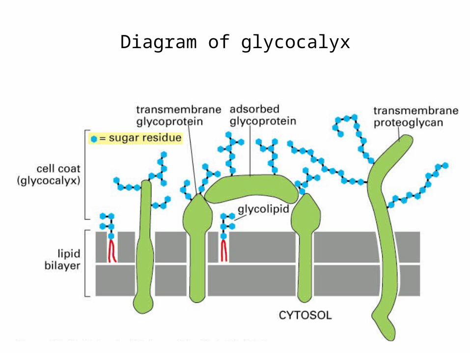
Diagram of glycocalyx
Summary
•membrane proteins and their anchoring models
•Methods to study membrane proteins, detergents
•diffusion, distribution, methods to study protein motion and distribution
•glycocalyx, proteoglycan

• Membrane Transport of Small Molecules and the Electrical Properties of
Membranes

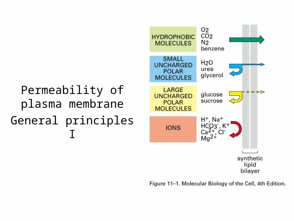
Permeability of plasma membraneGeneral principles I
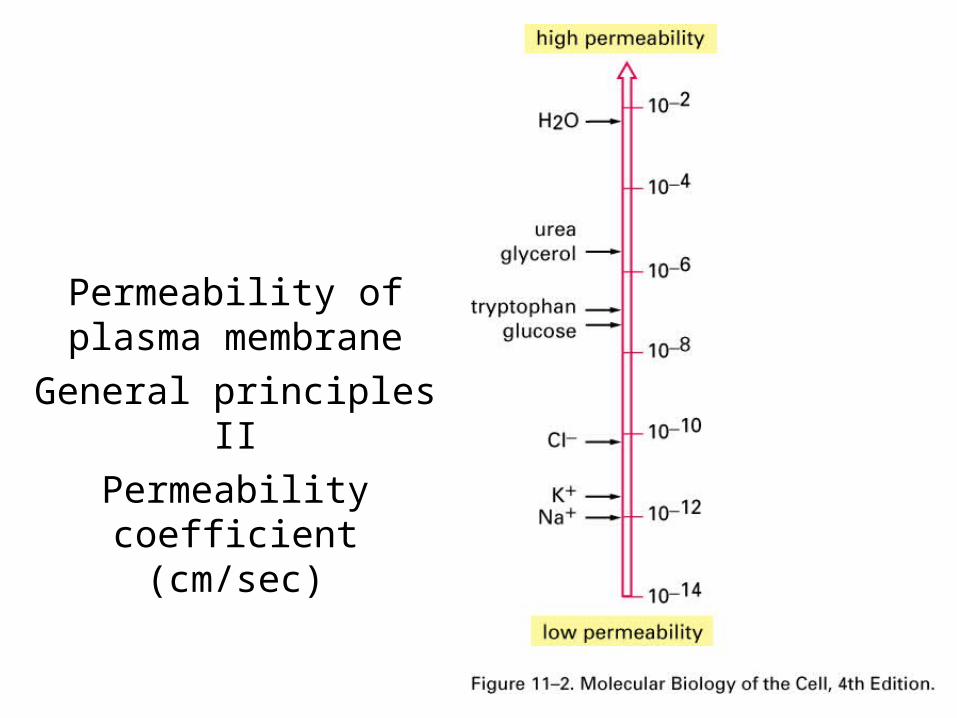
Permeability of plasma membraneGeneral principles II
Permeability coefficient (cm/sec)
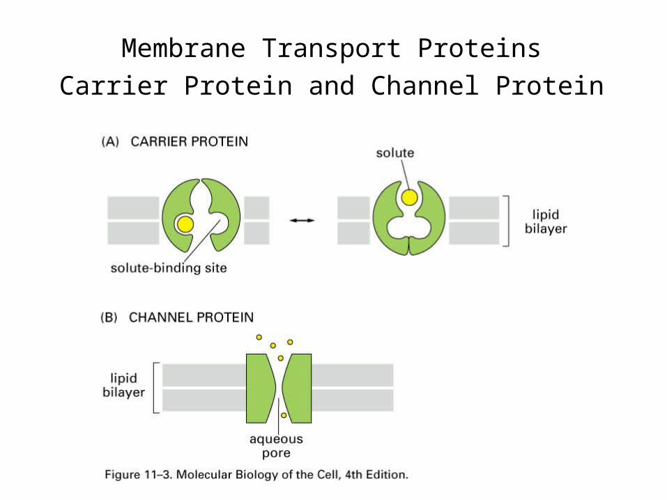
Membrane Transport ProteinsCarrier Protein and Channel Protein

Transportation ModelsPassive and Active Transport
Electrochemical and concentration gradient, membrane potential
Carrier proteins: passive and activeChannels: always passive

Electrochemical GradientIs the combinatory effect of concentration
gradient and membrane potentials
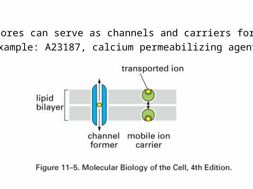
Ionophores can serve as channels and carriers for ionsExample: A23187, calcium permeabilizing agent
Carrier Proteins and Active Membrane Transportation

Conformational change of a carrier proteinMediates passive transport
Change is spontaneous and random, so dependent on concentration
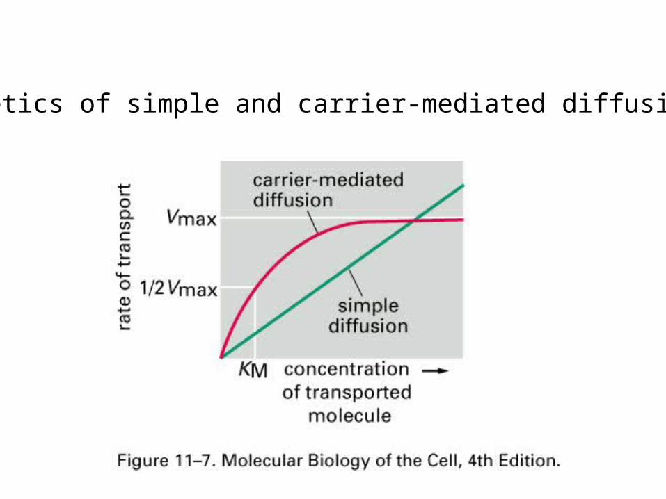
Kinetics of simple and carrier-mediated diffusions

3 ways of driving active transportation utilizing passive carriers
1. Coupled carriers2. ATP-driven pumps3. Light-driven pumps
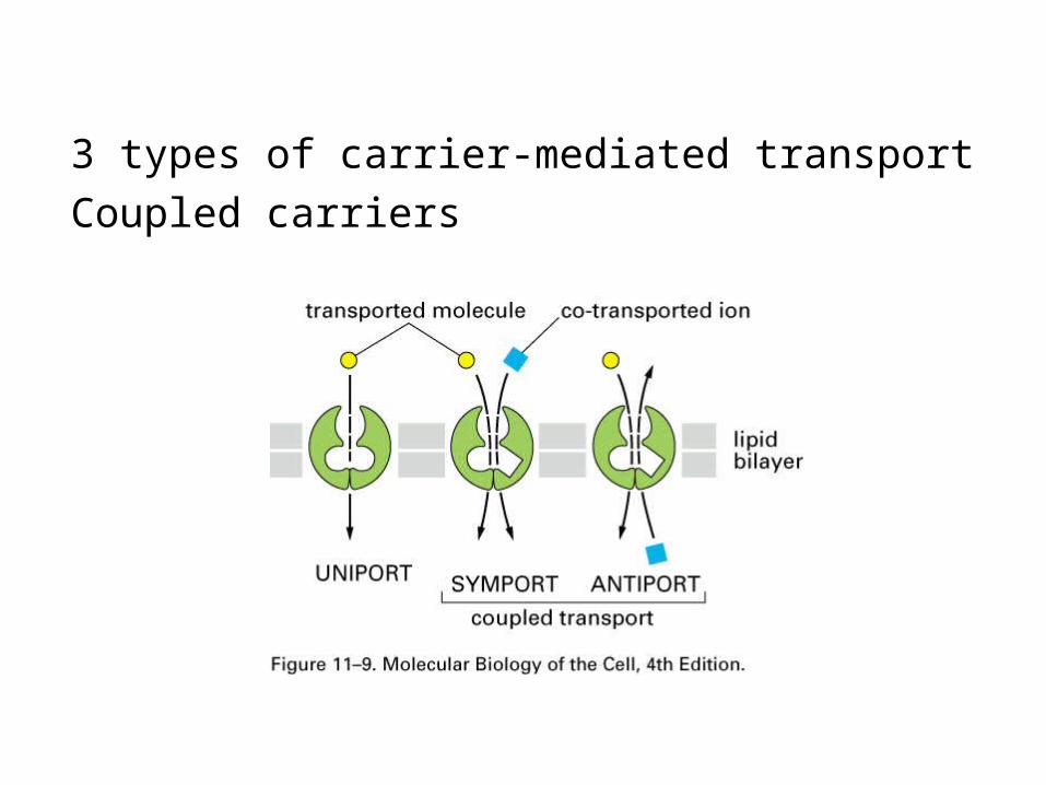
3 types of carrier-mediated transportCoupled carriers
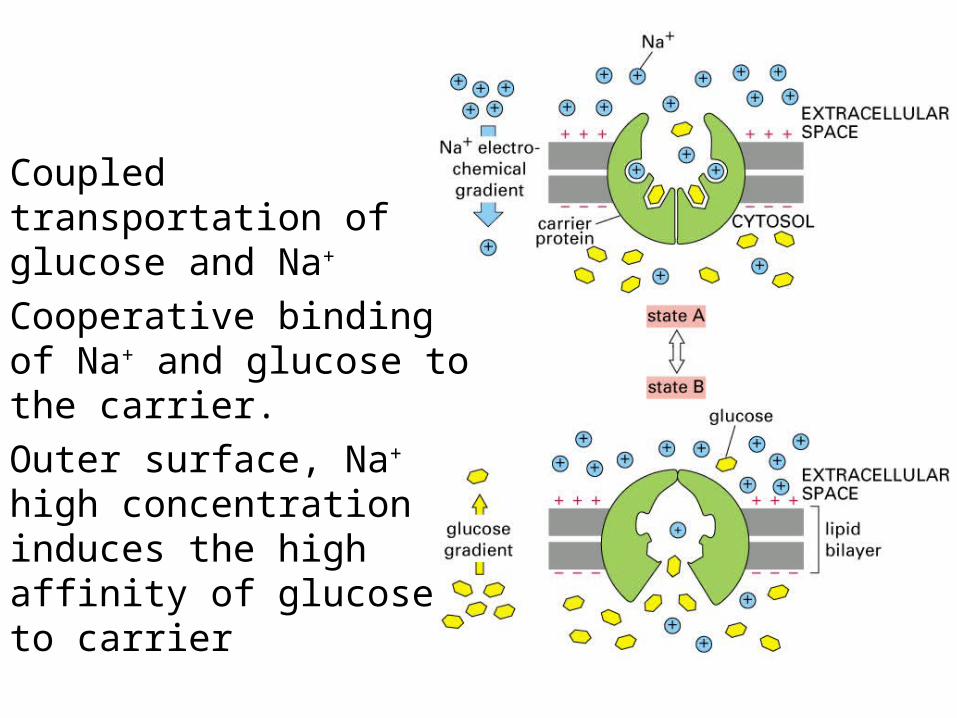
Coupled transportation of glucose and Na+
Cooperative binding of Na+ and glucose to the carrier. Outer surface, Na+ high concentration induces the high affinity of glucose to carrier

Transcellular transportTight junction separates apical and basal/lateral spaces
Apical: glucose and Na+ coupling; basal/lateral: glucose is passive, Na+ maintained by ATP-driven pump

Na+-K+ Pump, ATPaseP-type transport ATPase (dependent on phosphorylation)

Cycles of Na+-K+ Pump

Calcium PumpATP binding and hydrolysis can push calcium inside by bring
N and P domain together

A typical Ion Channel1. selectivity, 2. Gated (close and open)
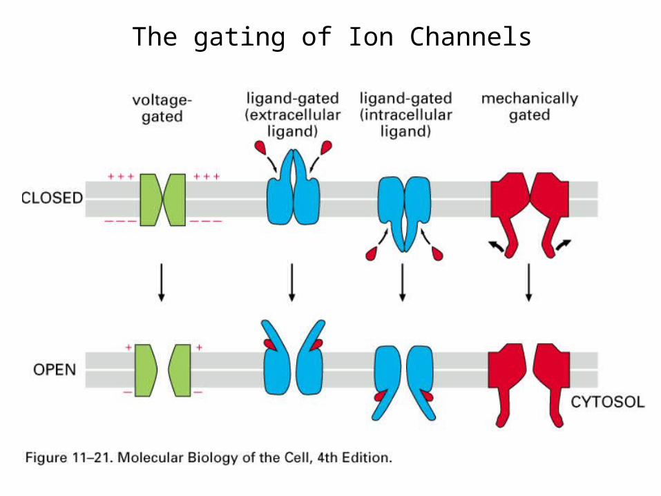
The gating of Ion Channels

The Structure of bacterial K+ channelSelectivity 10,000 fold over Na, although K+
0.133nm, Na+ 0.095 nm
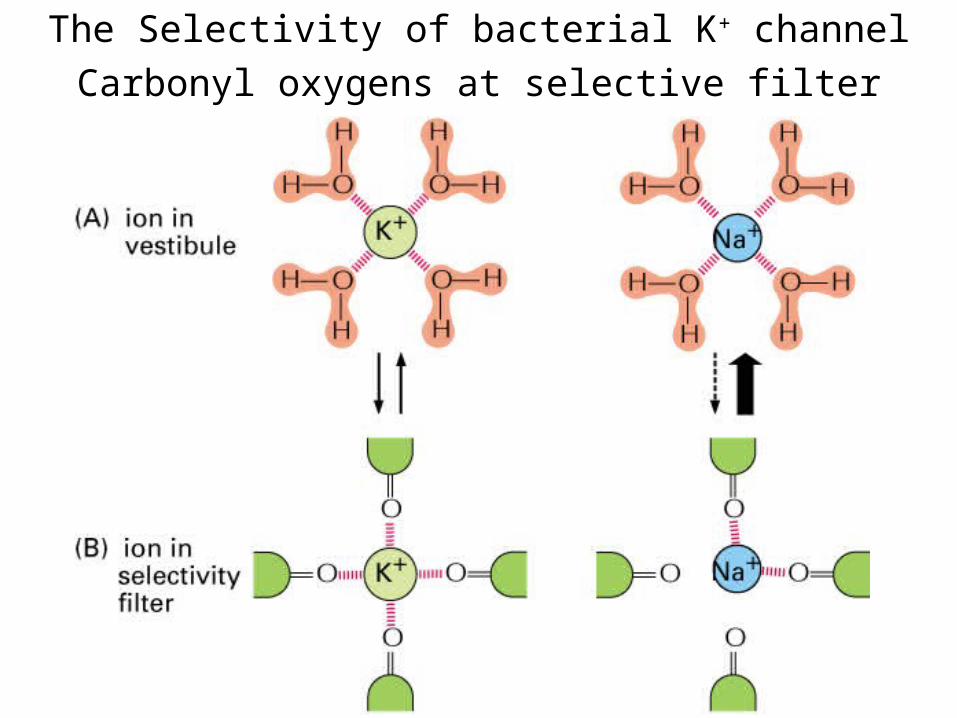
The Selectivity of bacterial K+ channelCarbonyl oxygens at selective filter
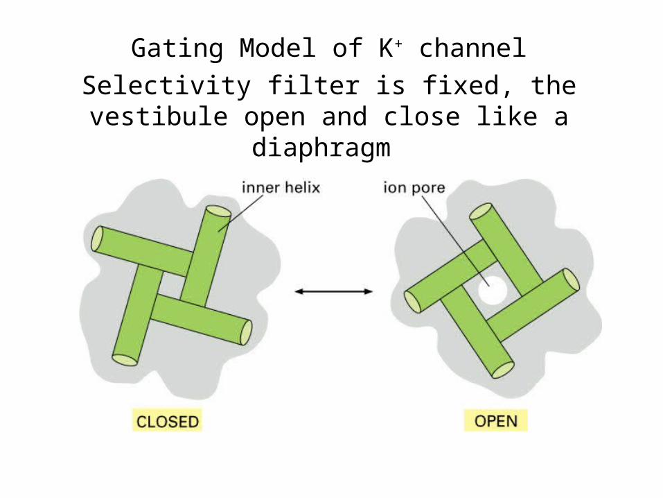
Gating Model of K+ channelSelectivity filter is fixed, the vestibule open and
close like a diaphragm
Summary
• Membrane transportation, carrier protein, channel protein
• Active transportation, passive transportation
• Carrier Proteins, coupled carriers, ATPases, Na+-K+ Pump
• Gating mechanisms of Ion Channels, K+ channel selectivity

• Intracellular Compartments and Protein Sorting
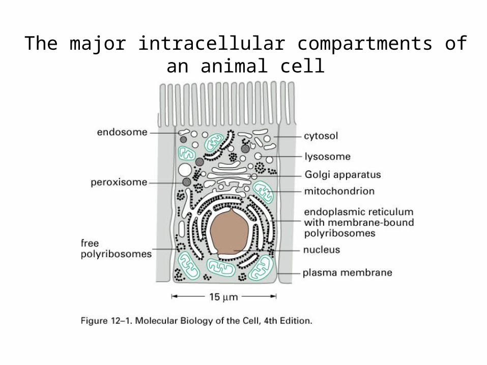
The major intracellular compartments of an animal cell
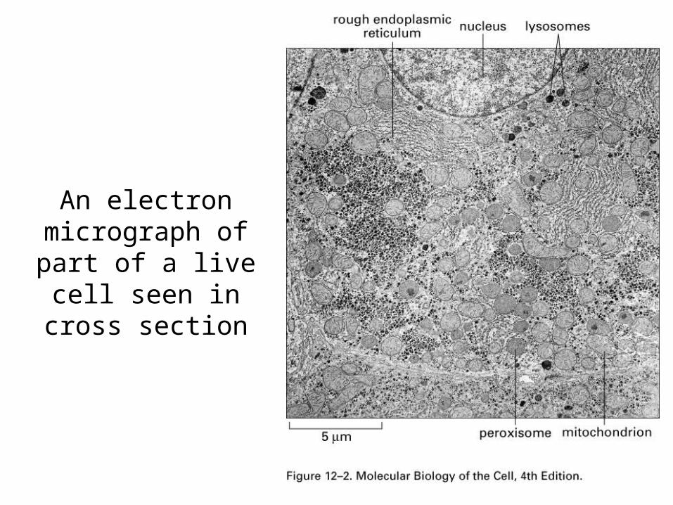
An electron micrograph of
part of a live cell seen in cross
section

Hypothetical schemes for
the evolutionary
origins of organelles

Topological relationships between compartments of the secretory and endocytic pathways in a
eucaryotic cell

A schematic roadmap of protein traffic
Red: gated transportBlue: transmembrane
transportGreen: vesicular
transport

Vesicle budding and fusion during
vesicular transport

Two ways in which a sorting signal can be built into a proteina.Signal sequence
b.Signal patch

• The transport of molecules between the nucleus and the cytosol


The nuclear envelope
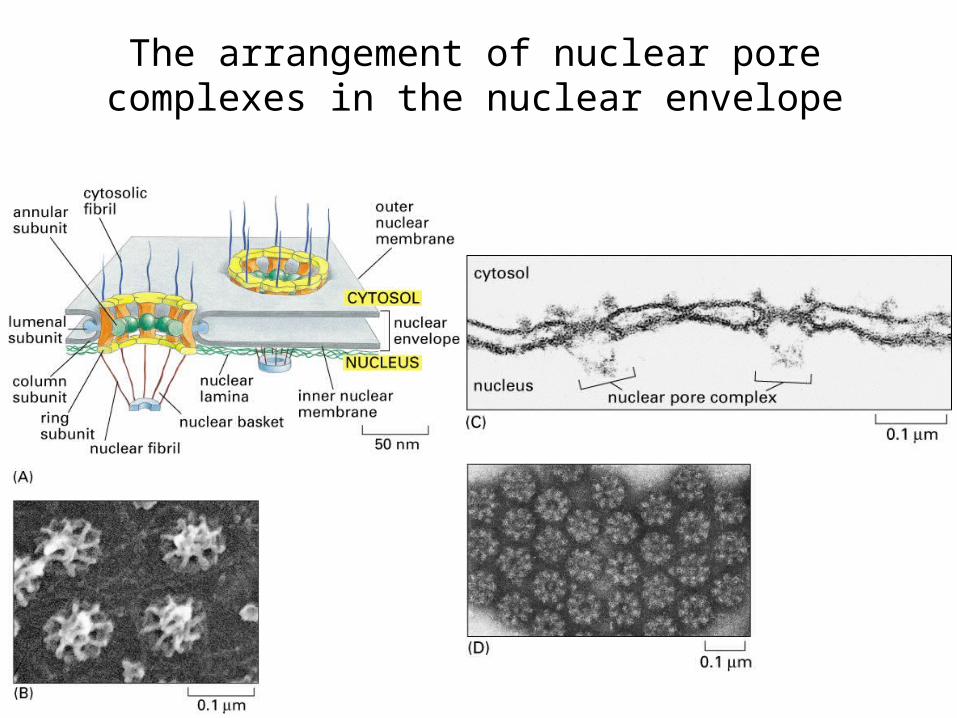
The arrangement of nuclear pore complexes in the nuclear envelope
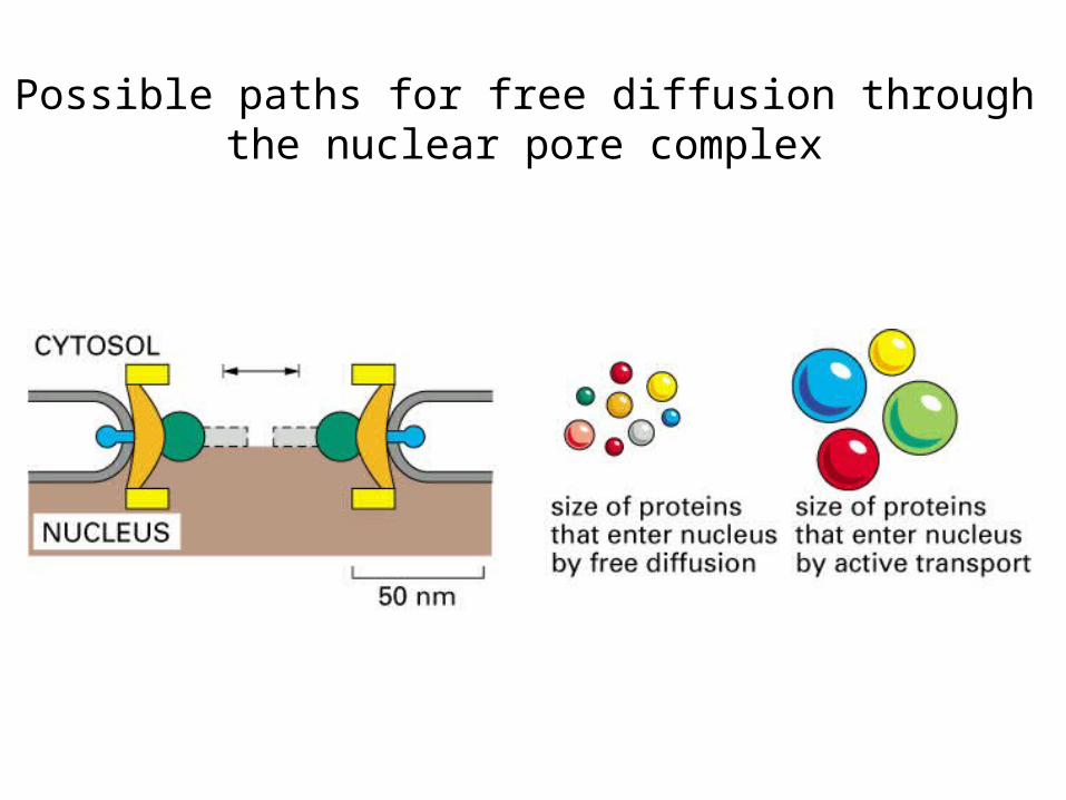
Possible paths for free diffusion through the nuclear pore complex

The function of a nuclear localization
signala.Nuclear localization
signal: NLSb.Nuclear export
signal: NES

Nuclear import receptors
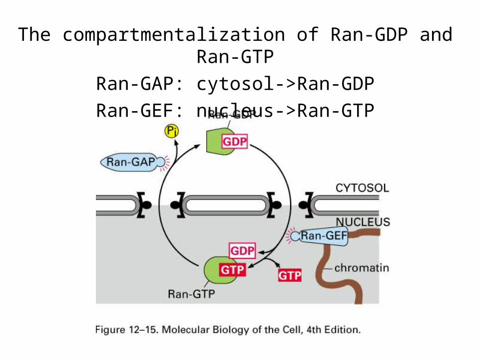
The compartmentalization of Ran-GDP and Ran-GTP
Ran-GAP: cytosol->Ran-GDPRan-GEF: nucleus->Ran-GTP

A model for how GTP hydrolysis by Ran provides directionality for nuclear transport

A model for how Ran-GTP binding might cause nuclear import receptors to release their cargo

The control of nuclear import during T-cell activation
• The endoplasmic reticulum


Fluorescent micrographs of the endoplasmic reticulum
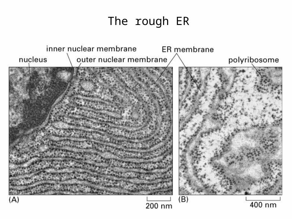
The rough ER
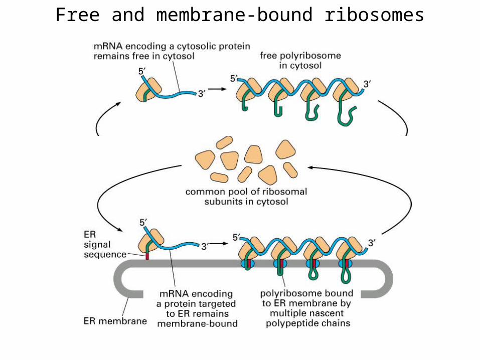
Free and membrane-bound ribosomes
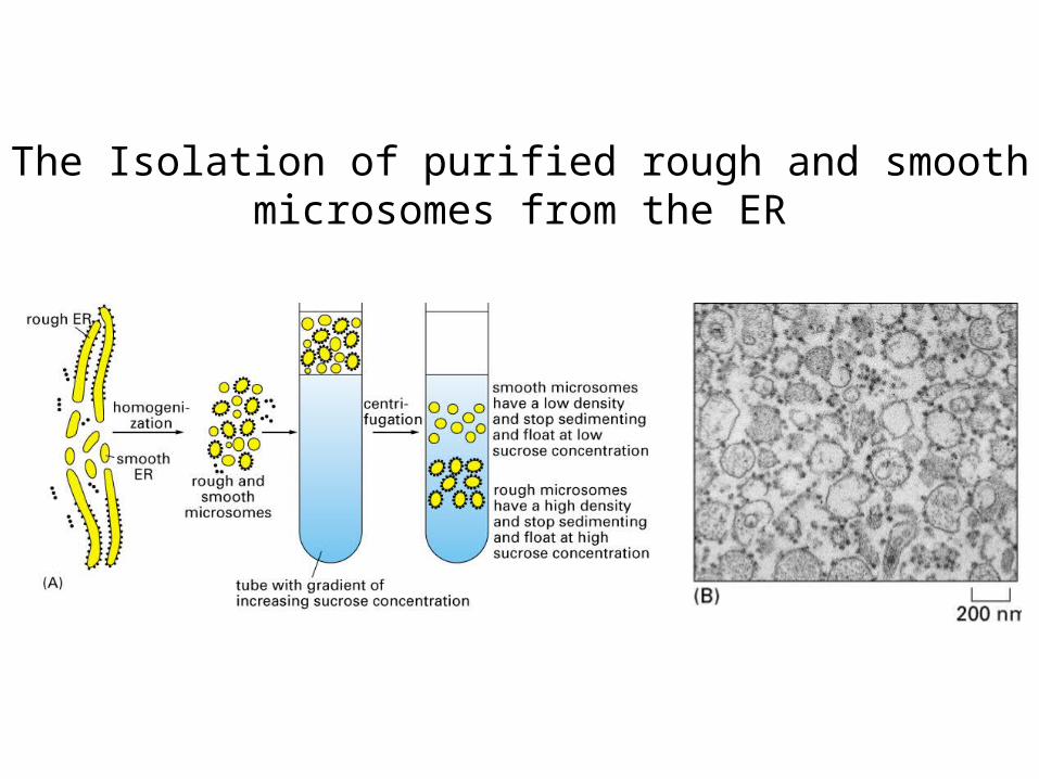
The Isolation of purified rough and smooth microsomes from the ER

The signal hypothesis

The signal-recognition particle (SRP)

How ER signal sequences and SRP direct ribosomes to the ER membrane
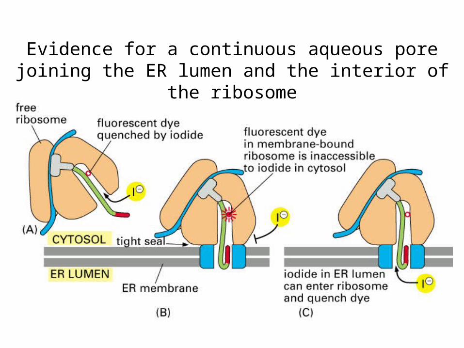
Evidence for a continuous aqueous pore joining the ER lumen and the interior of the ribosome

Three ways in which protein translocation can be driven through structurally similar translocators

A model for how a soluble protein is translocated across the ER membrane

How a single-pass transmembrane protein with a cleaved ER signal sequence is integrated into the
ER membrane
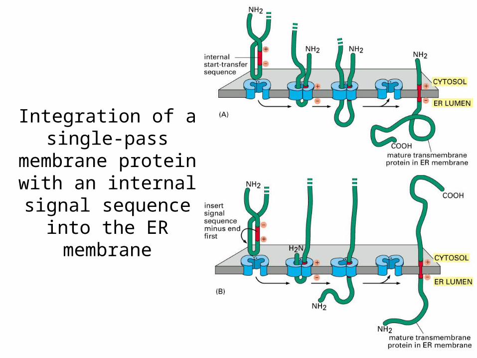
Integration of a single-pass
membrane protein with an internal
signal sequence into the ER membrane

Integration of a double-pass membrane protein with an internal signal sequence into the ER
membrane

The insertion of the multipass membrane protein rhodopsin into the ER membrane
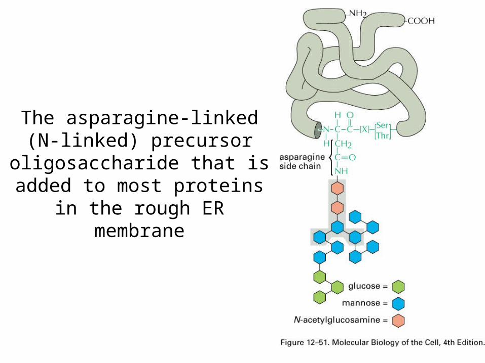
The asparagine-linked (N-linked) precursor
oligosaccharide that is added to most proteins in the rough ER membrane

Protein glycosylation in
the rough ER

The role of N-linked glycosylation in ER protein folding
Calnexin: membrane-bound chaperone proteinCalreticulin: soluble chaperone protein

The export and degradation of misfolded ER proteins

The unfolded protein response in yeast
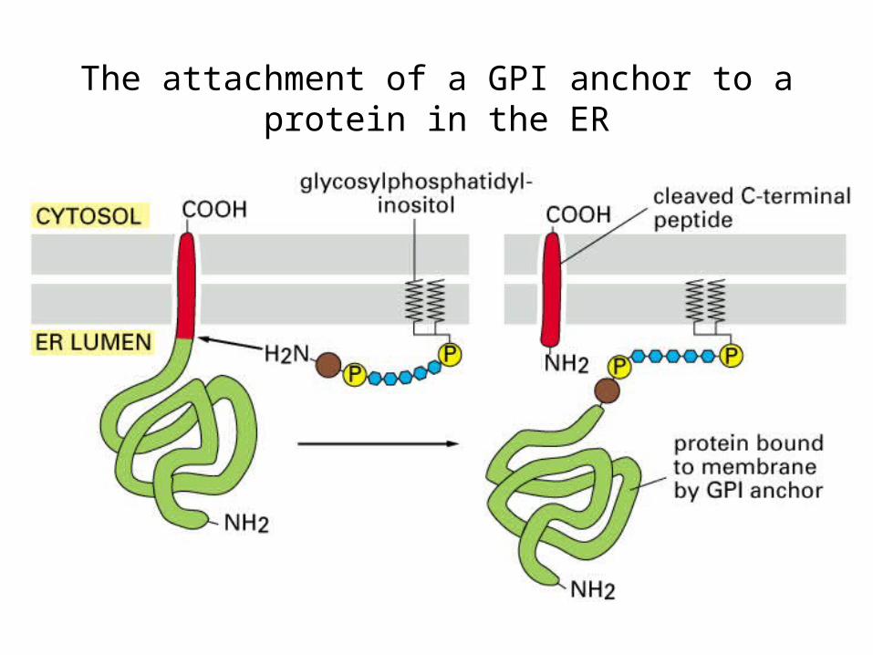
The attachment of a GPI anchor to a protein in the ER

The synthesis of phosphatidylcholine

The role of phospholipid translocation in lipid bilayer synthesis
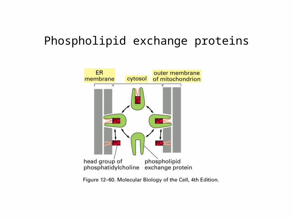
Phospholipid exchange proteins
Summary
•Nucleus translocation, NLS, NES, nuclear pore complex, Ran-GTP
•Endoplasmic reticulum, rough ER, smooth ER,
•SRP, soluble and membrane proteins in ER,
•Glycosylation in ER, folding,
•Membrane lipid bilayer assembly