Laryngopharyngeal reflux – development and refinement … · Laryngopharyngeal reflux ......
75
Laryngopharyngeal reflux - development and refinement of diagnostic tools Olle Andersson Institute of Clinical Sciences at the Sahlgrenska Academy University of Gothenburg University of Gothenburg
Transcript of Laryngopharyngeal reflux – development and refinement … · Laryngopharyngeal reflux ......

Laryngopharyngeal reflux
- development and refinement of diagnostic
tools
Olle Andersson
Institute of Clinical Sciences
at the Sahlgrenska Academy
University of Gothenburg
University of Gothenburg

2
Correspondence to: Olle Andersson Division of Otorhinolaryngology Sahlgrenska University Hospital SE-413 45 Göteborg, Sweden [email protected] © Olof Andersson 2009 ISBN 978-91-628-7850-0 Printed by Intellecta DocuSys AB. V. Frölunda, Sweden 2009
3
Abstract
Laryngopharyngeal reflux (LPR), characterized by symptoms of chronic cough,
hoarseness, throat clearing, globus, laryngospasm, throat pain and excessive mucus has in recent
years been recognized as an extra-esophageal manifestation of gastroesophageal reflux disease
(GERD). There are still many questions to be answered regarding how to diagnose the LPR
disease and how to effectively select patients that may benefit from treatment.
The aim of this thesis was to develop and refine diagnostics of LPR. In study I, an upper
limit of normality (ULN) for hypopharyngeal acid exposure with a cut-off level of pH5 instead
of the traditional pH 4 was established. Re-evaluation of ambulatory two-level 24-hour pH-
registrations of 35 healthy volunteers showed an ULN of 1.5% of the pH registration. In study II
we investigated the natural history of LPR and if asymptomatic pharyngeal reflux is a risk factor
for the development of LPR disease. Twenty-four healthy volunteers were re-evaluated after 13
years with pH-monitoring, symptom registration and a larynx examination. Upper airway
symptoms had developed in 10 of 24 (42%) subjects and pathological laryngeal findings in 9
(39%) subjects. However, the portion of subjects with pathological acid exposure at pH 4 in the
hypopharynx had decreased from 42 to 13%. Study III describes the Swedish translation and
adaption of the questionnaire Laryngo Pharyngeal Reflux – Health Related Quality of Life
Questionnaire (LPR-HRQL). LPR-HRQL was psychometrically evaluated in a population of
228 patients with upper airway symptoms. The Swedish translated version of LPR-HRQL
proved to be a statistically valid instrument to assess HRQL in patients with LPR disease. Study
IV described the development and psychometric evaluation of the Pharyngeal Reflux Symptom
Questionnaire (PRSQ) in the same cohort. After analysis and item-reduction it was found to be a
valid and reliable instrument.
The present thesis reports that the presence of asymptomatic hypopharyngeal reflux do
not constitute a risk factor for future development of LPR. Although upper airway symptoms
and pathological laryngeal findings seem to develop over time in a sample of healthy volunteers,
there was only a weak correlation between symptoms, laryngeal findings and pH-monitoring
results. The thesis also reports on normal values for hypopharyngeal reflux with a pH 5 which
may potentially improve upon diagnosis since weakly acidic reflux has been implicated in
mucosal damage and symptom generation. The thesis also presents validated questionnaires in
Swedish for health related quality of life (LPR-HRQL) and diagnosis (PRSQ) of the LPR
disease. Correctly developed and validated patient reported outcome (PRO) questionnaires have
the potential to sharpen the diagnosis and to capture a treatment effect, both in research and in
the clinic.

4
List of publications
I. Andersson O, Ylitalo R, Finizia C, Bove M, Ruth M. Pharyngeal reflux episodes at
pH 5 in healthy volunteers. Scand J Gastroenterol 2006;41:138-143.
II. Andersson O, Moller RY, Finizia C, Ruth M. A more than 10-year prospective,
follow-up study of esophageal and pharyngeal acid exposure, symptoms and
laryngeal findings in healthy, asymptomatic volunteers. Scand J Gastroenterol
2009;44:23-31.
III. Andersson O, Rydén A, Ruth M, Moller RY, Finizia C. Validation of the Swedish
translation of LPR-HRQL (Submitted).
IV. Andersson O, Rydén A, Ruth M, Moller RY, Finizia C. Development and validation
of a laryngopharyngeal reflux questionnaire, PRSQ (Submitted).

5
Contents
1 Abbreviations 7
2 Introduction 8
3 Background 10
3.1 Definitions and demographics of reflux disease 10
3.2 Established extraesophageal conditions associated with GERD 11
3.2.1 Asthma 11
3.2.2 Chronic cough 12
3.2.3 Dental erosions 12
3.2.4 Reflux laryngitis 12
3.3 Proposed extraesophageal associations with GERD 13
3.4 Physiology and patophysiology of LPR 13
3.5 Diagnosis of LPR 15
3.5.1 Laryngeal examination 16
3.5.2 Histology 17
3.5.3 24-hour pH monitoring 17
3.5.4 Esophageal manometry 19
3.5.5 Esophagogastroduodenoscopy 19
3.5.6 Other considerations 20
3.6 Treatment of LPR 20
3.7 Patient reported symptoms - Outcome variables 22
3.8 The validation process 23
4 Aims of the thesis 26
5 Methodology 27
5.1 Subjects and design 27
5.1.1 Study I and II 27
5.1.2 Study III and IV 28
5.2 24-hour pH monitoring 29
5.3 Laryngoscopic examination 31
5.4 Questionnaires 31
5.5 Statistical methods 34

6
6 Results 37
6.1 Study I 37
6.2 Study II 37
6.3 Study III 39
6.4 Study IV 39
7 Discussion 40
8 Conclusion 46
9 Future research and goals 47
10 Acknowledgments 48
11 Summary in Swedish 50
12 References 51
13 Original papers I-IV 61
14 Appendix A-B

7
1 Abbreviations
BID bis in die, Latin for twice daily
EER Extraesophageal reflux
EGD Esophagogastroduodenoscopy
FDA Food and Drug Administration
GERD Gastroesophageal reflux disease
HRM High resolution manometry
HRQL Health Related Quality of Life
LES Lower esophageal sphincter
LPR Laryngopharyngeal reflux
LPR-HRQL Laryngo Pharyngeal Reflux – Health Related Quality of Life
NERD Non-erosive reflux disease
PPI Proton pump inhibitor
PRO Patient Reported Outcome
PRSQ Pharyngeal Reflux Symptom Questionnaire
RCT Randomized controlled trial
RSI Reflux Symptom Index
SF-36 v2 Short Form -36 version 2
TLESR Transient lower esophageal sphincter relaxations
UES Upper esophageal sphincter
UGDQ Upper Gastrointestinal Disease Questionnaire
ULN Upper limit of normality

8
2 Introduction
Reflux is the retrograde flow of gastric contents from the stomach into the
esophagus. Reflux may in some instances also pass the upper esophageal sphincter and
into the hypopharynx/larynx as well as the lower aerodigestive tract [1]. While
gastroesophageal reflux disease (GERD) is readily recognised by the typical symptoms
of heartburn and acid regurgitation, the symptomatology of laryngopharyngeal reflux
(LPR) or extraesophageal reflux (EER) is more diverse and less patognomonic [2,3].
The prevalence of troublesome symptoms of heartburn and/or regurgitations on a
weekly basis in the western world vary in different reports between 10-20% [4]. The
prevalence of LPR is less established [5,6] and the natural history of the LPR disease is
largely unknown, mainly because a golden standard for how to diagnose LPR does not
exist. Long-term studies following a GERD population over time have, however, shown
it to be a chronic disease [7,8].
The main diagnostic tools are patient reported symptoms, 24-hour pH monitoring, and
laryngoscopic examination [9].
The symptoms associated with LPR are chronic cough, hoarseness, throat clearing,
globus, laryngospasm, throat pain and excessive mucus [3]. Koufman et al found that
patients with these symptoms represent around 10% of the patients presenting in an
ENT-clinic [3]. However, the symptoms of LPR may be caused by other conditions such
as smoking, allergy, voice abuse or airway infections [10] and according to several
studies, lack sufficient diagnostic specificity and sensitivity [11,12]. An important aspect
in the process of creating guidelines for diagnosis in this context is to develop and
evaluate psychometrically tested, patient reported symptom questionnaires.
24-hour pH monitoring is usually performed in the distal esophagus and either in
the proximal esophagus just below the upper esophageal sphincter (UES) or in the
hypopharynx within 1-2 cm above the UES. Not only is the optimal positioning of the
catheter debated, but also what should be regarded as pathological acid exposure in the
hypopharynx [6,13]. The classical cut-off limit at esophageal pH monitoring, pH 4, has
traditionally been used also in hypopharyngeal pH monitoring. It has been suggested,
however, that a higher limit should be used in the hypopharynx, for example pH 5, as the
laryngeal mucosa is reported to be more sensitive to pepsin and acid than the esophageal
mucosa [14]. Furthermore, pepsin has been reported to be active up to pH 6 [15].
9
Typical laryngeal findings associated with LPR are posterior laryngitis including edema
and erythema. Others findings are, vocal fold nodules, granuloma or even cancer.
However, the signs have shown poor specificity for the LPR-disease [16].
The aim of this thesis was therefore to aid in the understanding of the disease and
to develop and refine the diagnostic tools of LPR. This was done by investigating the
occurrence of LPR over time in healthy volunteers, by investigating reference intervals
of pharyngeal pH monitoring at the pH 5 level, by translating and validating a Swedish
version of the Laryngo Pharyngeal Reflux – Health Related Quality of Life (LPR-
HRQL) questionnaire [17], as well as by creating and validating a new diagnostic
instrument, the Pharyngeal Reflux Symptom Questionnaire (PRSQ).

10
3 Background
3.1 Definitions and demographics of reflux disease
Heartburn as a predominant symptom has been shown to have a high diagnostic
specificity for GERD [18]. The diagnosis of GERD, proposed in the Genval workshop
report from 1999 [19], i.e. heartburn at least 2 times a week, was challenged when it was
shown that 37% of patients with esophagitis did not have classical reflux symptoms [20].
Therefore the World Congress of Gastroenterology in Montreal, Canada 2005 defined
GERD as “a condition which develops when the reflux of stomach contents causes
troublesome symptoms and/or complications” [21]. This definition also includes patients
without classical symptoms but with complications or syndromes of both esophageal and
extraesophageal nature. Extraesophageal syndromes were divided into established and
proposed associations. The established associations were deemed to be reflux cough,
reflux laryngitis, reflux asthma and reflux dental erosions while pharyngitis, sinusitis,
pulmonary fibrosis and otitis media were deemed as proposed associations,
Figure 1. The existence of GERD increases the likelihood of laryngeal signs and
symptoms [2,22]. The reflux up to the hypopharynx/larynx area is referred to as LPR.
LPR is a more restricted definition than EER, where the latter also includes the lower
aerodigestive tract.

11
Figure 1. Montreal classification
Adaption from Vakil et al. 2006. With permission from Am J Gastroenterology.
The prevalence of GERD varies with geographical location according to a recently
published systematic review of 15 studies. The study showed that 8-27% of the adult
population in the western society had heartburn and/or acid regurgitation one or more
times per week. In Asia the reported prevalence is significantly lower, i.e. 3-5% [4].
Due to the lack of consensus in how to diagnose LPR and the disparate methodologies
used by investigators, the true prevalence of LPR is not well known. Connor et al.
reported that symptoms commonly attributed to LPR were as high as 49% in a normal
community dwelling [2,6]. In patients with GERD, extraesophageal symptoms were
reported by 33% [2].
3.2 Established extraesophageal conditions associated with GERD
3.2.1 Asthma
There is strong evidence linking asthma to GERD [23,24]. Several studies have
indicated that acidity in the trachea generates increased pulmonary resistance [25,26].
Harding et al. demonstrated that between 50-80% of the asthma patients also had GERD

12
symptoms and up to 75% also had pathological acid exposure [23]. A study of over
15.000 primary care patients showed that there was up to a twofold risk of receiving a
GERD diagnosis following a first diagnosis of asthma [27].
The cause and effect relationship between asthma and reflux is however not clear
as both conditions may be the result of the other. Reflux may cause asthma through
microaspirations into the bronchial tree or through a vagal mediated nerve reflex which
causes an asthmatic reaction. Furthermore, during an asthma attack you get a negative
intrathoracic pressure which may give reflux. [28].
3.2.2 Chronic cough
Chronic cough is defined as cough with a duration of more than 3 weeks.
According to Irwing et al. and Pratter et al., GERD together with postnasal drip
syndrome and asthma are responsible for over 90% of chronic cough diseases out of
which 10-30% of the cases could be directly related to GERD [29,30].
These results are in accordance with double-blind placebo controlled studies with GERD
patients, showing a significant improvement in cough scores in the PPI treated patient
group [31,32].
3.2.3 Dental erosions
The prevalence of dental erosion among individuals with GERD has been
estimated to 20-55%, compared with a prevalence of 2-19% in the general population
[33]. Munos et al. showed that GERD patients, verified with pH-registrations and
endoscopy, had significantly more dental erosions, compared with controls, 47.5% vs.
12.5% [34].
3.2.4 Reflux laryngitis
Laryngitis or laryngeal inflammation is relatively common, unspecific and often
resolves spontaneously [35]. When laryngitis is persistent the underlying causing agent
may be allergy, infection, vocal trauma, postnasal drip or LPR.
LPR may be suspected in the presence of symptoms of hoarseness, globus, throat
clearing, dysphagia, cough, laryngospasm, throat pain and excessive mucus [3,9,36,37].
Symptoms of heartburn and acid regurgitation are often absent in LPR, i.e. in 60 %

13
according to Koufman et al. [3]. However, more recently, the Montreal definition stated
that unexplained asthma and laryngitis are unlikely to be related to GERD in the absence
of heartburn or regurgitation.
Laryngeal findings indicating LPR are posterior laryngitis with erythema, edema
and presence of interarytenoid thickening in the posterior wall of glottis [38]. Medical or
surgical therapy on reflux laryngitis has in observational trials demonstrated a partial
improvement of upper airway symptoms and to some extent of laryngeal findings
[39,40]. However, the only randomized controlled trial demonstrating a treatment effect,
was on patients with classical GERD symptoms in addition to laryngitis. Whereas other
recent trials that excluded patients with frequent heart-burn demonstrated no benefit of a
PPI over placebo in treating the laryngitis [41,42].
3.3 Proposed extraesophageal associations with GERD
Adequate evidence of causal linkage is lacking between LPR and sinusitis,
pulmonary fibrosis, pharyngitis and recurrent otitis media [43,44].
However, a moderate increased risk for sinusitis (Odds ratio 1.6) and idiopathic
pulmonary fibrosis (Odds ratio 1.36) was seen in a epidemiological study on U.S.
military veterans with reflux esophagitis [45].
3.4 Physiology and patophysiology of LPR
Physiology: Gastroesophageal as well as gastro-esophago-hypopharyngeal reflux
occur due to the pressure gradient between the positive intra-abdominal pressures and the
negative pressures in the thorax/hypopharynx.
Gastroesophageal physiological reflux occurs predominantly in conjunction with
transient relaxations of the lower esophageal sphincter (TLESR) [46]. TLESR are
triggered by gastric distention, mainly in the postprandial period and are activated by
stretch receptors in the gastric wall. The reflex arch includes a vagus mediated impulse
to the N Tractus Solitarius in the brain stem, vagal efferents to the LES and a non-
cholinergic, non–nitergic inhibitory interneurone, which relaxes the sphincter [46]. The
role of these relaxations is to release swallowed air by belching [47]. They occur
independently from swallowing, are longer (5-30 seconds), and are typically followed by
an after-contraction of the LES.

14
Reflux to the hypopharynx occurs predominantly postprandially and in the upright
position [48,49]. LPR is, in contrast with GERD, often not associated with heartburn and
regurgitation [50].
Components: The agents responsible for producing upper airway symptoms and
laryngeal pathology are acid, pepsin, bile acids and trypsin. The relative importance of
the agents is, however, presently under debate. Pepsin combined with acid has been
found to be the most injurious agents with a significant association with laryngeal lesions
[15]. Pepsin has in animal experiments and in vitro been shown to be active and cause
laryngeal cell injury up to pH 6 [14].
The reflux can either be gas, liquid or mixed (gas/liquid). The vast majority of the
pharyngeal reflux is gaseous without pH drops and is seen equally in healthy subjects
and laryngitis patients, while mixed and liquid refluxes and also gas reflux with pH
drops are significantly more common in LPR patients [51]. The nocioceptive damage of
biliary reflux on laryngeal structures has also been demonstrated [52,53]. Impedance
testing together with bilitec may here improve upon diagnostics.
Protective mechanisms: There are 4 principal protective physiological barriers
against reflux
1. The lower esophageal sphincter (LES)
2. Acid clearance, through esophageal motor function and gravity
3. Esophageal mucosal tissue resistance
4. The upper esophageal sphincter (UES)
Active bicarbonate production is pumped into the extracellular space in the esophagus
but not into the larynx, which thus has less capacity to neutralize the nocioceptive
influence of acid. Recent investigations suggest that laryngeal tissues are also protected
from reflux damage by a carbonic anhydrase in the mucosa of the posterior larynx. The
carbonic anhydrase enzyme catalyzes hydration of carbon dioxide to produce
bicarbonate, which neutralizes the acid in the refluxate. Carbonic anhydrase isoenzyme
III, expressed at high levels in normal laryngeal epithelium, was however shown to be
absent in 64% of biopsy specimens from laryngeal tissues of LPR patients [54].
Patophysiology: There are two dominating theories about how gastric acid
provokes extra esophageal pathological symptoms and/or findings. The first implies
direct acid-pepsin injury to the larynx and surrounding tissues [15,26]. The second

15
proposes that acid in the distal esophagus stimulates vagal-mediated reflexes that result
in bronchoconstriction leading to chronic throat clearing and coughing, which in turn
provokes mucosal lesions [28,55]. In fact, they may both play an essential part in
conjunction [56]. Symptoms may arise either from direct mucosal injury or because of
damage to cilias, leading to mucous stasis and chronic throat clearing and cough.
Also the level of acidity corresponds to the degree of mucosal damage where pH 0-4 is
the most damaging [15]. Weakly acidic reflux episodes (pH4-pH7), not detected by the
classic cut-off limit at pH 4 in 24-hour pH monitoring, may pass through the esophagus
without symptoms and signs, but doing harm to the more sensitive mucosa of the larynx
[14,15,57]. The ciliated respiratory epithelium of the larynx has been shown to be more
sensitive to acid, activated pepsin and bile salts than the esophageal mucosa [14,15].
The time and frequency of acid exposure necessary to create disease is also under
debate. Koufman et al. postulated that one single reflux episode is enough [3]. This was
concluded after an animal experiment where he subjected the arythenoid region of
animal larynx to acid and pepsin 3 times a week and discovered that this was enough to
create mucosal damage. In later years however, many investigators agree that a larger
amount of reflux is needed to cause disease, (Table 1). This is due to the fact that around
20-50% of healthy symptom-free subjects have, in several studies, been reported to have
hypopharyngeal reflux episodes. A recent review by Joniau et al. compared 11 studies
with normal controls and 13 studies with reflux laryngitis, and found in the pooled data
that reflux events were detected in 23% of the normal controls and in 38% of the LPR-
patients [58]. In another meta analysis of upper-probe measurements by Merati et al. the
pooled data gave that 31% of the normal subjects had hypopharyngeal reflux events, as
opposed to 51% of the LPR patients. Bove et al. found the ULN of acid exposure at pH 4
in 40 healthy subjects to be 0.2% or 6 reflux episodes during the 24-hour pH-monitoring
[59].
3.5 Diagnosis of LPR
One challenge in diagnosing LPR is that the symptoms of the LPR disease lack
sufficient specificity to confirm LPR and thus to rule out other causative agents.
In fact, several studies have shown a poor correlation between LPR symptoms, laryngeal
findings and findings from hypopharyngeal pH registrations [9,13,60,61].
16
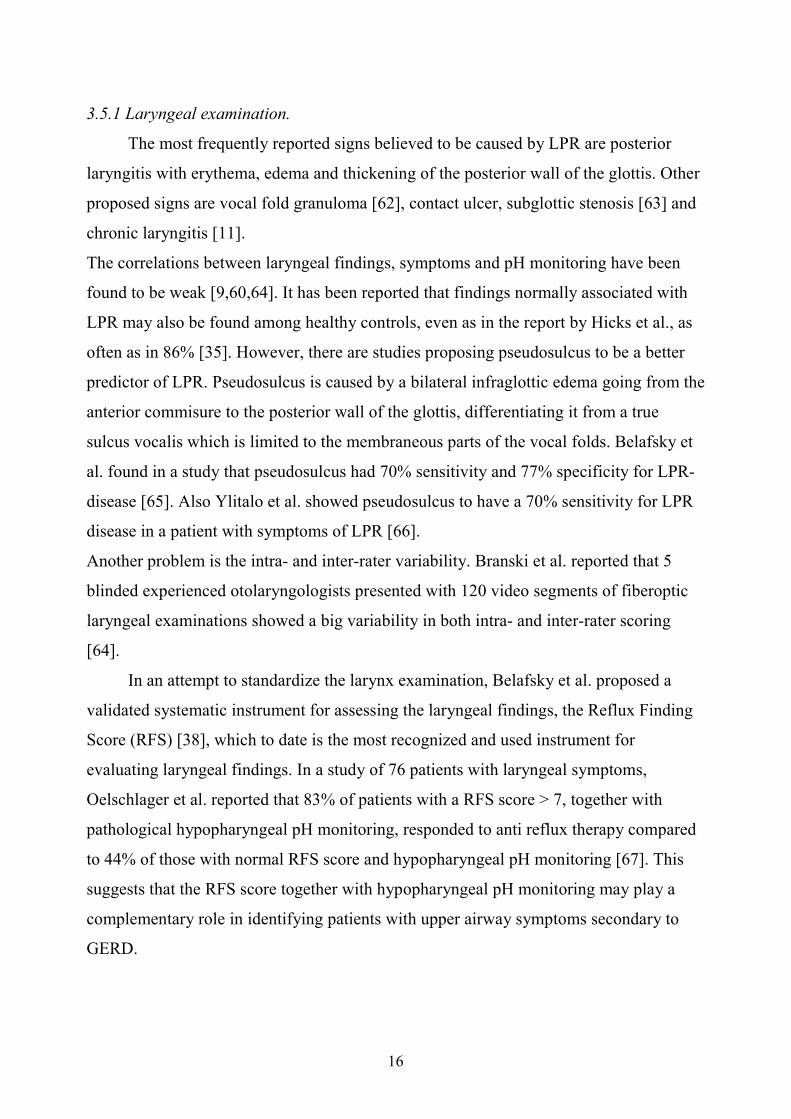
3.5.1 Laryngeal examination.
The most frequently reported signs believed to be caused by LPR are posterior
laryngitis with erythema, edema and thickening of the posterior wall of the glottis. Other
proposed signs are vocal fold granuloma [62], contact ulcer, subglottic stenosis [63] and
chronic laryngitis [11].
The correlations between laryngeal findings, symptoms and pH monitoring have been
found to be weak [9,60,64]. It has been reported that findings normally associated with
LPR may also be found among healthy controls, even as in the report by Hicks et al., as
often as in 86% [35]. However, there are studies proposing pseudosulcus to be a better
predictor of LPR. Pseudosulcus is caused by a bilateral infraglottic edema going from the
anterior commisure to the posterior wall of the glottis, differentiating it from a true
sulcus vocalis which is limited to the membraneous parts of the vocal folds. Belafsky et
al. found in a study that pseudosulcus had 70% sensitivity and 77% specificity for LPR-
disease [65]. Also Ylitalo et al. showed pseudosulcus to have a 70% sensitivity for LPR
disease in a patient with symptoms of LPR [66].
Another problem is the intra- and inter-rater variability. Branski et al. reported that 5
blinded experienced otolaryngologists presented with 120 video segments of fiberoptic
laryngeal examinations showed a big variability in both intra- and inter-rater scoring
[64].
In an attempt to standardize the larynx examination, Belafsky et al. proposed a
validated systematic instrument for assessing the laryngeal findings, the Reflux Finding
Score (RFS) [38], which to date is the most recognized and used instrument for
evaluating laryngeal findings. In a study of 76 patients with laryngeal symptoms,
Oelschlager et al. reported that 83% of patients with a RFS score > 7, together with
pathological hypopharyngeal pH monitoring, responded to anti reflux therapy compared
to 44% of those with normal RFS score and hypopharyngeal pH monitoring [67]. This
suggests that the RFS score together with hypopharyngeal pH monitoring may play a
complementary role in identifying patients with upper airway symptoms secondary to
GERD.

17
3.5.2 Histology
Dilation of intercellular space (DIS) between squamous epithelial cells has been
put forward as the earliest microscopic marker of acid damage both in the esophagus and
the larynx [68,69]. Biopsies from the interarytenoid area in LPR patients were
demonstrated to have a significantly larger DIS than in healthy controls [69].
Intracellular concentrations of pepsin and depletion of carbonic anhydrase isoenzyme III
has also been demonstrated in laryngeal vocal fold and ventricle biopsies from LPR-
patients, whereas the healthy controls had low levels of pepsin and higher levels of
isoenzyme III [57].
3.5.3 24-hour pH monitoring
24-hour pH monitoring is considered the most reliable test for LPR. Two pH-
electrodes are introduced transnasally and positioned 5 cm above the LES and 0.5-2 cm
above the UES with the help of manometry. The probe placed 2 cm above the UES is
considered the best at representing this. With the probe placed in the proximal esophagus
the amount of reflux is greater and will not accurately reflect the acid exposure of the
hypopharynx/larynx [70]. Acid exposure in the esophagus and the hypopharynx is
registered by antimony, glass or ISFET probes and is stored on a digital recorder,
commonly with a sampling frequency of 0.25Hz. Symptoms, meals and body position
may be registered simultaneously by the patient by pressing a button on the same digital
recorder and by specifying the event in a manual diary.
Using pH < 4 as the cut-off value, a meta analysis of dual probe pH metry of 16
studies showed that 10-30% of normal subjects had acid reflux events at the upper probe
(in the UES or up to 2 cm above the UES) [13]. Of the patients with diagnosed LPR
disease, only 51% had demonstrated reflux to pharynx [13]. The mean percentage of acid
exposure time in healthy subject was less than 0.01% of the 24-hour pH monitoring. The
authors concluded that acid exposure time was the most consistent indicator to detect
LPR and that measurements between the UES and 2 cm into the hypopharynx give the
most accurate and consistent information [13]. Shaker et al. used a simultaneous 3-site
pH monitoring (distal and proximal esophagus and hypopharynx) and demonstrated that
hypopharyngeal acid exposure occurred more frequently and in greater amount among
LPR patients than in GERD patients or controls. Koufman, in his groundbreaking study,

18
considered 1 single reflux to be enough to create signs or symptoms. A more recent
approach is to consider LPR confirmed when the total acid exposure time (%) < pH 4
during the 24-hour pH monitoring is more than 1% [51].
The sensitivity of esophageal pH-monitoring in the diagnosis of GERD reported in
the literature has ranged from 79% to 96% [71-74] and the specificity from 86% to 100%
[74,75] with a reproducibility of 89% [76]. The specificity and sensitivity of the
hypopharyngeal pH recordings are, however, less impressive. Ahmed et al. reported a
sensitivity of hypopharyngeal pH monitoring of only 40% sensitivity as compared to
70% and 50% for distal and proximal esophageal measurements [11], comparing the
correlations between clinical diagnosis of GERD and LPR with pH measurements.
Table 1 show the degree of reflux found in healthy controls and LPR patients in different
studies at 2 cm above the UES.
Table 1. Hypopharyngeal reflux in healthy subjects and LPR-patients
Hypoph reflux healthy Hypoph reflux LPR pat Study n Reflux
mean n Acid expos
mean % n Reflux
mean n Acid expos
mean % Koufman 91 12 0 0 Shaker 95 12 0.17 0 14 4.36 0.24 Smit 98 20 1.8 0.01 Toohill 98 12 2.42 0.009 Ulualp 99 17 0.2 0 20 2.65 0.13 Bove 00 40 1 0 Ylitalo 01 19 0.7 0 26 1.50 0.034 Powitzky 03 15 1 NR Oelschlager 02 76 3.4 NR
n = number, Reflux mean n = mean number of reflux events, Acid expos mean % = mean percentage of acid exposure, NR = not reported
The clinical importance of weakly acidic reflux (pH 4 – pH 7) has gained
increasing interest over the recent years, further enhanced with the introduction of 24-
hour combined impedance-pH monitoring. Combined multi-channel intraluminal
impedance and pH-metry is a technique that enables monitoring of gastroesophageal
reflux independent of its acidity and the relation to typical and atypical symptoms. It
allows the recognition of major acid, minor acid, non-acid, and gas reflux events

19
[51,77,78] which may improve on diagnostics. Recorded symptoms can be correlated
with all reflux events (e.g. acid, minor acid, non-acid, and gas). Impedance evaluation of
bolus transit might also investigate the functional relevance of manometric
abnormalities. Patients with persistent symptoms of gastroesophageal reflux in spite of
adequate treatment with proton pump inhibitors may still have weakly acidic reflux
and/or bile reflux associated with their symptoms. It has been shown that non-acid or
weakly acid reflux can be associated with symptoms in patients with GERD or LPR
symptoms [79]. Sifrim et al. could significantly correlate a subgroup of chronic cough to
weakly acidic reflux [77].
3.5.4 Esophageal manometry
Manometry is used to evaluate UES function, esophageal peristalsis and LES
function. There are several motor function disturbances associated with GERD,
(esophageal dysmotility and variation of either the LES or the UES sphincter tonus [80])
but no significant casual linkage have been found [81]. The UES pressure has with
standard manometry shown large variability in pressure values, which is why no
conclusions regarding the risk to develop LPR can be drawn [82]. Kahrilas et al. did
however find a marked decrease in UES resting pressure during sleep in normal controls
which may have significance in that it diminishes the barrier for nocturnal reflux [82].
Today, manometry in conjunction with pH metry is therefore mainly used to correctly
position the pH probes. High-resolution manometry (HRM), may however, potentially
improve on diagnostics of manometric abnormalities associated with LPR in the future.
3.5.5 Esophagogastroduodenoscopy (EGD)
Patients with symptoms of heartburn and acid regurgitation (GERD) but without
visible ulcers or erosive damage to the esophageal mucosa, are referred to as non-erosive
reflux disease patients (NERD). Between 50-70% of the GERD population are without
visible morphologic change to the esophageal mucosa, i.e. NERD patients [20,83,84]
while up to one third of the esophagitis patients lack symptoms [20].
Furthermore, Martinez found that only 45% of the NERD population have pathological
24-hour pH monitorings [85]. Tutuian et al. reported that the 24-hour pH monitorings of
fifty percent of patients with symptoms of GERD and negative endoscopy was negative,

20
thus suggesting that in some cases symptoms are not caused by abnormal esophageal
acid exposure [78]. In the LPR group approximately 10-20% of the patients have been
found to have esophagitis which is similar to the prevalence of esophagitis in the general
population [12,20,86]. EGD thus has a low predictive value for the diagnosis of LPR
disease.
3.5.6 Other considerations
Possible future improvements in the diagnosis of LPR could be had through a
pepsin assay of sputum/saliva. Pepsin is a proteolytic enzyme produced only in the
stomach and is initially secreted in zymogen form as either pepsinogen I or pepsinogen II
by gastric chief cells and mucous neck cells of the fundic gland mucosa [87]. The
presence of pepsin in salivary or pulmonary secretions would therefore be a direct
evidence of reflux of gastric contents into the oropharynx or lungs. In the presence of
acid, pepsinogen would be converted to pepsin and could cause mucosal injury to the
esophageal, oropharyngeal, and/or tracheal mucosa [88].
Pepsin in the larynx has been shown to result in depletion of carbonic anhydrase
isoenzyme III (CAI III) and squamous epithelial stress protein (Sep70) which are
laryngeal protective proteins [57,89]. This might explain that LPR symptoms and signs
are seen also with weakly acidic reflux as pepsin has been found to be active up to pH
6.5 [90]. This test might have the potential of facilitating diagnosis with less discomfort
for the patients.
Further recent developments of pH diagnostics are the use of reflux area index
(RAI) which is calculated from the number and duration of proximal reflux events, both
at pH 4 and 5 [91], and wireless upper esophageal monitoring using the Bravo system,
which may allow longer registration periods with less discomfort for the patient, though
the positioning in the upper esophagus instead of in the hypopharynx has been
questioned [92].
3.6 Treatment of LPR-disease
Behavioural changes including dietary modification, weight loss, smoking
cessation and alcohol avoidance is commonly recommended in LPR disease as well as in
GERD [93,94]. Although the scientific foundation for such recommendations is limited

21
there are reports that could support the practise. Steward et al. found that lifestyle
modification for 2 months, with or without PPI therapy, significantly improved chronic
laryngitis symptoms [94]. The current treatment recommendation is BID PPI for 3-6
months for suspected LPR patients [95]. LPR is believed to require a more aggressive
and prolonged therapy than GERD [96], which is why high dose, twice daily is
recommended. This is in part also motivated to reduce nocturnal acid breakthrough,
which is believed to be more common in LPR-patients than in GERD [39]. There are
many advocates of an empirical 2-month trial PPI treatment in suspected LPR patients
[97,98]. However, the practice of trial PPI-treatment has been questioned, as randomized
controlled trial (RCT) studies in suspected LPR-disease have not shown a convincing
treatment outcome with PPI compared to placebo in LPR-patients [41,42,94,99,100],
Table 2. This practice also entails considerable economical costs both for the health
system and the patients.
Table 2. RCT’s comparing PPI-treatment with placebo in LPR-patients.
Study Number ≥ 50% reduction in laryngeal symptoms
(PPI/Placebo) PPI (n/%) Placebo (n/%) Risk Ratio *
El-Serag -01 12/10 6 (50) 1 (10) 5.00
Noordzij -01 15/15 9 (60) 6 (40) 1.50
Steward -04 21/21 8 (38) 9 (43) 0.89
Wo -05 20/19 8 (40) 8 (42) 0.95
Vaezi -06 95/51 40 (42) 23 (46) 0.93
* A risk ratio of more than 1 favours PPI compared to Placebo (adapted from Qadeer, permission to reprint granted)
Numerous observational trials of medical or surgical therapy of reflux laryngitis
report a partial improvement in upper airway symptoms and to some extent laryngeal
findings [39,40]. A recent Cochrane review by Hopkins et al. on acid reflux treatment for
hoarseness identified 302 studies, among which 6 were RCT’s [101]. The authors
concluded that sufficient evidence based on randomized controlled trials is lacking and

22
that no reliable conclusions could be drawn due to differences in inclusion criteria,
symptom registration, short assessment periods and small numbers of patients. They also
stated the need for a carefully designed prospective placebo controlled study to
determine whether anti-reflux treatment is effective on hoarseness.
Similarly, a recent meta-analysis showed that PPI treatment is not significantly better
than placebo treatment for suspected chronic laryngitis [102]. The poor treatment
outcomes may at least in part be explained by difficulties in patient selection underlined
by the fact that abnormal findings of pH monitoring do not predict response to therapy
[102].
This creates a problem of patient selection which in turn may explain the poor treatment
outcomes [102]. Better diagnostic tools such as pepsin assays, impedance monitoring,
validated questionnaires and diagnostic guidelines are therefore much needed in order to
improve the selection of patients and treatment outcomes.
3.7 Patient Reported Symptoms - Outcome variables
In 2006 the Food and Drug Administration (FDA) presented a set of guidelines for
the development and evaluation of patient-reported outcome (PRO) instruments used as
endpoints in clinical trials [103]. A PRO is described as a measurement of any aspect of
patient health status that comes directly from the patient. In clinical trials, a PRO
instrument can be used to measure the impact of an intervention on one or more aspects
of patients’ health status, ranging from the symptomatic (e.g. heartburn) to more
complex concepts such as activities of daily living, to extremely complex concepts such
as health-related quality of life (HRQL) which includes physical, social and
psychological components. Health-related quality of life is defined as the subjective
perception of the degree of physical, psychosocial and social well-being. A HRQL
instrument attempts to measure both the impact of disease and the treatment effect. PRO
concepts can be general (e.g. physical function, psychological well-being) or specific
(e.g. frequency and severity of specific symptoms). It can also be generic (applicable in a
broad scope of diseases) or disease-specific.
To ensure the reliability of obtained data there must be evidence that the PRO
instrument effectively measures the concept/s being studied, i.e. has the ability to
measure the claimed treatment benefit and that it is specific and relevant to the intended
23
patient population as well as to the condition treated [104]. It can be argued that for
concepts such as pain intensity, the patient is the only source of data with a unique
perspective, which objective measurements cannot give. Patient-reported questionnaires
give a more direct response than observer-reported measures, which is an interpretation
of the experienced pain and therefore often is affected by inter-observer variability. It is
recommended that patients should be assessed using two kinds of PROs: generic and
disease specific. The generic measures, designed to measure e.g. domains of general
health, overall disability and HRQL, are important for comparisons across patient
populations and with a normative population. This often occurs at the expense of the
responsiveness to clinically relevant change in specific diseases. Therefore, disease-
specific instruments measuring e.g. attributes of symptoms and functional status relevant
to a particular disease or condition have been developed and they are often more
responsive to the target condition when compared to generic measures [105]. To
understand and document the impact of LPR on HRQL and the effect of treatment, it is
also important to measure functional health status [106,107]. Patient views are important
in order to understand and document both symptoms and impact of reflux on health-
related quality of life and the effect of treatment in LPR patients.
3.8 The validation process
Questionnaires have to be carefully validated to ensure their accuracy.
Four factors are of importance when evaluating the validity of a questionnaire:
1. Content validity, i.e. the instrument covers the relevant aspects of the disease.
2. The reliability/stability of the instrument.
3. The discriminative ability of the instrument, i.e. the ability to distinguish
between different patient groups.
4. Sensitivity to change/the evaluative ability, i.e. adequate detection of change in
relation to an intervention.
The development and validation of a patient reported symptom questionnaire includes
the following principal steps [108]:

24
1. Face and content validity. Item generation is conceived through literature review, the
experience of specialized physicians and input from patients, including cognitive
interviews. This process is to ensure that all relevant symptoms are included. Also,
careful attention is placed on how the questions are perceived by the patients, i.e. that
they are easily understood.
2. Stability. Test-retest procedure to determine the intraclass correlation coefficient and
the reliability. To ensure that the instrument is accurate when measuring the same
condition on repeated occasions.
3. Construct validity refers to whether the hypothesised scales are actually measuring the
underlying construct. Exploratory factor analysis is used to identify and confirm the
subscale structure, i.e. to find clusters of symptoms that are logically connected. Often
used is the principal component factor analysis (with Varimax rotation) to identify
possible meaningful and homogenous factors. Factors and items are retained according
to well-established criterias [108].
4. Internal consistency is usually measured with Cronbach's alpha, where the
recommended level is 0.7 or more [109]. Cronbach's alpha measures how well a set of
items measures a single unidimensional latent construct. Cronbach's alpha will generally
increase when the correlations between the items increase. When data have a
multidimensional structure, Cronbach's alpha will usually be low.
5. Item reduction is done based on validity and reliability assessment, and individual
item performance. Items with poor statistical performance (floor or ceiling effects) and
with poor subscale internal consistency reliability (scaling errors) are eligible for
elimination.
6. Convergent validity is analysed with Spearman non-parametric correlation by
correlating the scales in the questionnaire being analysed with the similar scales in other
already validated questionnaires. Convergent validity shows that the scale is related to
what it should theoretically be related to.
7. Predictive validity is the extent to which a score on a scale or of the entire instrument
predicts scores on some criterion measure. Predictive validity is achieved when scales
correlate adequately against other relavant external sources, e.g. a specialist diagnosis.

25
8. Responsiveness is tested before and after treatment and analysed e.g. with student t-
test or ANOVA [108]. This is to ensure that the instrument is sensitive enough to
display a significant change following intervention.

26
4 Aims of the thesis
The overall aim of the present thesis was to develop and refine diagnostic tools for the
LPR disease.
More specifically:
• To establish normal values for pharyngeal reflux episodes and exposure time at
pH 5 in healthy controls and to correlate these to the pH monitoring results from
the esophagus and the hypopharynx when a cut-off limit of pH 4 was used.
• To evaluate whether acid exposure in the esophagus and pharynx increase over
time in healthy subjects and to describe the relation to symptom occurrence as
well as laryngeal findings.
• To evaluate whether asymptomatic pharyngeal reflux is a risk factor for the
development of LPR disease.
• To translate and validate the Swedish version of a disease related HRQL
questionnaire for patients with LPR disease; Laryngo Pharyngeal Reflux – Health
Related Quality of Life.
• To develop and validate a disease specific patient reported questionnaire for LPR
disease, the Pharyngeal Reflux Symptom Questionnaire (PRSQ).

27
5 Methodology
5.1 Subjects and design
5.1.1 Study I and II
The investigation originated from a cross-sectional study by Bove et al. 1993 in
which 40 healthy hospital employees and their relatives and friends, volunteered to
participate in a normative 24-hour pH monitoring study with the aim of creating
reference boundaries [59].
In study I, 35 of the pH monitorings were retrospectively re-analysed with a new cut-off
limit, i.e. pH 5. Four of the pH monitorings were not retrievable and one was excluded
due to technical reasons.
In study II, 33 of the subjects included in study I were asked to participate in a
prospective follow-up examination, which took place after a mean time of 14 years. Two
subjects from study I were not available due to residency outside of Sweden and a
further 3 subjects were excluded due to health issues (dementia, pregnancy and chronic
severe illness). After commencing the study a further 4 subjects withdrew due to
personal matters and 2 subjects were excluded from the analysis because of technical
artifacts in the pH measurements. Thus, evaluable data was retrieved from 24 subjects
who completed the follow-up study.
At baseline the symptoms were assessed with the Upper Gastrointestinal Disease
Questionnaire (UGDQ) and a clinical examination. At follow-up, the subjects completed
the questionnaires UGDQ, PRSQ and Short Form -36 (SF-36), were re-examined with a
dual 24-hour pH monitoring (esophagus and hypopharynx) and a videolaryngoscopic
examination.

28
Table 3. Subjects in study I, II and the study Bove et al. [59]
n = number
5.1.2 Study III and IV
All patients who had performed a 2-level 24-hour pH-monitoring between year
2000 and 2006 at either Sahlgrenska University hospital, Göteborg or Karolinska
University Hospital, Huddinge due to symptoms presumed to be caused by LPR (e.g.
hoarseness, chronic cough, globus, or chronic throat clearing during ≥ 1 month) were
asked to participate in the study. A letter of introduction and a battery of questionnaires
including one for sociodemographic data, the LPR-HRQL, the Reflux Symptom Index
(RSI) and the SF-36 were sent to 372 identified patients. In case of no reply, two
reminders were mailed to the patients with an interval of 1 month.
Exclusions were made of 27 patients due to interfering illnesses that might confound or
inhibit an assessment of the effect of LPR on HRQL (e.g. cancer, major psychiatric
illness, previous anti-reflux surgery and/or unstable chronic illnesses).
The patients were classified according to the RSI cut-off score [110], i.e. 102
patients with a RSI score >13 were defined as abnormal; LPR+, i.e. having LPR disease
and 126 patients with a score between 0-13 were defined as normal controls; LPR-.
Subjects Age Gender Weight BMI Smokers n Mean
(range) n
male/female Mean
(range) Mean
(range) n
(%) Study -93 40 43
(23-64) 20/20 74
(51-102) 24.2
(20.0-32.6) 7 (18)
Study I 35 44 (24-64)
16/19 74 (51-102)
24.6 (20.0-32.6)
4 (17)
Study II Baseline 24 44
(26-64) 9/15 75
(52-102) 25.0
(20.8-32.6) 3 (12)
Follow-up 24 57 (38-76)
9/15 78 (51-110)
26.0 (20.2-35.1)
3 (12)

29
Table 4. Subjects in study III and IV
n = number, M = male, F = female, SD = standard deviation
5.2 24-hour pH monitoring
Figure 2. Manometric positioning of pH electrodes.
In all the studies (I-IV) the patients performed a stationary pull through manometry
determining the pressure characteristics and the exact locations of the LES and the UES,
followed by an ambulatory dual-probe 24-hour pH monitoring, depicted in Figure 2. The
pH recordings were done with 2 antimony electrodes positioned 5 cm above the upper
limit of LES and 2 cm above the superior limit of UES respectively. The relative
position of the electrodes was secured by taping them together and the assembly was
then fixed to the nose. The recordings were stored in a portable data logger (Digitrapper,
Synectics, Stockholm, Sweden) and computed using a commercial software program
Patients Gender BMI Age Study Contacted
n Responses n
(%) Included
n (%) M/F
n Mean (SD)
kg/m² Mean (SD)
years III & IV 372 255 (69) 228 (61) 101/127 26.1 (5.2) 59 (13)

30
(Polygram, Synectics, Stockholm, Sweden). Drugs affecting gastric acidity (PPI) were
not allowed the week prior to the investigation. During the sampling period the subject
was asked to lead a normal life but to avoid food with a low pH, carbonized beverages
and alcohol. The subjects were asked to mark symptom occurrences, meals as well as
time spent in the supine position by pressing registration buttons at the Digitrapper. The
event was also registered by the patient in a diary.
Figure 3. Graphic presentation of hypopharyngeal (upper curve) and esophageal (lower curve) pH measurement
The beginning of a reflux episode in the distal esophagus was defined as a
decrease in pH below 4 and the end when the pH rose above pH 5. A pharyngeal reflux
episode at the proximal probe with cut-off at pH 4 was considered to start when the pH
fell below 4 and end with the subsequent rise above pH 5. An acid event with cut-off at
pH 5 in the proximal probe was considered when pH decreased below pH 5 and end with
the following rise above pH 5. All registrations were manually analyzed to exclude meal
periods and technical artifacts. To qualify as a pharyngeal acid event the decrease in pH
at the pharyngeal probe had to be abrupt and either simultaneous with or preceded by an
esophageal reflux event within 20 seconds prior to the pharyngeal reflux event. An
example of a pH measurement can be seen in Figure 3. The analyzed parameters in the
hypopharynx were; the percentage of time < pH 4 and pH 5 respectively (study I and II),
the number of reflux events < pH 4 and pH 5 (study I and II). These were further

31
subclassified as total values and according to occurrence in upright, supine position or
during the postprandial 2 hour period.
Corresponding parameters were used also for the esophageal registration of reflux with a
pH < 4 [73,76].
5.3 Laryngoscopic examination
In study II a videolaryngoscopic examination was performed of the subjects with a
rigid endoscope (n=23, Storz 8706 CJ, 70°, Tuttlingen, Germany) or if not possible, with
a flexible fiberscope (n=2, Olympus ENF-P3, 3.5 mm, Tokyo, Japan). The endoscopes
were connected to a light source (Wolf 5052, Knittlingen, Germany) and to video
equipment (camera: Panasonic KS152; Super-VHS video recorder: Panasonic MD 835,
Tokyo, Japan; monitor: Sony CBM-1810E, Tokyo, Japan). The examinations were
performed using a standardized protocol [22]
In order to analyze the results of the laryngeal examinations, all video recordings
were copied in a random order to a separate super-VHS (S-VHS) tape. An S-VHS video
recorder (monitor: Sony CVM-1810E) with the option to view the recordings in slow
motion or frame-by-frame was used. Two experienced phoniatricians, who remained
blinded to subject data, examined the video tapes using a standardized evaluation
protocol, where the different sections of the glottis were evaluated separately according
to previously published definitions [22]. Prior to the evaluation, a training session was
organized to accustom the investigators to the definitions. A consensus procedure was
used, i.e. the investigators agreed on the ratings. The investigators were asked to
categorise the findings as either “larynx without mucosal abnormalities”, “posterior
laryngitis (PL)”, “chronic laryngitis” or “other additional diagnosis”.
5.4 Questionnaires
Upper Gastrointestinal Disease Questionnaire (UGDQ) study II.
The UGDQ has 21 items, with several multiple-choice questions addressing
esophageal symptoms and gastro-esophageal reflux. The questionnaire has an open recall
period. While not formally validated, its content relevance has been shown by extensive
use in clinical routine at the esophageal laboratory, Department of Otolaryngology,

32
Sahlgrenska University Hospital. Additionally, the UGDQ has been applied previously
in two epidemiological studies [111,112].
Pharyngeal Reflux Symptom Questionnaire (PRSQ) study II and IV.
In its initial version the disease-specific, self-administered PRSQ consisted of 24
items, including both frequency and severity aspects within the following domains;
Cough (9 questions), Voice/Hoarse (5 questions), Dysphagia (4 questions), Reflux (3
questions) and Chest (3 questions). The patients were asked to respond to each item on a
6-point Likert scale for frequency and severity with a 4 week recall period. The
frequency ranged from 0 (never) to 5 (7 days a week) and the severity ranged from 0 (not
at all/no bother) to 4 (very bothersome). Additional items regarding medical conditions
and medications, used to identify factors that might impact patient symptoms, were also
included. After psychometric analyses performed in study IV a slimmed
psychometrically valid version was achieved. The final PRSQ version contains 17 items
with the domains; Cough (6 questions), Voice/Hoarse (5 questions), Dysphagia (3
questions) and Reflux (3 questions), Appendix A
Laryngo Pharyngeal Reflux – Health Related Quality of Life (LPR-HRQL) study III
and IV.
The LPR-HRQL is a 43 item disease-specific, self-administered questionnaire with
a 4 week recall period addressing HRQL domains affected by the LPR disease. The
LPR-HRQL is using a standard 7-point Likert scale for reporting frequency of symptoms
from 0 (never) to 6 (6-7 days per week). It contains 5 domains; Voice/Hoarse (12
questions), Cough (6 questions), Clear throat (6 questions) and Swallow (5 questions).
Additionally, each domain is followed by a question, addressing the way the domain
affects overall HRQL. The final domain Overall Impact of Acid Reflux (OIAR) has 9
questions assessing the combined impact of acid reflux-related symptoms. The last
question in each domain and the OIAR questions scores ranges from 1 (no effect on
HRQL) to 10 (enormous effect on HRQL). In the original study by Carrau et al. the
questionnaire has shown promising psychometrical results [17]. The LPR-HRQL
questionnaire was translated into Swedish using a formal forward–backward translation

33
method, pre-tested on LPR patients and reviewed by clinicians and patient focus groups,
Appendix B.
Reflux Symptom Index (RSI) study III and IV.
The RSI is a validated 9-item, self-administered questionnaire designed to measure
the severity of laryngeal symptoms that may be secondary to LPR. The questions
included are; hoarseness or voice problems, throat clearing, excess mucus or postnasal
drip, difficulty swallowing, coughing after meal or lying down, breathing difficulties or
choking episodes, troublesome cough, sensation of sticking or lump in throat, heartburn,
chest pain or regurgitation. The RSI has a recall period of 4 weeks and each item
response ranges from 0 (no problem) to 5 (severe problem), giving a maximum total
score of 45 points. The psychometrical properties of the questionnaire have been found
to be satisfactory. Patients with an RSI score > 13 were defined as abnormal, i.e. having
LPR disease [110].
Short Form-36 version 2 (SF-36 v2) study II, III and IV.
The SF-36 is a widely used generic questionnaire for measuring HRQL with a
recall period of 4 weeks [113]. The instrument contains 36 items in 8 domains: Physical
Functioning (PF, 10 items), Role limitations due to Physical problems (RP, 4 items),
Bodily Pain (BP, 2 items), General Health (GH, 5 items), Vitality (VT, 4 items), Social
Functioning (SF, 2 items), Role limitations due to Emotional problems (RE, 3 items),
Mental Health (MH, 5 items) and a question concerning perceived health during the last
year. A score for each domain between 0 (worst possible HRQL) to 100 (best possible
HRQL) is calculated using a standardized scoring system The Swedish version has a
well documented reliability and validity [114].

34
5.5 Statistical methods
Study I. The results are expressed as medians and percentiles since the values of
the pharyngeal pH measurements were skewed. The 95th percentiles were used as the
upper limit of normality (ULN). The Mann-Whitney test was used for comparisons
between groups. The Spearman correlation coefficient was used to calculate correlations
between the reflux parameters.
Study II. Fisher’s exact test (dichotomous variables), Mantel-Haenszel chi-square
test (ordered categorical variables) and Mann-Whitney U-test (continuous variables)
were used for tests between groups. For tests of changes from baseline to follow-up, the
Sign test (dichotomous and non-continuous variables) and the Wilcoxon signed rank test
(continuous variables) were used. Correlation between continuous variables was tested
with Spearman correlation and the corresponding p-value. All tests were two-tailed and
conducted at the 1% significance level due to the large number of tests. To control for
multiple significance, the upper limit of the expected number of false significances was
calculated. The upper limit of the expected number is denoted by =alpha (N-n [alpha]) /
(1-alpha), where N= number of tests, n (alpha) = number of significances on level alpha,
alpha=significance level. The calculation was done separately for the primary variable
and the secondary variables to avoid mutual influence. Primary analyses were pH
variables at baseline compared to symptoms, laryngeal findings and pH at follow-up
respectively. All other analyses were either secondary or exploratory. The expected
numbers of false significant results for the primary and secondary variables respectively,
were 1.6 / 4 and 2.6 / 21. Given the population, n=24, a difference of +/- 2 reflux
episodes in the hypofarynx at pH 4 between baseline and follow-up was found to give a
statistical power of 89.2%.
Study III, IV. The principal steps in the psychometric evaluation of the LPR-HRQL
and PRSQ were a) convergent and discriminant validity assessment; and b) stability and
reliability assessment. Prior to this the PRSQ was also subjected to item reduction and
domain development. Item reduction was done based on validity and reliability
assessment and individual item performance. Items with poor statistical performance
(floor or ceiling effects) and with poor domain internal consistency reliability (scaling

35
errors) were eligible for elimination. Descriptive statistics and histograms for distribution
were used to assess statistical validity.
Exploratory factor analysis was used to identify and confirm the domain structure of the
LPR-HRQL and the PRSQ. We used factor analysis with Varimax rotations to identify
possible meaningful and homogenous factors. Factors were retained if they met Kaiser’s
criterion (eigenvalue >1) [108] and if the factor loadings were >|0.4| and conceptually
relevant in that factor.
Confirmation of the structure of domains was achieved using multitrait scaling
analysis, which was based on item domain correlation. Evidence of item convergent
validity was defined as a correlation of 0.40 or greater between an item and its own
domain (corrected for overlap). Item discriminant validity was based on the comparison
of the correlation with its hypothesised domain compared with other domains. If the
correlation between an item and its own domain was more than two standard errors
below its correlation with another scale a “definite” scaling error was established. A
correlation within two standard errors was regarded as a “probable” scaling error.
Convergent validity was analysed in study III by correlating the LPR-HRQL with
the RSI and the SF-36 questionnaires, using the Spearman correlation coefficient. The
PRSQ domains in study IV were correlated with the RSI questions, the LPR-HRQL and
the SF-36 domains. Discriminant validity of the questionnaire domains, e.g. known
group validity was achieved dividing the patients into groups with or without a current
LPR-disease. Reliability of the domains was assessed by internal consistency
(Cronbach’s alpha coefficient), which measures the overall correlation between items
within a domain. A level of 0.7 or higher is considered desirable.
Mean, SD and range were used for descriptive purposes. In the case of domains
with missing items, non-missing items within that given domain are rescaled to generate
a value comparable to subjects responding to all items. However, if more than 50% of
the items within the domain are missing, the domain score will be set as missing.
Tests for comparing the two groups established through RSI scores were performed
using Mann-Whitney U-test for continuous variables, Chi-square tests for nominal
(categorical) variables and Fisher’s exact test for binary variables. The Spearman
correlation coefficient was used for correlations between corresponding domains in
different questionnaires.

36
All tests were two-tailed and the significance level was set to 5% throughout. Prior to
analysis of response patterns, assumptions of inter-relationships were made, thereby
minimizing the risk of overestimation of the number of significant tests.
Ethics The studies were approved by the Ethical Committee for Human research at
Sahlgrenska University Hospital and were conducted in accordance with the Declaration
of Helsinki.

37
6 Results
6.1 Study I
Pharyngeal pH 5 reflux episodes occurred in 32/35 subjects (91%). The median
number of pharyngeal reflux episodes at pH 5 was 4.0. A vast majority of these reflux
episodes (92%) occurred in the upright position, especially in the postprandial period.
The median time pH < 5 in pharynx was 0.1% while the upper limit of normality was
1.5% of the registered time. Pharyngeal pH 5 reflux episodes were 5 times more
common than pH 4 reflux episodes. There was a significant positive correlation between
hypopharyngeal and esophageal acid exposure time at both pH 4 and pH 5 (p < 0.01).
Table 5. 24 hour pH registration at pH 4 and pH 5 in the esophagus and the hypopharynx
(n=35)
pH < 4 hypopharynx pH < 5 hypopharynx pH < 4 esophagus % n % n % n
Mean 0.1 1.7 0.3 7.7 2.6 26.1
Median 0 1 0.1 4 1.1 22
ULN * 95th percentile
0.2 9 1.5 34 7.6 68
* ULN; upper limit of normality, % total time
6.2 Study II
Twenty-four subjects completed the follow-up after a mean time of 14 years.
The number of subjects with pathological esophageal reflux, percentage of time per 24
hour < pH 4 exceeding 4.2%, increased from 5 (21%) at baseline to 8 (33%) at follow-up
(p=0.38), whereas the proportion with pharyngeal acid exposure of at least 0.1%
decreased from 10 (42%) to 3 (13%) (p=0.04).

38
Table 6. Distal esophageal pH monitoring results at baseline and follow-up Values expressed as medians and (5th and 95th percentiles)
Table 7. Hypopharyngeal 24-hour pH monitoring at baseline and follow-up
Baseline (n=24)
Follow-up (n=24)
pH 4 pH 5 pH 4 pH 5
Total number of reflux
episodes 2 (0-10) 5 (0-25) 0 (0-4) 3 (0-28)
- Upright 2 (0-8) 5 (0-24) 0 (0-4) 3 (0-16)
- Supine 0 (0-1) 0 (0-2) 0 (0, 0) 0 (0-5)
- Postprandial 2 (0-6) 2 (0-13) 0 (0-2) 1 (0-10)
Total % time 0 (0-0.2) 0.1 (0-1.5) 0 (0- 0.1) 0.1 (0-1.3)
- Upright 0 (0- 0.3) 0.2 (0-2.7) 0 (0- 0.2) 0.1 (0-1.8)
- Supine 0 (0) 0 (0-0.5) 0 (0) 0 (0- 0.1)
- Postprandial 0 (0-0.4) 0.2 (0-6.3) 0 (0- 0.2) 0 (0-3.6)
Values expressed as medians and (5th and 95th percentiles)
Baseline (n=24)
Follow-up (n=24)
Total number of reflux
episodes 26 (2-68) 21 (3-48)
- Upright 25 (2-62) 20 (1- 44)
- Supine 1 (0-12) 1 (0-22)
- Postprandial 17 (2-51) 9 (0-24)
Total % time with pH < 4 2.3 (0-7.6) 1.7 (0-14.2)
- Upright 2.8 (0.1-11.7) 2.2 (0-14.1)
- Supine 0.1 (0-7.7) 0.1 (0-14.2)
- Postprandial 3.6 (0.2- 13) 2.1 (0-16.6)

39
Laryngeal pathology was found in 9 of 23 subjects (39%) at follow-up.
The presence of airway symptoms was similar among subjects with or without laryngeal
findings or with or without pharyngeal reflux.
6.3 Study III
In this validation study a total of 228 patients were included and classified
according to the RSI cut-off score. Patients with an RSI score > 13 were defined as
abnormal; LPR+, i.e. having LPR disease (n=102) and those with a score between 0-13
as normal controls (n= 126); LPR-. The questionnaire was well accepted by the patients,
compliance was satisfactory, and missing item values were low. However, the data on all
scales and single items were skewed toward low values (few problems), especially in the
LPR- group, but responses covered the full range of scores for most of the scales. The
psychometric tests performed fulfilled the criteria for structural integrity, validity and
reliability. Discriminant validity was satisfactory as the questionnaire discriminated
between patients with and without LPR disease.
6.4 Study IV
The PRSQ was developed based on empirical evidence from literature review,
expert input from physicians and patients and tested in a pilot study. The PRSQ was well
accepted by the 228 patients. Compliance was satisfactory and missing item rates low. In
its initial version the PRSQ had 24 questions in 5 domains. After item reduction, due to
items not being conceptually relevant or scaling errors and/or low factor loadings, a
construct was achieved with no scaling errors and high internal consistency (Cronbach’s
alpha 0.79-0.93). The final PRSQ version contains 17 items with the domains of; Cough
(6 questions), Voice/Hoarse (5 questions), Dysphagia (3 questions) and Reflux (3
questions).
The correlations between the PRSQ and similar dimensions in the RSI and the LPR-
HRQL were generally strong. Discriminant validity was satisfactory as the questionnaire
discriminated between patients with and without LPR disease.

40
7 Discussion
There is an ongoing debate whether LPR is an entity of its own, separate from
GERD or if they in fact are two steps on the same ladder. There is, however, increasingly
more evidence suggesting that LPR and GERD belong together in a continuous spectra.
Groome et al. found that the prevalence of LPR increases with the severity of GERD
[115]. Another study which included both GERD patients and non-GERD patients with
LPR symptoms showed that when treated with PPI, laryngitis symptoms and signs
improved only in the GERD group [116]. Numerous studies have reported that healthy
subjects and LPR patients overlap in exposure of LPR [58,60,117]. This is in accordance
with the results from study 2 where laryngeal findings and upper airway symptoms were
found to increase; even though correlation between pH-monitoring, airway symptoms
and laryngeal findings was weak. The exact limits to where normality ends and disease
begins are still unclear. These results suggest that LPR, as well as GERD, is a continuous
spectrum, where progression and regression occurs [7,118]. Furthermore, the fact that
healthy volunteers have LPR contradicts Kaufman’s statement that one single reflux
episode would be enough to create symptoms.
The current lack of consensus regarding how to confirm the diagnosis of LPR [58] and
consequently how to successfully treat LPR [102], is due to the fact that neither
symptom registration, pH-monitoring or laryngoscopic examination have been sensitive
and specific enough to successfully select the patients who can benefit from treatment.
This thesis aimed to develop tools for the diagnostics of LPR through establishing
reference boundaries for pH-registration of weakly acidic reflux, i.e. at pH 5 and to
potentially improve upon symptom diagnosis with validated patient-reported symptom
questionnaires.
Weakly acidic reflux
Diagnosis of LPR-disease through pH-monitoring have traditionally used the
classical pH cut-off limit of pH 4 which misses patients with weakly acidic reflux (pH >
4) that is potentially nocioceptive to the laryngeal tissue. Furthermore, mucosal damage
in the larynx secondary to pepsin exposure up to pH 6 has been demonstrated in a
porcine model by Johnston et al. [57]. Weakly acidic reflux with a cut-off at pH 5 was in
study I found to be 5 times more common than with the traditional cut-off at pH 4. The
41
upper limit of normality for exposure was 34 episodes, or 1.5% of the registered time.
This cut-off level enables a higher sensitivity for capturing potentially nocioceptive
reflux. In fact, intervention studies with PPI have shown weak results in the treatment of
LPR disease [42,94,102,119]. In these studies pH 4 has been used as cut-off to identify
the LPR patients. The inclusion of weakly acidic reflux could in future studies improve
the selection of patients and thus the treatment results.
Study 1 is the first study that sets reference limits for pharyngeal reflux at pH 5. The
generalizability of the study may, however, be hampered by the limited cohort size of 35
subjects, although in comparison with other existing studies on the subject, the number
of study patients is not small.
Problems with pH monitoring
There are several evident problems with 24-hour pH monitoring. The sensitivity of
standard pharyngeal pH monitoring has been questioned and is found to be as low as
around 50% [11,120]. This can be contrasted to the higher distal esophageal sensitivity
of at least 70%. In part this has to do with the limited reliability of the hypopharyngeal
pH probe.
The pharynx is a wide cavity and the probe may lose contact with the mucosa
causing it to become dry, especially during sleep when swallowing is less frequent. This
results in false pH declines i.e. pseudoreflux events. As a consequence, manual analysis
of pharyngeal pH measurements is important. A total of 49% of the pH-drops in study I
were, in fact, manually detected pseudoreflux, which is similar to what other authors
have found [9,60]. Further problems with standard pH probe-monitoring are failure to
identify gaseous and non acid reflux events. This may be of even more importance in
LPR patients as gases are more diffusible and can reach higher. It is expected that the
diagnostic sensitivity and specificity may be improved by the recent introduction of
combined pH-metry and multichannel intraluminal impedance monitoring. This
technique enables monitoring of gastroesophageal reflux and the relation to typical and
atypical symptoms independent of the acidity of the refluxate. The technique thus allows
the recognition of acidic (pH < 4), weakly acidic (pH 4-6.5), weakly alkaline (pH > 6.5),
as well as gas, liquid and mixed reflux events [51,77,78]. However, impedance testing as
well as traditional pH monitoring suffers from lack of established norms, pseudoreflux

42
events and artefacts [58]. Furthermore, the double probe pH-monitoring (study 1 and 2),
in which the retrograde direction of the reflux can be manually visualized; give the same
information regarding weakly acidic or weakly alkaline reflux as impedance monitoring.
The impedance monitoring can however, in addition, also differentiate between liquid,
mixed and gaseous refluxes. This can be of importance since there have been reports of
acid gaseous reflux provoking symptoms [51,78].
The exact positioning of the proximal probe has also been debated. A placement 1-
2 cm above UES is judged to give more accurate and consistent information,
differentiating normal subjects from patients with LPR disease, than probe-locations in
the proximal esophagus [13]. A higher placement reduces contact of the probe with the
mucosa with drying and false-positive readings, whereas reflux at or below the sphincter
correlate less well with LPR symptoms. Furthermore, acid exposure percentage was
found to be more reliable than the number of reflux events [13].
Hypopharyngeal reflux in healthy volunteers
Twenty-four of the subjects from study I were re-examined after a mean time of 14
years which makes study II the first study to describe the natural history of the
esophageal and hypopharyngeal reflux in healthy subjects. It also reports that the
presence of asymptomatic hypopharyngeal reflux (silent reflux) do not constitute a risk
factor for future development of LPR disease. Airway symptoms were found to increase
over time and laryngeal pathological findings developed in 39% of the subjects. The
increase in symptoms and laryngeal abnormalities were however not related to a
significant increase in acid exposure. An analogy can be made to non-erosive GERD
(NERD) patients. Fass et al. discovered that 50% of the patients with NERD had normal
pH-monitoring despite a typical symptomatology [84]. In the case of esophageal reflux
two explanations to this phenomenon have been suggested; the first is a hypersensitive
esophagus (functional heartburn) which Martinez et al found 40% of the NERD
population to have [85]. These patients partially respond to PPI treatment and it is
believed that hypersensitive esophagus is caused by weakly acidic or weakly alkaline
reflux, [85]. Secondly, the causing factor in the remaining group is considered to be not
reflux related.

43
Laryngeal findings
The unspecificity of laryngeal findings has been demonstrated in several studies
[22,60,64] as they can be caused by many other conditions such as allergies, sinusitis,
voice abuse, smoking etc. This is in accordance with the results in study 2. There are
however, attempts to find specific mucosal lesions that might be more patognomonic,
such as pseudosulcus, granuloma and interarytenoid thickening [9,65]. Another problem
is the weak inter- and intra-rating scoring seen when experienced laryngologists examine
the larynx and compare the results [64]. A route to circumvent this is through
standardized evaluation protocols with training sessions and consensus agreement to
achieve consistency and reproducibility [64]. To properly evaluate the value of different
laryngeal findings in LPR diagnostics, large prospective placebo-controlled double-
blinded treatment studies are needed.
Symptom questionnaires
Diagnosis of LPR disease through symptom registration has suffered the lack of
properly developed and evaluated symptom questionnaires valid for use in the intended
patient population [17,110,121]. The PRO in an intervention study should be a primary
efficacy variable. A recent review by Hopkins et al. highlights the lack of reliable data in
randomized controlled trials (RCT) studies in the PPI treatment of hoarseness, and only
6 RCT’s among 302 studies were identified. The authors concluded that sufficient
evidence based on randomised controlled trials is lacking and no reliable conclusions
could be drawn. Inclusion and diagnostic criteria varied which rendered subsequent
meta-analyses deficient [101]. Their conclusion was that there is a need for high-quality
randomized controlled trials to evaluate the efficacy of antireflux therapy. The fact that
RCT-studies have not shown conclusive results with PPI-treatment may partly be due to
the lack of use of such questionnaires in these studies.
Apart from the Reflux Symptom Index (RSI) designed to measure only the
severity of laryngeal symptoms [110], there is a shortage of validated symptom
questionnaires. Only recently; the Supraesophageal Reflux Questionnaire (SERQ) [121],
a disease specific self-administered questionnaire, and the Laryngopharyngeal Reflux-
Health Related Quality of Life questionnaire (LPR-HRQL) [17,122], a questionnaire that
addresses specific HRQL domains affected by the disorder, were presented.

44
Shortcomings such as limited psychometric evaluations or excessive questionnaire
length may limit the success in previously developed instruments [106,110,121]. It is
desirable that a patient-reported instrument should have a defined recall period, be short
enough to facilitate compliance and detailed enough to be sufficiently sensitive and
specific. Although the mentioned questionnaires in general have demonstrated
satisfactory psychometric properties, the RSI lacks aspects concerning frequency and
duration of symptoms and the SERQ have not presented data on internal consistency. In
fact, there is presently no available disease specific validated questionnaire for LPR
symptoms that includes both frequency and severity dimensions, which could improve
the sensitivity of the instruments.
In study III the LPR-HRQL was successfully translated, adaptated and found to
have good psychometrical validity, confirming the results found by Carrau et al. in the
original LPR-HRQL version [17]. Even though all responses covered the full range of
scores for most of the scales, suggesting that the response categories to some extent were
sensible, the data on all scales and single items were skewed toward low values (few
problems). This finding was in accordance with the findings in the original study by
Carrau et al. An item reduction could possibly be undertaken in a future prospective
study to limit this tendency. However, this is to date the only disease-specific LPR
questionnaire to include the HRQL aspect of the disease and might be considered a
valuable tool in future studies as it includes HRQL aspects of the LPR disease. Possibly
in a revised version with fewer items due to the tendency towards a floor effect in the
response profiles.
In study IV we reported the development and validation of a comprehensive, LPR
disease specific PRO, the Pharyngeal Reflux Symptom Questionnaire (PRSQ) that
includes both frequency and severity dimensions. The PRSQ was well accepted by the
patients, compliance was satisfactory and number of missing items low. After analysis
with item reduction and scale development, it was found to be psychometrically valid
with good convergent and discriminant validity. This is the first formally validated
disease specific instrument that includes both frequency and severity aspects for the LPR
disease. The instrument has the potential of facilitating the standardization of PRO’s in
studies to come. Drawbacks are the lack of test-retest reliability and responsiveness to
intervention, aspects that will be addressed in a future prospective study.

45
Also important in the process of validating a PRO instrument is the interpretability.
How will the instrument be used in a clinical setting and which is the smallest difference
that is considered clinically important [104]. A possible future strategy to achieve a
higher interpretability of the PRSQ could be through creating a composite PRSQ score.
In order to find a clinically useful cut-off score the standard deviation or the upper 95%
confidence interval limit of the mean total product PRSQ score in the LPR- group might
be used. The total mean product score (frequency x severity) and standard deviation (SD)
was 100.7 (65.2) and 23.9 (30.4) in the LPR+ and LPR- groups respectively. The upper
95% confidence interval limit of the LPR- group was 83.5, hence a total mean product
score (frequency x severity) ≥ 84 might be interpreted as having LPR disease. This has
however to be further investigated in a prospective study before actual implementation.
There are many unanswered questions as to how to best identify the LPR patients.
The three legs of the diagnosis of the LPR disease; pH-monitoring, symptom
evaluation and larynx examination have in previous studies reported a low predictive
value. At the same time, large prospective studies such as the ProGerd study has shown a
high odds ratio for airway disorders in GERD patients [7] thus indicating that a clinically
significant relationship does exist. The dilemma is how to best identify these patients. To
do this we need large prospective intervention RCT’s with validated symptom
questionnaires, standardized evaluation protocols of laryngeal findings, with consensus
agreement between trained laryngologists and also reference intervals for pH-
monitoring, including weakly acidic reflux.

46
8 Conclusion
Weakly acidic reflux with a cut-off limit at pH 5 in the pharynx is 5 times more common
than at pH 4. This cut-off level might enable a higher sensitivity for capturing potentially
nocioceptive reflux.
A significant correlation was observed between the hypopharyngeal and the esophageal
reflux exposure time at both pH 4 and pH 5.
Healthy individuals do not exhibit increased LPR or esophageal reflux, according to pH-
monitoring, over time. The presence of asymptomatic hypopharyngeal reflux, i.e. silent
reflux does not constitute a risk factor for future development of LPR.
However, upper airway symptoms and pathological findings upon laryngeal examination
increase. Alternative explanations to why this occurs have to be sought.
Psychometrically tested, stable patient reported symptom questionnaires such as PRSQ
and LPR-HRQL may contribute to clearer criteria for diagnosing LPR. Hopefully this
can contribute to improved patient selection and thus treatment results.

47
9 Future research and goals
I. A prospective intervention study of patients seeking medical assistance for upper-
airway symptoms is ongoing. The aim is to investigate the test-retest reliability of the
LPR-HRQL and PRSQ questionnaires as well as their responsiveness to treatment.
II. To evaluate the occurrence of salivary/sputum pepsin in healthy volunteers and
patients with reflux symptoms and laryngitis. To relate the pepsin contents to laryngeal
acid exposure and to evaluate the effect of PPI treatment on the acid/pepsin relationship.
The study will also include impedance monitoring of pharyngeal reflux.

48
10 Acknowledgments
I wish to express my sincere gratitude to the people who have supported me in the
completion of this work at the department of Otolaryngology, Sahlgrenska University
Hospital and in particular:
Caterina Finizia; Associate Professor, supervisor and co-author, for your tremendous
work capacity and fabulous general knowledge in research, your continuous encouraging
and swift support, and enthusiasm.
Magnus Ruth; Associate Professor, assistant supervisor and co-author, for your scientific
knowledge in the field of reflux, friendly encouragements and swift and fantastic
support.
Riitta Ylitalo; Associate Professor, assistant supervisor and co-author, for your scientific
knowledge in the field of LPR, invaluable help and encouraging support.
Anna Rydén; co-author, for proof-reading and sharing of expert assistance in the field of
questionnaire validation .
Magnus Bove; co-author, for friendly encouragement.
Gösta Granström; Professor at the department of Otolaryngology at Sahlgrenska, for
interest and support.
Hasse Ejnell; head of the department of Otolaryngology at Sahlgrenska, for interest and
support.
Ann-Christine Mjörnheim; for skilful technical assistance and practical, caring and
friendly support.

49
Aldina Pivodic, Gunnar Ekeroth and Nils-Gunnar Persson at Statistiska Konsultgruppen
AB; for skilful help with statistics and friendly and swift responses.
Per-Åke Lindestad, Department of Otolaryngology, Karolinska University Hospital
Huddinge; for evaluation of laryngeal video recordings.
Henrik Bergquist; for sharing everyday life at the ”esofagus laboratory” with a friendly
smile and a listening ear.
Stefan Skrtic; room partner, for sharing mutual space and friendly acceptance.
Anders Ebenfelt; for testing me at the running course and for numerous humoristic and
friendly comments.
Ingrid Parada; My wife, without whom all of this would have been impossible.
The studies were supported by grants from the Göteborgs Läkarsällskap, Acta Oto-
Laryngologica, Nachmanssons fond, Assar Gabrielssons fond and Astra Zeneca,
The Research and Development Council (FoU), Västra Götaland County and the
Medical Faculty of Göteborg University
Göteborg 091005
Olle Andersson

50
11 Summary in Swedish
Laryngofaryngeal reflux (LPR), dvs. regurgitation av maginnehåll till svalget, anses kunna ge
upphov till respektive försämra sjukdomar inom de övre och nedre luftvägarna. Symptom från
struphuvudområdet som associeras med LPR är heshet, harkling, ökad slembildning, ont i
halsen, klumpkänsla och hosta. Undersökningsfynden omfattar bl.a. rodnad, ödem och
förtjockning av slemhinnan på de äkta, falska stämbanden och på arybrosken. Tydliga kriterier
för hur LPR skall diagnostiseras saknas emellertid. Konsensus saknas bl.a. avseende vilket
gränsvärde som skall gälla för normal syraexponering i farynx och vilka larynxfynd och
symptom som är av betydelse, där bristen på psykometriskt testade symptomfrågeformulär
förstärkt problematiken. Denna avhandling inkluderar 4 delarbeten med mål att utforska
diagnostiska gränser för pH-registrering och beskriva naturalförloppet för sjukdomen samt
validera ett nyskapat (PRSQ) och ett översatt (LPR-HRQL) symptomfrågeformulär.
I arbete I re-analyserades pH-registreringar från 35 friska frivilliga med pH 5 som cut-off värde
istället för det klassiska pH 4. Den övre normalgränsen befanns vara 1.5% < pH 5 av
dygnsregistreringen och 34 refluxepisoder. I arbete II följdes 24 av de friska frivilliga
försökspersonerna longitudinellt dvs. 14 år senare med förnyad pH mätning,
symptomregistrering och larynx bedömning. Populationen hade vid uppföljningen utvecklat
övre luftvägssymptom i 42 % och patologiska larynxfynd i 39 % medan fraktionen med
patologisk syraexponering i hypofarynx gick ner från 42 % till 13 %. I arbete III och IV
genomfördes psykometrisk testning av två symtomfrågeformulär, den svenska översättningen av
LPR-HRQL (III) och det nyskapade frågeformuläret PRSQ (IV) i en population av 228 patienter
med övre luftvägs symptom. Exploratorisk och konfirmatorisk faktoranalys av den svenska
versionen av frågeformuläret LPR-HRQL visade acceptabla psykometriska egenskaper och
resultat jämförbara med den amerikanska originalversionen. PRSQ var lätt att fylla i och var väl
accepterad av patienterna. Genomförd faktoranalys av PRSQ resulterade i att 7 av formulärets
frågor togs bort och gav en slutlig PRSQ version (2) med 4 subskalor. PRSQ 2 visade goda
psykometriska egenskaper med bl.a. signifikant förmåga att diskriminera på gruppnivå.
Avhandlingen visar att övre luftvägssymptom och patologiska larynxfynd i ett friskt
normalmaterial är vanliga och ökar över tid, samt att några signifikanta samband mellan
symptom, larynxfynd och pH-registreringar inte kunde säkerställas. Vidare, att ett cut-off värde
på pH 5 kan fånga svagt sur reflux och förbättra diagnostiken vid LPR sjukdom och att
validerade diagnosspecifika symptomfrågeformulär bör användas i framtida studier för att
förbättra diagnostiken och därmed patientselektionen.

51
12 References
1. Castell DO, Murray JA, Tutuian R, Orlando RC, Arnold R. Review article: the
pathophysiology of gastro-oesophageal reflux disease - oesophageal manifestations.
Aliment Pharmacol Ther 2004;20 Suppl 9:14-25
2. Jaspersen D, Kulig M, Labenz J, et al. Prevalence of extra-oesophageal manifestations in
gastro-oesophageal reflux disease: an analysis based on the ProGERD Study. Aliment
Pharmacol Ther 2003;17:1515-1520
3. Koufman JA. The otolaryngologic manifestations of gastroesophageal reflux disease
(GERD): a clinical investigation of 225 patients using ambulatory 24-hour pH
monitoring and an experimental investigation of the role of acid and pepsin in the
development of laryngeal injury. Laryngoscope 1991;101:1-78
4. Dent J, El-Serag HB, Wallander MA, Johansson S. Epidemiology of gastro-oesophageal
reflux disease: a systematic review. Gut 2005;54:710-717
5. Andersson O, Moller RY, Finizia C, Ruth M. A more than 10-year prospective, follow-
up study of esophageal and pharyngeal acid exposure, symptoms and laryngeal findings
in healthy, asymptomatic volunteers. Scand J Gastroenterol 2009;44:23-31
6. Connor NP, Churas KL, Cohen SB, Leverson GE, Bless DM. Symptoms of
Extraesophageal Reflux in a Community-Dwelling Sample. J Voice 2006
7. Labenz J, Nocon M, Lind T, et al. Prospective follow-up data from the ProGERD study
suggest that GERD is not a categorial disease. Am J Gastroenterol 2006;101:2457-2462
8. Pace F, Bollani S, Molteni P, Bianchi Porro G. Natural history of gastro-oesophageal
reflux disease without oesophagitis (NERD)--a reappraisal 10 years on. Dig Liver Dis
2004;36:111-115
9. Ylitalo R, Lindestad PA, Ramel S. Symptoms, laryngeal findings, and 24-hour pH
monitoring in patients with suspected gastroesophago-pharyngeal reflux. Laryngoscope
2001;111:1735-1741
10. Vakil N. The frontiers of reflux disease. Dig Dis Sci 2006;51:1887-1895
11. Ahmed TF, Khandwala F, Abelson TI, et al. Chronic laryngitis associated with
gastroesophageal reflux: prospective assessment of differences in practice patterns
between gastroenterologists and ENT physicians. Am J Gastroenterol 2006;101:470-478
12. Vaezi MF. Laryngitis and gastroesophageal reflux disease: increasing prevalence or poor
diagnostic tests? Am J Gastroenterol 2004;99:786-788

52
13. Merati AL, Lim HJ, Ulualp SO, Toohill RJ. Meta-analysis of upper probe measurements
in normal subjects and patients with laryngopharyngeal reflux. Ann Otol Rhinol
Laryngol 2005;114:177-182
14. Johnston N, Bulmer D, Gill GA, et al. Cell biology of laryngeal epithelial defenses in
health and disease: further studies. Ann Otol Rhinol Laryngol 2003;112:481-491
15. Adhami T, Goldblum JR, Richter JE, Vaezi MF. The role of gastric and duodenal agents
in laryngeal injury: an experimental canine model. Am J Gastroenterol 2004;99:2098-
2106
16. Vaezi MF. Sensitivity and specificity of reflux-attributed laryngeal lesions: experimental
and clinical evidence. Am J Med 2003;115 Suppl 3A:97S-104S
17. Carrau RL, Khidr A, Gold KF, et al. Validation of a quality-of-life instrument for
laryngopharyngeal reflux. Arch Otolaryngol Head Neck Surg 2005;131:315-320
18. Berstad A, Hatlebakk JG. The predictive value of symptoms in gastro-oesophageal
reflux disease. Scand J Gastroenterol Suppl 1995;211:1-4
19. An evidence-based appraisal of reflux disease management--the Genval Workshop
Report. Gut 1999;44 Suppl 2:S1-16
20. Ronkainen J, Aro P, Storskrubb T, et al. High prevalence of gastroesophageal reflux
symptoms and esophagitis with or without symptoms in the general adult Swedish
population: a Kalixanda study report. Scand J Gastroenterol 2005;40:275-285
21. Vakil N, van Zanten SV, Kahrilas P, Dent J, Jones R. The Montreal definition and
classification of gastroesophageal reflux disease: a global evidence-based consensus. Am
J Gastroenterol 2006;101:1900-1920; quiz 1943
22. Ylitalo R, Lindestad P, Hertegard S. Pharyngeal and laryngeal symptoms and signs
related to extraesophageal reflux in patients with heartburn in gastroenterology practice:
a prospective study. Clin Otolaryngol 2005;30:347-352
23. Harding SM. Recent clinical investigations examining the association of asthma and
gastroesophageal reflux. Am J Med 2003;115 Suppl 3A:39S-44S
24. Sontag SJ. Why do the published data fail to clarify the relationship between
gastroesophageal reflux and asthma? Am J Med 2000;108 Suppl 4a:159S-169S
25. Jack CI, Calverley PM, Donnelly RJ, et al. Simultaneous tracheal and oesophageal pH
measurements in asthmatic patients with gastro-oesophageal reflux. Thorax
1995;50:201-204
26. Tuchman DN, Boyle JT, Pack AI, et al. Comparison of airway responses following
tracheal or esophageal acidification in the cat. Gastroenterology 1984;87:872-881

53
27. Ruigomez A, Rodriguez LA, Wallander MA, et al. Gastroesophageal reflux disease and
asthma: a longitudinal study in UK general practice. Chest 2005;128:85-93
28. Ing AJ, Ngu MC, Breslin AB. Pathogenesis of chronic persistent cough associated with
gastroesophageal reflux. Am J Respir Crit Care Med 1994;149:160-167
29. Irwin RS, Baumann MH, Bolser DC, et al. Diagnosis and management of cough
executive summary: ACCP evidence-based clinical practice guidelines. Chest
2006;129:1S-23S
30. Pratter MR. Chronic upper airway cough syndrome secondary to rhinosinus diseases
(previously referred to as postnasal drip syndrome): ACCP evidence-based clinical
practice guidelines. Chest 2006;129:63S-71S
31. Kiljander TO, Salomaa ER, Hietanen EK, Terho EO. Chronic cough and gastro-
oesophageal reflux: a double-blind placebo-controlled study with omeprazole. Eur
Respir J 2000;16:633-638
32. Ours TM, Kavuru MS, Schilz RJ, Richter JE. A prospective evaluation of esophageal
testing and a double-blind, randomized study of omeprazole in a diagnostic and
therapeutic algorithm for chronic cough. Am J Gastroenterol 1999;94:3131-3138
33. Jailwala JA, Shaker R. Oral and pharyngeal complications of gastroesophageal reflux
disease: globus, dental erosions, and chronic sinusitis. J Clin Gastroenterol 2000;30:S35-
38
34. Munoz JV, Herreros B, Sanchiz V, et al. Dental and periodontal lesions in patients with
gastro-oesophageal reflux disease. Dig Liver Dis 2003;35:461-467
35. Hicks DM, Ours TM, Abelson TI, Vaezi MF, Richter JE. The prevalence of
hypopharynx findings associated with gastroesophageal reflux in normal volunteers. J
Voice 2002;16:564-579
36. Ulualp SO, Toohill RJ, Hoffmann R, Shaker R. Pharyngeal pH monitoring in patients
with posterior laryngitis. Otolaryngol Head Neck Surg 1999;120:672-677
37. Woo P, Noordzij P, Ross JA. Association of esophageal reflux and globus symptom:
comparison of laryngoscopy and 24-hour pH manometry. Otolaryngol Head Neck Surg
1996;115:502-507
38. Belafsky PC, Postma GN, Koufman JA. The validity and reliability of the reflux finding
score (RFS). Laryngoscope 2001;111:1313-1317
39. Kamel PL, Hanson D, Kahrilas PJ. Omeprazole for the treatment of posterior laryngitis.
Am J Med 1994;96:321-326

54
40. So JB, Zeitels SM, Rattner DW. Outcomes of atypical symptoms attributed to
gastroesophageal reflux treated by laparoscopic fundoplication. Surgery 1998;124:28-32
41. El-Serag HB, Lee P, Buchner A, et al. Lansoprazole treatment of patients with chronic
idiopathic laryngitis: a placebo-controlled trial. Am J Gastroenterol 2001;96:979-983
42. Noordzij JP, Khidr A, Evans BA, et al. Evaluation of omeprazole in the treatment of
reflux laryngitis: a prospective, placebo-controlled, randomized, double-blind study.
Laryngoscope 2001;111:2147-2151
43. Groen JN, Smout AJ. Supra-oesophageal manifestations of gastro-oesophageal reflux
disease. Eur J Gastroenterol Hepatol 2003;15:1339-1350
44. Weaver EM. Association between gastroesophageal reflux and sinusitis, otitis media,
and laryngeal malignancy: a systematic review of the evidence. Am J Med 2003;115
Suppl 3A:81S-89S
45. el-Serag HB, Sonnenberg A. Comorbid occurrence of laryngeal or pulmonary disease
with esophagitis in United States military veterans. Gastroenterology 1997;113:755-760
46. Holloway RH, Penagini R, Ireland AC. Criteria for objective definition of transient
lower esophageal sphincter relaxation. Am J Physiol 1995;268:G128-133
47. Pandolfino JE, Zhang QG, Ghosh SK, et al. Transient lower esophageal sphincter
relaxations and reflux: mechanistic analysis using concurrent fluoroscopy and high-
resolution manometry. Gastroenterology 2006;131:1725-1733
48. Eubanks TR, Omelanczuk PE, Maronian N, et al. Pharyngeal pH monitoring in 222
patients with suspected laryngeal reflux. J Gastrointest Surg 2001;5:183-190; discussion
190-181
49. Sifrim D. Acid, weakly acidic and non-acid gastro-oesophageal reflux: differences,
prevalence and clinical relevance. Eur J Gastroenterol Hepatol 2004;16:823-830
50. Koufman JA. Laryngopharyngeal reflux is different from classic gastroesophageal reflux
disease. Ear Nose Throat J 2002;81:7-9
51. Kawamura O, Aslam M, Rittmann T, Hofmann C, Shaker R. Physical and pH properties
of gastroesophagopharyngeal refluxate: a 24-hour simultaneous ambulatory impedance
and pH monitoring study. Am J Gastroenterol 2004;99:1000-1010
52. Galli J, Cammarota G, De Corso E, et al. Biliary laryngopharyngeal reflux: a new
pathological entity. Curr Opin Otolaryngol Head Neck Surg 2006;14:128-132
53. Sasaki CT, Marotta J, Hundal J, Chow J, Eisen RN. Bile-induced laryngitis: is there a
basis in evidence? Ann Otol Rhinol Laryngol 2005;114:192-197

55
54. Gill GA, Johnston N, Buda A, et al. Laryngeal epithelial defenses against
laryngopharyngeal reflux: investigations of E-cadherin, carbonic anhydrase isoenzyme
III, and pepsin. Ann Otol Rhinol Laryngol 2005;114:913-921
55. Field SK, Evans JA, Price LM. The effects of acid perfusion of the esophagus on
ventilation and respiratory sensation. Am J Respir Crit Care Med 1998;157:1058-1062
56. Burton LK, Jr., Murray JA, Thompson DM. Ear, nose, and throat manifestations of
gastroesophageal reflux disease. Complaints can be telltale signs. Postgrad Med
2005;117:39-45
57. Johnston N, Knight J, Dettmar PW, Lively MO, Koufman J. Pepsin and carbonic
anhydrase isoenzyme III as diagnostic markers for laryngopharyngeal reflux disease.
Laryngoscope 2004;114:2129-2134
58. Joniau S, Bradshaw A, Esterman A, Carney AS. Reflux and laryngitis: a systematic
review. Otolaryngol Head Neck Surg 2007;136:686-692
59. Bove M, Ruth M, Cange L, Mansson I. 24-H pharyngeal pH monitoring in healthy
volunteers: a normative study. Scand J Gastroenterol 2000;35:234-241
60. Noordzij JP, Khidr A, Desper E, et al. Correlation of pH probe-measured
laryngopharyngeal reflux with symptoms and signs of reflux laryngitis. Laryngoscope
2002;112:2192-2195
61. Shaker R, Bardan E, Gu C, et al. Intrapharyngeal distribution of gastric acid refluxate.
Laryngoscope 2003;113:1182-1191
62. Ylitalo R, Ramel S. Gastroesophagopharyngeal reflux in patients with contact
granuloma: a prospective controlled study. Ann Otol Rhinol Laryngol 2002;111:178-183
63. Maronian NC, Azadeh H, Waugh P, Hillel A. Association of laryngopharyngeal reflux
disease and subglottic stenosis. Ann Otol Rhinol Laryngol 2001;110:606-612
64. Branski RC, Bhattacharyya N, Shapiro J. The reliability of the assessment of endoscopic
laryngeal findings associated with laryngopharyngeal reflux disease. Laryngoscope
2002;112:1019-1024
65. Belafsky PC, Postma GN, Koufman JA. The association between laryngeal pseudosulcus
and laryngopharyngeal reflux. Otolaryngol Head Neck Surg 2002;126:649-652
66. Ylitalo R, Lindestad PA, Hertegard S. Is pseudosulcus alone a reliable sign of
gastroesophago-pharyngeal reflux? Clin Otolaryngol Allied Sci 2004;29:47-50
67. Oelschlager BK, Eubanks TR, Maronian N, et al. Laryngoscopy and pharyngeal pH are
complementary in the diagnosis of gastroesophageal-laryngeal reflux. J Gastrointest
Surg 2002;6:189-194
56
68. Calabrese C, Fabbri A, Bortolotti M, et al. Dilated intercellular spaces as a marker of
oesophageal damage: comparative results in gastro-oesophageal reflux disease with or
without bile reflux. Aliment Pharmacol Ther 2003;18:525-532
69. Franchi A, Brogelli B, Massi D, et al. Dilation of intercellular spaces is associated with
laryngo-pharyngeal reflux: an ultrastructural morphometric analysis of laryngeal
epithelium. Eur Arch Otorhinolaryngol 2007;264:907-911
70. Shaker R, Milbrath M, Ren J, et al. Esophagopharyngeal distribution of refluxed gastric
acid in patients with reflux laryngitis. Gastroenterology 1995;109:1575-1582
71. Euler AR, Byrne WJ. Twenty-four-hour esophageal intraluminal pH probe testing: a
comparative analysis. Gastroenterology 1981;80:957-961
72. Fuchs KH, DeMeester TR, Albertucci M. Specificity and sensitivity of objective
diagnosis of gastroesophageal reflux disease. Surgery 1987;102:575-580
73. Johnson LF, Demeester TR. Twenty-four-hour pH monitoring of the distal esophagus. A
quantitative measure of gastroesophageal reflux. Am J Gastroenterol 1974;62:325-332
74. Rosen SN, Pope CE, 2nd. Extended esophageal pH monitoring. An analysis of the
literature and assessment of its role in the diagnosis and management of
gastroesophageal reflux. J Clin Gastroenterol 1989;11:260-270
75. Stanciu C, Hoare RC, Bennett JR. Correlation between manometric and pH tests for
gastro-oesophageal reflux. Gut 1977;18:536-540
76. Wiener GJ, Morgan TM, Copper JB, et al. Ambulatory 24-hour esophageal pH
monitoring. Reproducibility and variability of pH parameters. Dig Dis Sci
1988;33:1127-1133
77. Sifrim D, Dupont L, Blondeau K, et al. Weakly acidic reflux in patients with chronic
unexplained cough during 24 hour pressure, pH, and impedance monitoring. Gut
2005;54:449-454
78. Tutuian R, Castell DO. Review article: complete gastro-oesophageal reflux monitoring -
combined pH and impedance. Aliment Pharmacol Ther 2006;24 Suppl 2:27-37
79. Sifrim D, Castell D, Dent J, Kahrilas PJ. Gastro-oesophageal reflux monitoring: review
and consensus report on detection and definitions of acid, non-acid, and gas reflux. Gut
2004;53:1024-1031
80. Demeester TR, Johnson LF, Joseph GJ, et al. Patterns of gastroesophageal reflux in
health and disease. Ann Surg 1976;184:459-470

57
81. Kahrilas PJ, Clouse RE, Hogan WJ. American Gastroenterological Association technical
review on the clinical use of esophageal manometry. Gastroenterology 1994;107:1865-
1884
82. Kahrilas PJ, Dodds WJ, Dent J, et al. Effect of sleep, spontaneous gastroesophageal
reflux, and a meal on upper esophageal sphincter pressure in normal human volunteers.
Gastroenterology 1987;92:466-471
83. El-Serag HB. Time trends of gastroesophageal reflux disease: a systematic review. Clin
Gastroenterol Hepatol 2007;5:17-26
84. Fass R. Erosive esophagitis and nonerosive reflux disease (NERD): comparison of
epidemiologic, physiologic, and therapeutic characteristics. J Clin Gastroenterol
2007;41:131-137
85. Martinez SD, Malagon IB, Garewal HS, Cui H, Fass R. Non-erosive reflux disease
(NERD)--acid reflux and symptom patterns. Aliment Pharmacol Ther 2003;17:537-545
86. Koufman JA, Belafsky PC, Bach KK, Daniel E, Postma GN. Prevalence of esophagitis
in patients with pH-documented laryngopharyngeal reflux. Laryngoscope
2002;112:1606-1609
87. Tobey NA, Hosseini SS, Caymaz-Bor C, et al. The role of pepsin in acid injury to
esophageal epithelium. Am J Gastroenterol 2001;96:3062-3070
88. DeRosa J, Marcinkiewicz M, Sarosiek J, Edmunds M, McCallum RW. Modulatory
impact of acid and pepsin on esophageal hydrophobicity in humans. Am J Gastroenterol
1995;90:2020-2024
89. Johnston N, Dettmar PW, Lively MO, et al. Effect of pepsin on laryngeal stress protein
(Sep70, Sep53, and Hsp70) response: role in laryngopharyngeal reflux disease. Ann Otol
Rhinol Laryngol 2006;115:47-58
90. Johnston N, Dettmar PW, Bishwokarma B, Lively MO, Koufman JA. Activity/stability
of human pepsin: implications for reflux attributed laryngeal disease. Laryngoscope
2007;117:1036-1039
91. Reichel O, Issing WJ. Impact of different pH thresholds for 24-hour dual probe pH
monitoring in patients with suspected laryngopharyngeal reflux. J Laryngol Otol
2008;122:485-489
92. Friedman M, Schalch P, Vidyasagar R, et al. Wireless upper esophageal monitoring for
laryngopharyngeal reflux (LPR). Otolaryngol Head Neck Surg 2007;137:471-476
93. Ford CN. Evaluation and management of laryngopharyngeal reflux. Jama
2005;294:1534-1540

58
94. Steward DL, Wilson KM, Kelly DH, et al. Proton pump inhibitor therapy for chronic
laryngo-pharyngitis: a randomized placebo-control trial. Otolaryngol Head Neck Surg
2004;131:342-350
95. Vaezi MF. Therapy Insight: gastroesophageal reflux disease and laryngopharyngeal
reflux. Nat Clin Pract Gastroenterol Hepatol 2005;2:595-603
96. Park W, Hicks DM, Khandwala F, et al. Laryngopharyngeal reflux: prospective cohort
study evaluating optimal dose of proton-pump inhibitor therapy and pretherapy
predictors of response. Laryngoscope 2005;115:1230-1238
97. Fass R. Empirical trials in treatment of gastroesophageal reflux disease. Dig Dis
2000;18:20-26
98. Williams RB, Szczesniak MM, Maclean JC, et al. Predictors of outcome in an open
label, therapeutic trial of high-dose omeprazole in laryngitis. Am J Gastroenterol
2004;99:777-785
99. Vaezi MF, Richter JE, Stasney CR, et al. Treatment of chronic posterior laryngitis with
esomeprazole. Laryngoscope 2006;116:254-260
100. Wo JM, Koopman J, Harrell SP, et al. Double-blind, placebo-controlled trial with single-
dose pantoprazole for laryngopharyngeal reflux. Am J Gastroenterol 2006;101:1972-
1978; quiz 2169
101. Hopkins C, Yousaf U, Pedersen M. Acid reflux treatment for hoarseness. Cochrane
Database Syst Rev 2006:CD005054
102. Qadeer MA, Phillips CO, Lopez AR, et al. Proton pump inhibitor therapy for suspected
GERD-related chronic laryngitis: a meta-analysis of randomized controlled trials. Am J
Gastroenterol 2006;101:2646-2654
103. Administration. FD. Guidance for Industry. Patient-Reported Outcome Measures: Use in
Medical Product Development to Support Labeling Claims. .
http://wwwfdagov/cder/guidance/5460dftpdf
104. Guidance for industry: patient-reported outcome measures: use in medical product
development to support labeling claims: draft guidance. Health Qual Life Outcomes
2006;4:79
105. Patrick DL, Chiang YP. Measurement of health outcomes in treatment effectiveness
evaluations: conceptual and methodological challenges. Med Care 2000;38:II14-25
106. Carrau RL, Khidr A, Crawley JA, et al. The impact of laryngopharyngeal reflux on
patient-reported quality of life. Laryngoscope 2004;114:670-674

59
107. Lenderking WR, Hillson E, Crawley JA, et al. The clinical characteristics and impact of
laryngopharyngeal reflux disease on health-related quality of life. Value Health
2003;6:560-565
108. Nunally J, Bernstein I. Psychometric theory. New York: McGraw Hill; 1994
109. Cronbach LJ. Coefficient alpha and the internal structure of tests. Psychometrika
1951;16:297-334
110. Belafsky PC, Postma GN, Koufman JA. Validity and reliability of the reflux symptom
index (RSI). J Voice 2002;16:274-277
111. Ruth M, Finizia C, Lundell L. Occurrence and future history of oesophageal symptoms
in an urban Swedish population: results of a questionnaire-based, ten-year follow-up
study. Scand J Gastroenterol 2005;40:629-635
112. Ruth M, Mansson I, Sandberg N. The prevalence of symptoms suggestive of esophageal
disorders. Scand J Gastroenterol 1991;26:73-81
113. Ware JE, Jr., Gandek B. Overview of the SF-36 Health Survey and the International
Quality of Life Assessment (IQOLA) Project. J Clin Epidemiol 1998;51:903-912
114. Taft C, Karlsson J, Sullivan M. Performance of the Swedish SF-36 version 2.0. Qual
Life Res 2004;13:251-256
115. Groome M, Cotton JP, Borland M, et al. Prevalence of laryngopharyngeal reflux in a
population with gastroesophageal reflux. Laryngoscope 2007;117:1424-1428
116. Qua CS, Wong CH, Gopala K, Goh KL. Gastro-oesophageal reflux disease in chronic
laryngitis: prevalence and response to acid-suppressive therapy. Aliment Pharmacol Ther
2007;25:287-295
117. Tauber S, Gross M, Issing WJ. Association of laryngopharyngeal symptoms with
gastroesophageal reflux disease. Laryngoscope 2002;112:879-886
118. Fullard M, Kang JY, Neild P, Poullis A, Maxwell JD. Systematic review: does gastro-
oesophageal reflux disease progress? Aliment Pharmacol Ther 2006;24:33-45
119. Eherer AJ, Habermann W, Hammer HF, et al. Effect of pantoprazole on the course of
reflux-associated laryngitis: a placebo-controlled double-blind crossover study. Scand J
Gastroenterol 2003;38:462-467
120. Vaezi MF. Review article: the role of pH monitoring in extraoesophageal gastro-
oesophageal reflux disease. Aliment Pharmacol Ther 2006;23 Suppl 1:40-49
121. Dauer E, Thompson D, Zinsmeister AR, et al. Supraesophageal reflux: validation of a
symptom questionnaire. Otolaryngol Head Neck Surg 2006;134:73-80

60
122. Andersson O, Rydén A, Ruth M, Ylitalo R, Finizia C. Validation of the Swedish
translation of LPR-HRQL. Manuscript 2009

61
I

62
II

63
III

64
IV

65
PRSQ
Hur ofta har Du under den senaste månaden:
Hur besvärad har du varit vid varje tillfälle?
1) hostat dagtid
Aldrig
Mindre än 1 dag i veckan
1-2 dagar i veckan
3-4 dagar i veckan
5-6 dagar i veckan
7 dagar i veckan
Inte aktuellt
Inte alls
Lite
Måttligt
Mycket
Väldigt mycket
2) hostat nattetid
Aldrig
Mindre än 1 dag i veckan
1-2 dagar i veckan
3-4 dagar i veckan
5-6 dagar i veckan
7 dagar i veckan
Inte aktuellt
Inte alls
Lite
Måttligt
Mycket
Väldigt mycket
3) upplevt att Din röst
förändrats, låtit sämre
Aldrig
Mindre än 1 dag i veckan
1-2 dagar i veckan
3-4 dagar i veckan
5-6 dagar i veckan
7 dagar i veckan
Inte aktuellt
Inte alls
Lite
Måttligt
Mycket
Väldigt mycket
4) haft svårt att svälja
Aldrig
Mindre än 1 dag i veckan
1-2 dagar i veckan
3-4 dagar i veckan
5-6 dagar i veckan
7 dagar i veckan
Inte aktuellt
Inte alls
Lite
Måttligt
Mycket
Väldigt mycket
5) haft astmaattacker
Aldrig
Mindre än 1 dag i veckan
1-2 dagar i veckan
3-4 dagar i veckan
5-6 dagar i veckan
7 dagar i veckan
Inte aktuellt
Inte alls
Lite
Måttligt
Mycket
Väldigt mycket
Appendix A

66
Hur ofta har Du under den senaste månaden:
Hur besvärad har du varit vid varje tillfälle?
6) känt att det sticker, kliar i
halsen
Aldrig
Mindre än 1 dag i veckan
1-2 dagar i veckan
3-4 dagar i veckan
5-6 dagar i veckan
7 dagar i veckan
Inte aktuellt
Inte alls
Lite
Måttligt
Mycket
Väldigt mycket
7) känt att det ”bränner” i
halsen
Aldrig
Mindre än 1 dag i veckan
1-2 dagar i veckan
3-4 dagar i veckan
5-6 dagar i veckan
7 dagar i veckan
Inte aktuellt
Inte alls
Lite
Måttligt
Mycket
Väldigt mycket
8) hostat efter måltider
Aldrig
Mindre än 1 dag i veckan
1-2 dagar i veckan
3-4 dagar i veckan
5-6 dagar i veckan
7 dagar i veckan
Inte aktuellt
Inte alls
Lite
Måttligt
Mycket
Väldigt mycket
9) harklat Dig
Aldrig
Mindre än 1 dag i veckan
1-2 dagar i veckan
3-4 dagar i veckan
5-6 dagar i veckan
7 dagar i veckan
Inte aktuellt
Inte alls
Lite
Måttligt
Mycket
Väldigt mycket
10) haft sura uppstötningar
Aldrig
Mindre än 1 dag i veckan
1-2 dagar i veckan
3-4 dagar i veckan
5-6 dagar i veckan
7 dagar i veckan
Inte aktuellt
Inte alls
Lite
Måttligt
Mycket
Väldigt mycket

67
Hur ofta har Du under den senaste månaden:
Hur besvärad har du varit vid varje tillfälle?
11) haft bröstsmärtor
Aldrig
Mindre än 1 dag i veckan
1-2 dagar i veckan
3-4 dagar i veckan
5-6 dagar i veckan
7 dagar i veckan
Inte aktuellt
Inte alls
Lite
Måttligt
Mycket
Väldigt mycket
12) haft ont i halsen
Aldrig
Mindre än 1 dag i veckan
1-2 dagar i veckan
3-4 dagar i veckan
5-6 dagar i veckan
7 dagar i veckan
Inte aktuellt
Inte alls
Lite
Måttligt
Mycket
Väldigt mycket
13) varit hes
Aldrig
Mindre än 1 dag i veckan
1-2 dagar i veckan
3-4 dagar i veckan
5-6 dagar i veckan
7 dagar i veckan
Inte aktuellt
Inte alls
Lite
Måttligt
Mycket
Väldigt mycket
14) haft slem i halsen
Aldrig
Mindre än 1 dag i veckan
1-2 dagar i veckan
3-4 dagar i veckan
5-6 dagar i veckan
7 dagar i veckan
Inte aktuellt
Inte alls
Lite
Måttligt
Mycket
Väldigt mycket
15) haft baksnuva (rinner
bakom näsan)
Aldrig
Mindre än 1 dag i veckan
1-2 dagar i veckan
3-4 dagar i veckan
5-6 dagar i veckan
7 dagar i veckan
Inte aktuellt
Inte alls
Lite
Måttligt
Mycket
Väldigt mycket

68
Hur ofta har Du under den senaste månaden:
Hur besvärad har du varit vid varje tillfälle?
16) haft smärtor när Du sväljer
Aldrig
Mindre än 1 dag i veckan
1-2 dagar i veckan
3-4 dagar i veckan
5-6 dagar i veckan
7 dagar i veckan
Inte aktuellt
Inte alls
Lite
Måttligt
Mycket
Väldigt mycket
17) haft halsbränna
Aldrig
Mindre än 1 dag i veckan
1-2 dagar i veckan
3-4 dagar i veckan
5-6 dagar i veckan
7 dagar i veckan
Inte aktuellt
Inte alls
Lite
Måttligt
Mycket
Väldigt mycket
18) haft en ansträngd röst
Aldrig
Mindre än 1 dag i veckan
1-2 dagar i veckan
3-4 dagar i veckan
5-6 dagar i veckan
7 dagar i veckan
Inte aktuellt
Inte alls
Lite
Måttligt
Mycket
Väldigt mycket
19) fått ont i halsen när du
pratat
Aldrig
Mindre än 1 dag i veckan
1-2 dagar i veckan
3-4 dagar i veckan
5-6 dagar i veckan
7 dagar i veckan
Inte aktuellt
Inte alls
Lite
Måttligt
Mycket
Väldigt mycket
20) hostat i liggande läge
Aldrig
Mindre än 1 dag i veckan
1-2 dagar i veckan
3-4 dagar i veckan
5-6 dagar i veckan
7 dagar i veckan
Inte aktuellt
Inte alls
Lite
Måttligt
Mycket
Väldigt mycket

69
Hur ofta har Du under den senaste månaden:
Hur besvärad har du varit vid varje tillfälle?
21) hostat i upprätt läge
Aldrig
Mindre än 1 dag i veckan
1-2 dagar i veckan
3-4 dagar i veckan
5-6 dagar i veckan
7 dagar i veckan
Inte aktuellt
Inte alls
Lite
Måttligt
Mycket
Väldigt mycket
22) fått andnödsattacker
Aldrig
Mindre än 1 dag i veckan
1-2 dagar i veckan
3-4 dagar i veckan
5-6 dagar i veckan
7 dagar i veckan
Inte aktuellt
Inte alls
Lite
Måttligt
Mycket
Väldigt mycket
23) känt rösttrötthet, att det
varit jobbigt att prata
Aldrig
Mindre än 1 dag i veckan
1-2 dagar i veckan
3-4 dagar i veckan
5-6 dagar i veckan
7 dagar i veckan
Inte aktuellt
Inte alls
Lite
Måttligt
Mycket
Väldigt mycket
24) haft en klump i halsen
Aldrig
Mindre än 1 dag i veckan
1-2 dagar i veckan
3-4 dagar i veckan
5-6 dagar i veckan
7 dagar i veckan
Inte aktuellt
Inte alls
Lite
Måttligt
Mycket
Väldigt mycket
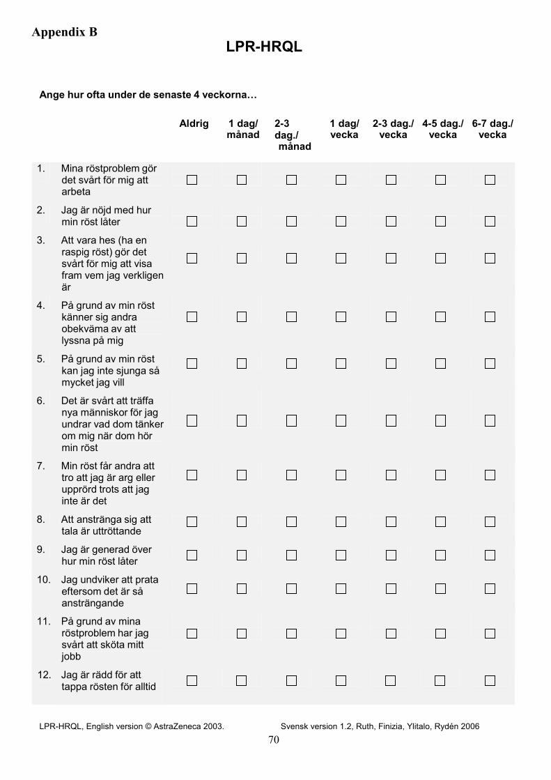
70
Appendix B
LPR-HRQL
Ange hur ofta under de senaste 4 veckorna…
Aldrig 1 dag/ månad
2-3 dag./ månad
1 dag/ vecka
2-3 dag./ vecka
4-5 dag./ vecka
6-7 dag./ vecka
1. Mina röstproblem gör det svårt för mig att arbeta
2. Jag är nöjd med hur
min röst låter 3. Att vara hes (ha en
raspig röst) gör det svårt för mig att visa fram vem jag verkligen är
4. På grund av min röst
känner sig andra obekväma av att lyssna på mig
5. På grund av min röst
kan jag inte sjunga så mycket jag vill
6. Det är svårt att träffa
nya människor för jag undrar vad dom tänker om mig när dom hör min röst
7. Min röst får andra att
tro att jag är arg eller upprörd trots att jag inte är det
8. Att anstränga sig att
tala är uttröttande 9. Jag är generad över
hur min röst låter 10. Jag undviker att prata
eftersom det är så ansträngande
11. På grund av mina
röstproblem har jag svårt att sköta mitt jobb
12. Jag är rädd för att
tappa rösten för alltid LPR-HRQL, English version © AstraZeneca 2003. Svensk version 1.2, Ruth, Finizia, Ylitalo, Rydén 2006
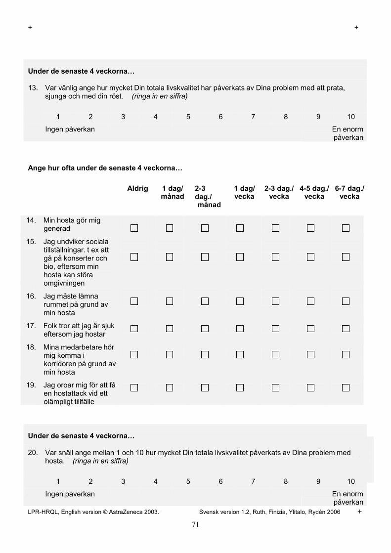
71
+ + Under de senaste 4 veckorna…
13.
Var vänlig ange hur mycket Din totala livskvalitet har påverkats av Dina problem med att prata, sjunga och med din röst. (ringa in en siffra)
1
2
3
4
5
6
7
8
9
10
Ingen påverkan En enorm påverkan
Ange hur ofta under de senaste 4 veckorna…
Aldrig 1 dag/ månad
2-3 dag./ månad
1 dag/ vecka
2-3 dag./ vecka
4-5 dag./ vecka
6-7 dag./ vecka
14. Min hosta gör mig generad
15. Jag undviker sociala
tillställningar. t ex att gå på konserter och bio, eftersom min hosta kan störa omgivningen
16. Jag måste lämna
rummet på grund av min hosta
17. Folk tror att jag är sjuk
eftersom jag hostar 18. Mina medarbetare hör
mig komma i korridoren på grund av min hosta
19. Jag oroar mig för att få
en hostattack vid ett olämpligt tillfälle
Under de senaste 4 veckorna…
20.
Var snäll ange mellan 1 och 10 hur mycket Din totala livskvalitet påverkats av Dina problem med hosta. (ringa in en siffra)
1
2
3
4
5
6
7
8
9
10
Ingen påverkan En enorm påverkan
LPR-HRQL, English version © AstraZeneca 2003. Svensk version 1.2, Ruth, Finizia, Ylitalo, Rydén 2006 +

72
+ + Ange hur ofta under de senaste 4 veckorna…
Aldrig 1 dag/ månad
2-3 dag./ månad
1 dag/ vecka
2-3 dag./ vecka
4-5 dag./ vecka
6-7 dag./ vecka
21. Folk lägger märke till hur ofta jag måste harkla mig
22. Mitt sexliv påverkas
negativt av att jag måste harkla mig
23. Relationen till mina
vänner påverkas negativt av att jag måste harkla mig
24. Behovet att harkla mig
gör det svårt att prata 25. Jag blir frustrerad av
att behöva harkla mig så ofta som jag gör
26. Jag undviker sociala
tillställningar. t ex att gå på konserter och bio, eftersom jag behöver harkla mig
Under de senaste 4 veckorna…
27.
Var snäll ange mellan 1 och 10 hur mycket Din totala livskvalitet påverkats av Dina problem med harkling. (ringa in en siffra)
1
2
3
4
5
6
7
8
9
10
Ingen påverkan En enorm påverkan
LPR-HRQL, English version © AstraZeneca 2003. Svensk version 1.2, Ruth, Finizia, Ylitalo, Rydén 2006 +

73
+ + Ange hur ofta under de senaste 4 veckorna…
Aldrig 1 dag/ månad
2-3 dag./ månad
1 dag/ vecka
2-3 dag./ vecka
4-5 dag./ vecka
6-7 dag./ vecka
28. Jag känner en klump i halsen som gör det svårt att svälja
29. Jag drar mig för att äta
offentligt, t ex på restauranger eller fester, eftersom jag har svårt att svälja
30. Jag är rädd för att
kvävas i sömnen 31. Jag besväras av en
brännande känsla i halsen
32. Jag vaknar av att jag
kippar efter luft Under de senaste 4 veckorna…
33.
Var snäll ange mellan 1 och 10 hur mycket Din totala livskvalitet påverkats av Dina halsproblem. (ringa in en siffra)
1
2
3
4
5
6
7
8
9
10
Ingen påverkan En enorm påverkan
Under de 4 senaste veckorna, hur mycket har de tidigare beskrivna symtomen med röst, hosta, harkling, sväljning/hals påverkat … (ringa in en siffra)
34. Din energi i allmänhet
1 2 3 4 5 6 7 8 9 10
Ingen påverkan En enorm
påverkan
35. Din arbetskapacitet
1 2 3 4 5 6 7 8 9 10
Ingen påverkan En enorm påverkan
LPR-HRQL, English version © AstraZeneca 2003. Svensk version 1.2, Ruth, Finizia, Ylitalo, Rydén +

74
+ + 36. Dina sociala relationer
1 2 3 4 5 6 7 8 9 10
Ingen påverkan En enorm
påverkan
37. Dina äktenskapliga/intima relationer
1 2 3 4 5 6 7 8 9 10
Ingen påverkan En enorm påverkan
38. Dina sexuella relationer
1 2 3 4 5 6 7 8 9 10
Ingen påverkan En enorm
påverkan
39. Din sömn
1 2 3 4 5 6 7 8 9 10
Ingen påverkan En enorm påverkan
40. Din möjlighet att ligga bekvämt i en säng
1 2 3 4 5 6 7 8 9 10
Ingen påverkan En enorm
påverkan 41. Din uppfattning om Dig själv
1 2 3 4 5 6 7 8 9 10
Ingen påverkan En enorm
påverkan 42. Din livsstil (t ex, rökning, alkoholintag, motion, matvanor)
1 2 3 4 5 6 7 8 9 10
Ingen påverkan En enorm
påverkan
43. Hur mycket har de tidigare beskrivna symtomen begränsat Din förmåga att göra sådant du tycker om? (ringa in en siffra)
1 2 3 4 5 6 7 8 9 10
Inte alls begränsad
Mycket
begränsad – oförmögen att
göra

75