Laparoscopic-Assisted Pull-Through for Congenital Rectal ...
5
Laparoscopic-Assisted Pull-Through for Congenital Rectal Stenosis Martin L. van Niekerk, MD, Andre Visser, MD, and Daniel J. Venter, MD Abstract Congenital rectal stenosis is a rare condition. In this article, we report on a 30-day-old infant with rectal stenosis who underwent a successful laparoscopic pull-through operation. The laparoscopic method allows the surgeon to mobilize the rectum within the muscle complex without the division of any muscles of continence. Introduction A norectal malformations include a wide spectrum of anomalies and occur in about 1 in 4000–5000 live births. 1 Rectal stenosis is one of the most rare types of anorectal malformations. In this anomaly, the stenotic rectum lies within the levator animuscle complex, the patient has a nor- mal anal canal, and there is usually no fistula. 2 In 2000, Keith Georgeson proposed a laparoscopic approach as part of a new technique to repair high imperforate anal defects. 3 This tech- nique allows excellent visualization and definition of the pelvic anatomy without the saggital division of muscles. In this article, we present what we believe to be the first case report of a laparoscopic-assisted pull-through procedure for a congenital rectal stenosis. Patient and Method A 2-week-old, 3.1-kg male baby was admitted 14 days after birth, with symptoms of bowel obstruction and abdominal dis- tension. There was a history of frequent passing of small liquid stools. On perineal inspection, the baby had a normal anus, but the rectal examination revealed a blind-ending anal canal (Fig. 1). Method The baby was resuscitated, and an emergency sigmoid colostomy was carried out. A subsequent cologram, using water-soluble contrast, showed a distended distal sigmoid colon with a rectal stenosis (Fig. 2). Surgical technique The patient was operated on 14 days after the colostomy procedure, using a laparoscopic and transanal approach. He was placed in a supine position transversely across the end of the table. The bladder was catheterized to prevent postoper- ative urinary retention and also to allow palpation of the urethra during the dissection. A 5-mm camera port was placed just above the umbilicus, and a pneumoperitoneum was established. One 3-mm port each was placed in the upper and lower left quadrants of the abdomen. Another 5-mm port was placed in the lower right quadrant. The dissection was started at the level of peritoneal reflection. The whole rectum with the distal stenotic part was mobilized by using mono- polar hook coagulation (Fig. 3). The rectum was mobilized as far as its connection with the blind-ending anal canal. A Lone Star retractor (Lone Star Medical Products, Houston, TX) was used to keep the anal canal open (Fig. 4). A circumferential incision was made in the proximal part of the blind-ending anal canal. The rectum was grasped and extracted through the anus. The stenotic rectum was then resected, and rectoanal anastomosis was accomplished with interrupted 5.0 Vicryl sutures (Johnson & Johnson, Johannesburg, South Africa) (Fig. 5). The total operating time was 88 minutes. The postoperative course was uneventful. The patient was discharged on the postoperative day 3. A single anal dilatation was done after the laparoscopic operation, and the colostomy was closed 2 weeks later. The patient is now 2 months postcolostomy closure and passes an average of three normal stools a day. Discussion Rectal atresia and stenosis are rare types of anorectal mal- formations. They comprise no more than 1–2% of the entire spectrum of anorectal malformations. 4 The exact pathogene- sis of rectal stenosis is not known, but it might be a secondary development from an intrauterine vascular accident involv- ing the middle and superior rectal arteries. Genetic and environmental factors may also be responsible. 5 In the main Wingspread classification, rectal stenosis has been tabulated as a rare malformation 6 and in the International Krickenbeck Classification as a rare variant of the major clinical groups. 7 Department of Pediatric Surgery, University of Pretoria, Pretoria, South Africa. JOURNAL OF LAPAROENDOSCOPIC & ADVANCED SURGICAL TECHNIQUES Volume 20, Number 1, 2010 ª Mary Ann Liebert, Inc. DOI: 10.1089=lap.2008.0343 107
Transcript of Laparoscopic-Assisted Pull-Through for Congenital Rectal ...

Laparoscopic-Assisted Pull-Throughfor Congenital Rectal Stenosis
Martin L. van Niekerk, MD, Andre Visser, MD, and Daniel J. Venter, MD
Abstract
Congenital rectal stenosis is a rare condition. In this article, we report on a 30-day-old infant with rectal stenosiswho underwent a successful laparoscopic pull-through operation. The laparoscopic method allows the surgeonto mobilize the rectum within the muscle complex without the division of any muscles of continence.
Introduction
Anorectal malformations include a wide spectrum ofanomalies and occur in about 1 in 4000–5000 live births.1
Rectal stenosis is one of the most rare types of anorectalmalformations. In this anomaly, the stenotic rectum lieswithin the levator animuscle complex, the patient has a nor-mal anal canal, and there is usually no fistula.2 In 2000, KeithGeorgeson proposed a laparoscopic approach as part of a newtechnique to repair high imperforate anal defects.3 This tech-nique allows excellent visualization and definition of thepelvic anatomy without the saggital division of muscles. Inthis article, we present what we believe to be the first casereport of a laparoscopic-assisted pull-through procedure for acongenital rectal stenosis.
Patient and Method
A 2-week-old, 3.1-kg male baby was admitted 14 days afterbirth, with symptoms of bowel obstruction and abdominal dis-tension. There was a history of frequent passing of small liquidstools. On perineal inspection, the baby had a normal anus, butthe rectal examination revealed a blind-ending anal canal (Fig. 1).
Method
The baby was resuscitated, and an emergency sigmoidcolostomy was carried out. A subsequent cologram, usingwater-soluble contrast, showed a distended distal sigmoidcolon with a rectal stenosis (Fig. 2).
Surgical technique
The patient was operated on 14 days after the colostomyprocedure, using a laparoscopic and transanal approach. Hewas placed in a supine position transversely across the end ofthe table. The bladder was catheterized to prevent postoper-ative urinary retention and also to allow palpation of the
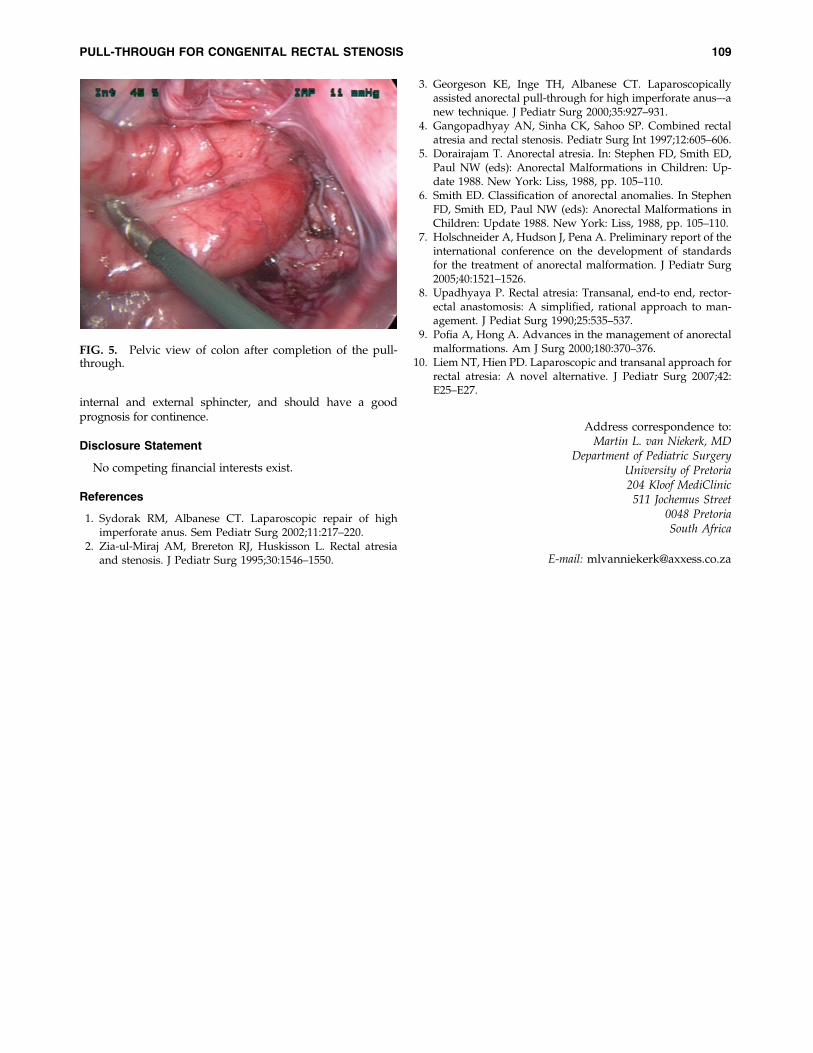
urethra during the dissection. A 5-mm camera port wasplaced just above the umbilicus, and a pneumoperitoneumwas established. One 3-mm port each was placed in the upperand lower left quadrants of the abdomen. Another 5-mm portwas placed in the lower right quadrant. The dissection wasstarted at the level of peritoneal reflection. The whole rectumwith the distal stenotic part was mobilized by using mono-polar hook coagulation (Fig. 3). The rectum was mobilized asfar as its connection with the blind-ending anal canal. A LoneStar retractor (Lone Star Medical Products, Houston, TX) wasused to keep the anal canal open (Fig. 4). A circumferentialincision was made in the proximal part of the blind-endinganal canal. The rectum was grasped and extracted through theanus. The stenotic rectum was then resected, and rectoanalanastomosis was accomplished with interrupted 5.0 Vicrylsutures (Johnson & Johnson, Johannesburg, South Africa)(Fig. 5).
The total operating time was 88 minutes. The postoperativecourse was uneventful. The patient was discharged on thepostoperative day 3. A single anal dilatation was done afterthe laparoscopic operation, and the colostomy was closed2 weeks later. The patient is now 2 months postcolostomyclosure and passes an average of three normal stools a day.
Discussion
Rectal atresia and stenosis are rare types of anorectal mal-formations. They comprise no more than 1–2% of the entirespectrum of anorectal malformations.4 The exact pathogene-sis of rectal stenosis is not known, but it might be a secondarydevelopment from an intrauterine vascular accident involv-ing the middle and superior rectal arteries. Genetic andenvironmental factors may also be responsible.5 In the mainWingspread classification, rectal stenosis has been tabulatedas a rare malformation6 and in the International KrickenbeckClassification as a rare variant of the major clinical groups.7
Department of Pediatric Surgery, University of Pretoria, Pretoria, South Africa.
JOURNAL OF LAPAROENDOSCOPIC & ADVANCED SURGICAL TECHNIQUESVolume 20, Number 1, 2010ª Mary Ann Liebert, Inc.DOI: 10.1089=lap.2008.0343
107

Patients with this anorectal anomaly have a well-developedanus. The internal and external sphincters are normal, but areoccasionally incompletely developed. The stenotic rectum lieswithin a normally developed levator animuscle complex.There are usually no fistulous communications of the urinarytract or vagina.4
Many innovative techniques have been used in the past totreat rectal stenosis,8 including posterior saggital anorecto-plasty (PSARP).9 Since the introduction of the laparoscopic-assisted anorectal pull-through operation in 2000, this methodhas been a safe, useful treatment for anorectal malformations.10
It accomplishes the same anatomic end result as the PSARPtechnique while minimizing the risk of injury to the sur-rounding anatomic structures. Liem and Hien10 published a
report on the laparoscopic and transanal approach for rectalatresia in 2 patients. In our report, we show that rectal stenosiscan also be treated successfully by the laparoscopic approach.
In our patient, the laparoscopic-assisted method allowedexcellent pelvic vision of the levator muscle complex and en-abled us to mobilize the rectum within the muscle complex. Bystaying close to the rectal wall during dissection, we minimizedthe chance of any injury to surrounding anatomic structuresand the muscles of continence. Laparoscopic mobilization wasvery easy because of the absence of fistulas in this patient.Additional advantages of the laparoscopic method include lesspain and potentially fewer perineal wound complications.
Conclusion
The laparoscopic-assisted repair of rectal stenosis is an idealmethod with which to treat this rare anorectal malformation.It allows proper placement of the pull-through rectum in thelevator muscle complex, without dividing any muscle of con-tinence. These patients usually have a normal anal canal and
FIG. 1. On perineal inspection, the anus appeared normal.
FIG. 2. Cologram through sigmoid colostomy showing rec-tal stenosis.
FIG. 3. Laparoscopic view of the mobilized rectum.
FIG. 4. Coloanal anastomosis done with the aid of the LoneStar retractor (Lone Star Medical Products, Houston, TX).
108 VAN NIEKERK ET AL.
internal and external sphincter, and should have a goodprognosis for continence.
Disclosure Statement
No competing financial interests exist.
References
1. Sydorak RM, Albanese CT. Laparoscopic repair of highimperforate anus. Sem Pediatr Surg 2002;11:217–220.
2. Zia-ul-Miraj AM, Brereton RJ, Huskisson L. Rectal atresiaand stenosis. J Pediatr Surg 1995;30:1546–1550.
3. Georgeson KE, Inge TH, Albanese CT. Laparoscopicallyassisted anorectal pull-through for high imperforate anus–-anew technique. J Pediatr Surg 2000;35:927–931.
4. Gangopadhyay AN, Sinha CK, Sahoo SP. Combined rectalatresia and rectal stenosis. Pediatr Surg Int 1997;12:605–606.
5. Dorairajam T. Anorectal atresia. In: Stephen FD, Smith ED,Paul NW (eds): Anorectal Malformations in Children: Up-date 1988. New York: Liss, 1988, pp. 105–110.
6. Smith ED. Classification of anorectal anomalies. In StephenFD, Smith ED, Paul NW (eds): Anorectal Malformations inChildren: Update 1988. New York: Liss, 1988, pp. 105–110.
7. Holschneider A, Hudson J, Pena A. Preliminary report of theinternational conference on the development of standardsfor the treatment of anorectal malformation. J Pediatr Surg2005;40:1521–1526.
8. Upadhyaya P. Rectal atresia: Transanal, end-to end, rector-ectal anastomosis: A simplified, rational approach to man-agement. J Pediat Surg 1990;25:535–537.
9. Pofia A, Hong A. Advances in the management of anorectalmalformations. Am J Surg 2000;180:370–376.
10. Liem NT, Hien PD. Laparoscopic and transanal approach forrectal atresia: A novel alternative. J Pediatr Surg 2007;42:E25–E27.
Address correspondence to:Martin L. van Niekerk, MD
Department of Pediatric SurgeryUniversity of Pretoria204 Kloof MediClinic511 Jochemus Street
0048 PretoriaSouth Africa
E-mail: [email protected]
FIG. 5. Pelvic view of colon after completion of the pull-through.
PULL-THROUGH FOR CONGENITAL RECTAL STENOSIS 109


Copyright of Journal of Laparoendoscopic & Advanced Surgical Techniques is the property of Mary Ann
Liebert, Inc. and its content may not be copied or emailed to multiple sites or posted to a listserv without the
copyright holder's express written permission. However, users may print, download, or email articles for
individual use.


![Retrospective Study Critical appraisal of laparoscopic vs ... · with colon and rectal cancer[1214]. This is suboptimal as the prognosis and recurrence pattern of colon and rectal](https://static.fdocuments.in/doc/165x107/5edc7da5ad6a402d66672bbb/retrospective-study-critical-appraisal-of-laparoscopic-vs-with-colon-and-rectal.jpg)