Langerhans cells down-regulate inflammation-driven … · Langerhans cells down-regulate...
6
Langerhans cells down-regulate inflammation-driven alveolar bone loss Moran Arizon a,1 , Itay Nudel a,1 , Hadas Segev a,1 , Gabriel Mizraji a , Mazal Elnekave a , Karina Furmanov a , Luba Eli-Berchoer a , Björn E. Clausen b , Lior Shapira c , Asaf Wilensky c,2 , and Avi-Hai Hovav a,2,3 a Institute of Dental Sciences and c Department of Periodontology, Hebrew University–Hadassah School of Dental Medicine, Jerusalem 91120, Israel; and b Department of Immunology, Erasmus Medical Center, University Medical Center, 3015 GE Rotterdam, The Netherlands Edited by John T. Potts, Massachusetts General Hospital, Charlestown, MA, and approved March 28, 2012 (received for review October 11, 2011) Excessive bone resorption is frequently associated with chronic infections and inflammatory diseases. Whereas T cells were demonstrated to facilitate osteoclastogenesis in such diseases, the role of dendritic cells, the most potent activators of naive T cells, remains unclear. Using a model involving inflammation- driven alveolar bone loss attributable to infection, we showed that in vivo ablation of Langerhans cells (LCs) resulted in enhanced bone loss. An increased infiltration of B and T lymphocytes into the tissue surrounding the bone was observed in LC-ablated mice, including receptor activator of NF-κB ligand (RANKL)-expressing CD4 + T cells with known capabilities of altering bone homeostasis. In addition, the absence of LCs significantly reduced the numbers of CD4 + Foxp3 + T-regulatory cells in the tissue. Further investiga- tion revealed that LCs were not directly involved in presenting antigens to T cells. Nevertheless, despite their low numbers in the tissue, the absence of LCs resulted in an elevated activation of CD4 + but not CD8 + T cells. This activation involved elevated production of IFN-γ but not IL-17 or IL-10 cytokines. Our data, thus, reveal a protective immunoregulatory role for LCs in inflam- mation-induced alveolar bone resorption, by inhibiting IFN-γ se- cretion and excessive activation of RANKL + CD4 + T cells with a capability of promoting osteoclastogenesis. osteoimmunology | Porphyromonas gingivalis | oral mucosa | experimental periodontitis A connection between inflammation and bone disease has been established in numerous clinical and animal studies (1). Bone loss attributable to inflammation could be found in many diseases, such as rheumatoid arthritis, multiple myeloma, diabetes mellitus, lupus erythematosus, and periodontal disease (2). Such diseases could develop because of infection or auto- immunity; they could be acute or chronic and either local or systemic. Although the mechanisms underlying the bone loss are complex, it appears that they involve disruption of the bone remodeling cycle. Bone formation and resorption is mediated by two cell types: osteoblasts and osteoclasts, respectively. Re- ceptor activator of NF-κB ligand (RANKL), its receptor RANK, and the natural antagonist osteoprotegrin (OPG) are key reg- ulators of the differentiation, activation, and survival of osteo- clasts (3). This bone-remodeling cycle is tightly regulated under physiological conditions to guarantee bone homeostasis. Under inflammatory conditions, however, the level of RANKL is in- creased because of its expression and secretion by inflammatory lymphocytes, which facilitate osteoclastogenesis and bone re- sorption (4). Dendritic cells (DCs) are a heterogeneous family of potent antigen-presenting cells with the capability to prime naïve T cells. With regard to inflammation-driven bone loss, attention was given to DCs as cells with an osteoclastogenic potential. DCs derive from the same myeloid precursor as osteoclasts, and the function of both cell types is modulated by common factors. Because of the high developmental and functional plasticity of myeloid cells such as DCs, it has been proposed that under certain inflammatory conditions, DCs could differentiate into osteoclast-like cells with a bone resorption capability (5). Nev- ertheless, as the major cells activating T cells, it is likely that DCs will also have a critical influence on the intensity and quality of the inflammation, thereby acting as potential osteo- immune players. Langerhans cells (LCs) are a unique DC subset expressing the C-type lectin receptor Langerin and are located in mucosal- stratified squamous epithelium and skin epidermis (6). The de- velopment of transgenic mice allowing conditional ablation of LCs in vivo has contributed to our understanding of the role of LCs in eliciting adaptive immune responses and in inducing and maintaining tolerance. The frontline localization of LCs makes them prone to probe the environment and orchestrate T-cell immunity against innocuous or infectious agents. This led us to hypothesize that LCs could play a role in inflammation-driven bone loss, by regulating the quality of the inflammatory reaction. Results In Vivo Ablation of LCs Aggravates Local Inflammation and Enhances Alveolar Bone Loss. Langerin–diphtheria toxin receptor (DTR) mice were administered i.p. with diphtheria toxin (DT) 7 and 5 d before infection to eliminate LCs, as previously shown (7). Infection with the pathogen Porphyromonas gingivalis was per- formed via oral gavage, a relevant model for inducing resorption of alveolar bone. Six weeks after the infection, the hemimaxillae were harvested and scanned using micro computed tomography (μCT) to measure alveolar bone volume. Fig. 1A presents rep- resentative μCT sections of the second upper molar following infection. The distance between the cemento-enamel junction (CEJ) and alveolar bone crest (ABC) in DT-treated mice was larger compared with mice exposed to vehicle alone or infected mice with no DT treatment. This indicates that the lack of LCs resulted in a considerable resorption of the alveolar crest. When bone morphology was evaluated, an irregular cortical plate with small radiolucent punched-out lesions was observed in the al- veolar bone where LCs were ablated, suggesting the occurrence of active bone loss (Fig. 1A). Such lesions were only seen in the ABC of infected mice in the absence of LCs and were not found in the carboxymethylcellulose (CMC)-treated control group. The role of LCs in down-modulating bone resorption was further demonstrated by a 3D quantification of the residual alveolar bone (P < 0.005 compared with infected mice with no DT treatment) (Fig. 1B). Author contributions: L.S., A.W., and A.-H.H. designed research; M.A., I.N., H.S., G.M., K.F., and L.E.-B. performed research; B.E.C. contributed new reagents/analytic tools; M.E. an- alyzed data; and A.W. and A.-H.H. wrote the paper. The authors declare no conflict of interest. This article is a PNAS Direct Submission. 1 M.A., I.N., and H.S. contributed equally to this work. 2 A.W. and A.-H.H. contributed equally to this work. 3 To whom correspondence should be addressed. E-mail: [email protected]. This article contains supporting information online at www.pnas.org/lookup/suppl/doi:10. 1073/pnas.1116770109/-/DCSupplemental. www.pnas.org/cgi/doi/10.1073/pnas.1116770109 PNAS | May 1, 2012 | vol. 109 | no. 18 | 7043–7048 IMMUNOLOGY
Transcript of Langerhans cells down-regulate inflammation-driven … · Langerhans cells down-regulate...

Langerhans cells down-regulate inflammation-drivenalveolar bone lossMoran Arizona,1, Itay Nudela,1, Hadas Segeva,1, Gabriel Mizrajia, Mazal Elnekavea, Karina Furmanova, Luba Eli-Berchoera,Björn E. Clausenb, Lior Shapirac, Asaf Wilenskyc,2, and Avi-Hai Hovava,2,3
aInstitute of Dental Sciences and cDepartment of Periodontology, Hebrew University–Hadassah School of Dental Medicine, Jerusalem 91120, Israel;and bDepartment of Immunology, Erasmus Medical Center, University Medical Center, 3015 GE Rotterdam, The Netherlands
Edited by John T. Potts, Massachusetts General Hospital, Charlestown, MA, and approved March 28, 2012 (received for review October 11, 2011)
Excessive bone resorption is frequently associated with chronicinfections and inflammatory diseases. Whereas T cells weredemonstrated to facilitate osteoclastogenesis in such diseases,the role of dendritic cells, the most potent activators of naive Tcells, remains unclear. Using a model involving inflammation-driven alveolar bone loss attributable to infection, we showedthat in vivo ablation of Langerhans cells (LCs) resulted in enhancedbone loss. An increased infiltration of B and T lymphocytes intothe tissue surrounding the bone was observed in LC-ablated mice,including receptor activator of NF-κB ligand (RANKL)-expressingCD4+ T cells with known capabilities of altering bone homeostasis.In addition, the absence of LCs significantly reduced the numbersof CD4+Foxp3+ T-regulatory cells in the tissue. Further investiga-tion revealed that LCs were not directly involved in presentingantigens to T cells. Nevertheless, despite their low numbers inthe tissue, the absence of LCs resulted in an elevated activationof CD4+ but not CD8+ T cells. This activation involved elevatedproduction of IFN-γ but not IL-17 or IL-10 cytokines. Our data, thus,reveal a protective immunoregulatory role for LCs in inflam-mation-induced alveolar bone resorption, by inhibiting IFN-γ se-cretion and excessive activation of RANKL+CD4+ T cells with acapability of promoting osteoclastogenesis.
osteoimmunology | Porphyromonas gingivalis | oral mucosa |experimental periodontitis
Aconnection between inflammation and bone disease hasbeen established in numerous clinical and animal studies
(1). Bone loss attributable to inflammation could be found inmany diseases, such as rheumatoid arthritis, multiple myeloma,diabetes mellitus, lupus erythematosus, and periodontal disease(2). Such diseases could develop because of infection or auto-immunity; they could be acute or chronic and either local orsystemic. Although the mechanisms underlying the bone lossare complex, it appears that they involve disruption of the boneremodeling cycle. Bone formation and resorption is mediatedby two cell types: osteoblasts and osteoclasts, respectively. Re-ceptor activator of NF-κB ligand (RANKL), its receptor RANK,and the natural antagonist osteoprotegrin (OPG) are key reg-ulators of the differentiation, activation, and survival of osteo-clasts (3). This bone-remodeling cycle is tightly regulated underphysiological conditions to guarantee bone homeostasis. Underinflammatory conditions, however, the level of RANKL is in-creased because of its expression and secretion by inflammatorylymphocytes, which facilitate osteoclastogenesis and bone re-sorption (4).Dendritic cells (DCs) are a heterogeneous family of potent
antigen-presenting cells with the capability to prime naïve Tcells. With regard to inflammation-driven bone loss, attentionwas given to DCs as cells with an osteoclastogenic potential. DCsderive from the same myeloid precursor as osteoclasts, and thefunction of both cell types is modulated by common factors.Because of the high developmental and functional plasticityof myeloid cells such as DCs, it has been proposed that undercertain inflammatory conditions, DCs could differentiate into
osteoclast-like cells with a bone resorption capability (5). Nev-ertheless, as the major cells activating T cells, it is likely thatDCs will also have a critical influence on the intensity andquality of the inflammation, thereby acting as potential osteo-immune players.Langerhans cells (LCs) are a unique DC subset expressing the
C-type lectin receptor Langerin and are located in mucosal-stratified squamous epithelium and skin epidermis (6). The de-velopment of transgenic mice allowing conditional ablation ofLCs in vivo has contributed to our understanding of the role ofLCs in eliciting adaptive immune responses and in inducing andmaintaining tolerance. The frontline localization of LCs makesthem prone to probe the environment and orchestrate T-cellimmunity against innocuous or infectious agents. This led us tohypothesize that LCs could play a role in inflammation-drivenbone loss, by regulating the quality of the inflammatory reaction.
ResultsIn Vivo Ablation of LCs Aggravates Local Inflammation and EnhancesAlveolar Bone Loss. Langerin–diphtheria toxin receptor (DTR)mice were administered i.p. with diphtheria toxin (DT) 7 and5 d before infection to eliminate LCs, as previously shown (7).Infection with the pathogen Porphyromonas gingivalis was per-formed via oral gavage, a relevant model for inducing resorptionof alveolar bone. Six weeks after the infection, the hemimaxillaewere harvested and scanned using micro computed tomography(μCT) to measure alveolar bone volume. Fig. 1A presents rep-resentative μCT sections of the second upper molar followinginfection. The distance between the cemento-enamel junction(CEJ) and alveolar bone crest (ABC) in DT-treated mice waslarger compared with mice exposed to vehicle alone or infectedmice with no DT treatment. This indicates that the lack of LCsresulted in a considerable resorption of the alveolar crest. Whenbone morphology was evaluated, an irregular cortical plate withsmall radiolucent punched-out lesions was observed in the al-veolar bone where LCs were ablated, suggesting the occurrenceof active bone loss (Fig. 1A). Such lesions were only seen in theABC of infected mice in the absence of LCs and were not foundin the carboxymethylcellulose (CMC)-treated control group. Therole of LCs in down-modulating bone resorption was furtherdemonstrated by a 3D quantification of the residual alveolarbone (P < 0.005 compared with infected mice with no DTtreatment) (Fig. 1B).
Author contributions: L.S., A.W., and A.-H.H. designed research; M.A., I.N., H.S., G.M., K.F.,and L.E.-B. performed research; B.E.C. contributed new reagents/analytic tools; M.E. an-alyzed data; and A.W. and A.-H.H. wrote the paper.
The authors declare no conflict of interest.
This article is a PNAS Direct Submission.1M.A., I.N., and H.S. contributed equally to this work.2A.W. and A.-H.H. contributed equally to this work.3To whom correspondence should be addressed. E-mail: [email protected].
This article contains supporting information online at www.pnas.org/lookup/suppl/doi:10.1073/pnas.1116770109/-/DCSupplemental.
www.pnas.org/cgi/doi/10.1073/pnas.1116770109 PNAS | May 1, 2012 | vol. 109 | no. 18 | 7043–7048
IMMUNOLO
GY
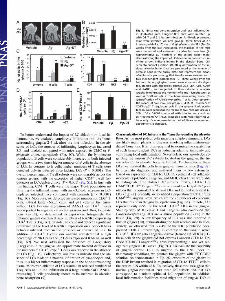
To better understand the impact of LC ablation on local in-flammation, we analyzed lymphocyte infiltration into the bone-surrounding gingiva 2–3 wk after the first infection. In the ab-sence of LCs, the number of infiltrating lymphocytes increased3.5- and twofold compared with mice exposed to CMC or P.gingivalis alone, respectively (Fig. 1C). Within the lymphocytepopulation, B cells were considerably increased in both infectedgroups, with a two times higher number of B cells in the absenceof LCs. In contrast to B cells, higher numbers of T cells weredetected only in infected mice lacking LCs (P < 0.0001). Theoverall percentages of T-cell subsets were comparable across thevarious groups, with the exception of higher CD4+ T-cell fre-quencies in LC-depleted mice (P < 0.001) (Fig. S1). In line withthis finding, CD4+ T cells were the major T-cell population in-filtrating the inflamed tissue, with an ∼3.2-fold increase in LC-depleted infected mice compared with controls (P < 0.0001)(Fig. 1C). Moreover, we detected increased numbers of CD8+ Tcells, natural killer (NKT) cells, and γδT cells in the tissuewithout LCs. Because expression of RANKL on CD4+ T cellswas reported to regulate osteoclastogenesis and, thus, facilitatebone loss (8), we determined its expression. Intriguingly, theinflamed gingiva contained large numbers of RANKL-expressingCD4+ T cells (Fig. 1D). Of note, we could not detect a significantdifference in the level of RANKL expression on a per-cell basisbetween infected mice in the presence or absence of LCs. Inaddition to CD4+ T cells, our analysis revealed that a highpercentage of NKT cells and γδT cells expressed RANKL as well(Fig. 1D). We next addressed the presence of T-regulatory(Treg) cells in the gingiva. An approximate twofold decrease inthe numbers of CD4+Foxp3+ T cells was detected in the absenceof LCs (Fig. 1E). Collectively, our results indicate that the ab-sence of LCs leads to a massive infiltration of lymphocytes and,thus, to a higher inflammatory response in the bone-surroundingtissue. Moreover, ablation of LCs resulted in reduced numbers ofTreg cells and in the infiltration of a large number of RANKL-expressing T cells previously shown to be involved in alveolarbone resorption (9).
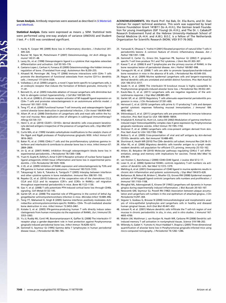
Characterization of DC Subsets in the Tissue Surrounding the AlveolarBone. As the most potent cells initiating adaptive immunity, DCsare likely major players in diseases involving inflammation-me-diated bone loss. It is, thus, essential to examine the capabilitiesof such tissue-resident DCs in inducing adaptive immunity andcontrolling local inflammation. Nevertheless, our knowledge re-garding the various DC subsets located in the gingiva, the tis-sue adjacent to alveolar bone, is limited. To characterize theseDCs, we isolated the cells from gingival tissue of mice (Fig. S2),by enzymatic digestion and analyzed them by flow cytometry.Based on expression of CD11c, CD103, epithelial cell adhesionmolecule (Ep-CAM), Langerin, and MHC class II, we were ableto distinguish three distinct DC subsets. Firstly, CD11c+Ep-CAMnegCD103negLangerinneg cells represent the largest DC pop-ulation that is equivalent to dermal DCs and termed interstitial (i)DCs (Fig. 2A). Secondly, we identified a population of CD11c+Ep-CAMbrightLangerin+ cells, which are the equivalents of epidermalLCs that reside in the gingival epithelium (Fig. 2A). Of note, LCsrepresent only 2–5% of the total CD11c+ DCs in the gingiva.Staining with MHC class II and Langerin also confirmed thatLangerin-expressing DCs are a minor population (∼3%) in thetissue (Fig. 2B). A low frequency of LCs was also reported inhuman gingiva (10), demonstrating the high resemblance to mice.Thirdly, we observed that ∼3–4% of the iDC population coex-pressed CD103. Interestingly, in contrast to the skin in whichCD103+ DCs are also Langerin-positive (termed Ln+dDCs) (11),these cells in the gingiva did not express Langerin (CD11c+Ep-CAM−CD103+Langerinneg), thus representing a not yet rec-ognized gingival DC subset (Fig. 2C). To evaluate the capabilityof gingival-derived DCs to migrate to the LNs under in-flammatory conditions, we painted the gingiva with FITC/DBPsolution. As demonstrated in Fig. 2D, exposure of the gingiva tothe DBP irritant resulted in migration of CD11c+FITC+ cells tothe cervical LN within 48 h. Collectively, these results suggest thatmurine gingiva contain at least three DC subsets and that LCscorrespond to a minor epithelial DC population. In addition,local inflammation facilitates rapid migration of gingival DCs to
A B
C
D E
Fig. 1. Enhanced bone resorption and local inflammationin LC-ablated mice. Langerin-DTR mice were injected i.p.with DT 7 and 5 d before infection. Antibiotic pretreatedmice were infected via oral gavage, three times at 2-dintervals, with 4 × 10⁹ cfu of P. gingivalis strain 381 (Pg). Sixweeks after the last inoculation, the maxillae of the micewere harvested and examined for alveolar bone loss. (A)Representative μCT sections of the second upper molardemonstrating the impact of LC deletion on bone structure.White arrows indicate lesions in the alveolar bone. CEJ,cemento-enamel junction. (B) 3D quantification of the re-sidual alveolar bone. Data are presented as the volume ofalveolar bone in the buccal plate and represent the meansof eight mice per group ± SEM. Results are representative oftwo independent experiments. (C) Three weeks after thelast inoculation, gingival tissues were enzymatically diges-ted, stained with antibodies against CD3, CD4, CD8, CD19,and RANKL, and subjected to flow cytometric analysis.Graphs demonstrate the numbers of B and T lymphocytes, aswell as T-cell subsets, in the bone-surrounding tissue. (D)Quantification of RANKL-expressing T cells. Data representthe means of five mice per group ± SEM. (E) Numbers ofCD4+Foxp3+ T regulatory cells in the gingiva 5 wk postin-fection. Data represent the means of five mice per group ±SEM. **P < 0.0001 compared with infected mice with noDT treatment; *P < 0.05 compared with mice receiving ve-hicle only. One representative out of three independentexperiments is depicted.
7044 | www.pnas.org/cgi/doi/10.1073/pnas.1116770109 Arizon et al.

the cervical draining lymph nodes (LNs) for subsequent activationof T cells.
Lack of LCs Facilitates the Elicitation of Pathogen-Specific CD4+
T Cells. We have demonstrated that the absence of LCs resul-ted in a massive infiltration of T cells into the bone surroundinggingiva. To better understand the role of LCs in T-cell activation,we first examined antigen-presenting capabilities of tissue-de-rived DCs. For this purpose, we have developed an ex vivo assayto evaluate T-cell activation following infection. Mice wereinfected once i.p. with P. gingivalis and used 4 wk later asa source of T cells in this assay. Another cohort of mice wasinfected orally as described in Material and Methods, and theirdraining cervical LNs were collected to isolate various DC sub-sets by flow cytometry. These purified DCs were cocultured for72 h with pathogen-specific CD4+ or CD8+ T cells, and secre-tion of IFN-γ was analyzed as a readout of DC-mediated T-cellactivation. Secretion of IFN-γ in this assay was P. gingivalis-specific, as demonstrated in our validation assay (Fig. S3). Be-cause the oral infection regimen consists of three constitutiveinoculations with the pathogen, we analyzed antigen-presentingactivity following the first and last inoculations. Fig. 3A illustratesour gating strategy for purifying each DC subsets. Tissue-derivedDCs were segregated from LN-resident DCs (LN-DCs) based onthe lack of CD8 expression, further divided into CD103+ andnegative DCs, and the latter were further separated into iDCsand LCs according to Ep-CAM expression (Fig. 3A). Our anal-ysis revealed that following the first infection, orally-derived DCsdiffer in their ability to stimulate CD4+ or CD8+ T cells (Fig.3B). Whereas iDCs were the only subset presenting antigen toCD4+ T cells, presentation to CD8+ T cells was mediated byiDCs and LCs. Limited presentation was detected by CD103+
DCs. Of note, we detected activation of CD4+ and CD8+ T cellsby LN-resident DCs. We next analyzed antigen presentation
following the third inoculation and observed only negligible T-cell activation, suggesting that T-cell priming was already com-pleted at this stage (Fig. S4).Next, we tested the impact of LC ablation on T-cell priming.
We used the ex vivo assay described above to measure T-cellactivation after elimination of LCs in orally infected mice. Fol-lowing the first inoculation, CD11c+ DCs were FACS-sorted andcocultured with pathogen-specific CD4+ or CD8+ T cells. Theabsence of LCs resulted in significantly increased secretion ofIFN-γ by pathogen-specific CD4+ T cells, indicating an elevatedactivation of these cells by migratory DCs (P < 0.05) (Fig. 3C). Incontrast, the lack of LCs had no effect on CD8+ T-cell activa-tion. This suggests that LCs are essential for downregulatingCD4+ T-cell responses.
LC Ablation Increased IFN-γ but Had No Impact on IL-17 or IL-10Production. Our data demonstrated that P. gingivalis-infectedmice developed more vigorous T and B-cell responses attribut-able to the lack of LCs. Therefore, we sought to determinewhether the nature of these elevated adaptive immune responseswas altered in the absence of LCs. To this aim, we collected sera6 wk after infection and examined the elicitation of pathogen-specific antibodies. High IgG titers were measured in P. gingi-valis-infected groups with a modest, but statistically significant,increase in IgG levels in LC-ablated mice (Fig. 4A). Furtheranalysis revealed that this increase was mainly attributable to anelevation in the titers of IgG2c (P < 0.005 at 1:104 dilution) andnot IgG1 (P = 0.1 at 1:104 dilution) (Fig. 4A). We also measuredthe levels of IgA in the saliva, the port of entry of P. gingivalis inour model. As demonstrated in Fig. S5, the levels of IgA werenot affected by the absence of LCs. We then analyzed cytokinesecretion by splenocytes upon in vitro exposure to a recombinantP. gingivalis antigen (RgpA-Ad) (12). A higher production ofIFN-γ was found by splenocytes derived from infected mice thatlack LCs compared with LC-competent infected animals (Fig.4B). Analysis of IL-10 secretion by splenocytes demonstratedthat the addition of RgpA-Ad induced a similar production ofIL-10 by all of the groups compared with unstimulated spleno-cytes (Fig. 4C). Finally, we observed no significant differences inthe levels of IL-17A secretion in the absence or presence of LCs(Fig. 4D). In concordance, neutrophil infiltration into the gingivawas not affected by the lack of LCs (Fig. S6). Taken together, ouranalysis suggests that LC ablation augmented the production ofIFN-γ in infected mice, whereas no significant impact was foundon the secretion of IL-10 and IL-17 cytokines.
DiscussionWe demonstrated in this study the critical role of LCs in con-trolling inflammatory-driven alveolar bone loss following P. gin-givalis infection. In the absence of LCs, infected mice developedreduced Treg cell numbers, produced elevated levels of IFN-γ,and activated high numbers of CD4+ T cells. Furthermore,considerable numbers of RANKL-expressing cells were found inthe bone-surrounding gingiva, in particular CD4+ T cells, whichresulted in enhanced destruction of alveolar bone. Our resultsare in line with the well-established role of RANK-RANKLinteractions, CD4+ T cells, and IFN-γ during experimentalperiodontitis, the model used in the present study (9, 13–15).Recent in vitro studies have questioned the role of IFN-γ in
inflammation-driven bone loss, because IFN-γ was shown tosuppress osteoclastogenesis by inhibiting RANK-RANKL sig-naling (16, 17). Nevertheless, the bone-destructive function ofIFN-γ in vivo seems to overcome these in vitro results (18–21).With regard to experimental periodontitis, IFN-γ was shown toincrease the number of RANKL-expressing CD4+ T cells andalveolar bone loss in vivo (21). Furthermore, another in vitrostudy has shown that IFN-γ-producing T cells were capable ofinducing monocyte differentiation into osteoclasts by RANK-
A B
C D
Fig. 2. Characterization of DC subsets in the bone-surrounding gingiva.Gingival and ear skin tissues were obtained from B6 mice and enzymaticallydigested to obtain single-cell suspension. The cells were treated by surfaceand intracellular staining using various panels of antibodies and analyzed byflow cytometry. (A) Assessment of Ep-CAM and Langerin expression onCD11c+ cells. (B) Analysis of MHC class II and Langerin expression on totalgingival cell suspension. (C) Analysis was restricted to CD11c+ cells and theexpression of CD103 versus Langerin was assessed. (D) Gingival tissues of B6mice (n = 4) were painted with FITC/DBP solution, and 2 d later, the drainingLNs were collected and pooled and CD11c+ cells were enriched and subjectedto flow cytometric analysis. FACS plots are presented confirming the mi-gration of gingiva-derived DCs (FITC+CD11c+) to the LNs. Results are repre-sentative of at least three independent experiments.
Arizon et al. PNAS | May 1, 2012 | vol. 109 | no. 18 | 7045
IMMUNOLO
GY

RANKL signaling, whereas non-IFN-γ producers failed to do so(22). This finding is in line with the vast presence of RANKL-expressing T cells in the gingiva of LC-depleted mice. Th17 cellswere also proposed to play a protective role in P. gingivalis-in-duced alveolar bone loss, by recruiting neutrophils (23). Never-theless, we observed no impact on IL-17 production in LC-ablated mice, nor on the frequencies of neutrophils in theinfected gingiva. It is, thus, likely that the protective role of LCsin our experimental system does not involve Th17 cells. We alsodetected an increased infiltration of RANKL-expressing NKTand γδT cells in the lack of LCs. These T-cell subsets have beenfound to be elevated in human periodontitis (24, 25), suggestingthat NKT and γδT cells might contribute to bone destruction asa source of RANKL.Besides T cells, our data identified large numbers of B cells in
the bone-surrounding gingiva of infected mice, which furtherincreased by the lack of LCs. Our findings are in concurrencewith a recent report demonstrating that B cells outnumber Tcells in periodontal lesions (26). In agreement with previousobservations regarding the phenotype of B cells in the inflamedgingiva, ∼30% of B cells accumulating in the gingiva of LC-ab-lated mice expressed RANKL (27). Still, the importance of theseB cells in mediating alveolar bone loss is uncertain, as a consid-erable number of B cells was also found in mice without DTtreatment, where no bone loss was observed. It was also dem-onstrated that B cells are not essential for LPS-induced boneloss, whereas T cells mediated this process (28). We, thus, arguethat B cells might contribute to bone loss but are not crucial forthis process. T cells, on the other hand, seem to play a criticalrole in alveolar bone loss, because their number increased only inLC-depleted mice, in which bone resorption was detected. In-terestingly, the absence of LCs resulted in higher production of
IgG2c antibodies. This is in concurrence with recent workdemonstrating higher IgG2c levels in LC-deficient mice follow-ing DNA vaccination (29). Of note, in this study, Ln+dDCs wereproposed to augment IFN-γ production by plasmid DNA-elicitedT cells. Although the gingival equivalent of this DC subset wasnot clearly identified in our analysis, it is intriguing to speculatewhether these cells present in extremely low numbers in thegingiva and can impact T-cell function.Our data suggest that the absence of LCs skewed, to some
extent, P. gingivalis-specific immunity toward a Th1-type re-sponse. This observation is in agreement with previous in vivostudies involving P. gingivalis-mediated bone loss (13, 30). Thecapability of LCs to negatively regulate Th1-type pathogen-spe-cific immune responses was recently demonstrated duringLeishmania infection (31). Similar to our study, the lack of LCsduring infection with Leishmania reduced the generation of Tregcells at the site of inflammation. Indeed, Treg cells were recentlyreported to attenuate alveolar bone loss (32). Depletion of LCswas also found to dampen cell-mediated immunity followingmucosal immunization (33). These observations are consistentwith a recent study proposing that LCs are precommitted toinduce tolerance (34). Of note, LC ablation had no impact on P.gingivalis clearance (Fig. S7), unlike Leishmania infection, whereLC-dependent Th1 response was important for pathogen clear-ance (31). This suggests that the quality of the inflammatoryreaction, rather than bacterial load, influences mostly bone loss,further supporting the concept of LCs as immune regulators ofadaptive immunity.The subsets of DCs mediating antigen presentation to CD4+
and CD8+ T cells following P. gingivalis infection seems to bequite different. Whereas iDCs play the major role in presentingantigens to both T-cell subsets, presentation to CD8+ T cells
A
B
C
Fig. 3. Role of LCs in T-cell activation. B6 mice wereinfected orally with P. gingivalis either once (n = 15) orthree times (n = 15). Three days after the infection, LNswere collected, pooled, and enriched for CD11c+ cells. Thecells were stained with antibodies against: CD11c, CD8α,CD103, and Ep-CAM, for further separation by flowcytometry. (A) Flow cytometric plots demonstrating ourstrategy to identify various DC subsets from the drainingLNs. (B) DC subsets purified after a single infection wereincubated with pathogen-specific CD8+ or CD4+ T cells for72 h. Secretion of IFN-γ to the supernatant, as an indicationfor T-cell activation by the DCs, were measured by ELISA.Data represent the means ± SD for each group. (C) Lan-gerin-DTR mice, either LC-depleted or not, were infectedorally once with P. gingivalis. Three days later, the drainingLNs were collected and CD11c+ cells were purified byFACSAria for the assay. CD11c+ cells from various experi-mental groups were incubated with purified pathogen-specific CD8+ or CD4+ T cells for 72 h. Secretion of IFN-γ tothe supernatant was measured by ELISA to quantify thelevel of T-cell activation. Data represent the means of fivemice per group ± SEM. One representative out of two in-dependent experiments is depicted.
7046 | www.pnas.org/cgi/doi/10.1073/pnas.1116770109 Arizon et al.

involved also LCs, LN-DCs, and CD103+ DCs. It is surprisingthat LCs present antigen to CD8+ but not to CD4+ T cells be-cause LCs reside close to the bacterial plaque, engulf thepathogens, and should be able to present bacterial peptides onMHC class II to CD4+ T cells. However, such presentation doesnot occur. A possible explanation could be that P. gingivalismembrane vesicles have the capability to inhibit IFN-γ-inducedMHC class II expression (35). On the other hand, CD8+ T-cellactivation may depend on cross-presentation, a function pre-viously reported for LCs (36), as well as LN-DCs (CD8+ DCs)and CD103+ DCs, thus explaining CD8+ T-cell activation bythese DC subsets in our system (37, 38). This conclusion is fur-ther supported by our inability to detect the pathogen in thedraining LNs, suggesting that LN-DCs obtained pathogen-de-rived antigen from incoming LCs/DCs, as has been demonstratedfor certain viral skin infections (39). Of note, LC ablation hadno impact on the level of antigen presentation to CD8+ T cells;nevertheless, an increased number of these cells was observedin the gingiva of LC-depleted mice. This could be explained bythe critical role of CD4-help in facilitating CD8+ T-cell respon-ses, as well as B-cell activation (40). Thus, inhibition of CD4+
T cells by LCs leads to reduced local inflammation and minimalbone destruction.The accumulation of CD4+ T cells in the gingiva for long
periods of time might influence resident DCs. Activated CD4+ Tcells express RANKL and CD40-ligand (CD40L), moleculeswith a potential to modulate immunity (41). Using transgenicmice that overexpress CD40L or RANKL in keratinocytes, ithas been shown that RANKL suppressed autoimmunity inducedby CD40L (42). Interestingly, in this system the numbers of
epidermal LCs were greatly reduced by CD40L (43), suggestingthat CD40L might regulate LC numbers, as was recently sug-gested also for RANKL (44). Oral DCs have the capability toexpress RANK (Fig. S8) and, thus, might directly interact withRANKL-expressing CD4+ T cells. Although the impact of in-filtrating CD4+ T cells on gingival LCs is unknown, the well-documented reduction in LC numbers during human perio-dontitis could be a result of an imbalance in CD40L/RANKLexpression by local CD4+ T cells (45–47). Gingival DCs mightalso regulate the function of T cells accumulating in the tissue.The gingiva was proposed to act as a tertiary lymphoid site duringperiodontitis, where local DCs can activate T cells (48). Suchperipheral activation of T cells was shown in the skin as well (49)and could enhance gingival inflammation and alveolar bone loss.In summary, this study reveals a protective role for LCs in in-
flammation-driven bone loss initiated by bacterial infection.Through increasing Treg numbers, LCs are proposed to inhibitIFN-γ production and reduce RANKL+ CD4+ T cells in theinflamed tissue. As a result, bone homeostasis is not disturbed bythe local inflammation, and alveolar bone remains intact. Addi-tional experiments involving blocking of RANK-RANKL inter-actions in the absence of LCs might further enforce the proposedmechanism by which LCs impact bone loss. Beyond increasingour understating of the role of LCs in inflammation-inducedbone loss, our data indicate that LCs are promising candidatesfor future approaches attempting to prevent inflammatory bonediseases without compromising protective antibacterial immunity.
Materials and MethodsAntibodies and Reagents. Antibodies and reagents are described in SIMaterials and Methods.
Mice. Six- to 12-wk-old knock-in mice expressing human DTR under tran-scriptional control of the endogenous Langerin/CD207 promoter (Langerin-DTR) were bred in our facility and maintained under specific pathogen-free(SPF) conditions (7). Langerin-DTR mice allow conditional ablation of Lan-gerin-expressing cells in vivo by the administration of DT. The identity of themice used for experiments was confirmed by genotyping with the followingPCR primers: forward, GCCACCATGAAGCTGCTGCCG; and reverse, ATAGT-TTA GCGGCCGCTTTACTTGTACAG. C57BL/6 (B6) mice were purchased fromHarlan and used at the age of 6–12 wk. Research on mice was approved bythe Hebrew University Institutional Animal Care and Ethic Committee.
Ablation of Langerin-Expressing Cells in Vivo. Langerin-DTR mice were treatedi.p. with 1 μg DT (Sigma-Aldrich) in 100 μL of PBS 7 and 5 d before the in-fection with P. gingivalis. Similar administration of DT into wild-type B6 micedid not influence the examined immune responses or bone properties.
Inflammation-Induced Bone Loss Model. The details of P. gingivalis infectionand μCT analysis of the alveolar bone were performed as described pre-viously (50) (SI Materials and Methods).
FITC Painting. Painting of the gingiva is described in SI Materials andMethods.
Antigen Presentation Assays. Antigen presentation assay is described in SIMaterials and Methods.
Isolation of Lymphocytes and DCs. Gingival and skin cells were isolated asexplained in SI Materials and Methods.
Cytokine Secretion by Splenocytes.Mice were infected orally with P. gingivalisas described above, and 2–3 wk later, the spleens were collected from eachmouse, and single-cell cultures were prepared. The samples were cultured incomplete RPMI medium 1640 (4 × 10⁶ cells/well) in a 96-well plate. TheRgpA-Ad antigen was added to the cultures (1 μg/well), the plates wereincubated for 72 h, and supernatants were collected. The level of IFN-γ, IL-17,and IL-10 in the supernatants was measured using an ELISA MAX mouse kits(BioLegend) according to the instructions of the manufacturer. Cytokinelevels were determined using standard curves of recombinant cytokines andare expressed as picograms per milliliter.
A B
C
D
Fig. 4. Production of higher IFN-γ levels and IgG2c antibodies due to thelack of LCS. (A) Six weeks after infection, sera or saliva from Langerin-DTRmice, either LC-depleted or not, were collected and analyzed for pathogen-specific IgG, IgG1, and IgG2c antibodies. Data represent the means of eightmice per group ± SEM. **P < 0.005 and *P < 0.05 compared with infectedmice with no DT treatment. (B–D) Three weeks after infection, splenocyteswere prepared and incubated with a recombinant adhesive fragment of theRgpA protein (RgpA-Ad) (1 μg/well) for 72 h. Levels of IFN-γ (B), IL-10 (C), andIL-17 (D) in the supernatants were measured by ELISA. Data represent themeans of five mice per group ± SEM. **P < 0.005 compared with infectedmice with no DT treatment; *P < 0.05 compared with mice receiving vehicleonly; #P < 0.05 compared with infected mice with DT treatment. Results arerepresentative of at three independent experiments.
Arizon et al. PNAS | May 1, 2012 | vol. 109 | no. 18 | 7047
IMMUNOLO
GY
Serum Analysis. Antibody responses were assessed as described in SI Materialsand Methods.
Statistical Analysis. Data were expressed as means ± SEM. Statistical testswere performed using one-way analysis of variance (ANOVA) and Studentt test. P < 0.05 was considered significant.
ACKNOWLEDGMENTS. We thank Prof. Itai Bab, Dr. Elia Burns, and Dr. DanLehman for expert technical assistance. This work was supported by IsraelScience Foundation Grant 1418/11 (to A.-H.H.), the German Israeli Founda-tion for young investigators (GIF Young) (A.-H.H.), and The Dr. I. CabakoffResearch Endowment Fund at the Hebrew University–Hadassah School ofDental Medicine (A.-H.H. and A.W.). B.E.C. is a fellow of The NetherlandsOrganization for Scientific Research (NOW; VIDI 917-76-365).
1. Hardy R, Cooper MS (2009) Bone loss in inflammatory disorders. J Endocrinol 201:309–320.
2. Rauner M, Sipos W, Pietschmann P (2007) Osteoimmunology. Int Arch Allergy Im-munol 143:31–48.
3. Lacey DL, et al. (1998) Osteoprotegerin ligand is a cytokine that regulates osteoclastdifferentiation and activation. Cell 93:165–176.
4. Caetano-Lopes J, Canhao H, Fonseca JE (2009) Osteoimmunology–the hidden immuneregulation of bone. (Translated from eng). Autoimmun Rev 8:250–255.
5. Alnaeeli M, Penninger JM, Teng YT (2006) Immune interactions with CD4+ T cellspromote the development of functional osteoclasts from murine CD11c+ dendriticcells. J Immunol 177:3314–3326.
6. Valladeau J, et al. (2000) Langerin, a novel C-type lectin specific to Langerhans cells, isan endocytic receptor that induces the formation of Birbeck granules. Immunity 12:71–81.
7. Bennett CL, et al. (2005) Inducible ablation of mouse Langerhans cells diminishes butfails to abrogate contact hypersensitivity. J Cell Biol 169:569–576.
8. Ju JH, et al. (2008) IL-23 induces receptor activator of NF-kappaB ligand expression onCD4+ T cells and promotes osteoclastogenesis in an autoimmune arthritis model. JImmunol 181:1507–1518.
9. Teng YT, et al. (2000) Functional human T-cell immunity and osteoprotegerin ligandcontrol alveolar bone destruction in periodontal infection. J Clin Invest 106:R59–R67.
10. Allam JP, et al. (2008) Distribution of Langerhans cells and mast cells within the hu-man oral mucosa: New application sites of allergens in sublingual immunotherapy?Allergy 63:720–727.
11. Henri S, et al. (2010) CD207+ CD103+ dermal dendritic cells cross-present keratino-cyte-derived antigens irrespective of the presence of Langerhans cells. J Exp Med 207:189–206.
12. Curtis MA, et al. (1999) Variable carbohydrate modifications to the catalytic chains ofthe RgpA and RgpB proteases of Porphyromonas gingivalis W50. Infect Immun 67:3816–3823.
13. Baker PJ, et al. (1999) CD4(+) T cells and the proinflammatory cytokines gamma in-terferon and interleukin-6 contribute to alveolar bone loss in mice. Infect Immun 67:2804–2809.
14. Jin Q, et al. (2007) RANKL inhibition through osteoprotegerin blocks bone loss inexperimental periodontitis. J Periodontol 78:1300–1308.
15. Yuan H, Gupte R, Zelkha S, Amar S (2011) Receptor activator of nuclear factor kappa Bligand antagonists inhibit tissue inflammation and bone loss in experimental perio-dontitis. J Clin Periodontol 38:1029–1036.
16. Ji JD, et al. (2009) Inhibition of RANK expression and osteoclastogenesis by TLRs andIFN-gamma in human osteoclast precursors. J Immunol 183:7223–7233.
17. Takayanagi H, Sato K, Takaoka A, Taniguchi T (2005) Interplay between interferonand other cytokine systems in bone metabolism. Immunol Rev 208:181–193.
18. Repeke CE, et al. (2010) Evidences of the cooperative role of the chemokines CCL3,CCL4 and CCL5 and its receptors CCR1+ and CCR5+ in RANKL+ cell migrationthroughout experimental periodontitis in mice. Bone 46:1122–1130.
19. Gao Y, et al. (2008) T cells potentiate PTH-induced cortical bone loss through CD40Lsignaling. Cell Metab 8:132–145.
20. Garlet GP, et al. (2008) The essential role of IFN-gamma in the control of lethal Ag-gregatibacter actinomycetemcomitans infection in mice. Microbes Infect 10:489–496.
21. Teng YT, Mahamed D, Singh B (2005) Gamma interferon positively modulates Acti-nobacillus actinomycetemcomitans-specific RANKL+ CD4+ Th-cell-mediated alveolarbone destruction in vivo. Infect Immun 73:3453–3461.
22. Kotake S, et al. (2005) IFN-gamma-producing human T cells directly induce osteo-clastogenesis from human monocytes via the expression of RANKL. Eur J Immunol 35:3353–3363.
23. Yu JJ, Ruddy MJ, Conti HR, Boonanantanasarn K, Gaffen SL (2008) The interleukin-17receptor plays a gender-dependent role in host protection against Porphyromonasgingivalis-induced periodontal bone loss. Infect Immun 76:4206–4213.
24. Gemmell E, Seymour GJ (1995) Gamma delta T lymphocytes in human periodontaldisease tissue. J Periodontol 66:780–785.
25. Yamazaki K, Ohsawa Y, Yoshie H (2001) Elevated proportion of natural killer T cells inperiodontitis lesions: A common feature of chronic inflammatory diseases. Am JPathol 158:1391–1398.
26. Gemmell E, Carter CL, Grieco DA, Sugerman PB, Seymour GJ (2002) P. gingivalis-specific T-cell lines produce Th1 and Th2 cytokines. J Dent Res 81:303–307.
27. Kawai T, et al. (2006) B and T lymphocytes are the primary sources of RANKL in thebone resorptive lesion of periodontal disease. Am J Pathol 169:987–998.
28. Yamaguchi M, et al. (2008) T cells are able to promote lipopolysaccharide-inducedbone resorption in mice in the absence of B cells. J Periodontal Res 43:549–555.
29. Nagao K, et al. (2009) Murine epidermal Langerhans cells and langerin-expressingdermal dendritic cells are unrelated and exhibit distinct functions. Proc Natl Acad SciUSA 106:3312–3317.
30. Sasaki H, et al. (2004) The interleukin-10 knockout mouse is highly susceptible toPorphyromonas gingivalis-induced alveolar bone loss. J Periodontal Res 39:432–441.
31. Kautz-Neu K, et al. (2011) Langerhans cells are negative regulators of the anti-Leishmania response. J Exp Med 208:885–891.
32. Garlet GP, et al. (2010) Regulatory T cells attenuate experimental periodontitis pro-gression in mice. J Clin Periodontol 37:591–600.
33. Hervouet C, et al. (2010) Langerhans cells prime IL-17-producing T cells and dampengenital cytotoxic responses following mucosal immunization. J Immunol 184:4842–4851.
34. Shklovskaya E, et al. (2011) Langerhans cells are precommitted to immune toleranceinduction. Proc Natl Acad Sci USA 108:18049–18054.
35. Srisatjaluk R, Kotwal GJ, Hunt LA, Justus DE (2002) Modulation of gamma interferon-induced major histocompatibility complex class II gene expression by Porphyromonasgingivalis membrane vesicles. Infect Immun 70:1185–1192.
36. Stoitzner P, et al. (2006) Langerhans cells cross-present antigen derived from skin.Proc Natl Acad Sci USA 103:7783–7788.
37. Bedoui S, et al. (2009) Cross-presentation of viral and self antigens by skin-derivedCD103+ dendritic cells. Nat Immunol 10:488–495.
38. Shortman K, Heath WR (2010) The CD8+ dendritic cell subset. Immunol Rev 234:18–31.39. Allan RS, et al. (2006) Migratory dendritic cells transfer antigen to a lymph node-
resident dendritic cell population for efficient CTL priming. Immunity 25:153–162.40. Ahlers JD, Belyakov IM (2010) Molecular pathways regulating CD4(+) T cell differ-
entiation, anergy and memory with implications for vaccines. Trends Mol Med 16:478–491.
41. van Kooten C, Banchereau J (2000) CD40-CD40 ligand. J Leukoc Biol 67:2–17.42. Loser K, et al. (2006) Epidermal RANKL controls regulatory T-cell numbers via acti-
vation of dendritic cells. Nat Med 12:1372–1379.43. Mehling A, et al. (2001) Overexpression of CD40 ligand in murine epidermis results in
chronic skin inflammation and systemic autoimmunity. J Exp Med 194:615–628.44. Barbaroux JB, Beleut M, Brisken C, Mueller CG, Groves RW (2008) Epidermal receptor
activator of NF-kappaB ligand controls Langerhans cells numbers and proliferation. JImmunol 181:1103–1108.
45. Moughal NA, Adonogianaki E, Kinane DF (1992) Langerhans cell dynamics in humangingiva during experimentally induced inflammation. J Biol Buccale 20:163–167.
46. Newcomb GM, Seymour GJ, Powell RN (1982) Association between plaque accumu-lation and Langerhans cell numbers in the oral epithelium of attached gingiva. J ClinPeriodontol 9:297–304.
47. Séguier S, Godeau G, Brousse N (2000) Immunohistological and morphometric anal-ysis of intra-epithelial lymphocytes and Langerhans cells in healthy and diseasedhuman gingival tissues. Arch Oral Biol 45:441–452.
48. Jotwani R, et al. (2001) Mature dendritic cells infiltrate the T cell-rich region of oralmucosa in chronic periodontitis: In situ, in vivo, and in vitro studies. J Immunol 167:4693–4700.
49. Wakim LM, Waithman J, van Rooijen N, Heath WR, Carbone FR (2008) Dendritic cell-induced memory T cell activation in nonlymphoid tissues. Science 319:198–202.
50. Wilensky A, Gabet Y, Yumoto H, Houri-Haddad Y, Shapira L (2005) Three-dimensionalquantification of alveolar bone loss in Porphyromonas gingivalis-infected mice usingmicro-computed tomography. J Periodontol 76:1282–1286.
7048 | www.pnas.org/cgi/doi/10.1073/pnas.1116770109 Arizon et al.