Landacre, F. L. - Ohio State University
16
THE MAJOR AND MINOR SULCI OF THE BRAIN OF THE SHEEP. F. L. LANDACRE, Department of Anatomy, Ohio State University. INTRODUCTION. A description of the constant and variable sulci of the cerebral hemispheres of the sheep brain is presented as a pre- liminary to the description of the cytoarchitecture. Owing to the lack of definite knowledge of the limits of cytoarchi- tectural zones the nomenclature of Krueg ('78) is followed. The same lack of knowledge makes it unsafe in many cases to name gyri and these have been omitted since a sulcus may lie on the border of a cell zone or within the zone. Krueg's excellent description covers a large series of ungulates indicating major, minor, and transient sulci as well as the order of appear- ance in embryonic stages along with a comparison with carni- vores. Krueg's paper does not include a large series of any one type and a description of a larger series of the sheep brain is needed in order to refer the description of the cytoarchitecture of a particular brain to the type or variations of the type. Fifty brains have been examined giving one hundred cases since the ordinary variations are as frequent on two hemi- spheres of the same brain as on the hemispheres of two different brains. For details of the insular area the papers and biblio- graphies of Clark ('96) and Holl ('00) should be consulted. The nomenclature of fissures, however, follows that of Krueg rather than that of Holl. THE DORSAL SURFACE. (Fig 1) The dorsal surface, aside from the f. longitudinalis, presents one tranverse fissure, the Sylvian, one transverse sulcus, ansatus (cruciatus), and five longitudinal sulci, coronalis, lateralis, entolateralis, ectolateralis and suprasylvius and one diagonal sulcus, the diagonal. The ansate and coronal sulci are described in detail under the rostral surface.
Transcript of Landacre, F. L. - Ohio State University

THE MAJOR AND MINOR SULCI OF THE BRAINOF THE SHEEP.
F. L. LANDACRE,Department of Anatomy, Ohio State University.
INTRODUCTION.
A description of the constant and variable sulci of thecerebral hemispheres of the sheep brain is presented as a pre-liminary to the description of the cytoarchitecture. Owingto the lack of definite knowledge of the limits of cytoarchi-tectural zones the nomenclature of Krueg ('78) is followed.The same lack of knowledge makes it unsafe in many cases toname gyri and these have been omitted since a sulcus may lieon the border of a cell zone or within the zone. Krueg'sexcellent description covers a large series of ungulates indicatingmajor, minor, and transient sulci as well as the order of appear-ance in embryonic stages along with a comparison with carni-vores.
Krueg's paper does not include a large series of any onetype and a description of a larger series of the sheep brain isneeded in order to refer the description of the cytoarchitectureof a particular brain to the type or variations of the type.Fifty brains have been examined giving one hundred casessince the ordinary variations are as frequent on two hemi-spheres of the same brain as on the hemispheres of two differentbrains. For details of the insular area the papers and biblio-graphies of Clark ('96) and Holl ('00) should be consulted.The nomenclature of fissures, however, follows that of Kruegrather than that of Holl.
THE DORSAL SURFACE.(Fig 1)
The dorsal surface, aside from the f. longitudinalis, presentsone tranverse fissure, the Sylvian, one transverse sulcus,ansatus (cruciatus), and five longitudinal sulci, coronalis,lateralis, entolateralis, ectolateralis and suprasylvius and onediagonal sulcus, the diagonal. The ansate and coronal sulciare described in detail under the rostral surface.

- 1 SULCI OF THE BRAIN OF SHEEP 37
Of the four longitudinal sulci on the caudal surface of thehemisphere, two, the lateral (Pig. 1, L) and the suprasylvian(Figs. 1, S. S. A, S. S. M., S. S. P.), are constant as to theirdepth but somewhat variable as to their position.' The s.entolateralis (Fig. 1, Enl.) (medialis Burkholder '04) and s.ectolateralis (Fig. 1, Eel.) (lateralis Burkholder '04) are shallowand vary both in length and position. A good deal of variationexists in the names of sulci associated with s. lateralis. Thename of s. lateralis follows the description of Krueg (78) and thesuggestion of Kappers ('21, p. 1152) that the s. lateralis is thedeepest sulcus on the caudal portion of the dorsal surface.
The s. entolateralis (Fig. 1, Enl.) is shallow and quitevariable in length but never entirely absent although it maybe represented by detached depressions. When best developedit lies next to the f. longitudinalis parallel to s. lateralis. Itsrostral end may join the s. lateralis or turn medially and endon the medial border of the hemisphere. Its caudal endusually extends to and sometimes completely around the caudalborder of the hemisphere where it may bifurcate over thedorsal end of ramus horizontalis posterior. When not presentas a continuous sulcus, the slight depressions occupying itsposition are too variable to describe.
The s. lateralis (Fig. 1, L.) (intermedius, Burkholder '04)is not more pronounced on superficial examination than ento-lateral and ectolateral but if it is examined in transverse sectionsor if it is opened it is found to be a deep sulcus with convolutedwalls throughout the. caudal two-thirds of its length while thelateral wall of the sulcus forms a well defined operculum overthe medial wall. It penetrates the brain wall in a ventrolateraldirection.
The s. lateralis is fairly constant in its position with referenceto the f. longitudinalis, i. e., in its distance from the medianborder of the hemisphere and in the fact that its caudal endextends except in rare cases (one in one hundred) around thecaudal border of the hemisphere where it may join or be con-tinued as the r. occipitotemporalis (Fig. 3, Oct.). In the casementioned the caudal end of lateralis is separated from thecaudal end of ectolateralis by a gyrus and ectolateralis extendsbeyond the caudal border of the hemisphere. Any reductionin the length or depth of s. lateralis is likely to be compensatedby changes in ectolateralis. The rostral end of lateralis is morevariable and shallower than the caudal two-thirds. It usually
38 F. L. LANDACRE Vol. X X X
extends to the middle of the hemisphere, shifting to a moremedial position and may end on the dorsal surface or extendaround the medial border of the hemisphere and appear on themedial surface (Fig. 5, L.).
The s. ectolateralis (Fig. 1, Eel.) (lateralis, Burkholder) isshallow and quite variable as to length and tends to appear as aseries of detached depressions. In its simplest form as a con-tinuous sulcus it occupies a position midway between lateralisand suprasylvius-with its rostral end bounded by the gyrusjust caudal to s. ansatus. In some brains its rostral endbifurcates, the medial branch ending at or near the medialborder of the hemisphere or even appearing on the medial sur-face. It does not join suprasylvius or lateralis in its rostralportion.
The caudal third of s. ectolateralis, even its simplest form,varies greatly. It may reach the caudal pole of the hemisphererarely, but is likely to be limited by a gyrus dorsal or medialto a ramus of s. suprasylvius (Fig. 4, S. S., 6) or be replaced bya series of shallow depressions quite variable in position. Thesimple form of the sulcus just described is in 50% of brainsexamined, replaced by a series of shallow depressions of whichonly the rostral portions have any degree of constancy. In nocases examined have two parallel depressions appeared repre-senting s. ectolateralis. Even detached depressions indicateroughly the position of the simple form.
The s. suprasylvius (Figs. 1 and 4, S. S. A., S. S. M., S. S. P.)is a constant deep sulcus with convoluted walls and lies nearthe lateral border of the hemisphere forming an arch in itsrostral third over the dorsal ramus of the f. of Sylvius and agentler curve throughout its caudal two-thirds. It penetratesthe brain wall in a ventromedial direction. In the rostral thirdof its extent it usually, just dorsal to the f. of Sylvius, gives off ashallow dorsal ramus (Figs. 1, S. S. 2) approaching or evenjoining s. ansatus and delimiting a triangular gyrus rostral towhich it curves laterally (ventrally) around the lateral portionof s. ansatus beyond which it again curves medially usuallyending in a bifurcation just caudal to s. diagonalis. Tworather frequent variations occur in this region. One due to theburied condition of the gyrus at the lateral border of s. ansatuswhich gives the appearance of a union of s. suprasylvius withthe lateral border of s. ansatus and the other the continuationthrough a shallow groove of the rostral end of s. suprasylvius

No. 1 SULCI OF THE BRAIN OF SHEEP 39
(Pigs. 1, S. S. 1) parallel with s. diagonalis. A connection be-tween these two sulci is present in at least 50% of the brainsexamined but when present it is so shallow as not to alter thevalidity of Krueg's (78) description of a constant s. diagonalisin the ungulates and invalidates Brodman's description (Kap-pers, p. 1187) of the s. diagonalis as an extension of the anteriorramus of s. suprasylvius in ungulates.
The s. suprasylvius presents a constant ventral ramus(Figs. 1 and 4, S. S. 5) varying in depth and sometimes con-nected by a shallow depression with the s. posticus. Thisventral branch of s. suprasylvius varies in length and apparentlyin depth depending on the extent and depth of a dorsal ramusof s. posticus which lies when present just caudal to it. Thereis constantly present between this ramus and the f. of Sylviusa shallow depression (Pig. 1). The caudal end of the s. supra-sylvius rarely reaches the caudal pole of the hemisphere. Itusually ends in a bifurcation on the dorsolateral surface, thedorsal ramus of which may join the caudal end of the s. ecto-lateralis, the ventral branch usually joining the caudal end ofthe s. posticus. Any change in the bifurcation of the caudalend of the s. suprasylvius or the absence of fusions with s.ectolateralis and s. posticus are likely to be replaced by slightdepressions of variable shape and depth.
THE ROSTRAL SURFACE.(Fig. 2).
The constant sulci appearing on the rostral surface are thetransverse sulci, ansatus (cruciatus) and splenialis, the hori-zontal sulci, coronalis, presylvius, diagonalis and the olfactorysulcus visible dh depressing or detaching the olfactory bulband tract.
The sulcus ansatus (Fig. 1 and 2 An.) is a deep sulcus withconvoluted walls extending from the fissura longitudinalis,where it is visible on the median surface of the hemisphere,laterally nearly to sulcus suprasylvius from which it is separatedby a curved gyrus which is sometimes buried giving the ansatesulcus the appearance of joining the sulcus suprasylvius. The s.ansatus penetrates the brain wall in a ventrocaudal directionso that the caudal wall forms an operculum over the rostralwall.
The sulcus splenialis (cruciatus? Krueg) described under themedial surface (Figs. 1 and 2 spl.) is quite constant and deep
40 F. L. LANDACRE Vol. X X X
varying only slightly in the degree to which it extends laterallyfrom the longitudinal fissure.
The sulcus coronalis (Figs. 1 and 2 Co.) is the deepestsulcus on the rostral surface. Both medial and lateral wallsare convoluted and the sulcus penetrates the brain wall in aventrolateral direction, the lateral wall forming an operculumover the median wall. It begins caudally at the sulcus ansatusfrom which it extends rostrally sometimes bifurcating intolateral and medial rami but in most brains having only thelateral ramus, the medial ramus when absent is usually repre-sented by a slight notch at the point of bifurcation. When themedian arm is absent its position is sometimes indicated by aslight indentation (Figs. 1 and 2 Co. 1). The lateral ramusis always present and usually lies parallel with the rostral endof the presylvian sulcus. The walls of these rami both medialwhen present and lateral are convoluted.
The presylvian sulcus (Figs. 1 and 2 Prs.) is constant withconvoluted walls and is nearly as deep as coronalis. It pene-trates the brain wall in a dorsomedial direction. It ends be-tween the rostral rami of coronalis when both are present ormedial to the lateral ramus when the medial ramus is absent.The dorsal end of presylvius may sometimes join the lateralramus of coronalis.
The olfactory sulcus (Fig. 2 Olf.) is visible on the rostralarea when the olfactory bulb and tract are depressed or removed.It is a shallow sulcus in which the olfactory tract lies and isquite constant.
The sulcus rostralis (paraolfactorius, Burkholder) sometimesappears on the medial border of the hemisphere as a notch in f.longitudinalis about midway between ansatus and the rostralend of the brain.
The sulcus diagonalis (Figs. 1 and 2, D. A., D. 1.) lying onthe lateral portion of the rostral area is always present but isquite variable in form. It is deep and its walls are usuallyconvoluted. It penetrates the brain wall in a slightly ventro-medial direction. Its simplest and most constant form is anearly straight line beginning near the pars dorsalis of theSylvian fissure and extending forward dorsally and mediallynearly to the s. coronalis. It is usually separated from Sylviusand always from coronalis by well defined gyri. It is rarelycontinuous with suprasylvius as in some ungulates (Brodmannand Kappers ('21) ). Continuity of these two sulci is some-
No. 1 SULCI Or THE BRAIN OF SHEEP 41
times indicated by a slight depression formed by a bloodvessel. A more complicated form of diagonalis is representedby the presence of a ventral ramus (Figs. 1 and 2, D. 1.). Thisventral ramus when present is almost as deep as the diagonalportion. When absent the ventral ramus is replaced by adetached sulcus. Sometimes the ventral ramus is continuouswith a dorsal ramus (Figs. 4, S. A. 1.) of pars anterior of Sylviusor may even be replaced by it.
THE LATERAL SURFACE.
On the lateral surface of the brain all the constant depres-sions except the vertically placed f. of Sylvius have a generalhorizontal direction and even the f. of Sylvius has pronouncedcaudal and rostral rami lying horizontally.
The s. rhinalis (Figs. 3 and 4, R.) is quite constant inposition and begins rostrally at the attachment of the olfactorytract to the ventral surface of the brain, extending caudally asa shallow groove to the level of the dorsal ramus of the f. ofSylvius where it becomes deep with convoluted walls andappears on the caudal surface of the hemisphere (Fig. 3, R.)extending sometimes almost to the dorsal border of the caudalsurface.
The fissure of Sylvius (Figs. 4, S. D., S. A., S. P. 1) presentsthree constant portions. The terminology of Krueg ('78) andKappers ('21) is adopted rather than that of Holl ('00). Thepars dorsalis (Fig. 4, S. D.) (processus acuminis Krueg) extendsalmost to the dorsal border of the hemisphere. Its lateral wallsare always deeply convoluted and sometimes show shortdepressions in the lateral walls running parallel with the floorof the main fissure. The caudal wall overlaps the rostral wallto some extent forming a slight operculum. The dorsal ex-tremity of pars dorsalis occasionally bifurcates. Two additionalanterior rami are sometimes found extending from the dorsalramus of the f. of Sylvius. The more dorsal (Fig. 4, S. D. 1)may join the ventral ramus of the bifurcated s. suprasylviusand the more ventral ramus (Fig. 4, S. D. 2) may join thecaudal end of the s. diagonalis.
The pars anterior is not only deep except at its anteriorend but is much convoluted on both dorsal and ventral walls.This ramus penetrates the brain wall in a ventro-medial direc-tion so that the buried portion of the dorsal wall is concealedby the ventral wall. The rostral end of this ramus usually
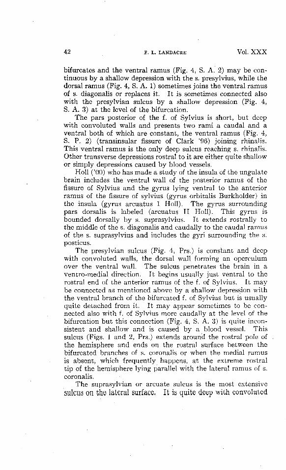
42 F. L. LANDACRE Vol. X X X
bifurcates and the ventral ramus (Fig. 4, S. A. 2) may be con-tinuous by a shallow depression with the s. presylvius, while thedorsal ramus (Fig. 4, S. A. 1) sometimes joins the ventral ramusof s. diagonalis or replaces it. It is sometimes connected alsowith the presylvian sulcus by a shallow depression (Fig. 4,S. A. 3) at the level of the bifurcation.
The pars posterior of the f. of Sylvius is short, but deepwith convoluted walls and presents two rami a caudal and aventral both of which are constant, the ventral ramus (Fig. 4,S. P. 2) (transinsular fissure of Clark '96) joining rhinalis.This ventral ramus is the only deep sulcus reaching s. rhinalis.Other transverse depressions rostral to it are either quite shallowor simply depressions caused by blood vessels.
Holl ('00) who has made a study of the insula of the ungulatebrain includes the ventral wall of the posterior ramus of thefissure of Sylvius and the gyrus lying ventral to the anteriorramus of the fissure of sylvius (gyrus orbitalis Burkholder) inthe insula (gyrus arcuatus 1 Holl). The gyrus surroundingpars dorsalis is labeled (arcuatus II Holl). This gyrus isbounded dorsally by s. suprasylvius. It extends rostrally tothe middle of the s. diagonalis and caudally to the caudal ramusof the s. suprasylvius and includes the gyri surrounding the s.posticus.
The presylvian sulcus (Fig. 4, Prs.) is constant and deepwith convoluted walls, the dorsal wall forming an operculumover the ventral wall. The sulcus penetrates the brain in aventro-medial direction. It begins usually just ventral to therostral end of the anterior ramus of the f. of Sylvius. It maybe connected as mentioned above by a shallow depression withthe ventral branch of the bifurcated f. of Sylvius but is usuallyquite detached from it. It may appear sometimes to be con-nected also with f. of Sylvius more caudally at the level of thebifurcation but this connection (Fig. 4, S. A. 3) is quite incon-sistent and shallow and is caused by a blood vessel. Thissulcus (Figs. 1 and 2, Prs.) extends around the rostral pole ofthe hemisphere and ends on the rostral surface between thebifurcated branches of s. coronalis or when the medial ramusis absent, which frequently happens, at the extreme rostraltip of the hemisphere lying parallel with the lateral ramus of s.coronalis.
The suprasylvian or arcuate sulcus is the most extensivesulcus on the lateral surface, It is quite deep with convoluted

No. 1 SULCI OF THE BRAIN OF SHEEP 43
walls. This sulcus penetrates the brain in a slightly ventro-medial direction especially in the pars media so that the ventralwall overlies the dorsal wall to some extent. It extends fromjust rostral to the pars dorsalis of the f. of Sylvius, in the formof an arch, almost to the caudal pole of the hemisphere. It isusually divided into a pars anterior (Figs. 1 and 4, S. S. A.)a pars media (Figs. 1 and 4, S. S. M.), a pars posterior (Figs. 1and 4, S. S. P.) portions. The terminations of the anteriorand posterior portions which are shallower than the pars media,being more variable in arrangement.
The pars media is the most constant and usually presentsone shallow dorsal ramus, (Figs. 3 and 4, S. S. 2) directlydorsal to the end of the f. of Sylvius. As mentioned in describ-ing the dorsal surface, the rostral portion of pars media (Fig.4, S. S. C.) seems sometimes to be continuous with the lateralend of the s. ansatus owing to the depressing of the gyrus atthe lateral termination of that sulcus.
The variations of pars anterior depend upon the form ofthe s. diagonalis. The pars anterior usually bifurcates into ananterior ramus (Fig. 4, S. S. 3) and a ventral ramus (Fig. 4,5. S. 4). Both rami are deep at the point of bifurcation andgradually become shallower toward their terminations. Theanterior ramus rarely joins s. diagonalis and when this occurs itis by a shallow depression. In four cases, however, out of onehundred examined the depression was pronounced. The shortventral ramus (Fig. 4, S. S. 4) of the bifurcated rostral end mayoccasionally join and sometimes be replaced by a ramus fromthe f. of Sylvius (Fig. 4, S. D. 1) in the same relative position.
The caudal border of the pars media of s. suprasylvius isindicated by a constant and fairly deep ventral ramus (Fig. 4,S. S. 5). It is never entirely absent but varies in length anddepth depending on the depth and form of s. posticus. Fromthis ventral ramus the pars posterior forms a gentle curvemaintaining its depth and convoluted wall almost to the caudalborder of the hemisphere. The caudal portion of this sulcusis quite variable but usually bifurcates into a dorsal ramus(Fig. 4, S. S. 6) which frequently reaches the caudal border andis connected sometimes with the caudal end of s. ectolateralis(Fig. 4, Eel.) or with detached depressions in the usual positionof the caudal end of that sulcus. The ventral ramus (Fig. 4,S. S. 7) is still more variable but rarely absent and may be
44 F, L. LANDACRE Vol. X X X
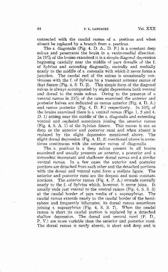
connected with the caudal ramus of s. posticus and whenabsent be replaced by a branch from s. posticus.
The s. diagonalis (Fig. 4, D. A., D. P.) is a constant deepsulcus and penetrates the brain in a ventfomedial direction.In 75% of the brains examined it is a simple diagonal depressionbeginning caudally near the middle of pars dorsalis of the f.of Sylvius and extending diagonally, rostrally and mediallynearly to the middle of s. coronalis with which it never forms ajunction. The caudal end of the sulcus is occasionally con-tinuous with the f. of Sylvius by a transient anterior ramus ofthat fissure (Fig. 4, S. D. 2). This simple form of the diagonalsulcus is always accompanied by slight depressions both ventraland dorsal to the main sulcus. Owing to the presence of aventral ramus in 25% of the cases examined the anterior andposterior halves are indicated as ramus anterior (Fig. 4, D. A.)and ramus posterior (Fig. 4, D. P.) respectively. In 25% ofthe brains examined there is a ventral ramus (Figs. 1, 2 and 4,D. 1) arising near the middle of the s. diagonalis and extendingventral and cephalad sometimes joining the anterior ramus(Fig. 4, S. A. 1) of the Sylvian fissure. This ramus is not asdeep as the anterior and posterior rami and when absent isreplaced by the slight depression mentioned above. Theslight dorsal depression (Fig. 4, D. 2) mentioned above is some-times continuous with the anterior ramus of diagonalis.
The s. posticus is a deep sulcus present in all brainsexamined and usually presents an anterior, a posterior and asomewhat inconstant and shallower dorsal ramus and a similarventral ramus. In a few cases the anterior and posteriorportions are detached from each other and the detached portionswith the dorsal and ventral rami form a stellate figure. Theanterior and posterior rami are the deepest and most constantportions. The anterior ramus (Fig. 4, P. A.) extends rostrallynearly to the f. of Sylvius which, however, it never joins. Itusually ends just ventral to the ventral ramus (Fig. 4, S. S. 5)at the caudal border of pars media of s. suprasylvius. Thecaudal ramus extends nearly to the caudal border of the hemi-sphere and frequently bifurcates, its dorsal ramus sometimesjoining s. suprasylvius (Fig. 4, S. S. 7). When the caudalramus is short its caudal portion is replaced by a detachedshallow depression. The dorsal and ventral rami (P. D.,P. V.) are more variable than the anterior and posterior rami.The dorsal ramus is rarely absent, is short and deep and is

No. 1 SULCI OF THE BRAIN OF SHEEP 45
situated more cephalad than the ventral ramus. The ventralramus (Fig. 4, P. P.) is sometimes absent and is replaced by adetached depression occupying the same relative position.
THE MEDIAL AND CAUDAL SURFACES.(Figs. 3 and 5).
The caudal and concealed portions of the medial surfacesof the hemisphere are exposed by a transverse incision andremoval of the brain stem at the level of the mammillary body.
On the buried medial surface of the hemisphere two depres-sions, the hippocampal fissure and fimbriodentate sulcus appearconstantly. Both are shallow even the hippocampal fissurecannot be readily opened.
The fimbriodentate sulcus (Fig. 5, Fd.) lies rostral to thehippocampal fissure between the fimbria and the gyrus dentatusand extends throughout the whole arch of the buried portion ofthe hemisphere appearing on the flat medial surface in frontof the more anterior of two prominent elevations of gray matterventral to and slightly rostral to the splenium of the corpuscallosum.
The hippocampal fissure (Fig. 5, H.) lies caudal to thefimbrio-dentate sulcus between the gyrus dentatus and thegyrus hippocampi and extends throughout the whole arch ofthe buried medial surface of the hemisphere. It appears onthe flat medial surface of the hemisphere when a medianlongitudinal section of the brain is made and is visible betweenthe two small gray elevations mentioned above. These twogray elevations are usually labeled in the text books as fasciolacinerea. The anterior of these two gray masses is the dorsallimit of the gyrus dentatus and the posterior elevation is acontinuation of the gyrus hippocampi.
On the flat medial surface of the hemisphere four sulci arealways present with slight variations to be mentioned laterwith the addition of transient sulci belonging to the dorsalsurface of the hemisphere.
The sulcus corporis callosi (Fig. 4, C.) is constant but shallowand extends throughout the length of the dorsal border of thecorpus callosum and well around both genu and splenium.
The rostral area of the medial surface presents two constantmoderately deep sulci: the s. genualis (Fig. 5, G.) of Kruegand Kappers (s. cinguli Burkholder) and the more rostral s.

46 F. L. LANDACRE Vol. X X X
rostralis (Fig. 5, Ro.) of Krueg and Kappers (parolfactoriusBurkholder).
The sulcus genualis (Fig. 5, G.) begins cephalad ventral tothe genu of the corpus callosum and arching around the corpuscallosum extends to the middle of that structure. It is some-times quite simple in structure but often presents a well defineddorsal ramus (Fig. 5, G. 1) just rostral to the anterior end of s.splenialis and sometimes a ventral ramus (Fig. 5, G. 2). Thisdorsal ramus may be absent or represented by a slight depressionand may occasionally be situated more rostral and invade thearea usually occupied by s. rostralis.
The s. rostralis (Fig. 5, Ro.) is more variable than genualis.In its most regular form it parallels the course of s. genualislying nearer the rostral border of the hemisphere forming anarch beginning ventral to the rostral end of s. genualis andarching around extends caudally to the gyrus just rostral tothe s. splenialis where it may reach the edge of the hemisphere.Its variations consist in its appearing as two detached depres-sions in some cases and occasionally in the detached portionsbeing separated by a ramus of s. genualis.
A constant sulcus (Fig. 5, Pc.) appears caudal to the spleniumof the corpus callosum occupying the same relation to thesplenium that s. genualis occupies with reference to the genu.It is a short sulcus and varies both in depth and length and whenleast pronounced is represented by a slight depression. It isnot named in any description of the sheep brain. It is indicatedin many ungulate brains by Krueg (78) and occupies theposition of the subparietoccipital fissure of the anthropoids.Since the sulcus is always present it is named from its positionsulcus post callosus, since the term retrosplenialis is preempted.Its position indicates that it is in an olfactory area (regioretrosplenialis of Campbell).
The sulcus splenialis (Fig. 5, Spl.) is the most extensive anddeepest sulcus on the medial surface of the hemisphere and ismuch convoluted in its caudal half. It has a well defineddiagonal direction ventrolaterally as it penetrates the hemi-sphere so that its ventral wall forms an operculum over itsdorsal wall. It begins rostrally on the dorsal surface of thehemisphere (Figs. 1 and 3, Spl.) just in front of the s. ansatusand extends in an arch below the dorsal border of the hemisphereto the caudal border where it bifurcates into a dorsal and ventralramus, the ventral ramus (Figs. 3 and 5, R. S.) being the sulcus

No. 1 SULCI OF THE BRAIN OF SHEEP 47
retrosplenialis of Kappers and the dorsal ramus (Figs. 3 and 5,H. P.), which is frequently shorter and shallower, being theramus horizontalis posterior of Kappers. These two rami arenever absent in the brains examined but vary somewhat inlength and depth. A rather common variation in the rostralportion of s. splenialis is the presence of a dorsal ramus (Fig. 5,Spl. 1) frequently reaching the dorsal border of the hemisphereand lying just caudal to the s. ansatus (Fig. 5, An.) and betweenthat sulcus and the s. lateralis when it reaches the medial borderof the hemisphere.
On the caudal surface of the hemisphere the s. rhinalis(Figs. 3 and 5, R.) is always present. Sometimes it ends welltoward the lateral surface of the caudal area and may becontinuous with the caudal end of s. suprasylvius. In othercases it lies more medial and is in one brain continuous withthe caudal end of s. lateralis of the dorsal surface. Thesevariations seem to be correlated with the form and extent of athird sulcus which Kappers ('21 p. 1126) calls the occipito-temporal sulcus. This is sometimes a detached sulcus in thesheep but is frequently represented by the caudal extension ofs. lateralis. The modeling of the caudal surface is usuallydetermined by the length and depth of the ramus horizontalisposterior, s. lateralis and s. rhinalis. When the simpler arrange-ments are not present, that is, well pronounced caudal extensionsof splenialis, lateralis and rhinalis detached depressions occur.The s. occipitotemporalis is best denned when lateralis isshort and does not appear on the caudal surface of the hemi-sphere. This indicates a close relation between occipito-temporalis when present and s. lateralis. When occipito-temporalis is best denned it lies parallel and between retro-splenialis (Fig. 3, Rs.) and rhinalis (Fig. 3, R.).
BIBLIOGRAPHY.
Bagley, C. 1922. Cortical motor mechanism of the sheep brain. Arch. Neur.Psychiat., Vol. 7, pp. 417-453.
Burkholder, J. F. 1904. Anatomy of the brain, Chicago, 1904.Clark, T. E. 1896. Comparative anatomy of the insula. Jour. Comp. Neur.,
Vol. VI.Fiske, E. W. 1913. An elementary study of the brain based on the dissection of
the brain of the sheep. New York, Macmillan Co.Herrick and Crosby. 1920. Laboratory outline of neurology, 2nd edition.Holl, M. 1900. Ueber die Insel des Ungulatengehirnes. Archiv fur Anatomie
und Entwickelungsgeschichte.

48 F. L. LANDACRE Vol. X X X
Kappers, C. U. Ariens. 1913. Cerebral Localization and the Significance ofSulci, XVIIth International Congress of Medicine, London, 1913, Sec. 1,Anat., Pt. 2, pp. 273-392, London, 1914.
< 1921. Vergleichenden Anatomie des Nervensystems, Haarlem, Vol. 2.Krueg, Julius. 1878. Ueber die Furchung der Grosshirnrinde der Ungulaten,
Zeitschrift fur Wissenschaftliche Zoologie, Vol. 31, 1878.Sutherland, S. and King, J. L. 1911. Localization of the motor area in the sheep.
Quar. Jour, of Exp. Zool., Vol. 4, 1911.
EXPLANATION OF PLATES.
In all figures primary or deep sulci and fissures are indicated by heavy lines,shallow sulci are indicated by fine lines, transient sulci are indicated by brokenlines. A shallow sulcus is likely to be transient, but when it is of value as a land-mark in localization it is sometimes indicated by a fine line and its transientcharacter is noted in text.
PLATE I.
Fig. 1. Dorsal surface X 1.75.Fig. 2. Rostral surface X 1.5.Fig. 3. Caudal surface X 1.5. Fig. 3, in order to show the relation of sulci on
the caudal surface to sulci on other surfaces includes more than the areain contact with the cerebellum which is taken as the caudal surface.The sulci shown in Fig. 3 are visible after the removal of thecerebellum.
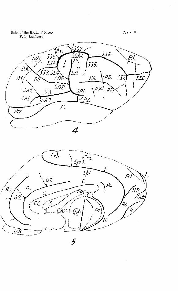
PLATE II.
Fig. 4. Lateral surface X 1.75.Fig. 5. Medial surface X 1.75. The brain stem was removed by a transverse
incision at the level of the mammillary body.
NOTE.—The determination of the relative depth of sulci on formalin fixedmaterial is easily accomplished by immersing brains in tap water for some time,when the hemisphere wall becomes flexible and gyri are easily opened.
No. 1 SULCI OF THE BRAIN OF SHEEP 49
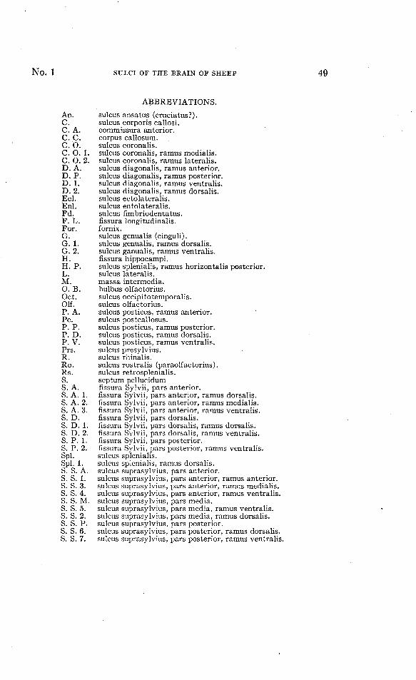
ABBREVIATIONS.
sulcus ansatus (cruciatus?).sulcus corporis callosi.commissura anterior.corpus callosum.sulcus coronalis.sulcus coronalis, ramus medialis.sulcus coronalis, ramus lateralis.sulcus diagonalis, ramus anterior.sulcus diagonalis, ramus posterior.sulcus diagonalis, ramus ventralis.sulcus diagonalis, ramus dorsalis.sulcus ectolateralis.sulcus entolateralis.sulcus fimbriodentatus.fissura longitudinalis.fornix.sulcus genualis (cinguli).sulcus genualis, ramus dorsalis.sulcus ganualis, ramus ventralis.fissura hippocampi.sulcus splenialis, ramus horizontalis posterior.sulcus lateralis.massa intermedia.bulbus olfactorius.sulcus occipitotemporalis.sulcus olfactorius.sulcus posticus, ramus anterior.sulcus postcallosus.sulcus posticus, ramus posterior.sulcus posticus, ramus dorsalis.sulcus posticus, ramus ventralis.sulcus presylvius.sulcus rhinalis.sulcus rostralis (paraolfactorius).sulcus retrosplenialis.septum pellucidumfissura Sylvii, pars anterior.fissura Sylvii, pars anterior, ramus dorsalis.fissura Sylvii, pars anterior, ramus medialis.fissura Sylvii, pars anterior, ramus ventralis.fissura Sylvii, pars dorsalis.fissura Sylvii, pars dorsalis, ramus dorsalis.fissura Sylvii, pars dorsalis, ramus ventralis.fissura Sylvii, pars posterior.fissura Sylvii, pars posterior, ramus ventralis.sulcus splenialis.sulcus splenialis, ramus dorsalis.sulcus suprasylvius, pars anterior.sulcus suprasylvius, pars anterior, ramus anterior.sulcus suprasylvius, pars anterior, ramus medialis.sulcus suprasylvius, pars anterior, ramus ventralis.sulcus suprasylvius, pars media.sulcus suprasylvius, pars media, ramus ventralis.sulcus suprasylvius, pars media, ramus dorsalis.sulcus suprasylvius, pars posterior.sulcus suprasylvius, pars posterior, ramus dorsalis.sulcus suprasylvius, pars posterior, ramus ventralis.
An.C.C AC. CC OC. 0C OD. AD. PD. 1.D. 2.Eel.Enl.Fd.F. L.For.G.G.I.G. 2.H.H. PL.M.0. BOct.Olf.P. A.Pc.P. P.P. DP. V.Prs.R.Ro.Rs.S.S. A.S. A.S. A.S. A.S. D.S. D.S. D.S. P.S. P.Spl.Spl. ]s. s.s. s.s. s.s. s.s. s.s. s.s. s.s. s.s. s.s. s.
'. 1.. 2 .
1.2.3.
1.2.1.2.
I.A.1.3.4.M.5.2.P.6.7.

Sulci of the Brain of SheepF. L. Landacre
PLATE I.
Prs.
Sulci of the Brain of SheepF. L. Landacre
PLATE II .


![F £-l],´F+|](https://static.fdocuments.in/doc/165x107/625ac2da5a426346697ed19b/f-lf.jpg)