Lack of prolactin receptor signaling in mice results in...
9
Introduction Human lactotroph adenomas (prolactinomas) are the most frequent functioning pituitary tumors (1), but their pathogenesis remains elusive. The primary regula- tion of prolactin (PRL) secretion is mediated by the inhibitory effects of dopamine released from the medi- an eminence of the hypothalamus and acting at the dopamine D2 receptor subtype (D2R) (2). Mice deficient in the D2R receptor (Drd2 –/– mice) have previously been shown to develop hyperprolactinemia, lactotroph hyperplasia, and prolactinomas (3, 4), confirming a crit- ical role of hypothalamic dopamine and dopamine receptor activation in the physiologic regulation of lac- totroph proliferation and PRL secretion. However, the involvement of a number of other regulatory factors has been postulated, including PRL itself (5). PRL increases hypothalamic–pituitary dopamine tone, i.e., constitutive inhibitory signaling by the D2R, and decreases PRL secretion (6–8) by altering the expression (9) and activi- ty (7) of tyrosine hydroxylase (TH), the rate-limiting enzyme in dopamine synthesis. In addition to its indirect effects, mediated by changes in hypothalamic dopamine, we hypothesize that PRL might have direct effects on the pituitary gland. PRL receptors (PRLRs) have been demonstrated in the anterior pituitary in several species, including mouse (10, 11), and have been found specifically on lac- totrophs in rats (12) and humans (13). Its effects on proliferation of other cell types are varied, but are pre- dominantly stimulatory (14–25) except in the ovary (26, 27), adrenal gland (28), and vascular endothelium (29). Although it has been postulated that PRL may play an autocrine or paracrine role in lactotroph pro- liferation (11), there is limited empirical evidence sup- porting this theory. Experiments to evaluate a direct pituitary effect of PRL in vivo are difficult because of its concurrent effects on hypothalamic dopamine. By The Journal of Clinical Investigation | October 2002 | Volume 110 | Number 7 973 Lack of prolactin receptor signaling in mice results in lactotroph proliferation and prolactinomas by dopamine-dependent and -independent mechanisms Kathryn G. Schuff, 1 Shane T. Hentges, 1 Michele A. Kelly, 1 Nadine Binart, 2 Paul A. Kelly, 2 P. Michael Iuvone, 3 Sylvia L. Asa, 4 and Malcolm J. Low 1,5 1 Vollum Institute, Oregon Health & Science University, Portland, Oregon, USA 2 Institut National de la Santé et de la Recherche Médicale (INSERM), Faculté de Necker, Paris, France 3 Department of Pharmacology, Emory University School of Medicine, Atlanta, Georgia, USA 4 Department of Pathology, University Health Network, Toronto, Ontario, Canada 5 Department of Behavioral Neuroscience, Oregon Health & Science University, Portland, Oregon, USA Hypothalamic dopamine inhibits pituitary prolactin secretion and proliferation of prolactin-pro- ducing lactotroph cells by activating lactotroph dopamine D2 receptors (D2Rs). Conversely, prolactin (PRL) stimulates hypothalamic dopamine neurons via PRL receptors (PRLRs) in a short-loop feedback circuit. We used Drd2 –/– and Prlr –/– mutant mice to bypass this feedback and investigate possible dopamine-independent effects of PRL on lactotroph function. The absence of either receptor induced hyperprolactinemia and large prolactinomas in females. Small macroadenomas developed in aged Prlr –/– males, but only microscopic adenomas were found in Drd2 –/– male mice. Pharmacologic stud- ies in Prlr –/– mice with D2R agonists and antagonists demonstrated a significant loss of endogenous dopamine tone, i.e., constitutive inhibitory signaling by the D2R, in the pituitary. However, Prlr –/– mice exhibited more profound hyperprolactinemia and larger tumors than did age-matched Drd2 –/– mice, and there were additive effects in compound homozygous mutant male mice. In vitro, PRL treatment markedly inhibited the proliferation of wild-type female and male Drd2 –/– lactotrophs, but had no effect on female Drd2 –/– lactotrophs, suggesting a downregulation or desensitization of PRLR in response to chronic hyperprolactinemia. We conclude that PRL inhibits lactotrophs by two distinct mechanisms: (a) indirectly by activation of hypothalamic dopamine neurons and (b) directly within the pituitary in a dopamine-independent fashion. J. Clin. Invest. 110:973–981 (2002). doi:10.1172/JCI200215912. Received for publication May 10, 2002, and accepted in revised form August 7, 2002. Address correspondence to: Malcolm Low, Vollum Institute L-474, Oregon Health & Science University, 3181 SW Sam Jackson Park Road, Portland, Oregon 97201-3098, USA. Phone: (503) 494-4672; Fax: (503) 494-4976; E-mail: [email protected]. Kathryn G. Schuff and Shane T. Hentges contributed equally to this work. Conflict of interest: No conflict of interest has been declared. Nonstandard abbreviations used: prolactin (PRL); dopamine D2 receptor (D2R); tyrosine hydroxylase (TH); PRL receptor (PRLR); growth hormone (GH); thyroid-stimulating hormone (TSH); follicle-stimulating hormone (FSH); luteinizing hormone (LH); adrenocorticotropic hormone (ACTH); 3,4-dihydroxyphenyl- alanine (DOPA); aromatic amino acid decarboxylase (AADC).
Transcript of Lack of prolactin receptor signaling in mice results in...

IntroductionHuman lactotroph adenomas (prolactinomas) are themost frequent functioning pituitary tumors (1), buttheir pathogenesis remains elusive. The primary regula-tion of prolactin (PRL) secretion is mediated by theinhibitory effects of dopamine released from the medi-an eminence of the hypothalamus and acting at thedopamine D2 receptor subtype (D2R) (2). Mice deficientin the D2R receptor (Drd2–/– mice) have previously beenshown to develop hyperprolactinemia, lactotrophhyperplasia, and prolactinomas (3, 4), confirming a crit-
ical role of hypothalamic dopamine and dopaminereceptor activation in the physiologic regulation of lac-totroph proliferation and PRL secretion. However, theinvolvement of a number of other regulatory factors hasbeen postulated, including PRL itself (5). PRL increaseshypothalamic–pituitary dopamine tone, i.e., constitutiveinhibitory signaling by the D2R, and decreases PRLsecretion (6–8) by altering the expression (9) and activi-ty (7) of tyrosine hydroxylase (TH), the rate-limitingenzyme in dopamine synthesis.
In addition to its indirect effects, mediated bychanges in hypothalamic dopamine, we hypothesizethat PRL might have direct effects on the pituitarygland. PRL receptors (PRLRs) have been demonstratedin the anterior pituitary in several species, includingmouse (10, 11), and have been found specifically on lac-totrophs in rats (12) and humans (13). Its effects onproliferation of other cell types are varied, but are pre-dominantly stimulatory (14–25) except in the ovary(26, 27), adrenal gland (28), and vascular endothelium(29). Although it has been postulated that PRL mayplay an autocrine or paracrine role in lactotroph pro-liferation (11), there is limited empirical evidence sup-porting this theory. Experiments to evaluate a directpituitary effect of PRL in vivo are difficult because ofits concurrent effects on hypothalamic dopamine. By
The Journal of Clinical Investigation | October 2002 | Volume 110 | Number 7 973
Lack of prolactin receptor signaling in mice results in lactotroph proliferation and prolactinomas by dopamine-dependent and -independent mechanisms
Kathryn G. Schuff,1 Shane T. Hentges,1 Michele A. Kelly,1 Nadine Binart,2 Paul A. Kelly,2
P. Michael Iuvone,3 Sylvia L. Asa,4 and Malcolm J. Low1,5
1Vollum Institute, Oregon Health & Science University, Portland, Oregon, USA2Institut National de la Santé et de la Recherche Médicale (INSERM), Faculté de Necker, Paris, France3Department of Pharmacology, Emory University School of Medicine, Atlanta, Georgia, USA4Department of Pathology, University Health Network, Toronto, Ontario, Canada5Department of Behavioral Neuroscience, Oregon Health & Science University, Portland, Oregon, USA
Hypothalamic dopamine inhibits pituitary prolactin secretion and proliferation of prolactin-pro-ducing lactotroph cells by activating lactotroph dopamine D2 receptors (D2Rs). Conversely, prolactin(PRL) stimulates hypothalamic dopamine neurons via PRL receptors (PRLRs) in a short-loop feedbackcircuit. We used Drd2–/– and Prlr–/– mutant mice to bypass this feedback and investigate possibledopamine-independent effects of PRL on lactotroph function. The absence of either receptor inducedhyperprolactinemia and large prolactinomas in females. Small macroadenomas developed in agedPrlr–/– males, but only microscopic adenomas were found in Drd2–/– male mice. Pharmacologic stud-ies in Prlr–/– mice with D2R agonists and antagonists demonstrated a significant loss of endogenousdopamine tone, i.e., constitutive inhibitory signaling by the D2R, in the pituitary. However, Prlr–/– miceexhibited more profound hyperprolactinemia and larger tumors than did age-matched Drd2–/– mice,and there were additive effects in compound homozygous mutant male mice. In vitro, PRL treatmentmarkedly inhibited the proliferation of wild-type female and male Drd2–/– lactotrophs, but had noeffect on female Drd2–/– lactotrophs, suggesting a downregulation or desensitization of PRLR inresponse to chronic hyperprolactinemia. We conclude that PRL inhibits lactotrophs by two distinctmechanisms: (a) indirectly by activation of hypothalamic dopamine neurons and (b) directly withinthe pituitary in a dopamine-independent fashion.
J. Clin. Invest. 110:973–981 (2002). doi:10.1172/JCI200215912.
Received for publication May 10, 2002, and accepted in revised formAugust 7, 2002.
Address correspondence to: Malcolm Low, Vollum Institute L-474, Oregon Health & Science University, 3181 SW SamJackson Park Road, Portland, Oregon 97201-3098, USA. Phone: (503) 494-4672; Fax: (503) 494-4976; E-mail: [email protected] G. Schuff and Shane T. Hentges contributed equally to this work.Conflict of interest: No conflict of interest has been declared.Nonstandard abbreviations used: prolactin (PRL); dopamine D2receptor (D2R); tyrosine hydroxylase (TH); PRL receptor (PRLR);growth hormone (GH); thyroid-stimulating hormone (TSH);follicle-stimulating hormone (FSH); luteinizing hormone (LH);adrenocorticotropic hormone (ACTH); 3,4-dihydroxyphenyl-alanine (DOPA); aromatic amino acid decarboxylase (AADC).

crossing PRLR-deficient (Prlr–/–) mice with Drd2–/–
mice, we were able to evaluate a possible contributionof dopamine-independent effects of PRL signaling onthe control of PRL secretion and lactotroph growth.
MethodsGeneration of receptor-deficient mice. Generation ofDrd2–/– and Prlr–/– mice by gene targeting in embryon-ic stem cells has been previously described (3, 30). Eachreceptor-deficient line of mice was independentlybackcrossed to inbred C57BL/6J mice for five or moregenerations to produce incipient congenic lines andreduce variability from gene epistasis. Because ofimpaired fertility, lactation (30, 31), and maternalbehavior (32) in Prlr–/– mice, a selective breeding strat-egy and inclusion of CD-1 foster mothers were used inthe intercrosses to produce sibling cohorts of wild-type, Drd2–/–, Prlr–/–, and compound mutant (Drd2–/–,Prlr–/–) mice for these studies. All animal care practicesand experimental procedures were approved by theOregon Health & Science University InstitutionalAnimal Care and Use Committee.
Determination of serum PRL. Serum was obtainedunder brief isoflurane anesthesia from a tail bleed andfrozen at –80°C until analysis. PRL radioimmunoassayused mouse PRL (mPRL) reference preparationAFP6476C, mPRL AFP1077D for iodination, and anti-mPRL antiserum AFP131078 (provided by A.F. Parlowand the National Hormone and Peptide Program, Har-bor–UCLA Medical Center, Torrance, California, USA).The assay was performed as directed, except that mPRLwas iodinated with 3 µg chloramine-T (Sigma-Aldrich,St. Louis, Missouri, USA) per µg peptide and purifiedwith Dowex 20-50 chloride mesh resin beads (Bio-RadLaboratories Inc., Hercules, California, USA); approxi-mately 20,000 cpm was added to each tube. Assay sen-sitivity was 15 ng/ml using 10-µl serum samples. Sam-ples were analyzed in duplicate at multiple dilutions.For drug responses, basal serum was collected from 15-month-old male mice, then 5 mg/kg 2-bromo-α-ergocryptine (bromocriptine; Sigma-Aldrich) or 10mg/kg haloperidol (Ortho-McNeil Pharmaceutical,Raritan, New Jersey, USA) was administered intraperi-toneally, and post-treatment serum samples wereobtained 1 hour later.
Histologic analyses. Mice were sacrificed by decapitationat ages 6, 12, 14, or 18–21 months, and their pituitaryglands were inspected visually with a dissecting micro-scope, weighed, and placed immediately in 10%buffered formalin. Paraffin-embedded sections 4–5 µmthick were stained with hematoxylin and eosin or theGordon-Sweet silver method for reticulin matrix.Immunohistochemical staining to identify adenohy-pophyseal hormones was performed using the strepta-vidin-biotin peroxidase technique. Primary antiseradirected against rat pituitary hormones were used atthe following dilutions: growth hormone (GH),1:2,500; PRL, 1:2,500; thyroid-stimulating hormone(TSH-β), 1:3,000; follicle-stimulating hormone–β
(FSH-β), 1:600; luteinizing hormone–β (LH-β), 1:2,500(all provided by A.F. Parlow and the National Hormoneand Peptide Program); and prediluted adrenocorti-cotropic hormone (ACTH), further diluted 1:20(DAKO Corp., Carpinteria, California, USA).
Evaluation of hypothalamic dopaminergic neuron function.TH gene expression was evaluated by in situ hybridiza-tion. Mice were sacrificed by decapitation, and brainswere removed and snap-frozen in isopentane over dryice and stored at –80°C. Ten-micrometer sections werecut on a cryostat and thaw-mounted onto gelatin-coat-ed slides. TH mRNA was detected by hybridization at60°C with 20 million cpm/ml of a 35S-UTP–labeledantisense riboprobe (provided by E. Lewis, Departmentof Biochemistry and Molecular Biology, Oregon Health& Science University, Portland, Oregon, USA). Theslides were exposed to NBT2 emulsion (EastmanKodak, Rochester, New York, USA) for 15 days, devel-oped, and counterstained with neutral red. Digitalimages were captured using both bright- and dark-fieldillumination on a Leica research microscope (LeicaMicrosystems Inc., Deerfield, Illinois, USA). TH activi-ty was estimated by the measurement of 3,4-dihydrox-yphenylalanine (DOPA) accumulation after inhibitionof aromatic amino acid decarboxylase (AADC) by m-hydroxybenzylhydrazine dihydrochloride (NSD1015; Sigma-Aldrich). Briefly, mice were sacrificed bydecapitation 30 minutes after an intraperitoneal injec-tion of 100 mg/kg NSD 1015. Whole hypothalamicblocks and a portion of dorsal striatum were snap-frozen in isopentane on dry ice and stored at –80°Cuntil analyzed. Hypothalami were sonicated in 20 vol-umes and striata in 50 volumes of homogenizationsolution and subjected to HPLC and electrochemicaldetection of DOPA as previously described (33).
Primary cultures of anterior pituitary cells. Anteriorpituitary glands were collected from randomlycycling C57BL/6J mice and placed into ice-cold HBSS(Sigma-Aldrich) and dissociated enzymatically inHBSS containing collagenase, deoxyribonuclease,and BSA as described previously (34). Cells were plat-ed at 250,000 cells/well on poly-L-lysine–coated glasscoverslips in 24-well plates in DMEM/Ham’s F12medium (1:1; Sigma-Aldrich) containing 50 U/mlpenicillin G, 50 µg/ml streptomycin, and 10% FBS for24 hours. The next day, 24 hours prior to PRL treat-ment, the medium was replaced with DMEM/F12supplemented with Serum Replacement 2 (Sigma-Aldrich). For the 48-hour PRL treatment, the medi-um was replaced with DMEM/F12/Serum Replace-ment 2 containing recombinant mPRL (0–10 µg/ml;provided by A.F. Parlow and the National Hormoneand Peptide Program). The media and treatment werechanged at 24 hours, and 0.1 mM bromodeoxyuri-dine (BrdU; Sigma-Aldrich) was added for the last 12hours of PRL treatment.
Lactotroph cell proliferation. Lactotroph cell prolifera-tion was determined by identifying the cells that dis-played immunoreactivity for both BrdU and PRL. After
974 The Journal of Clinical Investigation | October 2002 | Volume 110 | Number 7

treatment with mPRL and BrdU as above, cells werewashed and fixed in 99% ice-cold ethanol and processedfor immunofluorescence. The mouse monoclonalBrdU antibody (1:200; Becton Dickinson Immunocy-tometry Systems, San Jose, California, USA) and rabbitpolyclonal mouse-specific PRL antibody (1:3,000; giftof F. Talamantes, Biology, University of California atSanta Cruz, Santa Cruz, California, USA) were detect-ed with goat anti-mouse FITC-conjugated secondaryantibodies (1:200; BioSource International, Camarillo,California, USA) and goat anti-rabbit rhodamine-con-jugated secondary antibodies (1:200; American Qualex,San Clemente, California, USA). Cell nuclei were fluo-rescently labeled with Hoechst 33258 (Sigma-Aldrich).Cells immunoreactive for both BrdU and PRL werecounted as dividing lactotrophs. Cell counts were madeat 20× magnification on a Leica epifluorescence micro-scope. An investigator blind to the treatment and geno-type counted five random areas in each coverslip,approximately 250 cells/area. Six to eight coverslips pertreatment from three separate cultures were evaluatedfor each genotype.
Statistical analyses. Statistical analyses were performedusing StatView statistical analysis software (SAS Insti-tute Inc., Cary, North Carolina, USA). For comparisonswithin a genotype before and after drug treatment, one-tailed paired Student t tests were used because of thewell-described direction of change. For comparisonsamong multiple genotypes, ANOVA was used. Post hocanalyses were by the Fisher post hoc test. All data arepresented as mean ± SEM. P values less than 0.05 wereconsidered significant.
ResultsHyperprolactinemia and pituitary pathology. Serum PRLincreased exponentially with age in Drd2–/– mice, Prlr–/–
mice, and compound receptor-deficient (Drd2–/–,Prlr–/–) mice (Figure 1, a and b). Levels were moremarkedly elevated in mutant females than males at alltimepoints. In addition, there was earlier and moresevere hyperprolactinemia in both sexes of Prlr–/– micethan in Drd2–/– mice, and additive effects of the com-bined mutations were apparent at both 12 and 18months of age in males (Figure 1b).
Similar to our previous observations in Drd2–/– mice(3, 4), pituitary glands in Prlr–/– mice were significantlyenlarged compared with those in the wild type startingat 6 months, and continued to increase in size in theolder groups of female and male mice (Figure 1, c andd). However, at 6 months, the female Prlr–/– pituitaryglands already exhibited distinct lactotroph hyperpla-sia (data not shown), a histological feature delayed until10–12 months in female Drd2–/– mice (3). The pituitaryglands of 14-month-old female Prlr–/– and compoundreceptor-mutant mice contained massive, multifocaltumors histologically similar to, though significantlylarger than, those observed in female Drd2–/– mice of thesame age. Histopathology of these tumors demonstrat-ed that they were all monohormonal lactotroph adeno-mas characterized by extensive breakdown of the retic-ulin network, peliosis, and enlarged, pleomorphicnuclei with numerous mitotic figures (Figure 2). Hyper-trophied perinuclear Golgi networks were observed,indicating a hypersecretory state similar to thatobserved in human prolactinomas. Immunostaining
The Journal of Clinical Investigation | October 2002 | Volume 110 | Number 7 975
Figure 1Hyperprolactinemia and pituitary enlargement in Drd2–/– and Prlr–/– mice. Serum PRL increased with age in both female (a) and male (b)Drd2–/– and Prlr–/– mice and was more marked in Prlr–/– mice. Deficiency of both receptors produced an additive effect on hyperprolactinemiain male mice. Prlr–/– mice of both sexes developed hyperprolactinemia at younger ages than did Drd2–/– mice. P < 0.005 for ANOVA of maineffect of genotype at all timepoints. *P < 0.05 for pairwise comparisons by the Fisher post hoc test. n = 6–22 for ages 8 weeks and 6 months,and n = 4–8 for ages 12 months and older. (c) Female Drd2–/– mice and Prlr–/– mice had slightly enlarged pituitary glands at 6 months and mas-sively enlarged glands at 14 months (10- to 20-fold greater than wild-type). (d) Moderate pituitary enlargement occurred only at 18–21 monthsin Prlr–/– and compound homozygous mutant male mice. P < 0.001 for ANOVA of main effect of genotype for females at 14 months and malesat 18–21 months. *P < 0.05 for pairwise comparisons using the Fisher post hoc test; n = 3–4 for 6-month-old females and 12-month-old males(except n = 1 for Prlr–/– male and n = 2 for Drd2–/– males); n = 5–7 for 14-month-old females and 18- to 21-month-old males.
for GH, LH-β, ESHβ, TSH-β, and ACTH was negative inthe adenomas (data not shown). Male Prlr–/– and com-pound receptor-deficient (Drd2–/–, Prlr–/–) mice devel-oped smaller macroadenomas at 18–21 months, with anadditive effect in mice with the compound mutation(Figure 1d). Similar histological characteristics wereobserved in these tumors (data not shown).
Pharmacologic evaluation of hypothalamic dopamine. Weevaluated and compared the effect of the loss of PRL ordopamine signaling on hypothalamic dopamine neu-ron function in Drd2–/– and Prlr–/– mice and compoundreceptor-deficient (Drd2–/–, Prlr–/–) mice by threeapproaches: pharmacologic challenge with a D2R ago-nist and antagonist followed by measurement of serumPRL; in situ hybridization of mRNA for TH (the rate-limiting enzyme in the synthesis of dopamine); andassessment of TH activity by the quantitation of DOPAafter inhibition of AADC. Only male mice were used forthese experiments because of the early tumor develop-ment observed in female receptor mutant mice andconsequent secondary distortion of the overlyingstalk/median eminence and basal hypothalamus.
Wild-type male mice had low basal levels of serum PRL,which demonstrated a nonsignificant trend to reduction
by the D2R agonist bromocriptine (Figure 3a), butserum PRL levels were significantly increased by the D2Rantagonist haloperidol (Figure 3b), illustrating thedopamine inhibitory tone normally present on lac-totrophs. Drd2–/– and compound mutant (Drd2–/–,Prlr–/–) mice exhibited no response to either drug, asexpected from the absence of D2R. In contrast, Prlr–/–
mice demonstrated an average 85% decline in serum PRL1 hour after bromocriptine treatment (Figure 3a) and noresponse to haloperidol (Figure 3b), indicating the pres-ence of functional D2R on lactotrophs and low endoge-nous dopamine tone in the pituitary of these mice.
Hypothalamic TH expression. The loss of dopamine tonein the pituitaries of Prlr–/– mice could be due to devel-opmental reductions in the number of hypothalamicdopaminergic neurons (35) or decreased function of theneurons. We therefore performed in situ hybridizationto detect TH mRNA in hypothalamic neuronal soma ofmale mice. Compared with levels in wild-type mice (Fig-ure 4, a and d), levels of TH mRNA were qualitativelyincreased in the arcuate nucleus of Drd2–/– mice (Figure4, b and e). In contrast, arcuate nucleus TH mRNA lev-els appeared decreased but clearly still evident in Prlr–/–
mice (Figure 4, c and f). TH mRNA levels in dopamin-
976 The Journal of Clinical Investigation | October 2002 | Volume 110 | Number 7
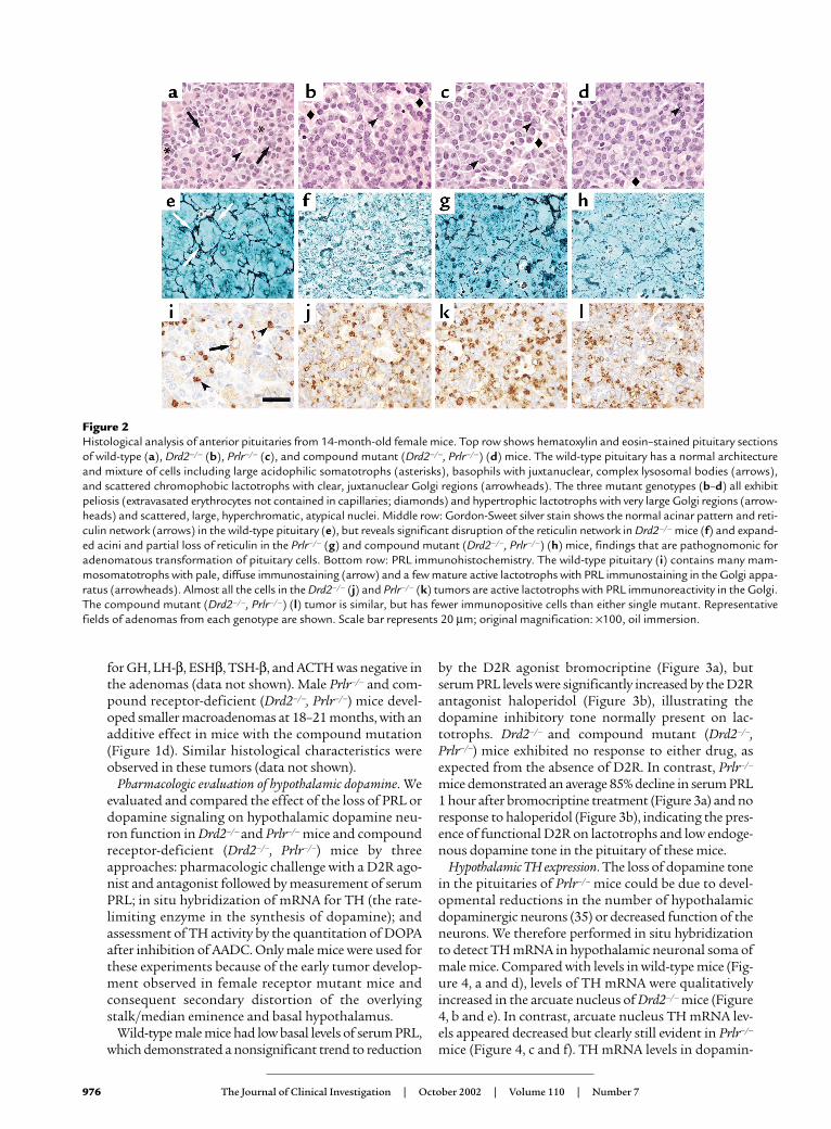
Figure 2Histological analysis of anterior pituitaries from 14-month-old female mice. Top row shows hematoxylin and eosin–stained pituitary sectionsof wild-type (a), Drd2–/– (b), Prlr–/– (c), and compound mutant (Drd2–/–, Prlr–/–) (d) mice. The wild-type pituitary has a normal architectureand mixture of cells including large acidophilic somatotrophs (asterisks), basophils with juxtanuclear, complex lysosomal bodies (arrows),and scattered chromophobic lactotrophs with clear, juxtanuclear Golgi regions (arrowheads). The three mutant genotypes (b–d) all exhibitpeliosis (extravasated erythrocytes not contained in capillaries; diamonds) and hypertrophic lactotrophs with very large Golgi regions (arrow-heads) and scattered, large, hyperchromatic, atypical nuclei. Middle row: Gordon-Sweet silver stain shows the normal acinar pattern and reti-culin network (arrows) in the wild-type pituitary (e), but reveals significant disruption of the reticulin network in Drd2–/– mice (f) and expand-ed acini and partial loss of reticulin in the Prlr–/– (g) and compound mutant (Drd2–/–, Prlr–/–) (h) mice, findings that are pathognomonic foradenomatous transformation of pituitary cells. Bottom row: PRL immunohistochemistry. The wild-type pituitary (i) contains many mam-mosomatotrophs with pale, diffuse immunostaining (arrow) and a few mature active lactotrophs with PRL immunostaining in the Golgi appa-ratus (arrowheads). Almost all the cells in the Drd2–/– (j) and Prlr–/– (k) tumors are active lactotrophs with PRL immunoreactivity in the Golgi.The compound mutant (Drd2–/–, Prlr–/–) (l) tumor is similar, but has fewer immunopositive cells than either single mutant. Representativefields of adenomas from each genotype are shown. Scale bar represents 20 µm; original magnification: ×100, oil immersion.

ergic neurons not regulated by PRL, such as the zonaincerta (data not shown) and ventral tegmentalarea/substantia nigra, were comparable in Drd2–/– mice,Prlr–/– mice, and wild-type mice (Figure 4, g–i).
Hypothalamic dopamine synthesis. To estimate changesin the rate of dopamine synthesis resulting fromdecreased levels of TH or decreased activity of TH,DOPA content of brain tissues was measured afterblockade of AADC. DOPA levels were significantlyhigher in striata from Drd2–/– mice than in striata fromwild-type mice (Figure 4k), but were not significantlyhigher in the whole hypothalamus (Figure 4j) of Drd2–/–
mice (although there was a trend toward an increase).In contrast, DOPA accumulation in the striatum andhypothalamus of Prlr–/– mice was not significantly dif-ferent from that in the wild type.
Effects of in vitro PRL treatment. Despite the pharmaco-logic and neurochemical evidence of reduced tubero-hypophyseal dopamine tone in Prlr–/– mice, the pheno-typic differences between Prlr–/– and Drd2–/– mice andadditive effects of the combined mutations in males onserum PRL levels and pituitary size suggest that PRLand PRLR have additional dopamine-independenteffects on lactotroph function. To determine whetherthere are direct pituitary effects of PRL on lactotrophgrowth, we assessed lactotroph proliferation in vitro byculturing dispersed pituitary cells from 10- to 12-month-old wild-type and Drd2–/– mice in the presenceof recombinant mPRL in doses of 0.1–10 µg/ml (Figure5). Absolute numbers of proliferating lactotrophs fromwild-type males were too low to reliably quantitate. PRLtreatment reduced the number of PRL-immunoreac-tive lactotrophs incorporating BrdU by 75% in pitu-itary cultures from wild-type female mice (Figure 5a).In contrast, there was no reduction in the number ofproliferating lactotrophs in cultures of pituitaries fromDrd2–/– female mice (Figure 5b), which in vivo alreadydemonstrate marked hyperprolactinemia, lactotrophhyperplasia, and adenomas at this age. An in vivo BrdUlabeling experiment performed in 6-month-old mice(data not shown) revealed a 5% labeling index of lac-totrophs from Drd2–/– females, similar to the in vitroresults; the figure was only 0.75% in wild-type females,indicating that the much higher basal index of wild-type lactotrophs in primary culture conditions is like-ly due to the acute loss of dopamine inhibition (2).Unlike the response in female Drd2–/– mice, PRLreduced the number of proliferating lactotrophs by73% in cultures from male Drd2–/– mice (Figure 5c),which have only very modest hyperprolactinemia com-pared with the females (3, 4).
DiscussionAlthough the finding of hyperprolactinemia in Prlr–/–
mice could be predicted based on the known feedbackloop of PRL on hypothalamic dopaminergic neurons,the finding of pituitary hyperplasia and adenoma for-mation was a surprise. Previously, both we (3) andanother group (11) concluded from our original stud-
ies of Drd2–/– mice that PRL most likely functioned asa mitogen for lactotrophs. In the majority of tissueswhere it has been evaluated, PRL is mitogenic (14–25),with only a few tissues demonstrating an antiprolifer-ative role (26–29). However, we show here that in Prlr–/–
mice, there are two factors contributing to the releaseof the lactotroph from its usual secretory and prolifer-ative controls: a decrease in the normally inhibitorydopaminergic control and a second, direct effect at thelevel of the pituitary that is most consistent with anantiproliferative action of PRL on lactotrophs.
We have demonstrated that a lack of PRLR signalingresults in decreased hypothalamic dopamine tone. Thiswas shown pharmacologically by the absence of antag-onizable dopamine action in male mice. The finding ofan acute decline in serum PRL after bromocriptinetreatment demonstrated that D2R signaling was intactin Prlr–/– mice, but the complete lack of response to theantagonist haloperidol suggests that dopaminergicinput to the pituitary gland was very low, despite themarked hyperprolactinemia in these mice.
There are several possible, and not mutually exclusive,mechanisms that must be considered to explain the lowlevels of tuberoinfundibular dopamine in the Prlr–/–
mice. These include the loss of the hypothalamic neu-ronal populations responsible for dopaminergic inputto the pituitary, decreased levels or activity of TH (therate-limiting enzyme in dopamine synthesis), anddecreased neuronal activity of these hypothalamic neu-rons. Experiments with Snell dwarf mice indicated arequirement for PRL early in development to promotethe normal survival and differentiation of the tuberoin-fundibular dopaminergic neurons (35). However, subse-quent studies of PRL peptide–deficient mice indicatedthat a lack of PRL predominantly affected the function
The Journal of Clinical Investigation | October 2002 | Volume 110 | Number 7 977
Figure 3Serum PRL responses to a dopamine D2R agonist and antagonist in15-month-old male mice measured one hour after treatment. (a)Bromocriptine decreased PRL levels in Prlr–/– mice, but not the othermutant genotypes. (b) Haloperidol increased PRL levels in wild-typemice but in none of the other genotypes. Because of their low basalvalues, the wild-type responses are shown magnified in the insets. Sta-tistical analyses were by one-tailed paired Student t test for each drugwithin genotype. *P < 0.05 compared with pretreatment; n = 5–8.

and not the survival of dopaminergic neurons in thehypothalamus (36). Consistent with these later data,arcuate TH mRNA levels in the Prlr–/– mice weredecreased but still present in a normal distribution inour study and were quantitatively normal in another(37), indicating grossly normal development of hypo-thalamic dopamine neurons. The reduction in levels ofTH mRNA is consistent with PRL’s known positive reg-ulatory feedback effects on transcription of the TH gene.
Whether the decreased mRNA levels resulted indecreased amounts of TH enzyme, and whether therewere additional changes in TH activity (mediated forexample by changes in phosphorylation), was evaluat-ed by measuring DOPA accumulation after blockadeof AADC by NSD 1015. An increase in DOPA afterNSD 1015 treatment has been shown to correlate withthe activity of TH and with dopaminergic neuronalactivity (38). As a control for the pharmacologic treat-ments, DOPA levels were measured and found to besignificantly higher in striata from Drd2–/– mice thanin striata from wild-type mice. This finding is consis-tent with increased TH activity secondary to the
absence of inhibitory D2R autoreceptorsfrom the mesostriatal dopamine neurons.However, DOPA levels also tended to behigher in the whole hypothalamus ofDrd2–/– mice. Because the A12 tuberoin-fundibular dopamine neurons do not nor-mally express dopamine autoreceptors(38), we believe TH mRNA and DOPAaccumulation in the hypothalamus ofDrd2–/– mice tended to be higher due toincreased PRL stimulation of these neu-rons (2, 5, 7). Notably, in other models ofchronic hyperprolactinemia, exposure oftuberoinfundibular dopamine neurons tovery high levels of PRL for extended peri-ods of time abrogates the increase indopaminergic activity of those neurons
(39). This may explain why statistically significantchanges were not seen in the current study.
In contrast, DOPA accumulation in neither striatumor hypothalamus of Prlr–/– mice was significantly dif-ferent from that in the wild type. It is likely that the lowbaseline levels of dopamine synthesis in male mice incombination with the presence of contaminating non-PRL-regulated catecholaminergic terminals present inthe hypothalamic samples precluded detection of anyreduction in DOPA accumulation in the hypothalamusof Prlr–/– mice. The arcuate nucleus has also beendemonstrated to contain a substantial population ofneurons that express TH but not AADC, and thereforenormally synthesize L-DOPA as an end product; theaccumulation of this L-DOPA would not be directlyaffected by the inhibition of AADC (40).
Clearly, however, the loss of PRL signaling on hypo-thalamic dopamine neurons does not entirely explainthe phenotypes observed in the two single-knockoutmutants. Specifically, hyperprolactinemia has an earli-er onset in both sexes of Prlr–/– mice than in Drd2–/– miceand is more severe in Prlr–/– mice than in Drd2–/– mice at
978 The Journal of Clinical Investigation | October 2002 | Volume 110 | Number 7
Figure 4TH expression and activity. (a–i) In situ hybridization forTH mRNA; dark-field (a–c and g–i) and bright-field (d–f)microscopy. (a and d) Wild-type male mice express THmRNA in the arcuate nucleus. TH mRNA is increased inDrd2–/– mice (b and e) and appears to be decreased,though clearly still evident, in Prlr–/– mice (c and f). Notecomparable levels of TH mRNA in the ventral tegmentalarea and substantia nigra among the three genotypes:wild type (g), Drd2–/– (h), and Prlr–/– (i). Two or threemice per group were evaluated. (j) Hypothalamic and (k)striatal content of DOPA after NSD 1015 treatment. Stri-atal DOPA is significantly increased, and hypothalamicDOPA shows a similar trend in Drd2–/– mice, consistentwith increased TH activity in dopamine neuronal termi-nals. DOPA levels did not differ between Prlr–/– and wild-type mice in either brain area. Scale bar represents 250µm. Comparison was by ANOVA and Fisher post hoctest. *P < 0.05 compared with both other groups; n = 4–7.

several timepoints. Furthermore, in males there is anadditive effect of the compound double knockout(Drd2–/–, Prlr–/–) with regard to both the degree of hyper-prolactinemia at various timepoints and the size ofpituitary adenoma formation. These differences in phe-notype suggest that despite the evidence of reduceddopaminergic input from the experiments discussedabove, there must be an additional mechanism for PRLin regulating lactotroph function.
A direct effect of PRL on the pituitary, and possiblyeven a specific direct effect on lactotrophs, is plausible.PRLRs have been localized to the pituitary gland in sev-eral species, including mouse (10, 11), but specificallyidentified on lactotrophs in only two species thus far —rat and human (12, 13). Our in vitro experiments pres-ent evidence of PRL action directly on mouse pituitarycells. Interestingly, PRL was observed to have aninhibitory effect on the proliferation of wild-typefemale lactotrophs. This effect appears to be complete-ly independent of dopamine, as this in vitro model usescultured pituitary cells grown in dopamine-free media.In addition, there was a similar inhibitory effect in pitu-itary cultures from male Drd2–/– mice. The lack ofresponse in cultures from female Drd2–/– mice, but per-sistent response in cultures from male Drd2–/– mice,suggests that the antecedent hyperprolactinemia in thefemales downregulates the response to further PRL sig-naling. This is similar to other models of chronic hyper-prolactinemia in which there is loss of the normalresponse of tuberoinfundibular dopaminergic neuronsto PRL (39). Indeed, there is evidence that in other tis-sues PRL regulates its own receptor, with downregula-tion observed at extremely high concentrations,although there does not appear to be a consistent PRLlevel at which this downregulation occurs (41–43). Thedichotomous response we observed appeared to occurwith preceding serum PRL levels of approximately 500ng/ml or greater. This level also represents a cutoff thatseparates the transition from lactotroph hyperplasia toadenoma in female Drd2–/– mice (3, 4) and distinguish-es the microscopic adenomas of male Drd2–/– mice
from the macroadenomas of male Prlr–/– mice andcompound mutant Drd2–/–, Prlr–/– mice.
The inhibitory growth response of lactotrophs toPRL is quite different from the proliferative effectexhibited in nonpituitary cell types (14–19, 21–25), inhuman prolactinoma cells (44), and in a transformedrat lactotroph cell line (20). It is, however, consistentwith models of PRL effects on EGF and TGF-α sig-naling cascades. Quijano and Sheffield demonstratedthat PRL can decrease the growth-promoting actionof EGF by increasing threonine phosphorylation anddecreasing tyrosine phosphorylation of EGFR (45).Therefore, it may be that PRL exerts an inhibitoryeffect on lactotroph growth by limiting the actions ofgrowth factors.
It was not possible in our experimental model todetermine whether the inhibitory response to PRL wasdue to a direct action on lactotrophs or instead wasmediated by juxtacrine or paracrine involvement ofanother anterior pituitary cell type. Possible mediationby one or more of the many other modulators of PRLsecretion and lactotroph proliferation also cannot beruled out. Evidence of both autocrine and paracrineregulation of lactotroph growth exists for TGF-β (46),EGF (47), galanin (48), and many other factors. Thus,there is abundant evidence for local pituitary controlof lactotroph proliferation.
Using the genetic models of Drd2–/– and Prlr–/– mice,we have provided new evidence that PRL is the majorphysiological regulator of tuberoinfundibular dopa-mine tone and that PRLR signaling is necessary tomaintain functionally relevant levels of dopaminerelease into the hypophyseal portal-venous circulation.However, the unanticipated phenotypic differencesbetween Drd2–/– and Prlr–/– mice, and the additive orpossibly synergistic effects of the two mutations on lac-totroph function in male mice, indicate an additionaldopamine-independent role for PRL in the inhibitionof PRL secretion and lactotroph growth. We demon-strated a profound in vitro inhibitory effect of PRL onlactotroph proliferation in primary mouse pituitary
The Journal of Clinical Investigation | October 2002 | Volume 110 | Number 7 979
Figure 5Effect of PRL treatment on lactotroph proliferation in primary pituitary cultures. Primary cultures were treated with increasing concentrations ofmPRL. The proliferative index was calculated as the percentage of cells demonstrating immunoreactivity for both PRL and BrdU relative to the totalnumber of PRL-immunoreactive cells. (a) PRL treatment reduced the proliferative index by a maximum of 75% in pituitary cultures from wild-typefemale mice. (b) PRL treatment had no effect on the lactotroph proliferative index in cultures from Drd2–/– female mice. (c) PRL treatment reducedthe proliferative index by a maximum of 73% in pituitary cultures from Drd2–/– male mice. P < 0.001 for ANOVA of main effect of PRL treatment forwild-type female and Drd2–/– male pituitary cultures. *P < 0.001 compared with control medium without recombinant PRL by Fisher post hoc test.

cultures, mediated either directly on lactotrophs or byparacrine or juxtacrine mechanisms through otherpituitary cells. Growth inhibition was diminished whenthere was pronounced preceding hyperprolactinemiain vivo, indicating that a loss of growth inhibition byPRL may precede or accompany adenoma formation.
Maximal growth of pituitary tumors and hyperpro-lactinemia in the mice was observed only when lac-totrophs were devoid of both D2R- and PRLR-mediat-ed inhibition. This finding of synergistic effects in maleDrd2–/– and Prlr–/– mice suggests possible implicationsfor the pathogenesis and management of invasive lac-totroph macroadenomas in humans. Furthermore, thefinding of such a profound pituitary phenotype ofmassive lactotroph adenomas in Prlr–/– mice argues forevaluation of PRLR defects or PRLR signaling defectsas a potential etiology of human macroprolactinomas.To date there are very few studies that have analyzedPRLR expression in prolactinomas (13, 49), and we areunaware of any studies assessing PRLR binding specif-ically to the plasma membrane in hyperprolactinemicstates. Rather than altering receptor expression, pro-longed hyperprolactinemia could alter PRLR signaling;such an evaluation has not yet been performed. Ourdata indicate the importance of determining specificfunctions and deficits in PRL signaling under hyper-prolactinemic conditions. Such findings could havesignificant implications for understanding the etiolo-gy and progression of these tumors and perhaps forproviding additional targets for pharmacologic inter-vention to complement D2R agonists.
AcknowledgmentsWe thank James Wessel and Kelvin So for technicalassistance. This work was supported by NIH grantsK08 DK-02477 (K.G. Schuff), T32 DK-07674 (S.T.Hentges), T32 DA-07262 (M.A. Kelly), R01 EY-04864(P.M. Iuvone), and P01 DK-55819 (M.J. Low), and by agrant from the Institut National de la Santé et de laRecherche Médicale (P.A. Kelly).
1. Asa, S.L., and Ezzat, S. 1998. The cytogenesis and pathogenesis of pitu-itary adenomas. Endocr. Rev. 19:798–827.
2. Ben-Jonathan, N., and Hnasko, R. 2001. Dopamine as a prolactin (PRL)inhibitor. Endocr. Rev. 22:724–763.
3. Kelly, M.A., et al. 1997. Pituitary hyperplasia and chronic hyperpro-lactinemia in dopamine D2 receptor-deficient mice. Neuron. 19:103–113.
4. Asa, S.L., Kelly, M.A., Grandy, D.K., and Low, M.J. 1999. Pituitary lac-totroph adenomas develop after prolonged lactotroph hyperplasia indopamine D2 receptor-deficient mice. Endocrinology. 140:5348–5355.
5. Freeman, M.E., Kanyicska, B., Lerant, A., and Nagy, G. 2000. Prolactin:structure, function, and regulation of secretion. Physiol. Rev.80:1523–1631.
6. DeMaria, J.E., Lerant, A.A., and Freeman, M.E. 1999. Prolactin activatesall three populations of hypothalamic neuroendocrine dopaminergicneurons in ovariectomized rats. Brain Res. 837:236–241.
7. Hentschel, K., et al. 2000. Prolactin regulation of tuberoinfundibulardopaminergic neurons: immunoneutralization studies. Brain Res.852:28–36.
8. Selmanoff, M. 1985. Rapid effects of hyperprolactinemia on basal pro-lactin secretion and dopamine turnover in the medial and lateral medi-an eminence. Endocrinology. 116:1943–1952.
9. Arbogast, L.A., and Voogt, J.L. 1991. Hyperprolactinemia increases andhypoprolactinemia decreases tyrosine hydroxylase messenger ribonu-cleic acid levels in the arcuate nuclei, but not the substantia nigra or zonaincerta. Endocrinology. 128:997–1005.
10. Tzeng, S.J., and Linzer, D.I.H. 1997. Prolactin receptor expression in thedeveloping mouse embryo. Mol. Reprod. Dev. 48:45–52.
11. Saiardi, A., Bozzi, Y., Baik, J.H., and Borrelli, E. 1997. Antiproliferativerole of dopamine: loss of D2 receptors causes hormonal dysfunction andpituitary hyperplasia. Neuron. 19:115–126.
12. Morel, G., Ouhtit, A., and Kelly, P.A. 1994. Prolactin receptor immunore-activity in rat anterior pituitary. Neuroendocrinology. 59:78–84.
13. Jin, L., et al. 1997. Prolactin receptor messenger ribonucleic acid in nor-mal and neoplastic human pituitary tissues. J. Clin. Endocrinol. Metab.82:963–968.
14. Vergani, G., Mayerhofer, A., and Bartke, A. 1994. Acute effects of ratgrowth hormone (GH), human GH and prolactin on proliferating ratliver cells in vitro: a study of mitotic behaviour and ultrastructural alter-ations. Tissue Cell. 26:457–465.
15. Dombrowicz, D., Sente, B., Reiter, E., Closset, J., and Hennen, G. 1996.Pituitary control of proliferation and differentiation of Leydig cells andtheir putative precursors in immature hypophysectomized rat testis. J. Androl. 17:639–650.
16. Boyles-Walsh, E., Shenkin, A., White, M.C., and Fraser, W.D. 1995. Effectof glycoprotein and protein hormones on human meningioma cell pro-liferation in vitro. J. Endocrinol. 145:155–161.
17. Matsuda, M., Mori, T., Park, M.K., Yanaihara, N., and Kawashima, S.1994. Enhanced cell proliferation by hyperprolactinemia in bothexocrine and endocrine pancreas in mice. Eur. J. Endocrinol. 130:187–194.
18. De Mello-Coelho, V., Savino, W., Postel-Vinay, M.C., and Dardenne, M.1998. Role of prolactin and growth hormone on thymus physiology. Dev.Immunol. 6:317–323.
19. Cheng, Y., Zhizhin, I., Perlman, R.L., and Mangoura, D. 2000. Prolactin-induced cell proliferation in PC12 cells depends on JNK but not ERKactivation. J. Biol. Chem. 275:23326–23332.
20. Krown, K.A., Wang, Y.F., Ho, T.W., Kelly, P.A., and Walker, A.M. 1992.Prolactin isoform 2 as an autocrine growth factor for GH3 cells.Endocrinology. 131:595–602.
21. Lee, R.C., Walters, J.A., Reyland, M.E., and Anderson, S.M. 1999. Consti-tutive activation of the prolactin receptor results in the induction ofgrowth factor-independent proliferation and constitutive activation ofsignaling molecules. J. Biol. Chem. 274:10024–10034.
22. Mangoura, D., Pelletiere, C., Leung, S., Sakellaridis, N., and Wang, D.X.2000. Prolactin concurrently activates src-PLD and JAK/Stat signalingpathways to induce proliferation while promoting differentiation inembryonic astrocytes. Int. J. Dev. Neurosci. 18:693–704.
23. Das, R., and Vonderhaar, B.K. 1996. Involvement of SHC, GRB2, SOSand RAS in prolactin signal transduction in mammary epithelial cells.Oncogene. 13:1139–1145.
24. Friedrichsen, B.N., Galsgaard, E.D., Nielsen, J.H., and Moldrup, A. 2001.Growth hormone- and prolactin-induced proliferation of insulinomacells, INS-1, depends on activation of STAT5 (signal transducer and acti-vator of transcription 5). Mol. Endocrinol. 15:136–148.
25. Lebrun, J.J., Ali, S., Sofer, L., Ullrich, A., and Kelly, P.A. 1994. Prolactin-induced proliferation of Nb2 cells involves tyrosine phosphorylation ofthe prolactin receptor and its associated tyrosine kinase JAK2. J. Biol.Chem. 269:14021–14026.
26. Kiya, T., et al. 1998. Apoptosis and PCNA expression induced by pro-lactin in structural involution of the rat corpus luteum. J. Endocrinol.Invest. 21:276–283.
27. Bignon, C., Daniel, N., Kermabon, A.Y., and Djiane, J. 1995. Prolactininduces growth inhibition and promotes differentiation of CHO cellsstably transfected with prolactin receptor complementary DNA. FEBSLett. 358:84–88.
28. Glasow, A., et al. 1998. Differential expression of prolactin receptor(PRLR) in normal and tumorous adrenal tissues: separation of cellularendocrine compartments by laser capture microdissection (LCM).Endocr. Res. 24:857–862.
29. Martini, J.F., et al. 2000. The antiangiogenic factor 16K PRL induces pro-grammed cell death in endothelial cells by caspase activation. Mol.Endocrinol. 14:1536–1549.
30. Ormandy, C.J., et al. 1997. Null mutation of the prolactin receptor geneproduces multiple reproductive defects in the mouse. Genes Dev.11:167–178.
31. Ormandy, C.J., Binart, N., and Kelly, P.A. 1997. Mammary gland devel-opment in prolactin receptor knockout mice. J. Mammary Gland Biol. Neo-plasia. 2:355–364.
32. Lucas, B.K., Ormandy, C.J., Binart, N., Bridges, R.S., and Kelly, P.A. 1998.Null mutation of the prolactin receptor gene produces a defect in mater-nal behavior. Endocrinology. 139:4102–4107.
33. Nir, I., Haque, R., and Iuvone, P.M. 2000. Diurnal metabolism ofdopamine in dystrophic retinas of homozygous and heterozygous reti-nal degeneration slow (rds) mice. Brain Res. 884:13–22.
34. Pastorcic, M., De, A., Boyadjieva, N., Vale, W., and Sarkar, D.K. 1995.Reduction in the expression and action of transforming growth factorbeta 1 on lactotropes during estrogen-induced tumorigenesis in theanterior pituitary. Cancer Res. 55:4892–4898.
980 The Journal of Clinical Investigation | October 2002 | Volume 110 | Number 7

35. Phelps, C.J., Romero, M.I., and Hurley, D.L. 1995. Role of prolactin indevelopmental differentiation of hypophysiotropic tuberoinfundibulardopaminergic neurons. Recent Prog. Horm. Res. 50:471–481.
36. Phelps, C.J., and Horseman, N.D. 2000. Prolactin gene disruption doesnot compromise differentiation of tuberoinfundibular dopaminergicneurons. Neuroendocrinology. 72:2–10.
37. Phelps, C.J., Estrada, I.J., Evdemon-Hogan, M., Binart, N., and Kelly, P.A.2002. Hypothalamic dopaminergic neurons in prolactin receptor-nullmice. In Program and abstracts: The Endocrine Society’s 84th Annual Meeting.The Endocrine Society Press. Bethesda, Maryland, USA. 192–193. (Abstr.)
38. Moore, K.E., and Lookingland, K.J. 1995. Dopaminergic neuronal sys-tems in the hypothalamus. In Psychopharmacology: the fourth generation ofprogress. F.E. Bloom and D.J. Kupfer, editors. Raven Press Ltd. New York,New York, USA. 245–256.
39. MohanKumar, P.S., MohanKumar, S.M.J., Kaleem Quadri, S., and Voogt,J.L. 1997. Chronic hyperprolactinemia and changes in dopamine neu-rons. Brain Res. Bull. 42:435–441.
40. Ershov, P.V., Ugrumov, M.V., Calas, A., Krieger, M., and Thibault, J. 2002.Differentiation of tyrosine hydroxylase-synthesizing and/or aromatic L-amino acid decarboxylase-synthesizing neurons in the rat mediobasalhypothalamus: quantitative double-immunofluorescence study. J. Comp.Neurol. 446:114–122.
41. Bowen, J.M., Telleria, C.M., Towns, R., and Keyes, P.L. 2000. Downregu-lation of long-form prolactin receptor mRNA during prolactin-inducedluteal regression. Eur. J. Endocrinol. 143:285–292.
42. Barash, I., Cromlish, W., and Posner, B.I. 1988. Prolactin (PRL) receptorinduction in cultured rat hepatocytes: dual regulation by PRL andgrowth hormone. Endocrinology. 122:1151–1158.
43. Rosa, A.A., Ferland, L.H., Djiane, J., Houdebine, L.M., and Kelly, P.A.1985. Maintenance of prolactin (PRL) binding sites in rat liver cells insuspension culture: effect of PRL and of inhibitors of various cellularfunctions. Endocrinology. 116:1288–1294.
44. Hosojima, H., and Wyche, J.H. 1985. Prolactin control of growth andprolactin autoregulation in cultured human pituitary cells. Horm. Res.21:240–245.
45. Quijano, V.J., and Sheffield, L.G. 1998. Prolactin decreases epidermalgrowth factor receptor kinase activity via a phosphorylation-dependentmechanism. J. Biol. Chem. 273:1200–1207.
46. Hentges, S., Pastorcic, M., De, A., Boyadjieva, N., and Sarkar, D. 2000.Opposing action of two TGF-β isoforms on pituitary lactotropic cellproliferation. Endocrinology. 141:1528–1535.
47. Felix, R., Meza, U., and Cota, G. 1995. Induction of classical lactotropesby epidermal growth factor in rat pituitary cell cultures. Endocrinology.136:939–946.
48. Hammond, P.J., et al. 1996. Signalling pathways mediating secretory andmitogenic responses to galanin and pituitary adenylate cyclase-activat-ing polypeptide in the 235-1 clonal rat lactotroph cell line. J. Neuroen-docrinol. 8:457–464.
49. Ciccarelli, E., et al. 1995. Prolactin receptors in human pituitary adeno-mas. Clin. Endocrinol. 42:487–491.
The Journal of Clinical Investigation | October 2002 | Volume 110 | Number 7 981