Lack of correlation between obturation limits and apical ... · Lack of correlation between...
5
Endodontics Braz Oral Res., (São Paulo) 2013 Jul-Aug;27(4):331-5 331 Ricardo Machado (a) Ulisses Xavier da Silva Neto (a) Sérgio Aparecido Ignácio (a) Rodrigo Sanches Cunha (b) (a) Postgraduate Program, School of Dentistry, Pontifícia Univ Católica do Paraná - PUCPR, Curitiba, PR, Brazil. (b) School of Dentistry, Univ of Manitoba, Winnipeg, MB, Canada. Corresponding Author: Ricardo Machado E-mail: [email protected] Lack of correlation between obturation limits and apical leakage Abstract: The aim of this paper was to evaluate a possible correlation between obturation limits and leakage. Thirty-six extracted human mandibular incisors were used, characterized by straight and single ca- nals, non-anatomical complexities, absence of previous endodontic treat- ment, complete root formation and patent foramen. For standardization of the specimens for the leakage analysis, foraminal instrumentation was performed up to a Flexofile #25 (Dentsply-Maillefer, Ballaigues, Swit- zerland). All specimens were instrumented and filled following the same protocol, and the obturation limits were measured using Axiovision 4.5 Software (Carl Zeiss Vision, Hallbergmoos, Germany). The specimens were then separated into three groups (n = 12) according to the following variables: Group I – obturation limits ranging from 0 mm to 0.76 mm of the main apical foramen. Group II – obturation limits ranging from 0.77 mm to 0.98 mm of the main apical foramen. Group III – obturation limits ranging from 0.99 mm to 1.68 mm of the main apical foramen. Apical leakage was quantified by fluid filtration. The analyses were con- fronted using Pearson’s test (p > 0.05). Groups I, II and III showed Pear- son correlation values (r 2 ) of − 0.152, − 0.186 and 0.058, respectively. No correlation was found between the obturation limits and apical leakage. Descriptors: Endodontics; Root Canal Obturation; Dental Leakage. Introduction Over the years, several studies have shown the importance of correct fillings for achieving higher success rates in endodontics. Most of these studies classify these fillings as appropriate when considering obturation limits ranging from 0 to 3 mm, among other factors. 1-4 Insofar as apical seals are the main barriers against tissue fluid leak- age and bacterial recontamination, the long-term success of endodontic therapy is directly dependent on the effectiveness of these seals. 3,4 Several techniques have been developed over the years to improve their proper- ties, including improved sealing of the apical filling. Although thermo- plastic techniques show a certain superiority in achieving gutta-percha density, compared to cold techniques, 5 neither technique can effectively prevent the leakage, which has been analyzed by several different meth- ods. 2-19 Considering that different filling techniques and sealers have re- sulted in similar apical seals, 20-22 it seems wise to investigate other rea- sons associated with greater or lesser leakage rates. To date, no research has evaluated the correlation between obturation Declaration of Interests: The authors certify that they have no commercial or associative interest that represents a conflict of interest in connection with the manuscript. Submitted: Dec 05, 2012 Accepted for publication: Apr 29, 2013 Last revision: May 13, 2013
Transcript of Lack of correlation between obturation limits and apical ... · Lack of correlation between...

Endodontics
Braz Oral Res., (São Paulo) 2013 Jul-Aug;27(4):331-5 331
Ricardo Machado(a)
Ulisses Xavier da Silva Neto(a)
Sérgio Aparecido Ignácio(a)
Rodrigo Sanches Cunha(b)
(a) Postgraduate Program, School of Dentistry, Pontifícia Univ Católica do Paraná - PUCPR, Curitiba, PR, Brazil.
(b) School of Dentistry, Univ of Manitoba, Winnipeg, MB, Canada.
Corresponding Author: Ricardo Machado E-mail: [email protected]
Lack of correlation between obturation limits and apical leakage
Abstract: The aim of this paper was to evaluate a possible correlation between obturation limits and leakage. Thirty-six extracted human mandibular incisors were used, characterized by straight and single ca-nals, non-anatomical complexities, absence of previous endodontic treat-ment, complete root formation and patent foramen. For standardization of the specimens for the leakage analysis, foraminal instrumentation was performed up to a Flexofile #25 (Dentsply-Maillefer, Ballaigues, Swit-zerland). All specimens were instrumented and filled following the same protocol, and the obturation limits were measured using Axiovision 4.5 Software (Carl Zeiss Vision, Hallbergmoos, Germany). The specimens were then separated into three groups (n = 12) according to the following variables: Group I – obturation limits ranging from 0 mm to 0.76 mm of the main apical foramen. Group II – obturation limits ranging from 0.77 mm to 0.98 mm of the main apical foramen. Group III – obturation limits ranging from 0.99 mm to 1.68 mm of the main apical foramen. Apical leakage was quantified by fluid filtration. The analyses were con-fronted using Pearson’s test (p > 0.05). Groups I, II and III showed Pear-son correlation values (r2) of −0.152, −0.186 and 0.058, respectively. No correlation was found between the obturation limits and apical leakage.
Descriptors: Endodontics; Root Canal Obturation; Dental Leakage.
Introduction Over the years, several studies have shown the importance of correct
fillings for achieving higher success rates in endodontics. Most of these studies classify these fillings as appropriate when considering obturation limits ranging from 0 to 3 mm, among other factors.1-4
Insofar as apical seals are the main barriers against tissue fluid leak-age and bacterial recontamination, the long-term success of endodontic therapy is directly dependent on the effectiveness of these seals.3,4 Several techniques have been developed over the years to improve their proper-ties, including improved sealing of the apical filling. Although thermo-plastic techniques show a certain superiority in achieving gutta-percha density, compared to cold techniques,5 neither technique can effectively prevent the leakage, which has been analyzed by several different meth-ods.2-19 Considering that different filling techniques and sealers have re-sulted in similar apical seals,20-22 it seems wise to investigate other rea-sons associated with greater or lesser leakage rates.
To date, no research has evaluated the correlation between obturation
Declaration of Interests: The authors certify that they have no commercial or associative interest that represents a conflict of interest in connection with the manuscript.
Submitted: Dec 05, 2012 Accepted for publication: Apr 29, 2013 Last revision: May 13, 2013
Lack of correlation between obturation limits and apical leakage
332 Braz Oral Res., (São Paulo) 2013 Jul-Aug;27(4):331-5
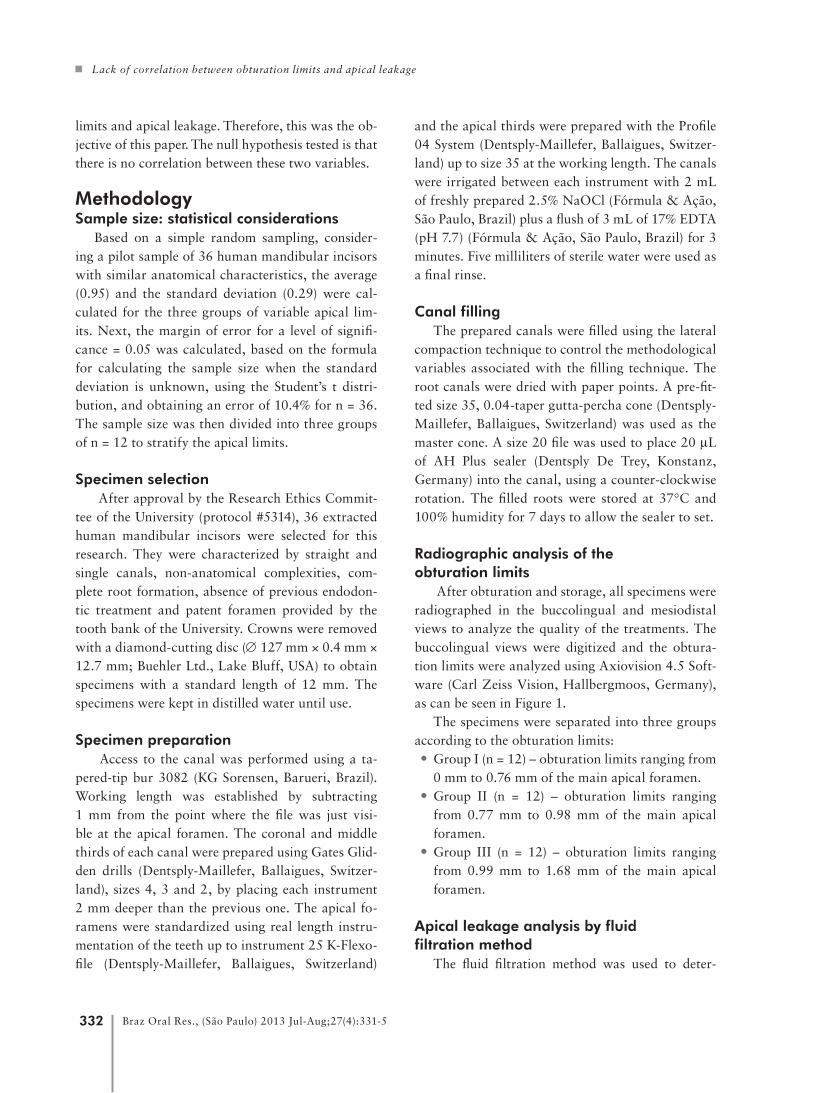
limits and apical leakage. Therefore, this was the ob-jective of this paper. The null hypothesis tested is that there is no correlation between these two variables.
Methodology Sample size: statistical considerations
Based on a simple random sampling, consider-ing a pilot sample of 36 human mandibular incisors with similar anatomical characteristics, the average (0.95) and the standard deviation (0.29) were cal-culated for the three groups of variable apical lim-its. Next, the margin of error for a level of signifi-cance = 0.05 was calculated, based on the formula for calculating the sample size when the standard deviation is unknown, using the Student’s t distri-bution, and obtaining an error of 10.4% for n = 36. The sample size was then divided into three groups of n = 12 to stratify the apical limits.
Specimen selection After approval by the Research Ethics Commit-
tee of the University (protocol #5314), 36 extracted human mandibular incisors were selected for this research. They were characterized by straight and single canals, non-anatomical complexities, com-plete root formation, absence of previous endodon-tic treatment and patent foramen provided by the tooth bank of the University. Crowns were removed with a diamond-cutting disc (∅ 127 mm × 0.4 mm × 12.7 mm; Buehler Ltd., Lake Bluff, USA) to obtain specimens with a standard length of 12 mm. The specimens were kept in distilled water until use.
Specimen preparation Access to the canal was performed using a ta-
pered-tip bur 3082 (KG Sorensen, Barueri, Brazil). Working length was established by subtracting 1 mm from the point where the file was just visi-ble at the apical foramen. The coronal and middle thirds of each canal were prepared using Gates Glid-den drills (Dentsply-Maillefer, Ballaigues, Switzer-land), sizes 4, 3 and 2, by placing each instrument 2 mm deeper than the previous one. The apical fo-ramens were standardized using real length instru-mentation of the teeth up to instrument 25 K-Flexo-file (Dentsply-Maillefer, Ballaigues, Switzerland)
and the apical thirds were prepared with the Profile 04 System (Dentsply-Maillefer, Ballaigues, Switzer-land) up to size 35 at the working length. The canals were irrigated between each instrument with 2 mL of freshly prepared 2.5% NaOCl (Fórmula & Ação, São Paulo, Brazil) plus a flush of 3 mL of 17% EDTA (pH 7.7) (Fórmula & Ação, São Paulo, Brazil) for 3 minutes. Five milliliters of sterile water were used as a final rinse.
Canal fillingThe prepared canals were filled using the lateral
compaction technique to control the methodological variables associated with the filling technique. The root canals were dried with paper points. A pre-fit-ted size 35, 0.04-taper gutta-percha cone (Dentsply-Maillefer, Ballaigues, Switzerland) was used as the master cone. A size 20 file was used to place 20 µL of AH Plus sealer (Dentsply De Trey, Konstanz, Germany) into the canal, using a counter-clockwise rotation. The filled roots were stored at 37°C and 100% humidity for 7 days to allow the sealer to set.
Radiographic analysis of the obturation limits
After obturation and storage, all specimens were radiographed in the buccolingual and mesiodistal views to analyze the quality of the treatments. The buccolingual views were digitized and the obtura-tion limits were analyzed using Axiovision 4.5 Soft-ware (Carl Zeiss Vision, Hallbergmoos, Germany), as can be seen in Figure 1.
The specimens were separated into three groups according to the obturation limits: • Group I (n = 12) – obturation limits ranging from
0 mm to 0.76 mm of the main apical foramen. • Group II (n = 12) – obturation limits ranging
from 0.77 mm to 0.98 mm of the main apical foramen.
• Group III (n = 12) – obturation limits ranging from 0.99 mm to 1.68 mm of the main apical foramen.
Apical leakage analysis by fluid filtration method
The fluid filtration method was used to deter-

Machado R, Silva Neto UX, Ignácio SA, Cunha RS
333Braz Oral Res., (São Paulo) 2013 Jul-Aug;27(4):331-5
DiscussionIn recent decades, different researchers have used
several leakage models. These models have been criticized for many factors mainly related to preclu-sion of direct clinical applicability of results.23-25
Several authors also observed divergent results when comparing different types of leakage tests. Barthel et al.26 applied the dye leakage test after
mine leakage.9,17 The root apex was connected to a Luer type metal needle by a plastic tube.4 The leak-age allowed by the tested groups was quantified ac-cording to the movement of a small air bubble inside a 25 µL micropipette (Fisher Scientific, Philadelphia, USA). The inside of the pipette and the entire sys-tem was filled with distilled water and a pressure of 10 psi was applied. After ensuring that there was no leakage at the connections, the system was activated and balanced for 4 minutes. The volume of fluid was calculated by observing the air bubble displace-ments, and was expressed in µL/min.10 psi. Mea-surements were made at 2-minute intervals over an 8-minute period.17
Results Tables 1 through 3 show the relevant statistical
data of the study.Groups I, II and III showed Pearson correlation
values (r2) of −0.152 (Table 1), −0.186 (Table 2) and 0.058 (Table 3), respectively. Furthermore, our re-sults showed that the leakage rates for a given obtu-ration limit, such as about 0.85, ranged from 0.10 to 0.89, indicating lack of correlation between obtura-tion limits and apical leakage.
Figure 1 - Obturation limits analysis by AxioVision 4.5 soft-ware (Carl Zeiss Vision, Hallbergmoos, Germany).
Table 3 - Correlation analysis of Group III.
Group III
Obturation limits
Apical leakage
Obturation limits
Pearson correlation 1 0.058
Sig. (2 - Tailed) 0.858
n 12 12
Apical leakage
Pearson correlation 0.058 1
Sig. (2 - Tailed) 0.858
n 12 12
Table 1 - Correlation analysis of Group I.
Group I
Obturation limits
Apical leakage
Obturation limits
Pearson correlation 1 −0.152
Sig. (2 - Tailed) 0.637
n 12 12
Apical leakage
Pearson correlation −0.152 1
Sig. (2 - Tailed) 0.637
n 12 12
Table 2 - Correlation analysis of Group II.
Group II
Obturation limits
Apical leakage
Obturation limits
Pearson correlation 1 −0.186
Sig. (2 - Tailed) 0.564
n 12 12
Apical leakage
Pearson correlation −0.186 1
Sig. (2 - Tailed) 0.564
n 12 12

Lack of correlation between obturation limits and apical leakage
334 Braz Oral Res., (São Paulo) 2013 Jul-Aug;27(4):331-5
the bacterial test on the same teeth and found no correlation between the tests. Pommel et al.27 also compared fluid filtration, electro-chemical and dye leakage tests in evaluating the sealing ability of sin-gle-cone and vertical condensation obturation tech-niques, using the same teeth, and found no correla-tion among the tests.
However, Wu et al.28 compared fluid filtration and dye penetration methods and found fluid trans-port was a more sensitive method for detecting voids along the root canal filling than dye penetration.
Moreover, Wu et al.29 showed a significant cor-relation between the quality of the fillings and leak-age rates. Of a total of 80 mesial roots of mandibu-lar molars observed in the buccolingual direction, 76% had well performed fillings, but this figure fell to 36% when the specimens were also analyzed in the mesiodistal direction. Because these specimens infiltrated less, although effectively, according to the analytic methodology used, we believe it is relevant to investigate others factors associated with leakage rates not just associated with radiographically evi-dent filling voids.
According to Karagenç et al.,14 the difference in results obtained when using various methods to as-sess leakage may be attributed to the differences in the working principles of various tests methods and the different nature of obturation materials.
A factor still not well studied in relation to apical leakage regards the obturation limits. Theoretically, when main cone obturation does not reach instru-mentation limits, it can predispose incorrect adapta-
tions in the apical, middle and cervical thirds, lead-ing to voids unfilled by lateral condensation.
Therefore, the purpose of this study was to eval-uate this possible predisposition according to fluid filtration tests. The results showed that there was no correlation between obturation limits and apical leakage. We believe these results are related to the effectiveness of the methodology. The fluid filtration test shows leakage only when there is at least one void extending from the apical to the coronal thirds. A root canal filling which looks badly condensed on the radiograph may contain many “cul de sac” type voids and no leakage. On the other hand, very small “through and through” type voids are invisible on radiographs but may be detected by the fluid filtra-tion test as having considerable leakage rates.16,28-30
Our results showed that there were no statisti-cal differences associated with apical leakage in the three groups analyzed. However, our obturation limits ranged from 0 to 1.68 mm of the main api-cal foramen, and these limits are in conformity with what are considered to be adequate limits in litera-ture.3 We believe that more research similar to that conducted in this study, using different obturation limits, is important to compare our results.
ConclusionsAccording to the methodology of this in vitro
study, we confirmed the null hypothesis that there is no correlation between the obturation limits and the apical leakage in roots filled with gutta percha and AH Plus sealer, in the three groups analyzed.
References1. Wu MK, Wesselink PR, Walton RE. Apical terminus location
of root canal treatment procedures. Oral Surg Oral Med Oral
Pathol Oral Radiol Endod. 2000 Jan;89(1):99-103.
2. Chueh LH, Chen SC, Lee CM, Hsu YY, Pai SF, Kuo ML, et
al. Technical quality of root canal treatment inTaiwan. Int
Endod J. 2003 Jun;36(6):416-22.
3. Schaeffer MA, White RR, Walton RE. Determining the op-
timal obturation length: a meta-analysis of literature. J En-
dod. 2005 Apr;31(4):271-4.
4. Moura MS, Guedes OA, Alencar AHG, Azevedo BC, Estrela
C. Influence of length of root canal obturation on apical peri-
odontitis detected by periapical radiography and cone beam
computed tomography. J Endod. 2009 Jun;35(6):805-9.
5. Collins J, Walker MP, Kulild J, Lee C. A comparison of three
gutta-percha obturation techniques to replicate canal irregu-
larities. J Endod. 2006 Aug;32(8):762-5.
6. Shakespeare RC, Donnelly JC. An in vitro comparison of api-
cal microleakage after obturation with JS Quick-fill or lateral
condensation. J Endod. 1997 May;23(5):312-4.
7. Von Fraunhofer JA, Fagundes DK, McDonald NJ, Dumsha
TC. The effect of root canal preparation on microleakage
within endodontically treated teeth: an in vitro study. Int
Endod J. 2000 Jul;33(4):355-60.

Machado R, Silva Neto UX, Ignácio SA, Cunha RS
335Braz Oral Res., (São Paulo) 2013 Jul-Aug;27(4):331-5
8. Carratù P, Amato M, Riccitiello F, Rengo S. Evaluation of leak-
age of bacteria and endotoxins in teeth treated endodontically
by two different techniques. J Endod. 2002 Apr;28(4):272-5.
9. Çobankara FK, Adanir N, Belli S, Pashley DH. A quantitative
evaluation of apical leakage of four root-canal sealers. Int
Endod J. 2002 Dec;35(12):979-84.
10. De Moor RJG, Hommez GMG. The long-term sealing ability
of an epoxy resin root canal sealer used with five gutta percha
obturation techniques. Int Endod J. 2002 Mar;35(3):275-82.
11. Robinson MJ, McDonald NJ, Mullally PJ. Apical extrusion of
thermoplasticized obturating material in canals instrumented
with Profile 0.06 or Profile GT. J Endod. 2004 Jun;30(6):418-
21.
12. Venturi M, Breschi L. Evaluation of apical filling after warm
vertical gutta-percha compaction using different procedures.
J Endod. 2004 Jun;30(6):436-40.
13. Diemer F, Sinan A, Calas P. Penetration depth of warm ver-
tical gutta-percha pluggers: impact of apical preparation. J
Endod. 2006 Feb;32(2):123-6.
14. Karagenç B, Gençoğlu N, Ersoy M, Cansever G, Külekçi G. A
comparison of four different microleakage tests for assessment
of leakage of root canal fillings. Oral Surg Oral Med Oral
Pathol Oral Radiol Endod. 2006 Jul;102(1):110-3.
15. Shemesh H, Wu M-K, Wesselink PR. Leakage along apical
root fillings with and without smear layer using two different
leakage models: a two-month longitudinal ex-vivo study. Int
Endod J. 2006 Dec;39(12):968-76.
16. Paqué F, Sirtes G. Apical sealing ability of Resilon/Epiphany
versus gutta-percha/AH Plus: immediate and 16-months leak-
age. Int Endod J. 2007 Sep;40(9):722-9.
17. Silva Neto UX, Moraes IG, Westphalen VPD, Menezes R,
Carneiro E, Fariniuk LF. Leakage of 4 resin-based root canal
sealers used with a single-cone technique. Oral Surg Oral
Med Oral Pathol Oral Radiol Endod. 2007 Aug;104(2):e53-7.
18. Veríssimo DM, Vale MS, Monteiro AJ. Comparison of apical
leakage between canals filled with gutta-percha/AH Plus and
the Resilon/Epiphany System, when submitted to two filling
techniques. J Endod. 2007 Mar;33(3):291-4.
19. Wedding JR, Brown CE, Legan JJ, Moore BK, Vail MM. An
in vitro comparison of microleakage between Resilon and
gutta-percha with a fluid filtration model. J Endod. 2007
Dec;33(12):1447-9.
20. Miletic I, Anic I, Pezelj-Ribaric S, Jukic S. Leakage of five root
canal sealers. Int Endod J. 1999 Sep;32(5):415-8.
21. Depraet FJHW, De Bruyne MAA, De Moor RJG. The seal-
ing ability of an epoxy resin root canal sealer after Nd:YAG
laser irradiation of the root canal. Int Endod J. 2005
May;38(5):302-9.
22. Qiong X, Ling J, Cheung GSP, Hu Y. A quantitative evalua-
tion of sealing ability of 4 obturation techniques by using a
glucose leakage test. Oral Surg Oral Med Oral Pathol Oral
Radiol Endod. 2007 Oct;104(4):e109-13.
23. Oliver CM, Abbott PV. Correlation between clinical success
and apical dye penetration. Int Endod J. 2001 Dec;34(8):637-
44.
24. Susini G, Pommel L, About I, Camps J. Lack of correlation
between ex vivo apical dye penetration and presence of apical
radiolucencies. Oral Surg Oral Med Oral Pathol Oral Radiol
Endod. 2006 Sep;102(3):e19-23.
25. De Bruyne MAA, De Moor RJG. Influence of cracks on leak-
age and obturation efficiency of root-end filling materials after
ultrasonic preparation: an in vitro evaluation. Quintessence
Int. 2008 Sep;39(8):685-92.
26. Barthel CR, Moshonov J, Shuping G, Orstavik D. Bacterial
leakage versus dye leakage in oburated root canals. Int Endod
J. 1999 Sep;32(5):370-5.
27. Pommel L, Jacquot B, Camps J. Lack of correlation among
three methods for evaluation of apical leakage. J Endod. 2001
May;27(5):347-50.
28. Wu MK, De Gee AJ, Wesselink PR. Fluid transport and
dye penetration along root canal filling. Int Endod J. 1994
Sep;27(5):233-8.
29. Wu MK, Bud MG, Wesselink PR. The quality of single cone
and laterally compacted gutta-percha fillings in small and
curved root canals as evidenced by bidirectional radiographs
and fluid transport measurements. Oral Surg Oral Med Oral
Pathol Oral Radiol Endod. 2009 Dec;108(6):946-51.
30. Wu MK, De Gee AJ, Wesselink PR, Moorer WR. Fluid trans-
port and bacterial penetration along root canal fillings. Int En-
dod J. 1993 Jul;26(4):203-8.