Lack of clinical AIDS in SIV-infected sooty mangabeys with...
9
Research article 1102 The Journal of Clinical Investigation http://www.jci.org Volume 121 Number 3 March 2011 Lack of clinical AIDS in SIV-infected sooty mangabeys with significant CD4 + T cell loss is associated with double-negative T cells Jeffrey M. Milush, 1,2 Kiran D. Mir, 1,3 Vasudha Sundaravaradan, 3 Shari N. Gordon, 4 Jessica Engram, 4 Christopher A. Cano, 1 Jacqueline D. Reeves, 5 Elizabeth Anton, 5 Eduardo O’Neill, 6 Eboneé Butler, 6 Kathy Hancock, 6 Kelly S. Cole, 7 Jason M. Brenchley, 8 James G. Else, 9 Guido Silvestri, 9 and Donald L. Sodora 1,3 1 Division of Infectious Disease, Department of Medicine, University of Texas Southwestern Medical Center, Dallas, Texas, USA. 2 Division of Experimental Medicine, Department of Medicine, UCSF, San Francisco, California, USA. 3 Seattle Biomedical Research Institute, Seattle, Washington, USA. 4 University of Pennsylvania, Philadelphia, Pennsylvania, USA. 5 Monogram Biosciences, South San Francisco, California, USA. 6 Influenza Division, National Center for Immunization and Respiratory Diseases, Centers for Disease Control and Prevention, Atlanta, Georgia, USA. 7 University of Pittsburgh School of Medicine, Pittsburgh, Pennsylvania, USA. 8 Laboratory of Molecular Microbiology, National Institute of Allergy and Infectious Diseases, NIH, Bethesda, Maryland, USA. 9 Yerkes National Primate Research Center, Emory University, Emory Vaccine Center, Atlanta, Georgia, USA. SIV infection of natural host species such as sooty mangabeys results in high viral replication without clinical signs of simian AIDS. Studying such infections is useful for identifying immunologic parameters that lead to AIDS in HIV-infected patients. Here we have demonstrated that acute, SIV-induced CD4 + T cell depletion in sooty mangabeys does not result in immune dysfunction and progression to simian AIDS and that a popula- tion of CD3 + CD4 – CD8 – T cells (double-negative T cells) partially compensates for CD4 + T cell function in these animals. Passaging plasma from an SIV-infected sooty mangabey with very few CD4 + T cells to SIV-negative animals resulted in rapid loss of CD4 + T cells. Nonetheless, all sooty mangabeys generated SIV-specific anti- body and T cell responses and maintained normal levels of plasma lipopolysaccharide. Moreover, all CD4- low sooty mangabeys elicited a de novo immune response following influenza vaccination. Such preserved immune responses as well as the low levels of immune activation observed in these animals were associated with the presence of double-negative T cells capable of producing Th1, Th2, and Th17 cytokines. These studies indicate that SIV-infected sooty mangabeys do not appear to rely entirely on CD4 + T cells to maintain immu- nity and identify double-negative T cells as a potential subset of cells capable of performing CD4 + T cell–like helper functions upon SIV-induced CD4 + T cell depletion in this species. Introduction Disease progression during pathogenic HIV/SIV infection has been defined historically by 2 strongly predictive parameters: plasma viral load and peripheral CD4 + T cell levels. However, it is becoming increasingly clear that HIV/SIV pathogenesis results from more than continuous virus replication and a coincident decline of target cells. For example, CD4 + T cells in mucosal com- partments are severely depleted very early after infection, and yet progression to AIDS is significantly delayed (1–5). Furthermore, the elevated levels of generalized immune activation observed dur- ing pathogenic infections are more predictive of disease progres- sion than viral load (6–8). The identification of natural host spe- cies such as sooty mangabeys and African green monkeys (AGMs) that replicate SIV to high levels but generally do not exhibit clinical signs of AIDS has been invaluable. Through comparison of patho- genic and nonpathogenic infections, we can infer the important pathogenic factors. For example, SIV-infected natural hosts expe- rience an early and rapid depletion of mucosal CD4 + T cells to an extent similar to that seen in HIV patients (9–12) as well as adap- tive immune responses that are comparable to those in pathogenic HIV/SIV infections (13, 14). One striking distinction of the natu- ral SIV infections is the maintenance of low levels of generalized immune activation during the chronic phase of the infection (at times after 28 dpi) (7, 8, 15–19). We previously observed that 2 SIV-infected sooty mangabeys infected by SIVsmm via plasma transfer in October 2000 exhibited a dramatic CD4 + T cell decline (< 100 cells/μl blood) yet maintained low levels of immune activation during chronic infection (19). The CD4 + T cell depletion occurred within all tissue samples examined, and the CD4-low phenotype in these mangabeys was associated with the presence of a multitropic (R5/X4/R8-using) SIVsmm (19). Unlike the inoculum, this multitropic SIVsmm was able to infect CD4 + T cells expressing CXCR4, which includes more than 90% of CD4 + T cells in sooty mangabeys (19). Importantly, these mangabeys have remained free of clinical signs of simian AIDS for the past 10 years. Here, we undertake a passage of the multitropic SIVsmm from a CD4-low mangabey to 3 additional mangabeys and investigate the immunologic mechanisms by which these SIV-infected mangabeys can remain free of disease despite AIDS-defining CD4 + T cell levels. Results Passage of multitropic SIVsmm results in rapid depletion of CD4 + T cells. To further investigate the CD4-low phenotype observed in 2 SIV- infected sooty mangabeys (19), we passaged SIVsmm from the pre- viously identified CD4-low mangabey SM2 through intravenous Authorship note: Jeffrey M. Milush and Kiran D. Mir contributed equally to this work. Conflict of interest: The authors have declared that no conflict of interest exists. Citation for this article: J Clin Invest. 2011;121(3):1102–1110. doi:10.1172/JCI44876.
Transcript of Lack of clinical AIDS in SIV-infected sooty mangabeys with...
Research article
1102 TheJournalofClinicalInvestigation http://www.jci.org Volume 121 Number 3 March 2011
Lack of clinical AIDS in SIV-infected sooty mangabeys with significant CD4+ T cell loss is associated with double-negative T cells
Jeffrey M. Milush,1,2 Kiran D. Mir,1,3 Vasudha Sundaravaradan,3 Shari N. Gordon,4 Jessica Engram,4 Christopher A. Cano,1 Jacqueline D. Reeves,5 Elizabeth Anton,5
Eduardo O’Neill,6 Eboneé Butler,6 Kathy Hancock,6 Kelly S. Cole,7 Jason M. Brenchley,8 James G. Else,9 Guido Silvestri,9 and Donald L. Sodora1,3
1Division of Infectious Disease, Department of Medicine, University of Texas Southwestern Medical Center, Dallas, Texas, USA. 2Division of Experimental Medicine, Department of Medicine, UCSF, San Francisco, California, USA.
3Seattle Biomedical Research Institute, Seattle, Washington, USA. 4University of Pennsylvania, Philadelphia, Pennsylvania, USA. 5Monogram Biosciences, South San Francisco, California, USA. 6Influenza Division, National Center for Immunization and Respiratory Diseases, Centers for Disease Control and Prevention, Atlanta, Georgia, USA. 7University of Pittsburgh School of Medicine, Pittsburgh, Pennsylvania, USA.
8Laboratory of Molecular Microbiology, National Institute of Allergy and Infectious Diseases, NIH, Bethesda, Maryland, USA. 9Yerkes National Primate Research Center, Emory University, Emory Vaccine Center, Atlanta, Georgia, USA.
SIVinfectionofnaturalhostspeciessuchassootymangabeysresultsinhighviralreplicationwithoutclinicalsignsofsimianAIDS.StudyingsuchinfectionsisusefulforidentifyingimmunologicparametersthatleadtoAIDSinHIV-infectedpatients.Herewehavedemonstratedthatacute,SIV-inducedCD4+TcelldepletioninsootymangabeysdoesnotresultinimmunedysfunctionandprogressiontosimianAIDSandthatapopula-tionofCD3+CD4–CD8–Tcells(double-negativeTcells)partiallycompensatesforCD4+Tcellfunctionintheseanimals.PassagingplasmafromanSIV-infectedsootymangabeywithveryfewCD4+TcellstoSIV-negativeanimalsresultedinrapidlossofCD4+Tcells.Nonetheless,allsootymangabeysgeneratedSIV-specificanti-bodyandTcellresponsesandmaintainednormallevelsofplasmalipopolysaccharide.Moreover,allCD4-lowsootymangabeyselicitedadenovoimmuneresponsefollowinginfluenzavaccination.Suchpreservedimmuneresponsesaswellasthelowlevelsofimmuneactivationobservedintheseanimalswereassociatedwiththepresenceofdouble-negativeTcellscapableofproducingTh1,Th2,andTh17cytokines.ThesestudiesindicatethatSIV-infectedsootymangabeysdonotappeartorelyentirelyonCD4+Tcellstomaintainimmu-nityandidentifydouble-negativeTcellsasapotentialsubsetofcellscapableofperformingCD4+Tcell–likehelperfunctionsuponSIV-inducedCD4+Tcelldepletioninthisspecies.
IntroductionDisease progression during pathogenic HIV/SIV infection has been defined historically by 2 strongly predictive parameters: plasma viral load and peripheral CD4+ T cell levels. However, it is becoming increasingly clear that HIV/SIV pathogenesis results from more than continuous virus replication and a coincident decline of target cells. For example, CD4+ T cells in mucosal com-partments are severely depleted very early after infection, and yet progression to AIDS is significantly delayed (1–5). Furthermore, the elevated levels of generalized immune activation observed dur-ing pathogenic infections are more predictive of disease progres-sion than viral load (6–8). The identification of natural host spe-cies such as sooty mangabeys and African green monkeys (AGMs) that replicate SIV to high levels but generally do not exhibit clinical signs of AIDS has been invaluable. Through comparison of patho-genic and nonpathogenic infections, we can infer the important pathogenic factors. For example, SIV-infected natural hosts expe-rience an early and rapid depletion of mucosal CD4+ T cells to an extent similar to that seen in HIV patients (9–12) as well as adap-tive immune responses that are comparable to those in pathogenic
HIV/SIV infections (13, 14). One striking distinction of the natu-ral SIV infections is the maintenance of low levels of generalized immune activation during the chronic phase of the infection (at times after 28 dpi) (7, 8, 15–19).
We previously observed that 2 SIV-infected sooty mangabeys infected by SIVsmm via plasma transfer in October 2000 exhibited a dramatic CD4+ T cell decline (< 100 cells/μl blood) yet maintained low levels of immune activation during chronic infection (19). The CD4+ T cell depletion occurred within all tissue samples examined, and the CD4-low phenotype in these mangabeys was associated with the presence of a multitropic (R5/X4/R8-using) SIVsmm (19). Unlike the inoculum, this multitropic SIVsmm was able to infect CD4+ T cells expressing CXCR4, which includes more than 90% of CD4+ T cells in sooty mangabeys (19). Importantly, these mangabeys have remained free of clinical signs of simian AIDS for the past 10 years. Here, we undertake a passage of the multitropic SIVsmm from a CD4-low mangabey to 3 additional mangabeys and investigate the immunologic mechanisms by which these SIV-infected mangabeys can remain free of disease despite AIDS-defining CD4+ T cell levels.
ResultsPassage of multitropic SIVsmm results in rapid depletion of CD4+ T cells. To further investigate the CD4-low phenotype observed in 2 SIV-infected sooty mangabeys (19), we passaged SIVsmm from the pre-viously identified CD4-low mangabey SM2 through intravenous
Authorshipnote: Jeffrey M. Milush and Kiran D. Mir contributed equally to this work.
Conflictofinterest: The authors have declared that no conflict of interest exists.
Citationforthisarticle: J Clin Invest. 2011;121(3):1102–1110. doi:10.1172/JCI44876.
research article
TheJournalofClinicalInvestigation http://www.jci.org Volume 121 Number 3 March 2011 1103
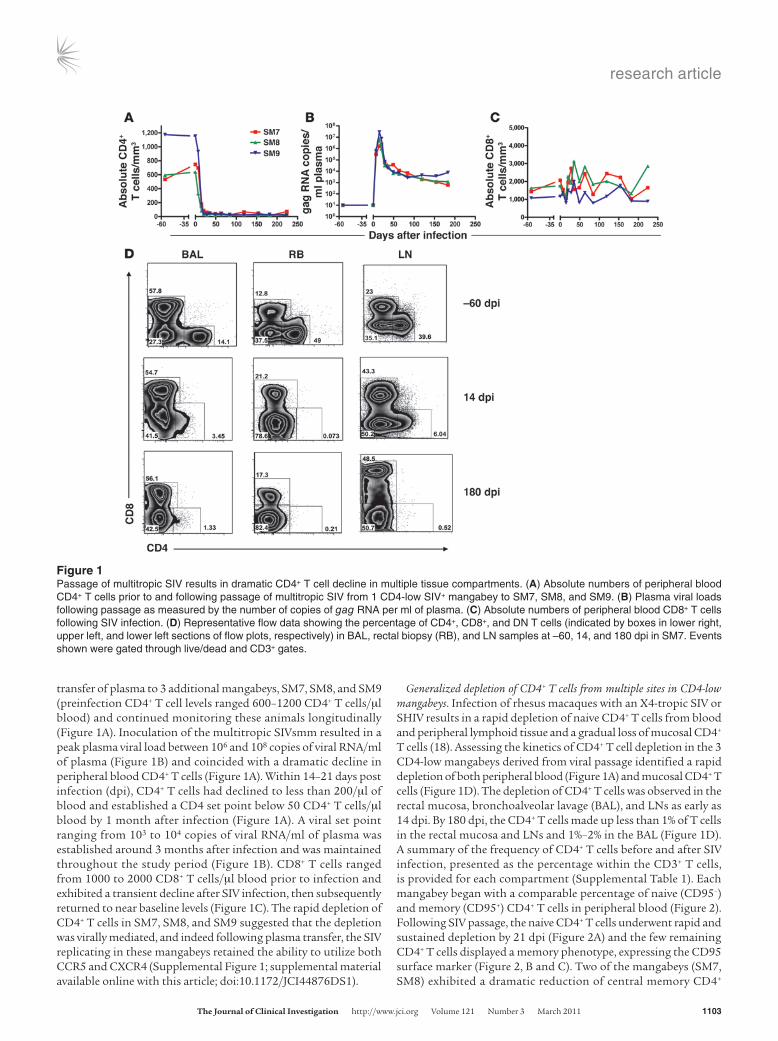
transfer of plasma to 3 additional mangabeys, SM7, SM8, and SM9 (preinfection CD4+ T cell levels ranged 600–1200 CD4+ T cells/μl blood) and continued monitoring these animals longitudinally (Figure 1A). Inoculation of the multitropic SIVsmm resulted in a peak plasma viral load between 106 and 108 copies of viral RNA/ml of plasma (Figure 1B) and coincided with a dramatic decline in peripheral blood CD4+ T cells (Figure 1A). Within 14–21 days post infection (dpi), CD4+ T cells had declined to less than 200/μl of blood and established a CD4 set point below 50 CD4+ T cells/μl blood by 1 month after infection (Figure 1A). A viral set point ranging from 103 to 104 copies of viral RNA/ml of plasma was established around 3 months after infection and was maintained throughout the study period (Figure 1B). CD8+ T cells ranged from 1000 to 2000 CD8+ T cells/μl blood prior to infection and exhibited a transient decline after SIV infection, then subsequently returned to near baseline levels (Figure 1C). The rapid depletion of CD4+ T cells in SM7, SM8, and SM9 suggested that the depletion was virally mediated, and indeed following plasma transfer, the SIV replicating in these mangabeys retained the ability to utilize both CCR5 and CXCR4 (Supplemental Figure 1; supplemental material available online with this article; doi:10.1172/JCI44876DS1).
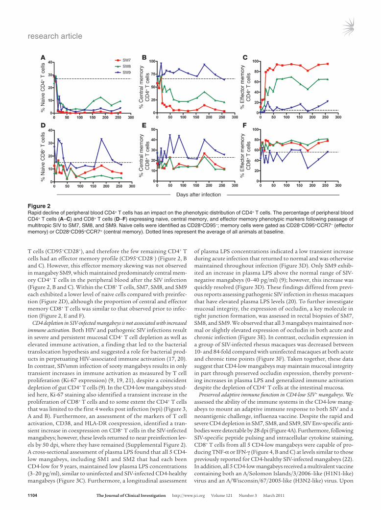
Generalized depletion of CD4+ T cells from multiple sites in CD4-low mangabeys. Infection of rhesus macaques with an X4-tropic SIV or SHIV results in a rapid depletion of naive CD4+ T cells from blood and peripheral lymphoid tissue and a gradual loss of mucosal CD4+ T cells (18). Assessing the kinetics of CD4+ T cell depletion in the 3 CD4-low mangabeys derived from viral passage identified a rapid depletion of both peripheral blood (Figure 1A) and mucosal CD4+ T cells (Figure 1D). The depletion of CD4+ T cells was observed in the rectal mucosa, bronchoalveolar lavage (BAL), and LNs as early as 14 dpi. By 180 dpi, the CD4+ T cells made up less than 1% of T cells in the rectal mucosa and LNs and 1%–2% in the BAL (Figure 1D). A summary of the frequency of CD4+ T cells before and after SIV infection, presented as the percentage within the CD3+ T cells, is provided for each compartment (Supplemental Table 1). Each mangabey began with a comparable percentage of naive (CD95–) and memory (CD95+) CD4+ T cells in peripheral blood (Figure 2). Following SIV passage, the naive CD4+ T cells underwent rapid and sustained depletion by 21 dpi (Figure 2A) and the few remaining CD4+ T cells displayed a memory phenotype, expressing the CD95 surface marker (Figure 2, B and C). Two of the mangabeys (SM7, SM8) exhibited a dramatic reduction of central memory CD4+
Figure 1Passage of multitropic SIV results in dramatic CD4+ T cell decline in multiple tissue compartments. (A) Absolute numbers of peripheral blood CD4+ T cells prior to and following passage of multitropic SIV from 1 CD4-low SIV+ mangabey to SM7, SM8, and SM9. (B) Plasma viral loads following passage as measured by the number of copies of gag RNA per ml of plasma. (C) Absolute numbers of peripheral blood CD8+ T cells following SIV infection. (D) Representative flow data showing the percentage of CD4+, CD8+, and DN T cells (indicated by boxes in lower right, upper left, and lower left sections of flow plots, respectively) in BAL, rectal biopsy (RB), and LN samples at –60, 14, and 180 dpi in SM7. Events shown were gated through live/dead and CD3+ gates.
research article
1104 TheJournalofClinicalInvestigation http://www.jci.org Volume 121 Number 3 March 2011
T cells (CD95+CD28+), and therefore the few remaining CD4+ T cells had an effector memory profile (CD95+CD28–) (Figure 2, B and C). However, this effector memory skewing was not observed in mangabey SM9, which maintained predominately central mem-ory CD4+ T cells in the peripheral blood after the SIV infection (Figure 2, B and C). Within the CD8+ T cells, SM7, SM8, and SM9 each exhibited a lower level of naive cells compared with preinfec-tion (Figure 2D), although the proportion of central and effector memory CD8+ T cells was similar to that observed prior to infec-tion (Figure 2, E and F).
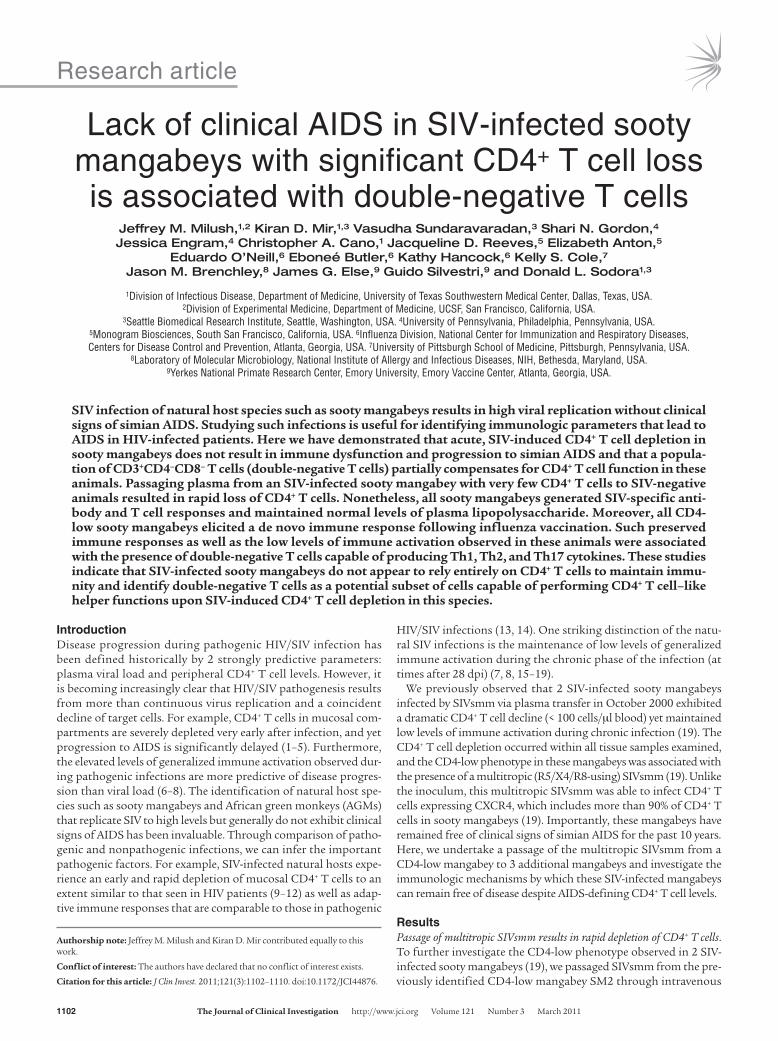
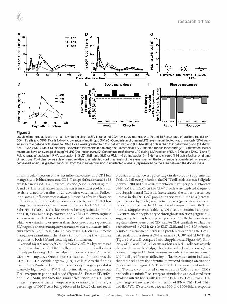
CD4 depletion in SIV-infected mangabeys is not associated with increased immune activation. Both HIV and pathogenic SIV infections result in severe and persistent mucosal CD4+ T cell depletion as well as elevated immune activation, a finding that led to the bacterial translocation hypothesis and suggested a role for bacterial prod-ucts in perpetuating HIV-associated immune activation (17, 20). In contrast, SIVsmm infection of sooty mangabeys results in only transient increases in immune activation as measured by T cell proliferation (Ki-67 expression) (9, 19, 21), despite a coincident depletion of gut CD4+ T cells (9). In the CD4-low mangabeys stud-ied here, Ki-67 staining also identified a transient increase in the proliferation of CD8+ T cells and to some extent the CD4+ T cells that was limited to the first 4 weeks post infection (wpi) (Figure 3, A and B). Furthermore, an assessment of the markers of T cell activation, CD38, and HLA-DR coexpression, identified a tran-sient increase in coexpression on CD8+ T cells in the SIV-infected mangabeys; however, these levels returned to near preinfection lev-els by 50 dpi, where they have remained (Supplemental Figure 2). A cross-sectional assessment of plasma LPS found that all 5 CD4-low mangabeys, including SM1 and SM2 that had each been CD4-low for 9 years, maintained low plasma LPS concentrations (3–20 pg/ml), similar to uninfected and SIV-infected CD4-healthy mangabeys (Figure 3C). Furthermore, a longitudinal assessment
of plasma LPS concentrations indicated a low transient increase during acute infection that returned to normal and was otherwise maintained throughout infection (Figure 3D). Only SM9 exhib-ited an increase in plasma LPS above the normal range of SIV-negative mangabeys (0–40 pg/ml) (9); however, this increase was quickly resolved (Figure 3D). These findings differed from previ-ous reports assessing pathogenic SIV infection in rhesus macaques that have elevated plasma LPS levels (20). To further investigate mucosal integrity, the expression of occludin, a key molecule in tight junction formation, was assessed in rectal biopsies of SM7, SM8, and SM9. We observed that all 3 mangabeys maintained nor-mal or slightly elevated expression of occludin in both acute and chronic infection (Figure 3E). In contrast, occludin expression in a group of SIV-infected rhesus macaques was decreased between 10- and 84-fold compared with uninfected macaques at both acute and chronic time points (Figure 3F). Taken together, these data suggest that CD4-low mangabeys may maintain mucosal integrity in part through preserved occludin expression, thereby prevent-ing increases in plasma LPS and generalized immune activation despite the depletion of CD4+ T cells at the intestinal mucosa.
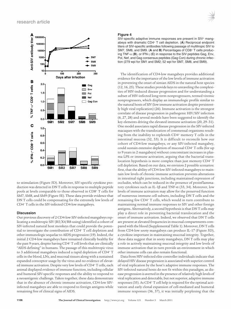
Preserved adaptive immune function in CD4-low SIV+ mangabeys. We assessed the ability of the immune systems in the CD4-low mang-abeys to mount an adaptive immune response to both SIV and a neoantigenic challenge, influenza vaccine. Despite the rapid and severe CD4 depletion in SM7, SM8, and SM9, SIV Env-specific anti-bodies were detectable by 28 dpi (Figure 4A). Furthermore, following SIV-specific peptide pulsing and intracellular cytokine staining, CD8+ T cells from all 5 CD4-low mangabeys were capable of pro-ducing TNF-α or IFN-γ (Figure 4, B and C) at levels similar to those previously reported for CD4-healthy SIV-infected mangabeys (22). In addition, all 5 CD4-low mangabeys received a multivalent vaccine containing both an A/Solomon Islands/3/2006–like (H1N1-like) virus and an A/Wisconsin/67/2005-like (H3N2-like) virus. Upon
Figure 2Rapid decline of peripheral blood CD4+ T cells has an impact on the phenotypic distribution of CD4+ T cells. The percentage of peripheral blood CD4+ T cells (A–C) and CD8+ T cells (D–F) expressing naive, central memory, and effector memory phenotypic markers following passage of multitropic SIV to SM7, SM8, and SM9. Naive cells were identified as CD28+CD95–; memory cells were gated as CD28–CD95+CCR7– (effector memory) or CD28+CD95+CCR7+ (central memory). Dotted lines represent the average of all animals at baseline.
research article
TheJournalofClinicalInvestigation http://www.jci.org Volume 121 Number 3 March 2011 1105
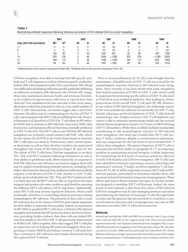
intramuscular injection of the first influenza vaccine, all 5 CD4-low mangabeys exhibited increased CD8+ T cell proliferation and 4 of 5 exhibited increased CD4+ T cell proliferation (Supplemental Figure 3, A and B). This proliferative response was transient, as proliferation levels returned to baseline by 21 days after vaccination. Follow-ing a second influenza vaccination (10 months after the first), an influenza-specific antibody response was detected in all 5 CD4-low mangabeys as measured by microneutralization for H1N1 and 4 of 5 for H3N2 (Table 1). The less sensitive hemagglutination inhibi-tion (HI) assay was also performed, and 3 of 5 CD4-low mangabeys seroconverted with HI titers between 40 and 453 (data not shown), which were similar to or greater than those previously reported in SIV-negative rhesus macaques vaccinated with a multivalent influ-enza vaccine (23). These data indicate that CD4-low SIV-infected mangabeys maintained the ability to mount adaptive immune responses to both SIV and neoantigenic stimulation.
Potential helper functions of CD3+CD4–CD8– T cells. We hypothesized that in the absence of CD4+ T cells, another immune cell subset is likely performing CD4-like helper functions in the SIV-infected CD4-low mangabeys. One immune cell subset of interest was the CD3+CD4–CD8– double-negative (DN) T cells due to the finding that both SIV-infected and uninfected sooty mangabeys exhibit relatively high levels of DN T cells primarily expressing the α/β T cell receptor in peripheral blood (Figure 5A). Prior to SIV infec-tion, SM7, SM8, and SM9 had similar frequencies of DN T cells in each respective tissue compartment examined with a larger percentage of DN T cells being observed in LNs, BAL, and rectal
biopsies and the lowest percentage in the blood (Supplemental Table 1). Following infection, the DN T cell levels increased slightly (between 200 and 500 cells/mm3 blood) in the peripheral blood of SM7, SM8, and SM9 as the CD4+ T cells were depleted (Figure 5 and Supplemental Table 1). Interestingly, the largest percentage increase in the DN T cell population was within the LNs (percent-age increased by 2-fold) and rectal mucosa (percentage increased almost 3-fold), while the BAL exhibited a more modest DN T cell increase (Supplemental Table 1). DN T cells maintained a primar-ily central memory phenotype throughout infection (Figure 5C), suggesting they may be antigen-experienced T cells that have down-regulated the expression of CD4 and/or CD8, similarly to what has been observed in AGMs (24). In SM7, SM8, and SM9, SIV infection resulted in a transient increase in proliferation of the DN T cells, with peak proliferation at 20 dpi, similar to CD8+ and CD4+ T cells (Figure 3, A and B, compared with Supplemental Figure 4A). Simi-larly, CD38 and HLA-DR coexpression on DN T cells was acutely elevated; however, by 28 dpi, it had returned to baseline levels (Sup-plemental Figure 4B). Furthermore, an early, transient increase in DN T cell proliferation following influenza vaccination indicated that these cells have the potential to respond during a vaccination (Supplemental Figure 4C). To assess the functionality of isolated DN T cells, we stimulated them with anti-CD3 and anti-CD28 antibodies to mimic T cell receptor stimulation and evaluated their cytokine mRNA levels with real-time PCR. DN T cells from CD4-low mangabeys increased the expression of IFN-γ (Th1), IL-4 (Th2), and IL-17 (Th17) cytokines between 300- and 8000-fold in response
Figure 3Levels of immune activation remain low during chronic SIV infection of CD4-low sooty mangabeys. (A and B) Percentage of proliferating (Ki-67+) CD4+ T cells and CD8+ T cells following passage of multitropic SIV. (C) Comparison of plasma LPS levels in uninfected and chronically SIV-infect-ed sooty mangabeys with absolute CD4+ T cell levels greater than 200 cells/mm3 blood (CD4-healthy) or less than 200 cells/mm3 blood (CD4-low; SM1, SM2, SM7, SM8, SM9 shown). Dotted line represents the average of 10 chronically SIV-infected rhesus macaques (20); Uninfected rhesus macaques have an average of 15 pg/ml LPS (20) (not shown). (D) Concentration of plasma LPS during SIV infection of SM7, SM8, and SM9. (E and F) Fold change of occludin mRNA expression in SM7, SM8, and SM9 or RMs 1–6 during acute (2–15 dpi) and chronic (184 dpi) infection or at time of necropsy. Fold change was determined relative to uninfected control animals of the same species; the fold change is considered increased or decreased when it is greater than 2 SD from the mean expression in uninfected animals (represented by the area between the dotted lines).
research article
1106 TheJournalofClinicalInvestigation http://www.jci.org Volume 121 Number 3 March 2011
to stimulation (Figure 5D). Moreover, SIV-specific cytokine pro-duction was detected in DN T cells in response to multiple peptide pools at levels comparable to those observed in CD8+ T cells for SM7, SM8, and SM9 (Figure 5E). These data provide evidence that DN T cells could be compensating for the extremely low levels of CD4+ T cells in the SIV-infected CD4-low mangabeys.
DiscussionOur previous discovery of 2 CD4-low SIV-infected mangabeys rep-licating a multitropic SIV (R5/X4/R8-using) identified a cohort of SIV-infected natural host monkeys that could provide the poten-tial to investigate the contribution of CD4+ T cell depletion and other immunologic sequelae to AIDS progression (19). Indeed, the initial 2 CD4-low mangabeys have remained clinically healthy for the past 9 years, despite having CD4+ T cell levels that are clinically “AIDS defining” in humans. The passage of this multitropic virus to 3 additional mangabeys induced a rapid depletion of CD4+ T cells in the blood, LNs, and mucosal tissues along with a sustained expanded coreceptor usage by the virus and no evidence of elevat-ed immune activation. Despite very low levels of CD4+ T cells, each animal displayed evidence of immune function, including cellular and humoral SIV-specific responses and the ability to respond to a neoantigenic challenge. Taken together, these data demonstrate that in the absence of chronic immune activation, CD4-low SIV-infected mangabeys are able to respond to foreign antigens while remaining free of clinical signs of AIDS.
The identification of CD4-low mangabeys provides additional evidence for the importance of the low levels of immune activation in preventing the onset of simian AIDS in the natural host species (12, 16, 25). These studies provide keys to unraveling the complexi-ties of HIV-induced disease progression and for understanding a subset of HIV-infected long-term nonprogressors, termed viremic nonprogressors, which display an immunologic profile similar to the natural hosts of SIV (low immune activation despite persistent-ly high viral replication) (26). Immune activation is the strongest correlate of disease progression in pathogenic HIV/SIV infection (6, 27, 28) and several models have been suggested to identify the key elements driving the elevated immune activation (20, 29–31). One model associates rapid disease progression in the SIV-infected macaques with the translocation of commensal organisms result-ing from the inability to replenish CD4+ memory T cells in the intestinal mucosa (32, 33). It is difficult to reconcile how our cohort of CD4-low mangabeys, or any SIV-infected mangabey, could sustain extensive depletion of mucosal CD4+ T cells (for up to 9 years in 2 mangabeys) without concomitant increases in plas-ma LPS or immune activation, arguing that the bacterial trans-location hypothesis is more complex than just memory CD4+ T cell depletion. Based on our data, we envision 2 possible scenarios: first, that the ability of CD4-low SIV-infected mangabeys to main-tain low levels of chronic immune activation prevents alterations in intestinal tight junctions, including maintained expression of occludin, which can be reduced in the presence of proinflamma-tory cytokines such as IL-1β and TNF-α (33, 34). Moreover, low levels of immune activation may allow for the preserved function of numerous immune cell subsets, including DN T cells and the remaining few CD4+ T cells, which would in turn contribute to maintaining normal immune responses to SIV and other foreign antigens. Alternatively, a second hypothesis is that DN T cells may play a direct role in preventing bacterial translocation and the onset of immune activation. Indeed, we observed that DN T cells are present at elevated frequencies in mucosal compartments com-pared with the blood (Supplemental Table 1). Moreover, DN T cells from CD4-low sooty mangabeys can produce IL-17 (Figure 5D), a cytokine important in maintaining mucosal integrity. Together, these data suggest that in sooty mangabeys, DN T cells may play a role in actively maintaining mucosal integrity and low levels of immune activation that in turn provide an environment in which other immune cells can also remain functional.
Data from HIV-infected elite controller individuals indicate that delayed HIV disease progression is associated with superior control of viral replication by the host’s adaptive immune responses. The SIV-infected natural hosts do not fit within this paradigm, as dis-ease progression is averted in the presence of relatively high levels of viral replication and detectable, but not superior, adaptive immune responses (35). As CD4+ T cell help is required for the optimal acti-vation and early clonal expansion of cell-mediated and humoral immune responses (36, 37), it was initially perplexing that the
Figure 4SIV-specific adaptive immune responses are present in SIV+ mang-abeys with dramatic CD4+ T cell depletion. (A) Reciprocal endpoint titers of SIV-specific antibodies following passage of multitropic SIV to SM7, SM8, and SM9. (A and B) Percentages of CD8+ T cells produc-ing TNF-α (B), or IFN-γ (C) in response to the SIV peptides Gag, Env, Pol, Nef, and Gag consensus peptides (Gag Con) during chronic infec-tion (279 wpi for SM1 and SM2; 52 wpi for SM7, SM8, and SM9).
research article
TheJournalofClinicalInvestigation http://www.jci.org Volume 121 Number 3 March 2011 1107
CD4-low mangabeys were able to develop both SIV-specific anti-body and T cell responses as well as influenza-specific antibodies. Indeed, HIV-infected patients with CD4 counts below 100 cells/μl have difficulties developing influenza-specific antibodies following an influenza vaccination (38). Moreover, the CD4-low SIV+ mang-abeys have maintained immune health and immune function, as no evidence of opportunistic infections or cancers have been observed. One explanation for this outcome is that sooty mang-abeys have evolved the potential to rely on a very small number of CD4+ T cells. Alternatively, our results suggest that a non-CD4+ immune cell subset may be compensating for the loss of CD4+ T cells, and we propose that DN T cells are fulfilling this role. Recent-ly Beaumier et al. described a CD3+CD4– T cell subset in SIV-infect-ed AGMs that is resistant to SIV infection, functional, MHC class II restricted, and maintains effector functions normally attributed to CD4+ T cells (24). The DN T cells in our CD4-low SIV-infected mangabeys are primarily central memory αβ-TCR+ cells, which do not express the γδ TCR or the Vα24 chain found on invariant NK T cells (data not shown). Furthermore, their central memo-ry phenotype as well as their absolute numbers are maintained throughout the course of SIV infection (Figure 5C and ref. 19). The ability of DN T cells from CD4-low mangabeys to produce Th1, Th2, and Th17 cytokines following stimulation as well as their ability to proliferate early, albeit transiently, in response to both SIV infection and influenza vaccination suggest these cells may be capable of performing multiple functions, including CD4 helper–like functions that may have enabled an influenza antibody response in the absence of CD4+ T cells. Similar to CD4+ T cells, which can be subdivided into Th1, Th2, and Th17 polarized cells, it is likely that not all DN T cells are able to respond to the same antigens and produce the same cytokines, and therefore assessing the different DN T cell subsets will be important. Additionally, some DN T cells may possess regulatory function, which could potentially contribute to controlling immune activation during nonpathogenic SIV infection. The potential of these cells to resist SIV infection due to the absence of CD4 is particularly important for their ability to maintain functionality. Indeed, an assessment of SIV DNA in purified DN T cells from a CD4-low SIV-infected mangabey determined that SIV levels were below the level of detec-tion, providing further evidence that these cells are indeed SIV-resistant (similar to the CD4–CD8+ T cells previously characterized in AGMs) (24). Taken together, these data suggest DN T cells play an important role in keeping SIV-infected mangabeys from pro-gressing to simian AIDS by providing a memory T cell repertoire that is resistant to SIV infection and remains functional during SIV-induced CD4+ T cell loss.
Prior to recent publications (9, 10, 19), it was thought that the maintenance of healthy levels of CD4+ T cells was critical for the nonpathogenic outcome observed in SIV-infected sooty mang-abeys. More recently, it has been shown that sooty mangabeys have limited expression of CCR5 on CD4+ T cells, which could be important for protecting specific subsets of CD4+ T cells (such as Tcm) from virus-mediated depletion, thus leading to a better preservation of the overall CD4+ T cell pool (39, 40). However, in our cohort of SIV-infected mangabeys, the multitropic nature of the virus permits the infection of essentially all CD4+ T cells, thereby inducing a profound depletion of CD4+ T cells from all immunologic sites. Despite extensive CD4+ T cell depletion, our cohort is able to maintain immunologic health and has resisted clinical disease progression despite 3 or 9 years of AIDS-defining CD4+ T cell numbers. While there are likely multiple mechanisms contributing to the nonpathogenic outcome in SIV-infected sooty mangabeys, this study has revealed that DN T cells pro-duce T helper cytokines, display a central-memory phenotype, and may compensate for the low levels of conventional CD4+ T cells in these mangabeys. The greater frequency of DN T cells in mucosal sites and their ability to upregulate IL-17, an important cytokine in maintaining mucosal integrity, is likely important for maintaining the low levels of immune activation observed in both CD4-healthy and CD4-low mangabeys. DN T cells have been identified in humans expressing a memory phenotype and possessing T cytotoxic, T helper, as well as regulatory T cell func-tions (39, 41, 42). It will be important to assess DN T cells in HIV-infected patients, particularly to determine whether these cells are preserved and functional in long-term nonprogressors. These efforts may lead to future immune therapies or vaccine modali-ties designed to modulate DN T cell function. Indeed, the main lesson we have learned to date from this cohort of SIV-infected CD4-low mangabeys may be that managing immune activation and bolstering the function of nontarget T cells through better vaccines and therapeutics has the potential to contribute to pre-served immune function and a nonprogressive outcome in HIV infection even when CD4+ T cell levels become low.
MethodsAnimals and viral infection. SM1 and SM2 were between 3 and 5 years of age when infected with SIV in the original study (19). They were inoculated intravenously with 1 ml of plasma transferred from mangabey FQi, a natu-rally SIVsmm-infected mangabey at the Yerkes primate colony. The infection occurred in October 2000 and has previously been described (19). Before SIVsmm infection, virologic assessment determined that none of the mang-abeys contained simian T cell lymphotropic virus or simian retrovirus. Fur-
Table 1Neutralizing antibody responses following influenza vaccination of SIV-infected CD4-low sooty mangabeys
SM1 SM2 SM7 SM8 SM9Daysafter2ndvaccination WIA SIB WI SI WI SI WI SI WI SI–7 5 5 5 5 10 20 5 5 5 400 5 5 5 5 10 20 5 5 7 287 160 80 5 40 640 453 40 80 160 128014 320 226 5 80 2560 > 1280 320 226 > 1280 256021 160 113 5 40 1280 > 1280 160 113 640 256028 80 80 5 40 640 > 905 80 80 320 1810
AAntibody titers to the vaccine component H3N2 (WI). BAntibody titers to the vaccine component H1N1 (SI).

research article
1108 TheJournalofClinicalInvestigation http://www.jci.org Volume 121 Number 3 March 2011
thermore, anti-sera analysis determined that all mangabeys except SM2 were seropositive for CMV/simian agent 6 and herpesvirus B. Passaging of virus from SM2 to 3 additional SIV-negative mangabeys (SM7, SM8, and SM9) was performed by intravenous inoculation of 1.5 ml of plasma from the 303 wpi time point in October 2007. The viral inoculum contained SIV that were multitropic, able to use multiple coreceptors including CCR5 and CXCR4 (19). All protocols were approved by the institutional animal care and use committee at Emory University.
LN biopsies, rectal biopsies, and BAL. LN (axillary or inguinal) and rectal mucosal (RM) biopsies as well as BALs were performed as previously described (19).
Viral load analysis. SIVsmm plasma viral load was determined as previously described (16, 43).
Immunophenotyping of T cell subsets. Immunophenotypes of total PBMC and the assessment of cellular proliferation were performed by flow cytometric analy-sis with antibodies from BD Biosciences — Pharmingen. Phenotypic analysis was undertaken utilizing the following: allophycocyanin-Cy7–conjugated (APC-Cy7–conjugated) anti-CD3 (clone SP34), peridinin-chlorophyll proteins-Cy5.5–conjugated (PerCP-Cy5.5-conjugated) anti-CD8 (clone SK1), APC-conjugated anti-CD4 (clone SK3), FITC-conjugated anti-panγδ (5A6.E9), and FITC-conju-gated anti-Vα24 (clone 6B11, gift from Lena Al-Harthi, Rush University Medi-cal Center, Chicago, Illinois USA). Naive and memory CD4+ T cells were defined utilizing PerCP-Cy5.5—conjugated anti-CD8 (clone SK1), FITC-conjugated anti-CD4 (clone SK3), PE-conjugated anti-CD28 (clone CD28.2), APC-conjugated anti-CD95 (clone DX2), and PE-Cy7–conjugated anti-CCR7 (clone 3D12). Cellular proliferation was assessed by intracellular staining with FITC-conju-gated anti–Ki-67 (clone B56). All cells were stained with LIVE/DEAD fixable dead cell stain in violet (Invitrogen), fixed in 2% paraformaldehyde in PBS, and collected on an LSRII (BD Biosci-ences) or CyAn flow cytometer (Dako-Cytomation) and analyzed with FlowJo software (Treestar).
Assessment of SIV-specific T cell and anti-body responses. SIV-specific peripheral blood CD8+ and DN T cell responses were assessed utilizing SIVsmm-spe-cific peptides for Gag, Env, Pol, Nef, and Gag con (a consensus sequence generated from gag clones of SIVsmm subtype-1 and -2) and intracellular cytokine staining on PBMC samples obtained at 279 wpi (SM1 and SM2) or 52 wpi (SM7, SM8, SM9) as previously described (19, 44). SIV-specific anti-
body titers were determined as previously described (19). Briefly, deter-gent-disrupted SIV envelope proteins from SIVsmB7 captured on the Con A plate were exposed for 1 hour at room temperature to plasma Abs, mAbs, or plasma from SIV control mangabeys. To determine endpoint titers, the plates were washed with PBS and developed using peroxidase-conjugated goat anti-monkey IgG antibody and TM blue (Serologicals) as the substrate. Endpoint titers represent the last 2-fold dilution with an OD450 twice that of SIV-negative control animals.
Influenza vaccination and assessment of influenza-specific antibody responses. SMs 1, 2, 7, 8, and 9 received two 0.5 ml doses (15 μg of HA per dose) of a trivalent, inactivated influenza vaccine comprising H1N1-like virus,
Figure 5DN T cells are present at high levels and are capable of producing multiple cytokines throughout SIV infection of sooty mangabeys. (A) Absolute numbers of DN T cells in uninfected and SIV+ sooty mang-abeys. (B) Absolute numbers of DN T cells in SM7, SM8, and SM9 are similar prior to and following passage of a multitropic SIV. (C) Percentage of DN T cells expressing a central memory (CM), effector memory (EM), or naive phenotype in SM7, SM8, and SM9 following infection (memory phenotypes determined as described in Figure 2 legend). Data points represent the mean of all 3 animals ± SEM for a particular phenotype. (D) Gene expression of IFN-γ, IL-4, and IL-17 by SIV-infected CD4-low sooty mang-abey DN T cells (n = 5). DN T cells were isolated (purity > 97%) from SIV-infected CD4-low mangabeys and stimulated by anti-CD3 and anti-CD28. Fold change of cytokine mRNA expression as measured by real-time PCR is defined as the change in expression of stimulated cells over unstimulated cells from the same animal, with a fold change of 1 indicating no change. Experiments were performed in duplicate. (E) SIV-specific TNF-α responses are detectable in DN T cells from SIV-infected CD4-low sooty mangabeys. PBMCs were stimulated with the SIV peptides Gag, Env, Pol, Nef, and Gag consensus (Gag Con) and the percentage of DN T cells producing TNF-α was measured by intracellular cytokine staining.

research article
TheJournalofClinicalInvestigation http://www.jci.org Volume 121 Number 3 March 2011 1109
H3N2-like virus, and B/Malaysia/2506/2004-like virus via intramuscular injection. Dose 1 was administered in November 2007, and dose 2 was given in September 2008. Influenza-specific antibodies were detected in plasma samples by microneutralization (MN) and HI assays as described previously (45). MN and HI titers greater than or equal to 4 times baseline titers were considered positive for seroconversion.
Determination of coreceptor usage. The coreceptor specificities of SIVsmm viruses were evaluated using methodology from the Trofile coreceptor tropism assay (Monogram Biosciences) as previously described (46, 47). Briefly, full-length envelope (env) genes were amplified from plasma sam-ples by RT-PCR and were used to generate Env expression vector libraries. Env expression vector libraries were cotransfected into 293 cells together with an env-deleted luciferase-reporter HIV vector to generate SIVsmm Env pseudoviruses. Luciferase-reporter pseudoviruses were added to U87/CD4 cells expressing either CCR5 or CXCR4 in the absence and presence of core-ceptor ligands as specificity controls. Infection of target cells was assayed by the addition of luciferase substrate and quantitation of luminescence, measured as RLUs. Specific infection of CCR5-positive cells only indicates that the viral population is R5 tropic. Specific infection via CXCR4 indi-cates exclusive X4 tropism, whereas infection via both CCR5 and CXCR4 indicates that the viral population is dual/mixed (D/M) tropic.
Assessment of occludin expression. Total RNA was extracted from the mucosal and LN biopsies as previously described, utilizing mechani-cal homogenization, followed by Trizol extraction (48). Real-time PCR utilizing gene-specific primer/probes was performed on an ABI 7700 or ABI 7300 (Applied Biosystems) analyzer, utilizing the default settings as described previously (48, 49). Changes in expression of occludin and the housekeeping gene GAPDH were calculated as previously described, uti-lizing Δ Ct values (48, 50).
Isolation and characterization of DN T cells. DN T cells were isolated by magnetic bead isolation as follows utilizing beads from Miltenyi Biotec: mangabey PBMCs were depleted of CD4+ cells, CD8+ cells, and CD16+ cells, and the remaining CD3+ cells were collected by positive selection. The cells were rested overnight. and purity was assessed on an LSRII flow cytometer
(BD Biosciences). Only greater than 97% pure DN T cells were used for stimulations. Briefly, 1 million DN T cells were stimulated for 4 hours with 0.5 μg/μl anti-CD3 (clone SP34) and 0.5 μg/μl anti-CD28 (clone CD28.2) antibodies (BD Biosciences). The stimulated cells were lysed in RLT buffer and RNA was extracted. After cDNA synthesis using random hexamers, the genes of interest were quantified by real-time PCR using gene-specific primer-probe sets. The mRNA expression of cytokines was normalized to an internal control (GAPDH), and fold-changes were estimated by the Ct method using Ct of specific cytokine genes in unstimulated and stimu-lated cells from the same animal.
Statistics. Statistical analyses were performed using GraphPad Prism 4.0 (GraphPad Software). A Mann-Whitney U test (nonparametric, 2-tailed, and unpaired) was performed to determine the differences between the values of absolute DN T cells for humans, rhesus macaques, and sooty mangabeys. Statistical significance was identified as P values less than 0.05 (95% CI).
AcknowledgmentsWe acknowledge the excellent animal care and veterinary staff at the Yerkes National Primate Center; F. Scott, A. Mobley, and S. Ehnert for technical assistance; M.P. Martin for helpful com-ments and discussions; L. Al-Harthi for discussions and supplying anti-Vα24 antibody; and A. Durudas, D. Kosub, M. Gasper, H. Jas-pan, L. Chen, and D. Fong for helpful comments. Work was sup-ported by the NIH, including grants to the Yerkes Primate Center (P51-RR00165) as well as to D. Sodora (R56 AI087468 and R01 DE017541), and the Pendleton Trust.
Received for publication August 24, 2010, and accepted in revised form December 15, 2010.
Address correspondence to: Donald L. Sodora, Seattle Biomedical Research Institute, 307 Westlake Ave. N, Suite 500, Seattle, Wash-ington, 98109, USA. Phone: 206.256.7413; Fax: 206.256.7229; E-mail: [email protected].
1. Veazey RS, et al. Gastrointestinal tract as a major site of CD4+ T cell depletion and viral replication in SIV infection. Science. 1998;280(5362):427–431.
2. Brenchley JM, et al. CD4+ T cell depletion dur-ing all stages of HIV disease occurs predomi-nantly in the gastrointestinal tract. J Exp Med. 2004;200(6):749–759.
3. Li Q, et al. Peak SIV replication in resting memory CD4+ T cells depletes gut lamina propria CD4+ T cells. Nature. 2005;434(7037):1148–1152.
4. Mattapallil JJ, Douek DC, Hill B, Nishimura Y, Martin M, Roederer M. Massive infection and loss of memory CD4+ T cells in multiple tissues during acute SIV infection. Nature. 2005;434(7037):1093–1097.
5. Guadalupe M, et al. Severe CD4+ T-cell depletion in gut lymphoid tissue during primary human immu-nodeficiency virus type 1 infection and substantial delay in restoration following highly active antiret-roviral therapy. J Virol. 2003;77(21):11708–11717.
6. Giorgi JV, et al. Shorter survival in advanced human immunodeficiency virus type 1 infection is more closely associated with T lymphocyte activation than with plasma virus burden or virus chemokine coreceptor usage. J Infect Dis. 1999;179(4):859–870.
7. Sodora DL, et al. Toward an AIDS vaccine: lessons from natural simian immunodeficiency virus infec-tions of African nonhuman primate hosts. Nat Med. 2009;15(8):861–865.
8. Silvestri G, Paiardini M, Pandrea I, Lederman MM, Sodora DL. Understanding the benign nature of SIV infection in natural hosts. J Clin Invest. 2007;117(11):3148–3154.
9. Gordon SN, et al. Severe depletion of mucosal
CD4+ T cells in AIDS-free simian immunodeficien-cy virus-infected sooty mangabeys. J Immunol. 2007; 179(5):3026–3034.
10. Pandrea I, et al. Acute loss of intestinal CD4+ T cells is not predictive of SIV virulence. J Immunol. 2007; 179(5):3035–3046.
11. Favre D, et al. Critical loss of the balance between Th17 and T regulatory cell populations in pathogen-ic SIV infection. PLoS Pathog. 2009;5(2):e1000295.
12. Paiardini M, et al. Perturbations of cell cycle con-trol in T cells contribute to the different outcomes of simian immunodeficiency virus infection in rhe-sus macaques and sooty mangabeys. J Virol. 2006; 80(2):634–642.
13. Barry AP, et al. Depletion of CD8+ cells in sooty mangabey monkeys naturally infected with simian immunodeficiency virus reveals limited role for immune control of virus replication in a natural host species. J Immunol. 2007;178(12):8002–8012.
14. Gaufin T, et al. Effect of B-cell depletion on viral replication and clinical outcome of simian immu-nodeficiency virus infection in a natural host. J Virol. 2009;83(20):10347–10357.
15. Estes JD, et al. Early resolution of acute immune activation and induction of PD-1 in SIV-infected sooty mangabeys distinguishes nonpathogenic from pathogenic infection in rhesus macaques. J Immunol. 2008;180(10):6798–6807.
16. Silvestri G, et al. Nonpathogenic SIV infection of sooty mangabeys is characterized by limited bystander immunopathology despite chronic high-level viremia. Immunity. 2003;18(3):441–452.
17. Brenchley JM, Price DA, Douek DC. HIV disease:
fallout from a mucosal catastrophe? Nat Immunol. 2006;7(3):235–239.
18. Ho SH, Shek L, Gettie A, Blanchard J, Cheng-Mayer C. V3 loop-determined coreceptor prefer-ence dictates the dynamics of CD4+-T-cell loss in simian-human immunodeficiency virus-infected macaques. J Virol. 2005;79(19):12296–12303.
19. Milush JM, et al. Virally induced CD4+ T cell deple-tion is not sufficient to induce AIDS in a natural host. J Immunol. 2007;179(5):3047–3056.
20. Brenchley JM, et al. Microbial translocation is a cause of systemic immune activation in chronic HIV infection. Nature Medicine. 2006;12(12):1365–1371.
21. Muthukumar A, et al. Timely triggering of homeo-static mechanisms involved in the regulation of T-cell levels in SIVsm-infected sooty mangabeys. Blood. 2005;106(12):3839–3845.
22. Wang Z, Metcalf B, Ribeiro RM, McClure H, Kaur A. Th-1-type cytotoxic CD8+ T-lymphocyte responses to simian immunodeficiency virus (SIV) are a consistent feature of natural SIV infection in sooty mangabeys. J Virol. 2006;80(6):2771–2783.
23. Aspinall R, et al. Old rhesus macaques treated with interleukin-7 show increased TREC levels and respond well to influenza vaccination. Rejuvenation Res. 2007;10(1):5–17.
24. Beaumier CM, et al. CD4 downregulation by memory CD4+ T cells in vivo renders African green monkeys resistant to progressive SIVagm infection. Nat Med. 2009;15(8):879–885.
25. Sumpter B, et al. Correlates of preserved CD4+ T cell homeostasis during natural, nonpathogenic simian immunodeficiency virus infection of sooty
research article
1110 TheJournalofClinicalInvestigation http://www.jci.org Volume 121 Number 3 March 2011
mangabeys: implications for AIDS pathogenesis. J Immunol. 2007;178(3):1680–1691.
26. Choudhary SK, et al. Low immune activation despite high levels of pathogenic human immuno-deficiency virus type 1 results in long-term asymp-tomatic disease. J Virol. 2007;81(16):8838–8842.
27. Deeks SG, et al. Immune activation set point dur-ing early HIV infection predicts subsequent CD4+ T-cell changes independent of viral load. Blood. 2004;104(4):942–947.
28. Liu Z, Cumberland WG, Hultin LE, Prince HE, Detels R, Giorgi JV. Elevated CD38 antigen expres-sion on CD8+ T cells is a stronger marker for the risk of chronic HIV disease progression to AIDS and death in the Multicenter AIDS Cohort Study than CD4+ cell count, soluble immune activation markers, or combinations of HLA-DR and CD38 expression. J Acquir Immune Defic Syndr Hum Retro-virol. 1997;16(2):83–92.
29. Schindler M, et al. Nef-mediated suppression of T cell activation was lost in a lentiviral lineage that gave rise to HIV-1. Cell. 2006;125(6):1055–1067.
30. Smed-Sorensen A, et al. Differential susceptibility to human immunodeficiency virus type 1 infection of myeloid and plasmacytoid dendritic cells. J Virol. 2005;79(14):8861–8869.
31. Eggena MP, et al. Depletion of regulatory T cells in HIV infection is associated with immune activation. J Immunol. 2005;174(7):4407–4414.
32. Picker LJ, et al. Insufficient production and tissue delivery of CD4+ memory T cells in rapidly pro-gressive simian immunodeficiency virus infection. J Exp Med. 2004;200(10):1299–1314.
33. Okoye A, et al. Progressive CD4+ central memory T cell decline results in CD4+ effector memory insuf-ficiency and overt disease in chronic SIV infection. J Exp Med. 2007;204(9):2171–2185.
34. Ma TY, et al. TNF-alpha-induced increase in intes-
tinal epithelial tight junction permeability requires NF-kappa B activation. Am J Physiol Gastrointest Liver Physiol. 2004;286(3):G367–G376.
35. Gordon S, et al. Short-lived infected cells support the bulk of virus replication in naturally SIV-infect-ed sooty mangabeys: implications for AIDS patho-genesis. J Virol. 2008;82(7):3725–3735.
36. Castellino F, Germain RN. Cooperation between CD4+ and CD8+ T cells: when, where, and how. Annu Rev Immunol. 2006;24:519–540.
37. Lanzavecchia A. Antigen-specific interaction between T and B cells. Nature. 1985;314(6011):537–539.
38. Iorio AM, et al. Immunogenicity of influenza vac-cine (1993-94 winter season) in HIV-seropositive and -seronegative ex-intravenous drug users. Vaccine. 1997;15(1):97–102.
39. Pandrea I, et al. Paucity of CD4+ CCR5+ T cells may prevent transmission of simian immunodefi-ciency virus in natural nonhuman primate hosts by breast-feeding. J Virol. 2008;82(11):5501–5509.
40. Paiardini M, Pandrea I, Apetrei C, Silvestri G. Les-sons learned from the natural hosts of HIV-related viruses. Annu Rev Med. 2009;60:485–495.
41. Brooks EG, Balk SP, Aupeix K, Colonna M, Strominger JL, Groh-Spies V. Human T-cell recep-tor (TCR) alpha/beta + CD4-CD8- T cells express oligoclonal TCRs, share junctional motifs across TCR V beta-gene families, and phenotypically resemble memory T cells. Proc Natl Acad Sci U S A. 1993;90(24):11787–11791.
42. Patel SS, Wacholtz MC, Duby AD, Thiele DL, Lip-sky PE. Analysis of the functional capabilities of CD3+CD4-CD8- and CD3+CD4+CD8+ human T cell clones. J Immunol. 1989;143(4):1108–1117.
43. Suryanarayana K, Wiltrout TA, Vasquez GM, Hirsch VM, Lifson JD. Plasma SIV RNA viral load determina-tion by real-time quantification of product generation in reverse transcriptase-polymerase chain reaction.
AIDS Res Hum Retroviruses. 1998;14(2):183–189. 44. Dunham R, et al. The AIDS resistance of natu-
rally SIV-infected sooty mangabeys is indepen-dent of cellular immunity to the virus. Blood. 2006;108(1):209–217.
45. Rowe T, et al. Detection of antibody to avian influ-enza A (H5N1) virus in human serum by using a combination of serologic assays. J Clin Microbiol. 1999;37(4):937–943.
46. Whitcomb JM, et al. Development and character-ization of a novel single-cycle recombinant-virus assay to determine human immunodeficiency virus type 1 coreceptor tropism. Antimicrob Agents Chemother. 2007;51(2):566–575.
47. Coakley E, et al. Comparison of human immuno-deficiency virus type 1 tropism profiles in clinical samples by the Trofile and MT-2 assays. Antimicrob Agents Chemother. 2009;53(11):4686–4693.
48. Abel K, et al. Simian-human immunodeficiency virus SHIV89.6-induced protection against intra-vaginal challenge with pathogenic SIVmac239 is independent of the route of immunization and is associated with a combination of cytotoxic T-lymphocyte and alpha interferon responses. J Virol. 2003;77(5):3099–3118.
49. Abel K, Alegria-Hartman MJ, Rothaeusler K, Mar-thas M, Miller CJ. The relationship between simian immunodeficiency virus RNA levels and the mRNA levels of alpha/beta interferons (IFN-alpha/beta) and IFN-alpha/beta-inducible Mx in lymphoid tis-sues of rhesus macaques during acute and chronic infection. J Virol. 2002;76(16):8433–8445.
50. Milush JM, Stefano-Cole K, Schmidt K, Durudas A, Pandrea I, Sodora DL. Mucosal innate immune response associated with a timely humoral immune response and slower disease progression after oral transmission of simian immunodeficiency virus to rhesus macaques. J Virol. 2007;81(12):6175–6186.