Novel Segmentation Method for Optical Braille Character Identification
Upload
trannguyetCategory
view
213download
1
CASE MANAGEMENT AND CONTACT INVESTIGATION INTENSIVE May 26-28, 2015
Curry International Tuberculosis Center, UCSF
300 Frank H. Ogawa Plaza, Suite 520 Oakland, CA; Office (510) 238-5100
LABORATORY TESTS FOR TB:
IMPLICATIONS FOR CASE MANAGEMENT
OBJECTIVES
Upon completion of this session, participants will be able to:
1. Describe three laboratory methods used in the diagnosis and control of TB resulting in a better
understanding of laboratory results and improved communication between the clinician, the
laboratory, and the patient
INDEX OF MATERIALS PAGES
1. Laboratory test for TB: Implications for case management – slide outline Presented by: Nicole Green, PhD, D(ABMM), MASCP
1-71
SUPPLEMENTAL READING MATERIALS
None

CASE MANAGEMENT AND CONTACT INVESTIGATION INTENSIVE May 26-28, 2015
Curry International Tuberculosis Center, UCSF
300 Frank H. Ogawa Plaza, Suite 520 Oakland, CA; Office (510) 238-5100
ADDITIONAL REFERENCES
CDC. Availability of an Assay for Detecting Mycobacterium tuberculosis, Including Rifampin- Resistant Strains, and Considerations for Its Use – United States, 2013. MMWR 2013;62 (No.41) October 18, 2013.
• CDC. Guide to the application of genotyping to tuberculosis prevention and control.
Page last updated September 1, 2012. www.cdc.gov/tb/programs/genotyping/manual.htm
• CDC. Report of an expert consultation on the uses of nucleic acid amplification tests for the diagnosis of tuberculosis. Page last updated September 1, 2012. www.cdc.gov/tb/publications/guidelines/amplification_tests/background.htm
WHO. Drug-resistant tuberculosis. Frequently asked questions. January 2012. http://www.who.int/tb/challenges/mdr/tdrfaqs/en/# (Accessed November 10, 2014).
• CDC. Updated Guidelines for Using Interferon Gamma Release Assays to Detect
Mycobacterium tuberculosis Infection, United States. MMWR 2010; 59 (No.RR-5) See CDC factsheet Interferon-Gamma Release Assays (IGRAs). Page last updated September 1, 2012. http://www.cdc.gov/tb/publications/factsheets/testing/IGRA.htm
• Chang-Hong, S., Xiao-Wu, W., Hai, Z., et al. Immune responses and protective efficacy
of the gene vaccine expressing Ag85B and ESAT6 fusion protein from Mycobacterium tuberculosis. DNA Cell Biol. 2008 Apr;27(4):199-207.
• Honscha, G., Von Groll, A., Valenca, M., et al. The laboratory as a tool to qualify
tuberculosis diagnosis. Int. J. Tuberc Lung Dis. 2008;12(2):218-20.
• Perez-Martinez, I., Ponce-De-Leon, A., Bobadilla, M., et al. A novel identification scheme for genus Mycobacterium, M.tuberculosis complex, and seven mycobacteria species of human clinical impact. Eur. J. Clin. Microbiol. Infect. Dis. 2008;27(6):451-459.
• Barman, P., Gadre, D., A study of phage based diagnostic technique for tuberculosis. Indian J. Tuberc. Jan. 2007; 54(1):36-40.
• Haldar, S., Chakravorty, S., Bhalla, M., De Majumdar, S., Tyagi, JS. Simplified detetion of Mycobacterium tuberculosis in sputum using smear microscopy and PCR with molecular beacons. J. Med. Microbiol. 2007;56(Pt.10):1356-62.
• Nahid, P., Pai, M., Hopewell, P. Advances in the diagnosis and treatment of tuberculosis. Proc. Am. Thorac. Soc. 2006; 3:103-110.

CASE MANAGEMENT AND CONTACT INVESTIGATION INTENSIVE May 26-28, 2015
Curry International Tuberculosis Center, UCSF
300 Frank H. Ogawa Plaza, Suite 520 Oakland, CA; Office (510) 238-5100
• Somoskovi, A., Dormandy, J., Mitsani, D., Rivenburg, J., Salfinger, M. Use of smearpositive samples to assess the PCR-based genotype MTBDR assay for rapid, direct detection of the Mycobacterium tubercuslosis complex as well as its resistance to isoniazid and rifampin. J. Clin. Microbiol. 2006;44(12):4459-4463.
• Lin, G., Probert, W., Lo, M., Desmond, E. Rapid detection of isoniazid and rifampin
resistance mutations in Mycobacterium tuberculosis complex from cultures or smearpositive sputa by use of molecular beacons. J. Cli. Microbiol. 2004;42(5):4204-4208.
Barnes, P., Cave, D. Molecular epidemiology of tuberculosis. NEJM. 2003;349(12): 1149-1155. Review article.
• Dowdy, D.W., Maters, A., Parrish, N., Beyer, C., Dorman, S.E. Cost-effectiveness
analysis of the gen-probe amplified mycobacterium tuberculosis direct test as used routinely on smear-positive respiratory specimens. J. Clin. Microbiol. 2003;41(3):948-53.
• Drobniewski, F.A., Caws, M., Gibson, A., Young, D. Modern laboratory diagnosis of
tuberculosis. Lancet Infect. Dis. 2003;3(3):141-47.
• Van Der Zanden, A.G., Te Kopppele-Vije, E.M., Vijaya Bhanu, N., et al. Use of DNA extracts from Ziehl-Neelsen-stained slides for molecular detection of rifampin resistance and spoligotyping of Mycobacterium tuberculosis. J. Clin. Microbiol. 2003;41(3):1101-08.
• Gillespie, S. Minireview. Evolution of drug resistance in Mycobacterium tuberculosis:
clinical and molecular perspective. Antimicrob. Agents Chemother. 2002;46(2):267-274.
• Mostowy, S., Behr, M.A., Comparative genomics in the flight against tuberculosis: diagnostics, epidemiology and BCG vaccination. Am. J. Pharmacogenomics. 2002;2(3):189-196.
• Somoskovi, A., Mester, J., Hale, Y.M., Parsons, L.M., Salfinger, M. Laboratory diagnosis of nontuberculous mycobacteria. Clin. Chest Med. 2002;23(3):585-597.
• Breese, P.E., Burman, W.J., Hildred, M., et al. The effect of changes in laboratory
practices on the rate of false-positive cultures for Mycobacterium tuberculosis. Arch Pathol. Lab Med. 2001;125(9):1213-1216.
• Somoskovi, A., Parsons, L.M., Salfinger, M. The molecular basis of resistance to
isoniazid, rifampin, and pyrazinamide in Mycobacterium tuberculosis. Respir. Res. 2001;2(3):164-168.
• Hale, Y.M., Desmond, E.P., Jost, K.C., Jr., Salfinger, M. Access to newer laboratory procedures: a call for action. Int. J. Tuberc Lung Dis. 2000;4(12 suppl 2):S171-175.

Laboratory Diagnostics for Mycobacterium tuberculosis
Nicole M. Green, PhD, D(ABMM)
Laboratory Director, Los Angeles County Department of Public Health
May 26, 2015

Disclosures/Disclaimers
• No potential conflicts of interest
• Financial disclosures – none
• Speaker conclusions do not necessarily represent the views of the Los Angeles County Public Health Department
• Use of brand names does not imply endorsement by the speaker or the Los Angeles County Public Health Department
1

Objectives
• Learn about the role of public health laboratory in TB diagnosis
• Discuss core TB functions for public health laboratories
• Review TB diagnostics
– Historical aspects
– Conventional methods for mycobacterial identification/DST
– Molecular methods for mycobacterial identification
– Molecular drug susceptibility testing
– Molecular epidemiology typing methods
• Provide case study example
2

Role of public health labs and core functions related to TB services
3

Role of the local PHL
• Rapid response to community needs and generate data
– Make decisions and policies
– Evaluate programs
– Provide testing at site of patient care
• Support mission of the local health department
• Serve as conduits for disease screening or control programs
• Serve as surge capacity for state PHLs
Wilson et al. 2010. Public Health Reports. S2 125: 118-125.4
TB services are an important part of public health laboratory work

WHO core elements of TB lab services
• Lab infrastructure, appropriate biosafety measures and maintenance
• Equipment validation and maintenance
• Specimen transport and referral mechanisms
• Management of laboratory commodities and supplies
• Lab information and data management systems
• Lab quality management systems
• Appropriate, adequate strategies and funding for laboratory HR development
• Coordination of technical assistance
• Integration of diagnostic algorithms in lab strengthening plans
http://www.who.int/tb/laboratory/en/ 5

APHL document: Core TB lab services for PHLs
• Defines core TB services that public health laboratories must provide or assure access (in-house and reference)
• Provide basic set of services locally to improve turnaround times and expertise for leadership and consultation
Core TB Laboratory Services for Public Health Laboratories. 2009. APHL TB Steering Committee. 6
Public health laboratories should have the ability to provide accurate and precise analytical results in a timely manner. Serve
as a “first line of defense” in the rapid recognition and prevention of the spread of communicable diseases. Serve as a
center of expertise for the detection and identification of biologic agents of importance in human disease.

Core TB lab services for PHLs
• DISEASE PREVENTION, CONTROL, AND SURVEILLANCE
• INTEGRATED DATA MANAGEMENT
• REFERENCE AND SPECIALIZED TESTING
• LABORATORY IMPROVEMENT AND REGULATION
• POLICY DEVELOPMENT
• PUBLIC HEALTH RELATED RESEARCH
• TRAINING AND EDUCATION
• PARTNERSHIP AND COMMUNICATION
7Core TB Laboratory Services for Public Health Laboratories. 2009. APHL TB Steering Committee.

TB diagnostics
8
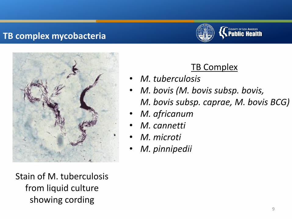
TB complex mycobacteria
9
Stain of M. tuberculosis from liquid culture
showing cording
TB Complex• M. tuberculosis• M. bovis (M. bovis subsp. bovis,
M. bovis subsp. caprae, M. bovis BCG)• M. africanum• M. cannetti• M. microti• M. pinnipedii

Discovery of the tubercle bacilli
Cambau and Drancourt. 2014. Clin. Microbiol. Infect. 20: 196-201.
10
From my numerous observations, I conclude that these tubercle bacilli occur in all tuberculous disorders, and that they are distinguishable from all other microorganisms.
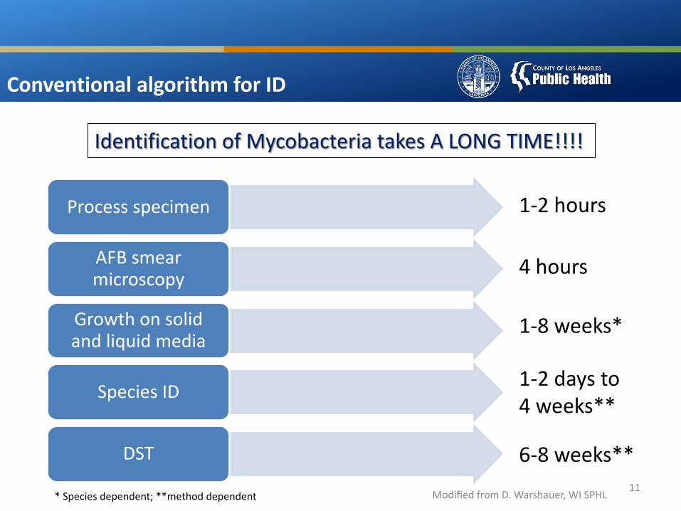
Conventional algorithm for ID
11
Process specimen
AFB smear microscopy
Growth on solid and liquid media
Species ID
DST
1-2 hours
4 hours
1-8 weeks*
1-2 days to 4 weeks**
6-8 weeks**
Identification of Mycobacteria takes A LONG TIME!!!!
* Species dependent; **method dependent Modified from D. Warshauer, WI SPHL

Modern algorithm for ID
12
Molecular based tests
Processed or raw specimen
AFB smear microscopy
Growth on solid and liquid media
Species ID
DST
Molecular based tests
Modified from D. Warshauer, WI SPHL
1 hour 1-3 days*
*method dependent

Acceptable specimen types
Pulmonary Extra-pulmonary
Sputum (expectorated, induced) Tissue
Bronchoalveolar lavage (BAL) Body fluids
Bronchial wash/brush Blood
Transtracheal aspirate Stool
Gastric lavage
Urine
CLSI M48-A 13
Swabs are generally not acceptable

Sputum specimen collection and transport
From Hector Rivas 14
Collect specimens before chemotherapy is begun
Collect 3-10 ml of specimen in a labeled sterile 50 ml conical tube
• Early morning specimens collected on successive days
• Specimens collected 8 hours apart with at least one being an early morning collection
Collect a minimum of 3 single patient specimens
Refrigerate and transport specimens to the laboratory as soon as possible
Seal and package specimen containers carefully to avoid leakage and breakage in transit

Extra-pulmonary specimens
Site Collection Time Volume Frequency Storage and transport
Gastric Aspirate
Early morning before eating and while in bed
5–10 ml One specimen per day on three consecutive days
Room temperature; if delayed >1 hour, neutralize with 100 mg sodium carbonate
Urine First morning specimen (mid-stream)
40 ml One specimen per day on three consecutive days
Refrigerate
Stool - Min. 1 gram - Refrigerate
CSF - 10 ml - As soon as possible at room temperature; do not refrigerate
Other Body Fluids (pleural, peritoneal, pericardial, synovial)
- 10-15 ml - Refrigerate
Tissues or Lymph Nodes
- As much as possible; add 2-3 ml sterile saline
- As soon as possible at room temperature
Blood - 10ml;SPS/heparin
- As soon as possible at room temperature
15

Decontamination, digestion and concentration
• Majority of the specimen types (sputum) are
contaminated with normal flora
• Many different procedures developed to
decontaminate specimens using acid or
alkaline chemical agents to kill contaminants
• NALC-NaOH, Z-TSP, NaOH, oxalic acid,
sulfuric acid, CPC
• Commercial and lab-made reagents
• High lipid content of cell walls (mycolic acid),
mycobacteria are able to resist bacteriocidal
effects of decontamination agents16

Decontamination, digestion and concentration
• Decontamination procedures digest or liquefy mucoid
specimens, free mycobacteria from clumps of protein, and
allow for organisms to sediment during centrifugation
• Decontamination/digestion procedures use centrifugation to
concentrate the specimen before smear culture
• Normally sterile specimens (e.g. CSF) can be concentrated by
direct centrifugation of the specimen without decontamination
• Concentration increases the sensitivity of both smear and
culture
17

Mycobacteria have unique cell wall structure made of mycolic acid
18
• In addition to peptidoglycan, the acid-fast cell wall of Mycobacteriumcontain a large amount of glycolipids, especially mycolic acids
• Stain poorly because cell wall structure and lipids interfere with gram stain
• Acid fast stains are used to force complex of dyes into mycobacterial cells Ghost Cells GPR

How do AFB stains work?
19

AFB smear microscopy
• Examination of specially stained smears for acid-fast bacteria
– Stains allow detection of organisms containing mycolic acid in the cell wall; resist decolorization with acid-alcohol and retain primary stain
• Direct and concentrated smears
• Two main types of AFB stains
– Fluorescent• Auramine O, Auramine Rhodamine
– Brightfield• Kinyoun
• Ziehl-Neelsen
20
CDC Public Health Image Library

How can AFB smear results be used?
• Clinical management
– Initiate patient therapy based on smear result and clinical presentation; presumptive diagnosis, allows dx weeks before culture results
– Monitoring response to therapy
• Laboratory testing
– Interpretation of NAAT tests
• Public health interventions
– Smear results identify most infectious cases
– Prioritization of contact investigations
– Decisions regarding respiratory isolation21

Limitations of AFB microscopy
• Does not distinguish between live and dead organisms
• Follow-up specimens from patients on treatment may be smear positive yet culture negative
• Limited sensitivity and requires relatively high bacillary load for detection
– AFB stain much less sensitive then culture
• 10-100/ml needed to detect by culture
• 5,000-50,000/ml to detect by AFB stain
• Limited specificity as all mycobacteria are acid fast; cannot determine species ID by AFB smears alone
• PPV of positive smears for TB should be taken into consideration with local prevalence of MTB and NTM in the patient population 22

AFB smear reporting
Number of AFB observed by fluorochrome at 250x
Report
0 No AFB Seen
1-2/30 Fields Doubtful, Repeat test
1-9/10 Fields 1+
1-9/Field 2+
10-90/Field 3+
>90/Field 4+
CDC Scale for AFB MicroscopyKent and Kubica. Public Health Mycobacteriology. 23
AFB microscopy reports should indicate magnification, scoring, and method of staining

Isolation of mycobacteria
• Most species are slow growing and require extended incubation times
• Can easily be overgrown; Require appropriate pre-treatment and processing
• Digestion and decontamination of non-sterile specimens vs. normally sterile specimens
• Isolation requires variety of media including selective and non-selective broth and solid media
24

Culture Media
• Solid media
– Egg based
• Lowenstein-Jensen
• Petragnani
• American Thoracic Society (ATS) medium
• Selective variants such as LJ-Gruft and Myctosel-LJ
– Agar based
• Middlebrook 7H10, Middlebrook 7H11
• Selective variants such as Mitchison’s selective 7H11 (7H11s)
• Liquid media
– Mycobacteria usually grow faster in liquid media and sensitivity for recovery is higher
• 7H9, 7H12, 7H13 25

Examples of media types
26
LJ 7H10 MGIT (7H9)

MGIT 960 instruments
• Used for liquid media tubes and to incubate cultures for primary growth and drug-susceptibility testing
• Middlebrook 7H9 Broth base with supplements
• Processes 960 tubes at once
• Uses fluorescent testing technology that detects the growth of mycobacteria in clinical specimens
• Continuously monitors samples to detect positive growth tubes every 60 minutes and check for fluorescence.
27
POS NEG

Biochemical identification
• Phenotypic identification of mycobacteria is based on the characteristics of the culture and biochemical features
• Comprises a large variety of tests and divides mycobacteria into two separate groups: TB complex and NTM
• Examples of biochemical tests
– Growth rate, colony morphology, pigment production, niacin test, nitrate reduction, catalase activity, formation of the cord factor, urease test, pyrazinamidase test, growth in the presence of p-nitrobenzoic acid, growth in the presence of hydrazide of thiophene-2-carboxylic acid, pigment, growth in MacConkey without crystal violet, growth in 5% NaCl, Tween-80, acid phosphatase test, arylsufatase test
28

Runyon classification
Slide from Hector Rivas 29
Photochromogen Scotochromogen Rapid GrowerNonphotochromogen

Examples of biochemical tests
CDC Public Image LibraryKent and Kubica. Public Health Mycobacteriology 30

DNA probe-based methods
• Target rRNA and performed on organisms grown in culture
• Identifies 4 groups: M. tuberculosis cplx , M. avium cplx., M. kansasii, or M. gordonae
www.hologic.com
31

HPLC analysis of mycolic acid
• FDA-approved method for mycobacterial species ID based on mycolic acid chain length
• Library contains >30 entries of TB complex and NTM and also includes other mycolic acid containing species of bacteria
• Utilizes liquid or solid growth and consists of a 2 day process
• Can separate MTB and M. bovis BCG
• Relatively inexpensive method
• Used often in conjunction with other methods
http://www.midi-inc.com/pages/mycobacterial_id.html
32

HPLC method for mycobacterial ID
Butler and Guthertz. 2001. Clin. Micro. Rev. 14(4): 704-726. 33

DNA sequencing
• rRNA (rrs) gene sequencing commonly used for bacterial taxonomy and identification
• 100% = genus and species
• 99-99.9 % = genus
• > 95% = unable to identify by 16s rRNA sequencing
• Liquid cultures or colonies from solid media
• Issue is that 16s rRNA gene nucleotide differences may not discriminate enough between species to provide identification
• Additional and alternative targets are needed for mycobacterial species (rpoB, hsp65, ITS, dnaA, gyrB, and recA)CLSI M18-A. 2008.
34
0 500 1000 1500250 750 1250

MALDI-TOF MS for microbiology
• Accurate, rapid, cost-effective, and reproducible method for identification of bacteria, fungi, and mycobacteria
• Ability to analyze organisms with relatively little sample preparation
• Potential for performing better than conventional biochemical systems for correct species identification and less mis-identifications
• Commercial databases are available and updated frequently; currently only RUO for mycobacteria
35
www.bruker.comhttp://www.biomerieux-
industry.com/biopharma/vitek-ms-0
Bruker Mycobacteria 2.0 Brochure

How does MALDI-TOF MS work?
36Emonet et al. 2010. Clin Microbiol Infect. 16: 1604–1613.
• Used on liquid cultures and colonies from solid growth
• Mycobacteria require inactivation and extraction using heat, EtoH, and bead beating
• Extract is spotted on target plate and coated with matrix to ionize proteins
• Compare to RUO system/databases and/or in-house reference spectral libraries

Comparison of common ID methods
Buchan et al. 2014. Am J Clin Pathol. 141:25-34.
37
• Increased confidence value for identification from liquid media
• Use of Mycobacterium-specific library had higher overall mean confidence scores for all groups/species tested compared to std library
• Highly concordant with DNA sequencing based methods
Rapid and accurate species-level identification may aid in guiding
management decisions

BD MGIT TBc
• BD MGIT TBc identification test is an immunochromatographic assay that utilizes growth positive MGIT liquid culture (not avail. in US: CE-approved)
• Detects MPB64 in liquid cultures using an MPT64-specific monoclonal antibody.
• LOD = 5x10^5 CFU/mL
http://www.bd.com/resource.aspx?IDX=11259
38Yu et al. 2011. JCM. 49(3): 802-807.

GenoType MTBC
• Not avail. in US; CE-marked
• Solid or liquid culture
• Allows differentiation of all M. tuberculosis complex members
• PCR-based assay with hybridization of amplicon to probe and detection by addition of conjugatehttp://www.hain-lifescience.de/
39

GenoType Mycobacterium CM/AS
• Not avail. in US; CE-marked
• Solid or liquid culture
• ID TB complex and 43 NTM species
• Able to identify organism from low growth and differentiation of mixed cultures from fast- and slow-growing mycobacteria
• PCR-based assay with hybridization of amplicon to probe and detection by addition of conjugate
http://www.hain-lifescience.de/ 40

Why are molecular methods needed for ID?
• Rapid diagnosis and detection
• Difficult to culture
• Inadequate biochemical tests
• Resistance screening/confirmation
• Patient management
• Cost effectiveness
• Relatively few FDA-approved tests for identification
41

TB NAAT
• NAATs have several advantages over culture:
– Higher positive predictive value (>95%) with AFB smear-positive specimens in settings in which NTM are common
– Rapidly confirm presence of MTB in 50%–80% of AFB smear-negative, culture-positive specimens
– Can detect TB in a specimen weeks earlier than culture for 80%–90% of patients suspected to have pulmonary TB
• FDA approved, modified FDA-approved, and LDTs
– Depending on test, may be used on direct patient specimens, concentrates, or isolates
– Pulmonary and extra-pulmonary specimens
42

Why use NAATs?
• Assist in decision to start therapy
• Reduce unnecessary treatment
• Respiratory isolation
• Cost savings
43
NAA testing should be performed on at least one respiratory specimen from each patient with signs and symptoms of
pulmonary TB for whom a diagnosis of TB is being considered but has not yet been established, and for whom the test result
would alter case management or TB control activities.
-CDC recommendations

CDC/MMWR NAAT guidelines
44
Collect respiratory specimens and perform AFB smear and culture. Testing
should not be delayed to await NAAT.
At least one specimen should be tested by NAAT
Interpret NAAT results in conjunction with AFB smear
Use of NAATs lead to earlier treatment
initiation, improved patient outcomes, and
increased opportunities to interrupt transmission
MMWR. Updated Guidelines for the Use of Nucleic Acid Amplification Tests in the Diagnosis of Tuberculosis. Jan. 16, 2009. 58(1): 7-10.

LAC TBCP NAAT guidelines
LA County TBCP NAAT Guidelines, 2012 45
Important points to rememberInterpret results on basis of clinical presentation
Single negative NAAT cannot be used to rule out TBCx and smear should always be ordered with NAAT
Sputum specimens may contain inhibitors NAATs detect live/dead TB bacteria

LAC TBCP NAAT guidelines
46

Amplified MTD
• Target-amplified nucleic acid probe test for detection of MTB complex rRNA in AFB smear positive and negative concentrated sediments
• Induced or expectorated sputum, BAL, BA, TA
• Useful for TB suspects who have not received therapy or less than 7 days treatment or have not received therapy in last 12 months
• Must be performed in addition to culture and smear; Neg. results do not exclude possibility of positive culture
www.hologic.com
47
MTD test can detect all TB complex

Xpert MTB/RIF
• Fully automated cartridge-based NAAT that uses real-time PCR
• Detects TB complex and mutations associated with RIF resistance
• Direct or concentrated sputum
• Use in conjunction with AFB smear and mycobacterial culture to address the risk of false negative results; recover MTB for further characterization and DST
Boeme et al. 2010. NEJM. 363:1005-1015.
48

Resistance mutations detected by RT- PCRshould be confirmedby
APHL Fact Sheet. Nov. 2013. Cepheid xpert mtb/rif assayMcAlister AJ et. al. 2015.J Clin Microbiol 53:1752–1753.
• Culture-based DST is necessary to complement molecular results
– Clinical relevance of some mutations is unknown
– Not all mechanisms of resistance are understood
• Potential low PPV for detection of RIF
• Assay does not provide the specific rpoBmutation detected

Molecular methods for resistance prediction
http://www.cdc.gov/tb/topic/laboratory/default.htmhttp://www.cdph.ca.gov/programs/mdl/Documents/MDL-PyroseqTBInfo.pdf
50
Method FDA approved
Where performed?
Specimenrequirement
Resistance detection
Cepheid XpertMTB/RIF
Yes Clinical and public health labs
Sputum Rifampin
Pyrosequencing No State PHLs Smear positive primary specimen or isolate
Rifampin IsoniazidFQsInjectables
Sanger sequencing No CDC Smear positive primary specimen or isolate
RifampinIsoniazidEthambutolPyrazinamideFQsKanamycinAmikacinCapreomycin

Example of mechanisms of drug resistance
A. Talenti. 1998. Thorax. 53:793–797. 51

Gene targets for molecular resistance
Drug Category Gene Target Mutation(s)
Rifampin 1st line rpoB 81 bp core region
Isoniazid 1st line inhAkatGahpC-oxyR
Promoter regionSer315Intergenic region
Pyrazinamide 1st line pncA Promoter and coding region
Ethambutol 1st line embB Met306, Gly604
FluoroquinolonesOfloxacin
2nd line gyrA Gly88, Ala90, Ser91, Asp94
Kanamycin 2nd line eis Promoter region
Capreomycin 2nd line tlyA Coding region
Kanamycin, Amikacin, Capreomycin,
2nd line rrs nt1401, 1402, 1484
Database of TB mutations can be found at: https://tbdreamdb.ki.se52

GenoType MTBDRplus/ GenoType MTBDRsl
• Not available in US (CE-marked)
• Pulmonary specimen and from culture isolates
• Detects mutations in genes for drug resistance (MDR/XDR)
• Nucleic acid isolation and amplification followed by product denaturation and hybridization to probe; detection by addition of conjugate
• Results in ~5 hrs. and can be used to confirm DST
http://www.hain-lifescience.de/ 53

Example of other methods in development
• Non-molecular
– MODS (Microscopic Observation Drug Susceptibility) assay is a broth microtiter method where wells are periodically examined for growth
• Molecular
– Loop mediated isothermal amplification is a form of nucleic acid amplification where DNA is generated to enable detection by visual fluorescence
– Oligonucleotide arrays allows for the simultaneous detection of multiple genetic sequences and may be useful to detect microorganisms or mutations conferring resistance
M.L. Wilson. 2011. CID. 52(11):1350–1355. 54

Purpose of TB molecular genotyping
• Detect and control TB outbreaks
• Identify incorrect TB diagnoses based on false-positive culture results
• Discover unknown relationships between cases
• Identify new and unusual transmission settings
• Detect transmission between patients who reside in different jurisdictions
• Evaluate routine contact investigations and progress toward TB elimination by monitoring measures of recent TB transmission
55http://www.cdc.gov/tb/programs/genotyping/chap1/intro_2_overview.htm

PH applications of molecular epi tools
• Supplement traditional contact tracing
• Enhance TB control activities through identification of unrecognized chains of transmission, monitoring disease trends, and allocation of resources
– Patient management
– Transmission dynamics
– Strain lineage and pathogenesis
Kata-Maeda et al. 2011. Future Microbiol. 6(2): 203–216. 56
For detailed information on TB genotyping methods, visit http://www.cdc.gov/tb/programs/genotyping/manual.htm

When is TB genotyping used?
• To detect or confirm an outbreak of TB
• When it is suspected that a cross-contamination has occurred in the TB laboratory, or other source of false-positive result
• To detect previously unsuspected patterns of transmission or outbreaks
• Three main methods currently used:
– Spoligotyping(spacer oligonucleotide typing)
– Mycobacterial Interspersed Repetitive Units (MIRU/VNTR)
– Restriction fragment length polymorphism (RFLP)
57
Comas et al. 2009. PLOS One. 4(11): e7815.

Laboratories that perform TB genotyping
58
Michigan Department of Community Health and at the CDC perform genotyping for all TB isolates submitted from California

Spoligotyping
• Spoligotyping is a PCR-based method which relies on the amplification of a highly polymorphic Direct Repeat (DR) locus
• DR region consists of multiple copies of a conserved 36-41bp sequence (direct repeats) separated by multiple unique spacer sequences; used for MTB complex strain differentiation
• Different M. tuberculosis strains have various complements of 43 spacers
• Isolates of MTC bacteria can be differentiated by the presence or absence of one or more spacers- Octal code designations
• Spoligotypes evolve by deletion of a single or multiple contiguous DVRs
59

Spoligotyping
60http://www.cdc.gov/tb/programs/genotyping/chap3/3_CDCLab_2Description.htm

MIRU-VNTR
• Multiple interspersed repeat unit (MIRU) typing is based on differences in repeats at specific loci
• Variable number of tandem repeat (VNTR) typing is based on analysis of DNA segments containing “tandem repeated” sequences in which the number of copies of the repeated sequence varies among strains
• Method relies on PCR amplification and calculation of the number of repeats on the basis of the size of the amplified product
• Total of 41 MIRU loci and 24 have been selected for genotyping; reported as 24-character designations, each character corresponding to the number of repeats at one of the 24 MIRU loci- 223225163324561333245623 61

MIRU-VNTR
62
MIRU locus name
02 04 10 16 20 23 24 26 27 31 39 40
No. of
repeats2 3 2 2 3 4 2 5 3 3 2 2
Ex.12- MIRU
designation:
232234253322
H37Rv
BCG Pasteur
392 bp
288 bp
Locus X
Locus X

IS6110 RFLP
• Southern blot of digested DNA
• IS6110-based RFLP genotyping detects variations generated by the insertion element IS6110
• Insertion elements are capable of making copies of themselves and then inserting the copy anywhere in the genome in a process known as transposition
• Strains can differ in both the number of copies of IS6110 and the positions of IS6110 in the bacterial DNA
• Performed when strains match by MIRU and spoligotyping 63
RE SITE
IS6110
Restriction Digest
Gel Electrophoresis
Southern blot IS6610 probe
IS6610 RFLP
Modified from Wong et al. 2011. RPE. Vol. 15. No. 1.

The future of molecular TB epidemiology
• Current methods are dependent on determining whether strains are clonal and if they could have originated from the same source or are genetically distinct and reflect independent transmission events
• Retrospective analysis performed months after initial patient dx
• Real-time molecular epi data is currently a challenge
• Ideal methods would lead to:
– Better understanding of genetic markers and association with clinical consequences and dynamics of epidemics
– Development of databases containing molecular, epidemiologic, sociological, and environmental data may lead to predictions of future outbreaks or predominant strains
– Patient and population level benefits 64

Whole genome sequencing and epi investigations
• Represents a relatively new method for molecular epidemiology• Increasingly accessible means for tracking disease outbreaks
• Can distinguish strains with single nucleotide polymorphisms and may provide greater resolution than current methods
• Current challenges include:
– No single platform/method protocol available
– Read length, data assembly, and analysis
– Criteria to define indistinguishable, closely related, possibly related, and different strains incl. outbreak thresholds
– No current informatic infrastructure for data processing and integration of results
– Technology remains out of reach for many clinical and public health lab due to cost and bioinformatics expertise needed 65

Will WGS be the future of TB contact tracing?
Gardy JL et al. 2011.NEJM. 364(8): 730-739. 66
Using WGS, several additional cases were recognized as related

Rapid WGS of M. tb directly from clinical samples
• Utilized biotinylated RNA baits designed specifically for M. tb DNA, captured genomic DNA directly from infected sputum samples; sequencing without culture
• Performed on 24 smear positive (1+ - 3+) sputum specimens and compared to culture and conventional molecular methods
• Sequence data was highly concordant for phenotype and predicted resistance based on genotype
Brown AC et al. 2015. JCM. May 13 2015. pii: JCM.00486-15. [Epub ahead of print]67
http://www.genomics.agilent.com/files/Media/SS_Halo/Magnet584.jpg

Summary
• TB complex mycobacteria are slow-growing organisms that can be identified using growth characteristics, biochemical tests, and molecular methods
• Rapid identification methods for TB and determination of drug resistance is important for TB control
• New molecular methods can be used for both species identification and TB epidemiology
• Communication between lab and TB control is important for results interpretation and follow-up
68

Acknowledgments
• Ed Desmond, PhD, D(ABMM)
• Hector Rivas, MPH and the LA County Public Health Laboratory Mycobacteriology and Molecular Epidemiology Units
• LA County Tuberculosis Control Program
• Brian Baker, MD, MPH
69
Our Mission:To protect health, prevent disease, and promote health and well-being

Questions?
70