Laboratory manual of elementary chemical physiology and ...
382
Transcript of Laboratory manual of elementary chemical physiology and ...


35-39 Randolph St.
fcHICAGO.
f'


Cornell University
Library
The original of this book is in
the Cornell University Library.
There are no known copyright restrictions in
the United States on the use of the text.
http://www.archive.org/details/cu31924000912133

/U'
t?ylT
Laboratory Manual :
Elementary Chemical Physiology
URINE ANALYSIS.
JOHN H. LONG, M. S., Sc. D.,
PROFESSOR OF CHEMISTRY AND DIRECTOR OF THE CHEMICAL LABORATORIES IN THE SCHOOLS OF
MEDICINE AND PHARMACY OF NORTHWESTERN UNIVERSITY.
WITH NUMEROUS ILLUSTRATIONS.
'
<r.
Chicago Mbdical Book Co.,35-37 Randolph Street,
1897

i
t
Entered according to act of Congress, in the year 1894.
By JOHN H. LONG,
in the office of the Librarian of Congress, at Washington.
Mo 5 5"<f
t rf<4(
1-'...

PREFACE.
This little book is an outgrowth of the course given to
second year students in the Medical School of Northwest-
ern University, and represents in its present form the prac-
tical laboratory work as now followed in the course of
study required for graduation.
The time devoted to the study of chemistry and chem-
ical physiology in our American medical colleges has been
altogether too short, and only within very recent years,
with a few notable exceptions, has any attempt been madeto impart systematic knowledge in this direction. But nowthe views of medical teachers are undergoing a rapid
change and among the scientific studies recognized as
forming a rational groundwork for the study of medicine
proper, physiological chemistry is justly regarded as of the
first importance.
It is the belief of the Author that instruction in chemis-
try should be given through two years of the course of the
medical student. While it may be urged that general
chemistry forms no proper part of the curriculum of the
medical school it is certainly true that no one of our schools
can afford to abandon it until it is generally agreed to makeit a requirement for admission. But this is a long way in
the future. In his first year work, therefore, the student
should be taught the elements of general chemistry both
in the classroom and the laboratory, the laboratory work
consisting of simple experiments followed by qualitative
analysis. Such a course opens the way for the laboratory
practice in chemical physiology in the second year, and this
should be made practical and simple.

In the set of experiments offered to students the Author
has aimed to select only such as illustrate some point of
importance, and which, at the same time, can be easily
performed. Complicated formulas and reactions which
are so largely introduced in some of our recent books on
medical chemistry, so called, the Author has thought
proper to exclude as not in any degree essential.
This book consists of two parts, the first dealing with
simple experiments in chemical physiology, while the second
takes up the subject of urine analysis as properly following
and partly illustrating the first. While intended primarily
for medical students, .it is believed that it contains not a
little matter of interest to the medical practitioner as it dis-
cusses fully all the practical processes of urine analysis of
value at the present time and in the chapters on chemical
physiology presents several features more in detail than is
customary in books of this class.
The Author can make no great claims of originality but
would freely acknowledge his indebtedness to such stand-
ard works as the Physiologies of Landois and Stirling and
Foster, the Physiological Chemistry of Hoppe-Seyler, the
Urine Analysis of Neubauer and Vogel and, others. Hewould extend his thanks to Messrs. E. H. Sargent & Co.,
of Chicago, to Mr. A. Kruess, of Hamburg and to Messrs.
Franz Schmidt and Haensch, of Berlin, for the use of
cuts used in illustrations. Finally, he wishes to express
his sense of obligation to his assistant, Mr. Chas. H.
Miller and also to his colleague, Professor Mark Powers,
for valuable aid rendered in the reading of proofs.
The Author.Chicago, Aug. 15, 1894.

TABLE OF CONTENTS.
Part I. Elementary Chemical Physiology.
Chapter I. Introduction 1
Chapter II. The Carbohydrates 19
Chapter III . The Fats : 59
Chapter IV. Proteids or Albumins 67
Chapter V. The Blood 91
Chapter VI. Bone Constituents, Saliva, Gastric
Juice, The Bile Ill
Chapter VII. Milk, Beef Extracts, Flour and Meal. . 121
Chapter VIII. Water and Air 134
Chapter IX. Special Problems 143
Part II. Urine Analysis.
Chapter X. Outline of Tests. Preliminary Tests. .. 177
Chapter XI. The Tests for Albumins 187
Chapter XII. The Tests for Sugar 213
Chapter XIII. The Coloring Matters in Urine.
Biliary Acids 242
Chapter XIV. Determination of Uric Acid 254
Chapter XV. Urea 263
Chapter XVI. The Determination of Phosphates and
Chlorides 282
Chapter XVII. The Sediment from Urine 298
Chapter XVIII. Unorganized Sediments and Calculi. 321
Appendix. Test Solutions and Tables 343


Part I.
Elementary Chemical Physiology.


Chapter I.
INTRODUCTION.
/"""HEMICAL Physiology is in the main concerned with^—* the study of certain organic compounds existing in the
animal body and with the changes which occur in the va-
rious food stuffs from the time they enter the stomach until
they leave the body, in the form of broken down excreta,
by the lungs or through the kidneys. Beginning with the
digestive changes in the stomach a series of complex
reactions take place which we are able to follow only to a
very limited extent. The formation of the important
elements in the blood, bile, saliva, the gastric and pancreatic
juices, the building up of the solid tissues of the body
and the subsequent breaking down of the same, the final
appearance of all the elements of these tissues or
secretions in the urea, uric acid, phosphates and other
products of the urine or in the carbon dioxide from the
lungs involve a number of chemical reactions of the
highest interest and importance. It is the province of
chemical physiology to investigate as far as possible the
conditions under which these changes take place, to study
the constitution of the food products at the one end of the
scale and of the excreta at the other and by means of
laboratory experiments to bridge over the space between
the two.
Chemical physiology attempts to duplicate in the test-
tube or beaker the changes taking place in the body
and to build up from the end products intermediate ones
and those at the beginning. Most of the problems sug-

2 ELEMENTARY CHEMICAL PHYSIOLOGY.
gested in this direction are as yet far from solution. Astudy of the urine, expired air and gastric juice has shown
the nature of these products under what may be consid-
ered normal conditions of the body. Further study has
developed certain facts connecting variations in the urine,
for instance, with variations in the condition of health in
the individual. Study has shown that in health certain
bodies are absent from the urine, while in certain diseases
they are present, and from this fact there has been
developed the very practical department of chemical
physiology, known as urine analysis, as an aid to diagnosis.
Much of the experimental work connected with the
investigation of problems in chemical physiology is of so
difficult a character as to be quite beyond presentation in
an elementary manual. Fortunately, however, other
problems, and among them very important ones, are moreeasily approached, so that they can be worked out in the
form of simple laboratory experiments.
The phenomena of salivary, gastric and pancreatic
digestion, of coagulation of blood and of oxidation andreduction of its most important element, of the emulsifi-
cation and absorption of fats and of diffusion and osmosis
are certainly among the most important and interesting in
the whole field of physiology. They have for the student
a new meaning when he learns them, not alone from the
printed page or the discourse of the lecturer, but also byhis own laboratory experiments. Chemical physiology
offers a vast field for the research of the specialist, but it
offers also abundant simple illustrations for the beginner.
Chemical physiology is, in a sense, a branch of applied
organic chemistry inasmuch as by far the larger number of
compounds discussed are compounds of carbon such as
are commonly termed organic. It was at one time sup-
posed, in fact was maintained as one of the fundamental

ELEMENTARY CHEMICAL PHYSIOLOGY. . 3
principles of chemical theory, that organic bodies, so-called,
are produced only through the agency of the animal or
vegetable cell, that is by what were termed vital forces, or
by simple transformation of compounds so formed. Thebuilding up of a complex carbon compound from the
elements of the earth or air was supposed to be a problem
which could be solved only by the plant or animal as a
function of its growth.
In 1828 Woehler showed that this view was a false one
and that characteristic organic compounds can actually
be built up in the chemist's laboratory. This was demon-
strated first by the production of a body of the greatest
interest to the physiologist, viz.: urea, which under the
old view would certainly be one of the last to be con-
sidered within the possibility of artificial production.
The discovery of Woehler marks the beginning of
modern scientific organic chemistry, but it proved of
scarcely less importance in the development of physiology,
as it paved the way for the preparation and study of a
great number of compounds, occurring in the animal body,
whose relations were imperfectly or not at all understood,
or whose existence even in many cases, was not suspected.
Nature of Organic Compounds.
Organic compounds contain carbon as their characteris-
tic element. Nearly all contain hydrogen. Oxygen is
present in a very large number and nitrogen is present in
many which are of importance to the physiologist. Sul-
phur is present in certain important organic bodies and in
a still smaller number there are found phosphorus, chlorine
and several other elements. Compounds of carbon are
characterized by the fact that when heated alone or with
certain oxidizing agents to a high temperature they are
decomposed with liberation of carbon dioxide. The

4 • ELEMENTARY CHEMICAL PHYSIOLOGY.
presence of hydrogen is shown under the same conditions
by the formation of water. Nitrogen in organic com-
pounds may be recognized in several ways but usually
most conveniently by conversion into ammonia. Sulphur,
phosphorus and chlorine are converted into sulphates,
phosphates and chlorides which may be detected by the
usual methods of qualitative analysis.
The proof of the presence of oxygen in an organic
compound is often indirect. Some of these reactions may
be illustrated by simple experiments, as follows:
For Carbon. Nearly all organic substances are
decomposed when strongly heated. If heated in the air
they give off inflammable vapors and often leave a residue
rich in carbon which, at a high temperature, burns, form-
ing carbon dioxide.
Solid organic substances when intimately mixed with
a large excess of fine granular black oxide of copper and
heated to a high temperature in a hard glass tube are
decomposed and completely oxidized at the expense of
the oxygen of the copper oxide. Carbon present is con-
verted into carbon dioxide and hydrogen into water. If
the combustion tube is furnished with a delivery tube
which dips beneath the surface of clear lime water the
characteristic reaction for carbonates is given. Organicliquids are characterized by being inflammable or byyielding inflammable vapors.
For Hydrogen. The presence of hydrogen may beshown by burning the substance in the air in such a man-ner as to condense the products of the combustion by a
cold porcelain or metallic surface. The appearance of
droplets of water is proof of the presence of hydrogen. In
the oxidation by copper oxide, if the process is properly
•conducted, drops of water condense on the cooler parts of
the combustion tube.

ELEMENTARY CHEMICAL PHYSIOLOGY. 5
For Nitrogen. Nitrogen in most organic substances
can be recognized by heating the latter with soda-lime to a
high temperature. The nitrogen is given off as ammonia,
which is recognized by the smell or reaction with litmus.
When a solid nitrogenous substance is heated in a nar-
row test-tube with some small fragments of clean metallic
sodium, cyanide of the metal is formed. Nitrogenous liq-
uids are mixed with dry sand and then with the sodium.
The cyanide is dissolved out from the fused mass with
distilled water in small amount, to the solution a drop of
ferrous sulphate solution and a drop of ferric chloride solu-
tion are added and then hydrochloric acid to acid reaction.
The appearance of the Prussian-blue color proves the cya-
nide and therefore the nitrogen. To recognize nitrogen in
nitro-compounds a mixture of soda-lime and sodium thio-
sulphate may be used.
For Sulphur. When organic compounds containing
sulphur are fused with a mixture of potassium nitrate and
sodium carbonate a sulphate is formed which may be recog-
nized in the dissolved mass by the usual tests.
Organic sulphur compounds, albumin for instance, give
up their sulphur as sulphide when heated in a glass tube
with metallic sodium. The sulphide can be detected in
the usual way.
Classification of the Physiologically Important
Organic Bodies.
From a strictly scientific point of view organic bodies
should be grouped here as elsewhere, that is as products
derived from fundamental hydrocarbons. But for manyreasons such a classification would be inconvenient or even
misleading for our purpose.
For the physiologist it is in most cases sufficient to con-
sider bodies as belonging to, or related to members of,

6 ELEMENTARY CHEMICAL PHYSIOLOGY.
three important groups of natural substances, viz. : the car-
bohydrates, the fats and the albuminoids.
Certain compounds appear properly as decomposition
products of members of one or the other of these groups.
By far the greater number of organic substances con-
sidered by the physiologist are bodies of natural origin, or,
at any rate, difficult of synthesis. While several of the car-
bohydrates have been produced artificially, and fats have
likewise been built up, no albuminoid has yet been made
by an artificial process. The plant alone seems to have
the power of building up proteid substances. In the ani-
mal body these are converted into new forms or digestion
products, but cannot be created outright. Carbohydrates
are produced also, in the main, in the vegetable cell, but
fats can be built up in the animal body from substances
not fat.
The animal, however, has not the power of producing
any organic substance directly from the elements.
In the following, attention will be paid primarily to
groups and derivatives as outlined.
General Methods of Experimentation.
It is assumed that the student beginning these experi-
ments is reasonably familiar with elementary general
chemistry and qualitative analysis. Most of the work fol-
lowing is of a qualitative nature, involving the use of very
simple methods only, and such apparatus as the student
has already used in other work.
A few volumetric quantitative processes are introduced,
the details cf which will be explained in the proper conec-
tion. The simple, general principles common to all volu-
metric methods may, however, be outlined here.
Quantitative determinations are made by gravimetric or
volumetric methods. In the first the substance sought, or

ELEMENTARY CHEMICAL PHYSIOLOGY. 7
some compound bearing a definite and known relation to
it, is precipitated, usually collected and weighed on a filter
or in a crucible or other small receptacle. In volumetric
methods the procedure is different. Advantage is taken of
the fact that many reactions in solution are completed in
some definite manner which permits the end-point to be
accurately observed. For instance, if to some dilute sul-
phuric acid in a beaker, to which a little litmus or phenol-
phthalein had been added, a dilute solution of potassium
hydroxide be next added a sudden change of color appears
as soon as enough alkali is present to neutralize the acid
and leave a trace in excess.
An alkali solution made blue with litmus becomes as
suddenly red on addition of an acid.
If a solution of salt is added to a solution of silver
nitrate, drop by drop, a precipitate appears. If the silver
solution is contained in a bottle which is violently shaken
after each addition of salt, the precipitate formed soon set-
tles out so that it is possible to observe the formation of a
new precipitate as fresh drops of salt are added. By work-
ing carefully the point can be determined where the addi-
tion of a drop of salt solution ceases to produce a precipi-
tate in the silver bottle, and this indicates the end of the
reaction.
Again, suppose we have a hot dilute solution of oxalic
acid containing a small amount of sulphuric acid. To this
is added, drop by drop, some solution of potassium per-
manganate, the deep color of which is very characteristic.
As the drops of permanganate touch the acid solution the
color disappears almost instantly, because of decomposition
of the compounds, (oxidation of the oxalic acid). The in-
stant the last trace of oxalic acid has been decomposed the
solution is almost colorless, but the addition of a drop moreof permanganate is sufficient to impart a pink color readily

8 ELEMENTARY CHEMICAL PHYSIOLOGY.
seen. In this case the end of the reaction is indicated by
a sudden change of color.
We determine in these experiments the relation between
acid and alkali, between silver and salt, between oxalic
acid and permanganate, in solution and in the same vessel
in which we began the test, and besides in a very short time.
It is evident that if we know the strength, grams
per liter, of the potassium hydroxide, of the salt solu-
tion and of the permanganate we determine the amount
of sulphuric acid in the first, of silver in the second and of
oxalic acid in the last of the above experiments by noting
simply the volumes of the alkali, salt and permanganate
solutions needed to give the characteristic end reaction.
This follows because we know that the several compoundsreact on each other according to the following equations:
2 K O H + H2S
4= K
2 S 4 + 2 H O H112 + 98 = 174 + 36
Na CI + Ag N Oa= Ag CI + Na N
3
58.4 -|- 169 = 142.4 + 85
2 K Mn 4 + 5 C2H
2 4 + 3 H2S 4
=316 + 450 -+- 294 =
2 Mn S 4 + K2S 4 + 8 H
2O + 10 C
3
302 -f 174 + 144 + 440
112 Gm. of potassium hydroxide neutralize 98 Gm. of
sulphuric acid; 58.4 Gm. of sodium chloride precipitate
169 Gm. of silver nitrate and 316 Gm. of potassium perman-ganate oxidize 450 Gm. of anhydrous oxalic acid.
The proportions hold good if we substitute here milli-
grams, grains, ounces or pounds for grams throughout.
If now we prepare solutions containing in one liter 112
Gm. of KOH, 58.4 Gm. of NaCl and 3. 16 Gm. of K Mn Q4

ELEMENTARY CHEMICAL PHYSIOLOGY. 9
one cubic centimeter of these solutions will react with 98
milligrams of H2S0
4 , 169 milligrams of Ag N Osand 4.5
milligrams of C2H
2 4 .
Solutions prepared in this manner are called standard
or volumetric solutions, because a definite volume of each
reacts with a certain weight of something to be measured.
A normal solution is usually defined as one which contains
a weight in grams equivalent to the molecular weight of the
substance in question, dissolved in one liter. Thus, 58.4
Gm. of Na CI dissolved in one liter would yield a normal
solution. A tenth-normal (/T ) solution of salt would con-
tain 5.84 Gm. in the liter. A twentieth-normal (j^) solution
would contain 2.92 Gm. of salt in a liter. A normal solu-
tion of sulphuric acid according to this definition would be
one with 98 Gm. of H2S0
4in the liter. But many chem-
ists take 49 Gm. of H2S0 4
(half the molecular weight) as
constituting here a normal solution. One cubic centimeter
of this would exactly neutralize one cubic centimeter of
normal potassium hydroxide (always taken as containing
56 Gm. to the liter) and would be equivalent in acidity to
one cubic centimeter of a normal hydrochloric acid with
36.4 Gm. to the liter. From this standpoint a normal acid
solution is one which furnishes in each cubic centimeter as
many replaceable hydrogen atoms as are furnished by
normal hydrochloric acid with 36.4 Gm. to the liter.
Many standard solutions are made empirically of such
a strength that one cubic centimeter will react with someconveniently small whole number of milligrams of the sub-
stance to be measured. A well-known test for sugars
depends on the reducing action of these compounds on
alkaline copper solutions. Based on this behavior wehave a volumetric process in which the standard solution
(Fehling's solution) is made of such a strength that one
cubic centimeter is completely reduced by 5 milligrams of

10 ELEMENTARY CHEMICAL PHYSIOLOGY.
dextrose. This important test will be explained in detail
later. Another volumetric solution of frequent use in
the physiological laboratory is the mercuric nitrate solution
for measurement of urea. Under certain conditions of
dilution urea and mercuric nitrate react with each other in
a perfectly definite manner, the urea falling as a precipitate
with the mercury. The end of the reaction is easily
determined and the solution of mercuric nitrate is made of
such a strength that one cubic centimeter precipitates
exactly 10 milligrams of urea.
In volumetric analysis indicators are very frequently
used to show the end of the reaction. These are usually
substances which give a characteristic color change as
soon as the principal reaction is completed. Litmus and
phenol-phthalein are indicators which show change of
reaction from acid to alkaline, or the reverse, and are
added, in small quantity, to the solution whose strength is
to be measured by a standard acid or alkali solution.
If to a salt solution whose strength is to be found bysilver nitrate, a few drops of neutral potassium chromate
solution be added, the chlorine is completely precipitated
first, on adding the silver, and then deep red silver chro-
mate. The appearance, therefore, of a faint shade of red
in making the test indicates the end of the principal
reaction.
In some cases the indicator is not added directly to
the solution to be measured, but is put in the form of
drops on a glass or porcelain plate ; and with these, fromtime to time, a drop of the solution to which the standard
is being added, is mixed. When enough of the standard
has been used a color reaction usually appears in the
mixed drops. The end of the reaction in measuring urea
by mercuric nitrate or phosphates by uranium solutions is
shown in this manner.

ELEMENTARY CHEM/CAL PHYSIOLOGY. 11
Measuring1 Apparatus. In volumetric analysis muchnaturally depends on the accuracy and convenience of the
measuring apparatus employed. This apparatus consists
of flasks, mixing cylinders, open cylinders, pipettes and
burettes. The usual forms of these are shown in the
accompanying figures. See next page.
Flasks are employed of the following dimensions : 1,000
Cc. (one liter), 500 Cc, 250 Cc, 100 Cc. and occasionally
others. They are made to contain these volumes, and
when correctly graduated deliver slightly less of all liquids
except mercury.
Mixing Cylinders are usually employed of the dimen-
sions given for flasks, but cannot be used where the great-
est accuracy is required.
Open Cylinders are used for measuring volumes of
liquids approximately, and are made to contain 500 Cc,250 Cc, 100 Cc, 50 Cc, 25 Cc, 10 Cc. and 5 Cc, usu-
ally.
Pipettes are made to accurately deliver small volumes of
liquids, and those most frequently used have these dimen-
sions : 100 Cc, 50 Cc. 25 Cc , 10 Cc. and 5 "Cc. Pipettes
are sometimes graduated to resemble burettes.
Burettes are made to deliver accurately small volumes
of liquid down to one-tenth or one-twentieth of a cubic
centimeter. They usually hold 50 Cc, sometimes 25 Cc.
or 100 Cc. They are frequently closed below with a
ground glass stop-cock. Sometimes with a rubber tube
compressed by means of a brass clamp, or filled by a little
glass ball inside. The ball is placed between the end of
the burette and the fine delivery tip, and when the rubber
tube is squeezed so as to form a channel on one side, the
liquid is allowed to pass.
These different measuring vessels are readily found in
the market graduated with accuracy sufficient for all prac-

12 ELEMENTARY CHEMICAL PHYSIOLOGY.
(4M
A represents a flask; B, a burette; C, an open cylinder; D, a pipette andE a glass-stoppered mixing cylinder.
FIG. 1.

ELEMENTARY CHEMICAL PHYSIOLOGY. 13
tical purposes.* If the user desires to further test their
accuracy, this may readily be done by weighing the amount
of distilled water at some standard temperature, they hold
or deliver.
Thermometers. While in ordinary qualitative analy-
sis the reactions are carred out with little regard to definite
temperatures, the room temperature or that of boiling
water being sufficiently close for practical purposes, in
chemical physiology, for a wide range of investigations,
the temperature must be maintained within rather narrow
limits. This is necessary because many chemico-physiolog-
ical changes are brought about by the activity of living
ferments or of enzymes which are destroyed by high tem-
perature, and are either destroyed or become inert at low
temperature. A certain class of phenomena are best ob-
served at a temperature near that of the human body, or
a little above, that is about 40° C, while for other reactions
a temperature of 65°—70° C. is found best, while a tem-
perature of 75°—80° C. would be destructive.
For the ordinary uses of the chemical physiological lab-
oratory a thermometer graduated in single degrees with a
range —6° C. to 110° C. is sufficient. Such thermometers
can be purchased from chemical dealers and are usually
made with accuracy enough for practical purposes. But this
is not always the case, and the user should test his instru-
ment to ascertain its approximate error.
First, the error at the zero point should be found by
immersing it in clean fresh snow which has been kept an
hour or more in a room with a temperature a little above
the freezing point, 0° C. This will insure that the snow has
the temperature of zero exactly if it has a chance to drain.
Snow in this condition packs easily when pressed. The
* Graduated glassware and other apparatus described in this book may be
obtained from Messrs. E. H. Sargent & Co., 106 and 108 Wabash Ave., Chicago.

14 ELEMENTARY CHEMICAL PHYSIOLOGY.
mercury of the thermometer left in about half an hour
sinks and finally becomes stationary. If the graduation is
correct the top of the column should be at the 0° mark.
Instead of employing snow, which is seldom found clean
enough in cities, and the use of which is limited to the winter
season, a better plan is to freeze some pure distilled water
in a test-tube by surrounding it with a mixture of crushed
ice and salt. When the water begins to freeze it is shaken
to produce ice in fine particles. The bulb of the ther-
mometer is now immersed in this loose ice, which, as long
as any water is present, has accurately the 0° temperature,
and stirred about. The level of the top of the mercury col-
umn soon becomes constant and can be read off. In manyof the cheaper thermometers this level is somewhat above
the zero mark.
The other fixed point on the scale is determined by sus-
pending the instrument in the steam of water boiling in a
tall metallic vessel. The bulb must not dip in the boiling
water, and the top of the mercury column must be a trifle
above the neck of the vessel.
The boiling point is dependent on the atmospheric pres-
sure and is by definition 100° C, under a pressure of
760 Mm. For temperatures near 100° the boiling point is
lowered 1° by a depression of 27 Mm. in the barometric
column, from which datum a correction can be made.The error of the thermometer at the two fixed points
having been determined it remains to determine the errors
in the intermediate parts of the scale. The readings at the
fixed points may be correct and yet wrong at points
between, because of lack of uniformity in the diameter of the
capillary tube. To determine the errors accurately between
the two fixed points is a matter of some practical difficulty,
and will not be explained here in detail. An idea of the
amount of the variations in the section of the capillary

ELEMENTARY CHEMICAL PHYSIOLOGY. 15
sufficient for our purpose can be obtained by shaking off
a short thread of mercury from the column and allowing it
to flow from one point to another, noting its length as
measured by degrees on the scale. As the volume of the
separated thread is constant a variation in the bore of the
capillary is indicated by the lengthening or shortening of
the thread as it moves through the tube. Large errors can
be detected in this way; small ones we are not concerned
with here.
Thermometers are graduated with the centigrade or
Fahrenheit scale in this country. For all scientific meas-
urements the former alone is used. The conversion of
temperatures on one scale to temperatures on the other is
a simple matter and can be done by these formulas.
Centigrade temperatures to Fahrenheit:
F° = 32°+|C°
Fahrenheit temperatures to centigrade :
C°=4(F°— 32°)
Separation by Dialysis. For the separation of cer-
tain classes of compounds the methods of dialysis are very
frequently employed. Many substances in solution pos-
sess the property of passing through animal membranes,
while other bodies, likewise soluble, do not. Sugar and
salt are types of the first class, while glue and albumin are
types of the second. The phenomenon depends ultimately
on a property common to all bodies in solution, the
property of diffusion, which may be illustrated in this way.
Suppose we partly fill a glass jar with a strong solution
of copper sulphate and then bring over this, with as little
disturbance as possible, a layer of pure water and leave the
jar so charged, several days in a perfectly quiet place at a
uniform temperature. It will soon be observed that the
blue color of the lower layer begins to diffuse upward,

16 ELEMENTARY CHEMICAL PHYSIOLOGY.
notwithstanding the fact that the copper sulphate solution
is specifically heavier than water. After a long time the
two layers become as one, molecules of water passing
down to take the place of the copper sulphate molecules
which ascended.
Something very similar takes place when a layer of
water is brought over a layer of solution of salts, acids,
alkalies or other substances. Here the change cannot be
followed by the eye, however, but must be detected in some
other manner. Solid substances in solution show the same
tendency to distribute themselves through all available
space that is characteristic of gases. The phenomena are
the same with miscible liquids, e. g., alcohol over water.
We speak, therefore, of diffusion of solids, liquids or gases.
This fundamentally simple phenomenon is greatly mod-ified if we separate the substances by an animal membrane,
as a piece of bladder. The rate of diffusion of many sub-
stances is retarded, while that of others is made so slow
as to practically prevent passage. Graham, who investi-
gated this subject carefully, divided substances roughly
into crystalloids and colloids, crystalloids being those bodies
which in solution diffuse through animal membranes, andcolloids (glue-like substances) those which cannot pass
through. It seems to hold true that the failure of bodies to
diffuse through membranes depends on the size of their
molecules, as the molecules of all those substances called
colloids are known to be large, in some cases enormouslylarge.
In physiological chemical analysis these principles are
applied in what was termed above dialysis, or separation bydiffusion through membranes.
This can be carried out by very simple appliances. Adialyser is easily made by stretching a piece of wet parch-ment over a hoop and tying it down close all around. This

ELEMENTARY CHEMfCAL PHYSIOLOGY. 17
makes a shallow vessel into which the substance to be in-
vestigated is poured. The dialyser is then floated on
water in a larger vessel. The water is renewed occasion-
ally and so hastens the passage of diffusible substance
through the parchment. As an illustration a solution of
albumin containing salt can be purified in this manner.
The salt diffuses while the albumin fails to.
Instead of using the common hoop dialyser a parch-
ment tube is now frequently employed. The tube is bent,
making a narrow double sack into which the liquid to bedialysed is poured. The filled sack is then suspended in
pure water which is renewed as before.
The cut above, Fig. 2, shows the manner of using the
first form of dialyser.
Determination of Specific Gravity. By the specific
gravity or density, or specific weight, of a substance we

18 ELEMENTARY CHEMICAL PHYSIOLOGY.
understand the ratio of the weight of a given volume of the
substance to the weight of the same volume of some other
substance considered as a standard. To make our defini-
tion exact we must suppose the volumes taken at some
normal or standard temperature. The standard substance
to which specific gravities of solids and liquids are referred
is water and usually at a temperature of 4° C.
The temperature of the substance compared with water
must be accurately known but may be 4°, 15°, 20°, 25° C,
or in fact any temperature, but usually one of these.
When we say the specific gravity of a liquid is 1.0154 wemean that a volume of it at some definite temperature is
that much heavier than the same volume of water at 4° C.
If taken at 20° C. we can express the true specific gravity
in this manner.
Sp. gr.2
4° = 1.0154.
Simple relations exist between the densities of solu-
tions and the amounts of dissolved substance, and the de-
termination of density is, therefore, usually made in order
to find amount of a body in solution or to detect variations
in amounts of substances dissolved. The progress of fer-
mentation in a sugar solution is readily followed in this
manner, and the amount of solid substance in urine is ap-
proximately found by making a determination of its density.
Special application of these facts will appear later andin the appendix general tables of specific gravities of solu-
tions will be given with methods of determination.

Chapter II.
CARBOHYDRATES.
The name carbohydrate, as commonly used, is given to
a peculiar group of bodies containing carbon, hydrogen and
oxygen combined in certain proportions. In this group
are included the sugars, the starches, the gums and someallied substances. The molecules of the compounds in
this group contain, ordinarily, six, or some multiple of six,
atoms of carbon with twice as much hydrogen as oxygen
and usually enough to yield five or more molecules of
water. In a strictly scientific classification certain mole-
cules with five or even four atoms of carbon should prob-
ably be included among the carbohydrates.
The carbohydrates are formed mainly in the vegetable
kingdom and are, almost without exception, important food
products. A few occur in the animal kingdom, but they
are in this case derived from similar substances produced
by plants.
In the experimental study of these bodies it is best to
begin with the starches, which are widely distributed in
nature and important in the highest degree.
Starch.
Ex. I. Prepare starch from the common potato as
follows: Grate a potato to a pulp by means of an ordinarytin grater, mix the pulp with water and squeeze it througha piece of coarse unbleached muslin. Moisten the pulpagain and repeat this operation several times, collecting
the strained liquids in a large beaker. Allow the mixtureto settle a half hour or longer and pour off the water,
which contains some soluble albuminous substances, some

20 ELEMENTARY CHEMICAL PHYSIOLOGY.
cellular floating matter, but very little starch. Most of
this will be found in the bottom of the beaker. Add somefresh water, stir up and allow to settle. Now pour the
water off again and repeat these operations until the starch
appears perfectly clean and white. Transfer this starch to
a clean shallow dish and allow what is not intended for im-
mediate use to dry spontaneously in an atmosphere free
from dust. The dried product will consist of minute glis-
tening particles resembling small crystals.
Corn starch has been made from green corn, by a
process quite similar to the above. Ordinary dry corn is
used in the United States for the production of starch on
the large scale, and to aid in separating the starch from
the accompanying materials, it is customary to add a little
acid or afkali to the water. Sometimes the meal is allowed
to stand with water until a slight fermentation begins,
which has the effect of disintegrating the cell walls hold-
ing the starch. Potato starch is the common starch of
continental Europe.
Starch consists of minute microscopic, granules varying
in size and shape with the root or seed employed. Thegranules of wheat starch are regular in outline, but vary
greatly in size, the smallest having a diameter of less than
.005 Mm., while the largest may have a diameter of .04
Mm. Corn starch granules have a mean diameter of about
.02 Mm., while those from the potato are often .06 Mm., or
more in diameter. See Figs. 3 and 4.
Ex. 2. Examine starch from several sources underthe microscope, employing a power of about 300 diameters.Clean a glass slide thoroughly, place in the center of it asmall drop of water, and stir into this by means of aneedle, or glass rod, a minute quantity of starch. Nowdrop on a thin, carefully cleaned cover glass, and in such amanner as to exclude air bubbles, and place under themicroscope for observation.

ELEMENTARY CHEMICAL P//YS/OLOGY. 31
FIG. 3.
Potato Starch. 300 diameters
FIG. 4.
Wheat Starch. 300 diameters.

22 ELEMENTARY CHEMICAL PHYSIOLOGY.
Ex. 3. Repeat -Experiment 2, using an aqueous solu-
tion of iodine instead of water. The starch granules will
now appear blue. For the detection of starch in mixtures
the use of iodine is often indispensable.
Starch can be recognized by a number of chemical
tests, the best of which are the following :
Ex. 4. Boil a small amount of starch with water so as
to make a thin paste. Allow this to cool, and add a few
drops of an aqueous, or dilute alcoholic solution of iodine.
A deep blue color is formed which disappears on boiling
the mixture. This test is exceedingly delicate and char-
acteristic, and serves for the detection of minute traces of
iodine as well as starch.
Ex. 5. Starch is insoluble in cold water, which canbe shown by stirring some with water in a beaker, allowingto settle, and pouring the liquid through a paper filter.
The filtrate tested with the iodine solution does not give a
blue color.
Action of Acids on Starch.
When boiled with dilute acids starch is converted into
soluble compounds. The nature of these compounds de-
pends on the acid used and on the duration of the heating.
Two experiments will illustrate these points.
Ex. 6. Make a paste by boiling about a gram of starchwith 100 Cc. of water in a glass flask. Add ten drops ofdilute sulphuric acid (1:5) and boil five to ten minutes.Now allow the liquid to cool, remove 5 Cc. with a pipette,dilute this to 25 Cc. with water and add a few drops ofiodine solution; a blue violet color results, showing thatstarch or a starch-like substance is still present. The re-mainder of the acid liquid in the flask is next boiledsteadily for one hour, a little water being added from timeto time to replace that lost by evaporation. At the end of
an hour remove 5 Cc, dilute and test with iodine solution

ELEMENTARY CHEMICAL PHYSIOLOGY. 23
as before. The characteristic starch reaction is now ab-
sent, while the liquid has become thin and transparent.
Save this for experiments below.
Ex. 7- Add 5 Cc. of strong sulphuric acid to a gram of
starch in a flask holding about 200 Cc. Heat to the boil-
ing point and observe that a black mass is soon produced.By prolonged heating this is further decomposed, whilefumes of sulphurous oxide escape, leaving finally a color-
less liquid. This experiment must be performed in a fumechamber.
Ex. 8. Add 5 Cc. of strong nitric acid to one gram of
starch in a flask holding 200 to 300 Cc, place this on a
sand-bath in a fume chamber and apply heat. After a time
copious red fumes are given off. Remove the lamp andallow the reaction to continue until the fumes cease to beevolved. Finally, transfer the liquid to a porcelain dish
and evaporate to a small volume. On cooling, a crystalline
residue remains which consists mainly of oxalic acid.
These experiments show in a striking manner the be-
havior of starch with acids. The action of dilute acids in
general resembles that of sulphuric as illustrated in Ex. 6.
This reaction is of great practical importance and should
be carefully observed by the student. Therefore, with the
remainder of the liquid left in Ex. 6 let the following ex-
periment be tried:
Ex. 9. Neutralize the free sulphuric acid by addition
of a slight excess of chalk or fine marble dust, heat gently
to complete the reaction. Then filter and evaporate the
filtrate nearly to dryness on a water-bath. Allow to cool
and notice that the residue has a sweet taste. It is, in fact,
dextrose and the experiment illustrates the method of man-ufacture on the large scale. Test it by dissolving a little in
water and adding a few drops of Fehling's solution, (see
appendix). On boiling, a yellowish precipitate appears

24 ELEMENTARY CHEMICAL PHYSIOLOGY.
which becomes bright yellow and finally red. Other tests
will be given below under the sugars.
Much of the value of starch depends on the readiness
with which it can be converted into sugar. In the animal
body this reaction takes place normally and is brought
about in two general ways, by the action of saliva and also
by the pancreatic juice. Starch, being insoluble in water
and very dilute acids, cannot be directly absorbed from the
alimentary tract. It must first be brought into soluble
form or converted into a soluble compound, and this is
just what takes place in digestion. What are com-
monly termed digestive processes, are carried on in the
body through the action of peculiar complicated chemical
compounds called soluble ferments or enzymes. These are
unorganized substances and not living cells as in the case
of yeast, which may be taken as a type of what are called
organizedferments.
The saliva contains an enzyme known as plyalin, which
has the power of converting starch into malt sugar or
maltose. A similar active principle is found in malted
grain, especially in common malt prepared from barley.
The bitter almond contains also an enzyme, called emulsin.
The behavior of these bodies will be shown by a few ex-
periments below.
In the pancreas is found a soluble ferment whichrapidly converts starch into sugar. The reaction is, in its
main features, similar to that produced by saliva, but goes
somewhat further. The ptyalin of saliva has been shownto yield maltose as the important product, even after pro-
tracted action. But in the case of the pancreatic ferment,
while maltose is at first formed, dextrose veiy soon appears
and may possibly be looked upon as a normal end product.
The action on starch, begun by the saliva, is continued by

ELEMENTARY CHEMICAL PHYSIOLOGY. 25
the amylolytic enzyme of the pancreatic juice and com-
pleted, as comparatively recent investigations seem to show,
by the secretion of the small intestine. This secretion has
only a slight action on starch itself, but rapidly converts
maltose into dextrose, which is a point of the highest im-
portance, since maltose seems to be only imperfectly
assimilable, while of dextrose the reverse is true.
Action of Saliva on Starch.
Experimentally the digestive behavior of saliva can be
shown without difficulty.
Ex. 10. After washing out the mouth thoroughly withwater, chew a piece of rubber or other neutral insoluble
substance to stimulate the secretion of saliva. Collect this
in a beaker and continue until about 25 Cc. has beensecured. Dilute the more or less turbid liquid with anequal volume of water and allow to stand a short time, andthen filter through a 7 or 8 Cm. filter into a small clean
flask.
Ex. II. Make a thin starch paste, about a gram to 200Cc. of water, and observe that it does not respond to the
Fehling sugar test referred to above. Mix 10 Cc. of this
paste with 5 Cc. of the filtered saliva and warm to a tem-perature not above 40° C. for about fifteen minutes. At the
end of the time apply the sugar test again. A yellow or
red precipitate will appear now, showing that the starch
has been converted, in part at least, into sugar.
The saliva alone fails to reduce the copper solution, as
should be shown by trial.
Ex. 12. Pour about 5 Cc. of the clear saliva in a test-
tube and boil a few minutes; add the starch paste andallow to stand as in the above experiment. On testing
with the copper solution no sugar will be found, showingthat heat destroys the activity of the ferment.

26 ELEMENTARY CHEMICAL PHYSIOLOGY.
The digesting power of the saliva is destroyed also, bythe addition of a small amount of strong acid or alkali
solution, which the student should prove by experiment.
Our common starchy foods are prepared for use by
cooking or baking in some manner. Application of heat
produces several interesting changes in starch, some
of which will be described below. One important effect is
to make it digestible by saliva, which is practically with-
out action on raw starch.
Ex. 13. Stir a small amount of uncooked starch into
5 Cc. of saliva, and allow to stand fifteen minutes at
35°-40°, and filter. Now apply the Fehling test, andnote the absence of precipitated copper suboxide.
Action of Malt Extract on Starch.
In the germination of various grains, the enzyme knownas diastase is formed, and this is exceedingly active in the
conversion of starch. The reaction is largely employed in
the arts, especially in the production of the sugar used by
the brewer and distiller, and is brought about practically
by the use of malt, which is commonly made from sprouted
barley.
The barley is steeped in lukewarm water, and then
spread out exposed to the air in a temperate atmosphere until
it sprouts. The rootlet fibrils grow very rapidly, and after
several days, when they have reached a certain length, the
process of growth is checked by thorough drying in hot air
kilns. The grain in this condition is rich in the starch
converting element, and can be kept for use at any future
time. When ground to a coarse meal, and mixed with
water, the ferment present goes into solution along with
other soluble products formed from the grain. When the

ELEMENTARY CHEMICAL PHYSIOLOGY. 27
solution is evaporated at a low temperature (best in vacuo)
it yields the commercial product known as malt extract,
which, however, is usually so carelessly made as to possess
but little of the diastatic power of the original liquid.
Like the ptyalin of saliva diastase is destroyed by heat,
and evaporation at an}' temperature above 70° C, would
result in rendering the product valueless.
The following experiments show properties of the true
malt extract
:
Ex. l4. Mix about 10 Gm. of pale ground malt with
25 Cc. of lukewarm water, and allow the mixture to standa short time, with frequent stirring or shaking. Then filter
and add the clear, bright filtrate to a thin starch pastemade of 10 Gm. of starch, with 250 Cc. of water. Thestarch paste must be cool when the malt extract is added.Now, place the mixture on the water-bath and warm to50°-60° C, and maintain at this temperature. Note that
the liquid gradually becomes thin and transparent. Fromtime to time remove a few drops by means of a small pipette,
and test with iodine solution. At. first a deep blue color
appears but this grows weaker, giving place to violet, then to
yellowish brown, and finally no color is obtained, indicat-
ing completion of the reaction. The starch paste is first
converted - into dextrin and finally into maltose. Evap-orate the solution to a very small volume and observe thetaste and appearance of the residue. Save it for a later
experiment.
Strong acids or alkalies and high heat destroy the
diastase as with ptyalin, and in its behavior with raw starch
malt extract resembles saliva.
Action of Extract of Pancreas on Starch.
As intimated above the pancreatic fluid contains a fer-
ment which converts starch into sugar, maltose at first andby prolonged action into dextrose.

28 ELEMENTARY CHEMICAL PHYSIOLOGY.
Several methods have been proposed for obtaining an
active extract, but all require that the minced gland should
remain a number of days in contact with the menstruum.
The extracting liquid may be water, alcohol (dilute) or
glycerol, and the reaction either acid or alkaline. It has
not been found practically possible to separate by any of
these processes the amylolytic from the other ferments pres-
ent, that is from those which digest proteids, which emulsify
fats and which curdle milk. A good extract which shows
the characteristic reactions can be made in this manner:
Ex. 15. Cut a hog's pancreas into small pieces and pass
these through a small sausage mill. Take about 10 Gm. of
the finely divided matter and cover it with 50 Cc. of dilute
alcohol of about 25 per cent strength. Allow the mixtureto stand a week, filter it and evaporate the alcohol at a lowtemperature. Take up the residue with 50 Cc. of water,
and use this solution for tests below.
Ex. 16. Prepare a starch paste with 5 Gm. of starch to
100 Cc. of water. Mix 10 Cc. of this paste, after cooling,
with 5 Cc. of the pancreatic extract, warm to a temperatureof 35°-40° and notice that the paste soon becomes thin
and nearly clear. After a time test for sugar. Repeat theexperiment, using pancreatic extract which has been boiled
before mixing with the starch. The sugar reaction nowfails to appear, showing that high temperature destroys theactivity of the enzyme, as in the case of saliva.
Ex. 17. Instead of extracting the pancreas with weakalcohol, glycerol may be used. Take 10 Gm. of minced sub-stance, cover with absolute alcohol in a small dish andallow to stand over night. Then pour off the alcohol andpress out the residue between folds of_ filter paper. Thistreatment removes water. Now put the material in a test-
tube and add 10 Cc. of pure glycerol. Cork and allow to
stand a week. At the end of the time pour off the liquid

ELEMENTARY CHEMICAL PHYSIOLOGY. 39
and use a little of it for tests on starch paste. Save therest foT tests on proteids.
A few drops should be sufficient to give the sugar reac-
tions above described.
Action of Heat on Starch.
An important article of commerce is made by heating
starch to a temperature of about 250° C, and is called
British gum, or dextrin. The same substance is produced
at a lower temperature, usually at 125° to 150° by previ-
ously moistening the starch with very dilute nitric acid.
The acid employed for this purpose need not be stronger
than 0.15 of one per cent. The product made in this man-
ner is lighter colored than that made in the dry way.
The dry preparation is illustrated in the following ex-
periment :
Ex. l8. Heat about 10 Gm. of starch in a porcelaindish on a sand-bath to a temperature short of the pointwhere it begins to scorch. It is necessary to stir well all
the time, and continue the heat ten minutes after the starch
has become uniformly yellowish brown. Then allow thedish to cool, add water and boil thoroughly, which bringspart of the product into solution. When sufficiently
diluted this solution can be filtered. The filtrate is pre-
cipitated by alcohol.
Dextrin, or British gum, with a small amount of water,
constitutes a valuable mucilage or paste which has manyuses in the arts. The solution, treated with extract of
malt, yields maltose. Dilute acids bring about the same
change, leaving dextrose, however, as the final product.
The Sugars.
Closely allied to the starches in origin, structure and
chemical properties are the sugars, which, for our purposes,

30 ELEMENTARY CHEMICAL PHYSIOLOGY.
may be divided into two principal groups, the glucoses and
the saccharoses. Representatives of both groups occur
widely distributed in nature, in the animal as well as in the
vegetable kingdom ; and the members of the second group,
the saccharoses, can readily be converted into glucoses by
treatment with dilute acids. For our purpose, in the glu-
cose group we need consider only dextrose.
This occurs as grape sugar in many fruits, as diabetic
sugar in the urine of persons afflicted with diabetes mel-
litus, normally in several animal tissues or secretions, in
honey and elsewhere. As intimated above, it is formed as
the final product in the complete digestion of starch by
the enzymes of the body, and on the commercial scale is
produced in immense quantities by the action of dilute
sulphuric acid on starch. Illustrations of these processes
have been given. We have now to consider some of the
important chemical properties of dextrose.
Reactions of Dextrose.
For experiments with dextrose, solutions made by dis-
solving crystallized commercial grape sugar in water maybe used.
An important property of dextrose is its power of de-
composing alkaline solutions of many metallic salts,
usually with precipitation of the metal or a low oxide.
Ex. 19. Add to a dilute solution of dextrose enoughcopper sulphate solution to impart a very faint greenishblue color and then a considerable excess of strong potas-sium hydroxide solution. This produces no precipitation,
but increases the color. On warming the solution a yellow-ish precipitate forms which grows bright red by boiling.
This is cuprous oxide, and the test is known as Trommer'stest. It is frequently employed to detect the presence of

ELEMENTARY CHEMICAL PHYSIOLOGY. 31
sugar in liquids, especially in urine, but on the whole is not
as satisfactory as the next one or Fehling's test.
Ex. 20. The use of Fehling's solution has already
been referred to. Its preparation is given in the appendix.Add to a small volume of the sugar solution some
Fehling solution, and as long as both liquids are cold anddilute no precipitate forms. On standing, however, in the
cold, a precipitate will in time form. Precipitation takesplace immediately on heating, the final reaction being the
same as in the Trommer test.
The Fehling solution is in many respects the most val-
uable sugar test we have and is used quantitatively as will
be described below.
In the method by the Trommer test if too much copper
sulphate is added the color that appears on heating is black
instead of red, from the formation of precipitated cupric
oxide in place of cuprous, by reduction.
Ex. 21. Add to a dextrose solution some strong po-tassium hydroxide solution and then a very small amountof bismuth subnitrate. For an ordinary test a few milli-
grams will be enough. On boiling, a black precipitate ap-pears which frequently forms a bright mirror on the walls
of the test-rube. This precipitate seems to be a mixtureof metallic bismuth with some oxide, and shows the strong
reducing power of the sugar.
Ex. 22. Prepare an alkaline solution of mercuric cya-
nide as described in the appendix. Heat two or three
cubic centimeters to boiling in a test-tube and add a small
amount of sugar solution. A reduction of the reagent takes
place with deposition of metallic mercury. This solution
is sometimes employed in the quantitative determination of
sugar, especially in urine.

32 ELEMENTARY CHEMICAL PHYSIOLOGY.
Among the sugar reactions not depending on the reduc-
tion of metallic salts, three may be given as of especial
interest. The first of these is the so-called fermentation
test and may be briefly explained as follows:
Under the influence of yeast a number of saccharine
substances have the property of splitting up into alcohol
and carbon dioxide, when in sufficiently dilute coridition.
The practical and theoretical importance of this reaction is
so great that it has been made the subject of a large num-
ber of investigations, which have established numerous
points regarding the kinds of sugars which may be fer-
mented, the by-products as well as principal products
formed, the best conditions as to temperature and dilution
of the fermenting solution, the isolation and peculiarities,
of various kinds of yeast and so on, but for our purpose a.
few experiments will show such points as may be looked
upon as most worthy of attention.
The yeast ferment differs from the enzymes referred to-
above in being a living plant cell, capable of reproduction
under proper conditions, but like the enzyme is active only
within certain limits of temperature and strength of solu-
tion. Strong acids or alkalies destroy both.
Ex. 23- Rub up a quarter of a cake of compressedyeast with a little water to a thick cream, and then add.
about 100 Cc. of water. Allow the mixture to stand in a
warm place some hours when it is ready for use. Now pre-pare a sugar solution by diluting 25 to 30 Cc. of commer-cial glucose syrup with an equal volume of water. Pourthis solution into a flask holding about 100 Cc. and addabout 25 Cc. of the yeast mixture. After mixing thoroughlydetermine the specific gravity. Close the flask with a goodstopper through which a twice bent delivery tube passes,and set aside in a moderately warm place, the temperatureof which should not go below 20° C. At the end of 24hours note the escape of gas bubbles from the surface of the:

ELEMENTARY CHEMICAL PHYSIOLOGY. 33
liquid. Now dip the delivery tube in lime water containedin a test-tube or small flask and observe the formation of
a precipitate as the gas bubbles pass into the liquid. Theprecipitate is calcium carbonate. Allow the liquid to standtwo days longer, that is about three days in all and observethat it has become much lighter in specific gravity and thatit has the odor of alcohol. It is, in fact, a weak solution of
alcohol, which may be obtained by distillation.
Ex.24. Phenyl hydrazine test. A characteristic reac-
tion of great practical value is given on the addition of
phenyl hydrazine to a solution of dextrose under certain
definite conditions. To a dilute dextrose solution addabout a gram of phenyl hydrazine hydro-chloride, and twograms of sodium acetate. Heat on a water bath half anhour, and then allow the liquid to cool. There will nowbe found a beautiful yellow crystalline precipitate of phenylglucosazone, the nature of which is best seen under the
microscope. This test is one of great delicacy, and hasbeen applied to the detection of traces of sugar in urine.
Another test which serves for the recognition of even
minute traces of dextrose and other sugars, is the following,
proposed by Molisch .
Ex. 25. To a small amount of a dilute sugar solution
add two drops of a solution of a-naphthol, containing about20 Gm. in 100 Cc. On shaking, the liquid becomes turbid.
Now add to it an equal, or slightly greater volume of purestrong sulphuric acid and shake. A deep violet color
appears, which gives place to a violet precipitate on addi-
tion of water. This reaction has been shown to be due to
the combination of the a-naphthol with furfurol producedby the action of sulphuric acid on the sugar present.
Saccharoses.
Three sugars deserve our attention in the saccharose
group, viz. : Cane sugar, milk sugar and malt sugar.

34 ELEMENTARY CHEMICAL PHYSIOLOGY.
Cane sugar, or saccharose, occurs widely distributed in
the vegetable kingdom, but on a large scale is obtained
mainly from several varieties of cane, from the sugar
beet, and from maple sap. It has not been produced syn-
thetically.
Several reactions distinguish it from dextrose, as illus-
trated in the following experiment
:
Ex. 26. Prepare a dilute solution of pure cane sugar
and boil it with Fehling solution in the usual manner.
Observe that no reduction of the copper compound takes
place. Next boil a similar cane sugar solution with a
few drops of dilute hydrochloric or sulphuric acid several
minutes, neutralize with sodium carbonate, and then apply
the Fehling test. The characteristic red precipitate nowappears. In this reaction the cane sugar is broken up bythe acid into a molecule of dextrose, and a molecule of
laevulose, both reducing sugars.
Cane sugar solutions do not undergo fermentation
directly with yeast, and differ from dextrose solutions also
by their behavior with polarized light, which will be ex-
plained below.
Milk sugar resembles cane sugar in some of its chemi-
cal properties, but reduces copper solutions. It does not
ferment with pure yeast. Dilute acids convert it into
galactose and dextrose. A method of preparing milk for
sugar tests will be given below. Tests applied directly
are unreliable because of the presence of albuminoids.
The formation of malt sugar has been referred to above.
It has few uses in the pure state, but is interesting as
being one of the final products of the digestion of starch.
Its action on Fehling's solution resembles that of dex-
trose.

ELEMENTARY CHEMICAL PHYSIOLOGY. 35
The Determination of Sugars.
Numerous methods have been employed for the deter-
mination of sugars in solutions. Two will be described in
detail here which depend on different principles and which
are generally followed in practice. One of these methods is
but a modification of the qualitative test. carried out with
the Fehling solution, while the other depends on the be-
havior of sugars with polarized light.
Method with Fehling's Solution.
Fehling's solution, as described in the appendix, is
made arbitrarily of such a strength that one cubic centi-
meter is reduced by 5 milligrams of dextrose, on the suppo-
sition that the sugar and copper salt react on each other in
the proportion of
5 (CuS4
. 5 H2O) to C
6H 1S 0„.
It was formerly held that the reaction was a perfectly
definite and simple one, and could be expressed in this
manner, but it is now known that the dilution of the solu-
tions is a very important factor in determining the amount
of copper reduced: The best conditions to be employed
in practice have been determined by Soxhlet, who found the
reducing power of several sugars to vary as follows, whenthey were tested in solutions of one per cent strength:
0.5 Gm. of invert sugar in 1 per cent solution reduces
101.2 Cc. of Fehling solution, undiluted.
0.5 Gm. of invert sugar in 1 per cent solution reduces
97.0 Cc. of Fehling solution, diluted with 4 volumes of
water.
0.5 Gm. of dextrose in 1 per cent solution reduces 105.2
Cc. of Fehling solution, undiluted.
0.5 Gm. of dextrose in 1 per cent solution reduces 101.
1
Cc. of Fehling solution, diluted with 4 volumes of water.

3(3 ELEMENTARY CHEMICAL PHYSIOLOGY.
0.5 Gm. of milk sugar in 1 per cent solution reduces 74
Cc. of Fehling solution, undiluted. The reducing power
in diluted solutions is the same.
0.5 Gm. of maltose in 1 per cent solution reduces 64.2
Cc. of Fehling solution, undiluted.
0.5 Gm. of maltose in 1 per cent solution reduces 6*7.5
Cc. of Fehling solution diluted with 4 volumes of water.
The oxidizing power of one cubic centimeter of Fehling
solution with each kind of sugar may be tabulated as fol-
lows, assuming the sugars to be in solutions of approxi-
mately one per cent, strength when acted upon.
One Cc. of Fehling solution oxidizes:
Wben When diluted with
undiluted. 4 vols, of water,
Dextrose 4.75 Mg. 4.94 Mg.
Invert sugar 4 94 " 5.15 "
Milk sugar 6.76 " 6.76 "
Maltose 7.78 " 7.40 "
The practical application of the test is best shown by
an experiment.
Ex. 27. Measure out accurately into a flask holdingabout 250 Cc, 25 Cc. of the copper solution and the samevolume of the alkaline tartrate. Heat the mixture, or
Fehling solution, on a wire gauze and note that it remainsclear. Fill a 50 Cc burette with a dilute dextrose solu-
tion and run 10 Cc. into the hot liquid. Boil two minutes,shaking the flask continuously, and allow the mixture to
settle. If the supernatant liquid appears yellow it indi-
cates that the sugar solution is much too strong and mustbe diluted with at least an equal volume of water beforebeginning another test. If, on the other hand, the liquidis still blue, add 2 Cc. more of the sugar solution, boilagain for two minutes and allow to settle. If the color is
now yellow an approximate value for the amount ofsugar in the solution becomes known, but if still blue, theoperation of adding solution and boiling must be continued

ELEMENTARY CHEMICAL PHYSIOLOGY. 37
until, after settling, a yellow color appears. Approximate-ly 250 Mg. of dextrose is required to reduce the Fehlingsolution taken, and this must be contained in the sugarsolution added. From this preliminary experiment calcu-
late the amount of sugar present in each cubic centimeter.
Ex. 28. With the data obtained in the above experi-
ment as a basis, make now a new sugar solution, having a
strength of about one per cent. Measure out 50 Cc. of the
Fehling solution, heat to boiling and run the new sugar so-
lution from the burette as before, the first addition beingabout 20 Cc. Boil and note the color after settling andthen cautiously continue the addition of sugar solution, a
few tenths of a Cc. at a time, boiling after each addition,
until the blue color gives place to a yellowish green andthen, by the addition of a drop or two, to a pale yellow.
Under the conditions of this experiment 4. 75 X 50 in-
dicates the number of milligrams of sugar in the volume
discharged from the burette. By dividing the product,
237.5, by this volume the number of milligrams of sugar
per cubic centimeter is given.
If the sugar solution taken for analysis is very dilute the
amount oxidized by each Cc. of the Fehling solution ap-
proaches five milligrams, as indicated by the table given
above.
The method of employing the test as just outlined is
sufficiently exact for the ordinary practical uses.
Sometimes the final disappearance of the copper from the
solution is determined by filtering a few drops through a
very small filter and adding a drop of acetic acid and a
drop of ferrocyanide solution to the filtrate, when the
characteristic color is given if a trace of copper is present.
With clear aqueous solutions this procedure can hardly
be considered necessary as the operator can determine the
end of the reaction with very great accuracy after a little

38 ELEMENTARY CHEMICAL PHYSIOLOGY.
practice. But in highly colored solutions or with diabetic
urine the cuprous oxide precipitate settles very slowly at
times, leaving the experimenter in doubt. It is then nec-
essary to resort to the ferrocyanide or other test to dis-
cover the progress of the reduction. Under the head of
urine analysis some modifications of the test will be given.
The direction is given above to shake the flask in which
the mixture of Fehling solution and sugar is boiling. This
precaution is necessary to prevent bumping. The neck of
the flask is held'by a test-tube holder (a strip of stiff paper
is a good substitute) and a continuous rotary motion given
to the liquid while it is being heated.
To determine cane sugar by the Fehling solution it must
first be inverted or converted into a mixture of dextrose
and lffivulose. If the sugar is in the dry condition the in-
version can be accomplished as follows: Weigh out 9.5
Gm., dissolve in 700 Cc. of water, add 20 Cc. of normal hy-
drochloric acid and heat for 30 minutes on the water bath.
Then neutralize with 20 Cc. of normal sodium hydroxide
solution and make up to 1000 Cc. on cooling. This gives
now a one per cent solution which is employed as given for
dextrose using the factor 4.94 instead of 4.75 as the
amount of sugar oxidized by each Cc. of the copper solu-
tion. On the completion of the experiment take 95
parts of cane sugar for each 100 parts of invert sugar found.
If the cane sugar is in solution it must be diluted to
a specific gravity of about 1.005 at 15° C, and then
heated with acid, using the proportions given above.
Malt sugar and milk sugar are determined directly by
the Fehling solution, using the proper factors as given in
the table.
If the analyst has under examination a solution con-
taining both cane sugar and dextrose the latter can be
determined first as outlined above, and then the sum of the

ELEMENTARY CHEMICAL PHYSIOLOGY. 39
two sugars after the inversion of the saccharose. By sub-
tracting the amount of Fehling solution required for a
given volume of the original from that required for the
same volume of the solution inverted, the equivalent of the
cane sugar in the original is found, expressed as invert
sugar.
Polarization Method.
Many substances, solids, liquids and gases, have the
power of rotating the plane of polarized light passed
through them, the amount, or number of degrees of rota-
tion, being proportional to the number of molecules the
light passes.
The observation of this phenomenon in solids and
gases is usually a matter of some difficulty, and not, there-
fore, practically useful. But in liquids, single substances
or solutions, the observation is one readily made, and has
become the basis of an important method in the qualita-
tive and quantitative examination of organic substances.
Instruments employed to determine the action of sub-
stances on polarized light, are, in general, called polari-
scopes, or polarimeters, and are made in a great variety of
forms. For the details of construction of these instru-
ments the student is advised to consult a special manual
or work on general physics. Enough will be given here
to render plain the construction and use of one instru-
ment which can be used for practically all tests.*
In an ordinary ray of light the vibrations of the parti-
cles of ether producing it take place in all directions and
perpendicular to the line of propagation of the light. Theprojections of these oscillations can be represented as
shown in Fig. 5.
Landolt's work, "The Optical Rotation of Organic Substances," is the
standard authority on the subject.

40 ELEMENTARY CHEMICAL PHYSIOLOGY.
Certain substances have the power of so changing a
ray of light passed through, or reflected from them, that
the oscillations no longer take place in all directions, but in
certain definite directions only. Fig. 6 represents these
vibrations as taking place in two directions, in a vertical
FIG. 5. FIG. 6.
plane, and in one at right angles to it. Light so modified
is spoken of as "plane polarized light," and for most pur-
poses is produced by the passage of an ordinary beam of
gFIG. 7.
light through a specially constructed prism of Iceland spar
known as a Nicol's prism. The construction and behavior
of this prism can be shown by the accompanying diagrams.

ELEMEN.TARY CHEMICAL PHYSIOLOGY. 41
Fig. 7 represents the natural crystal of Iceland spar
and a line drawn from d to // represents the principal axis.
A plane through b d h f is a principal plane with the
angles d b h and h/ d, Fig. 8, ='71° nearly. If we suppose
a ray of light to fall on the surface abed it will pass
FIG 8.
through and emerge as two rays, an ordinary and an extraor-
dinary as shown in Fig. 9.
The light here has suffered double refraction and the
ray which is bent the most from its course, o p, is the ordi-
FIG. 9.
nary ray and the other, o q, the extraordinary. These rays
are found by experiment to be both polarized, one in a
plane parallel to the principal plane or section of the
crystal b dfh, and the other at right angles to it. It is
assumed that the ordinary ray is the one polarized in the
direction parallel to the principal section.

42 ELEMENTARY CHEMICAL PHYSIOLOGY.
For the purpose before us, it is necessary to eliminate
one of these rays, best the ordinary, and this is accomp-
lished in the following manner. The four faces a be </and
e fg h of the crystal are ground down until the angles h b d
and h / d of the principal section are not 71 ° but 68°,
which leaves the angle b d h, as shown in Fig. 10,90°. Now
FIG. 10.
the crystal is sawed in two halves, the cut passing through
rfand h and perpendicular to the plane b d h f. The cut sur-
faces are covered with Canada balsam and pressed together
again, which gives us a. crystal exactly like the original one
in appearance, but with somewhat different physical
properties.
FIG. II.
The light enters the " Nicol" at o, Fig. 11, suffers
double refraction as before, giving the rays o m and o n.
The extraordinary ray, o m, passes through the balsam sur-
face d h, and emerges in the direction^, while the ordinary
ray fails to pass the balsam, but is thrown to one side as
it strikes it at a very oblique angle, and emerges from the
prism in the direction p. The ray at q is polarized but its

ELEMENTARY CHEMICAL PHYSIOLOGY. 43
intensity is, of course, only one-half of that of the original
ray at o.
Suppose, now, that two of these prisms are placed in
positions as shown in the following figure, Fig. 12, with
their principal sections in the same plane, that is perpen-
dicular to the plane of the paper.
* w V / \7 \ /»
T~.—
FIG. 12.
A ray of light entering a is polarized and emerges as a
ray of half the intensity, the plane of polarization being par-
allel to the plane of the paper. This ray now passes into b,
the position of whose planes is symmetrical with those of
a. The light then emerges at n practically as it entered
b, that is, as an extraordinary plane polarized ray and of
nearly half the intensity at m.
But, on the other hand, if the prism b is turned so that
its principal section is in a plane perpendicular to that of a
we have the condition shown in Fig. 13.
a 2
FIG. 13.
The extraordinary plane polarized ray leaving a enters
b and behaves here as an ordinary ray until it reaches
the balsam layer. Here it suffers total reflection and
is thrown to one side, no light whatever emerging
in the direction n. If the prism b has an interme-
diate position with reference to a any ray entering it
from a is broken up into two rays, an ordinary and an
extraordinary, of which the former is always lost by reflec-
44 ELEMENTARY CHEMICAL PHYSIOLOGY.
tion from the balsam layer. If the prism b is placed so
that its principal plane makes an angle of 45° with that of a
the ordinary and extraordinary rays have equal intensity
and the latter emerging finally in the direction n has just
half the intensity of the light entering b, or one-fourth the
intensity of that entering a. From this it follows that by
giving a a fixed position and moving b around a horizontal
axis parallel to the plane of the paper the light leaving it
in the direction n passes through all degrees of intensity
between thftt of the extraordinary ray of a and zero. It
would further follow that with the two prisms placed as in
Fig. 13, that is so that no light passes both, their principal
FIG. 14.
sections being at right angles to each other, a ray of someintensity can still be made to emerge from b after leaving a,
provided we place between the two something which is
capable of rotating the extraordinary polarized ray before
it reaches b. The rotation of the plane of vibration of this
ray to the right or left would be equivalent to the rotation
of b to the left or right through the same number of degrees.
Now, there are many substances, solids, liquids andgases, which have the power of twisting or rotating the
polarized ray in this manner, and the application of polar-
izing apparatus to the study of chemical phenomenadepends on this fact.
For convenience in handling, the Nicol prisms are fas-
tened in tubes of brass by the aid of pieces of blackened
cork. The mounted prism shows in section as Fig. 14.
Suppose now we support two of these Nicols on a

ELEMENTARY CHEMICAL PHYSIOLOGY. - 45
stand, and place between them a glass vessel with plane
parallel glass walls.
Allow a ray of light to pass in the direction indicated.
If the glass vessel is filled with clear water, and the Nicol
b is placed in position symmetrical with a, the light leav-
ing a will pass through the water and through b, as above
in Fig. 12. If b is placed as in Fig. 13 no light will pass
through ; the water is, therefore, without action. But if
instead of with water we fill the glass vessel with a sugar
solution, or with a solution of Rochelle salt, we observe
now that some light does pass through b, and that to reach
the condition of darkness again it will be necessary to
rotate the latter prism through a certain number of de-
grees.
•\zz\-FIG. 10.
By trying different solutions in the glass vessel it will
be further noticed that the number of degrees through
which b must be rotated to secure perfect darkness, varies
not only with the nature of the solution, but with its con-
centration.
In this combination of prisms a is called the polarizer,
and b the analyzer, because it permits us to detect and
measure the phenomena of "rotary" polarization.
The fact that a number of organic liquids and other
organic bodies in solution have the power of rotating the
plane of polarized light was discovered by the French
physicist, Biot, and at his suggestion several forms of
apparatus were constructed with which this phenomenon
could be observed. These pieces of apparatus are in gen-
eral called polariscopes, or polarimeters, and in their sim-

46 ELEMENTARY CHEMICAL PHYSIOLOGY.
plest form consist essentially of a polarizing and an analyz-
ing prism, a tube placed between them to hold the liquid
under investigation and some arrangement to measure the
number of degrees through which the analyzer may be
rotated.
Fig. 16 shows a stand arranged to hold two crystals or
two Nicol prisms, with which the general phenomena of
polarization may be studied, the rotation of one of the
prisms, as B, being measured on the graduated circle, C,
while Fig. 17 shows one of the first forms employed in the
practical examination of liquids.
The original Mitscherlich apparatus, shown in Fig. VI,
contains nothing more than this : a stout brass pillar sup-
ports a horizontal arm d, on one end of which is a brass

ELEMENTARY CHEMICAL PHYSIOLOGY. 47
tube containing the polarizing prism, a. At the other end
of the arm is a graduated circle fixed in position. In the
center of this is an opening in which rotates the tube
holding the analyzing prism, b. To this tube a handle, c, is
attached, and also a pointer moving over the graduated
circle. Between the two prisms a glass or metal tube,/,
with ends of plane glass plates perfectly parallel to each
FIG. 17.
other is placed. This tube may be 100 to 600 Mm. in
length, and holds the liquid to be examined.
In making an observation the apparatus is directed to-
ward a bright light, and the analyzing Nicol turned until
the condition of greatest darkness is reached. The tube,/,
with the plate at one end in place, is now filled with the
liquid to be tested, and the other glass end-plate screwed
down by means of the brass cap in such a manner as to ex-

48 ELEMENTARY CHEMICAL PHYSIOLOGY.
elude all air bubbles, leaving the tube quite full and clear.
It is placed in position, and if it contains a rotating sub-
stance it will be found necessary to turn the analyzer
through a certain angle, as indicated by the movement of
the pointer over the graduated circle, to obtain the darkened
field again. The number of degrees read off is called the
angle of rotation, and is characteristic of the substance, but
depends on the concentration of its solution and the length
of the tube.
This form of instrument cannot be used for exact quan-
titative measurements, but only for qualitative indications.
More complete instruments have been devised by Wild,
Laurent, Jellett, Landolt, and others, some of which can
HO
FIG. 18.
be used for polariscopic observations in general while
others are employed only in the examinations of saccharine
liquids. A very useful form suitable for all kinds of obser-
vations with homogeneous light is the Laurent, as made byseveral German and French firms.
The form made by Schmidt & Haensch, of Berlin, is
shown in Fig. 19, while the arrangement of the optical
parts is illustrated in Fig. 18.
Fig. 20 shows the lamp by which the yellow light is
produced, sodium carbonate or bromide being volatilized
in the flame.
Homogeneous yellow light is supposed to pass froma to g, Fig. 18. It first meets, at a, a thin plate of potas-sium bichromate, held between two glass plates. The ob-

ELEMENTARY CHEMICAL PHYSIOLOGY. 49
FIG. 19.
FIG. 20.

50 ELEMENTARY CHEMICAL PHYSIOLOGY.
ject of this is to further purify the sodium light by absorb-
ing any foreign rays. The pure yellow light then passes
into the polarizing Nicol b, which is mounted in a short
brass tube in such a manner that it can be slightly rotated
by means of a lever shown at H, of Fig. 19. At c there is
a round glass plate over half of which is mounted a thin
plate of quartz shown by the shaded part of c. This plate
of quartz is ground parallel to the axis of the crystal and is
of such a thickness that it produces a difference of half a
wave length in the rate of the two polarized rays of yellow
light passing through it. Following it is the tube d to hold
the liquid to be examined, then the analyzing Nicol e, and
finally the achromatic telescope eyepiece made up of /and g. The analyzing Nicol can be rotated around its
center and is firmly mounted in the middle of a graduated
circle which turns with it.
The part of this instrument which distinguishes it essen-
tially from the simple Mitscherlich and other forms is the
small quartz plate at c. When the polarizing prism b is
adjusted so that the plane of polarization is exactly paral-
lel to the axis of the quartz plate on c (or its vertical edge)
the two halves of c appear equally light or dark whenviewed through the analyzer; with the latter at right angles
to the polarizer there is absolute darkness. But if the adjust-
ing lever over b is turned slightly so as to rotate b, and there-
fore the plane of polarization, with reference to the vertical
line of the quartz plate on c, then there is no condition of
absolute darkness for any position of the analyzer, but a
condition approaching darkness with the shadow the sameon both halves of c when the analyzer is turned to cross the
polarizer. The slightest rotation of the analyzer to the
one side or the other now causes one-half of c to grow sud-
denly dark and the other light. Reversing the direction of
the rotation of the analyzer causes a reversion of light andshade on viewing c.
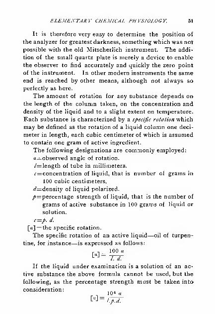
ELEMENTARY CHEMICAL PHYSIOLOGY. 51
It is therefore very easy to determine the position of
the analyzer for greatest darkness, something which was not
possible with the old Mitscherlich instrument. The addi-
tion of the small quartz plate is merely a device to enable
the observer to find accurately and quickly the zero point
of the instrument. In other modern instruments the sameend is reached by other means, although not always so
perfectly as here.
The amount of rotation for any substance depends on
the length of the column taken, on the concentration and
density of the liquid and to a slight extent on temperature.
Each substance is characterized by a specific rotation which
may be defined as the rotation of a liquid column one deci-
meter in length, each cubic centimeter of which is assumed
to contain one gram of active ingredient.
The following designations are commonly employed:
a=observed angle of rotation.
/=length of tube in millimeters.
*r=concentration of liquid, that is number of grams in
100 cubic centimeters.
(/^density of liquid polarized.
/=percentage strength of liquid, that is the number of
grams of active substance in 100 grams of liquid or
solution.
c—p. d.
[a]=the specific rotation.
The specific rotation of an active liquid—oil of turpen-
tine, for instance—is expressed as follows:
r -. 100 a
If the liquid under examination is a solution of an ac-
tive substance the above formula cannot be used, but the
following, as the percentage strength must be taken into
consideration:, „.10 4 «
[«] = l.p.d.

52 ELEMENTARY CHEMICAL PHYSIOLOGY
.
In using this formula it is necessary to know both the
percentage strength and the specific gravity, but as p. d
is the concentration of the solution, that is the number of
grams in 100 Cc, a result equally valuable for many pur-
poses is reached by weighing out the active substance in
grams and dissolving it to make 100 Cc. of solution.
In this case the formula to be used is
10 4 a
As an illustration of the use of the polariscope deter-
mine the specific rotation of .several sugar solutions.
Ex. 29. Weigh out 10 Gm. of cane sugar and dissolve
it in distilled water in a 100 Cc. flask. When solution is
•complete, fill to the mark and shake well.
Close one end of the 200 Mm. polarizing tube with its
glass cap and fill with the solution to be tested. Now put
on the other glass end so as to exclude all bubbles of air
and screw it down. Place the filled tube in its receptacle
in the polariscope and direct this toward a bright sodiumlight in an otherwise dark room.
If the instrument had been adjusted so that the zero of
the scale corresponded to the condition of equal shadowson the circular plate it will be found that with the sugarsolution between the polarizer and analyzer, the latter,
with the divided circle, must be rotated through a certain
number of degrees to reach the same shadow effect. Notethe direction and amount of this rotation, and calculate
the specific rotation according to the last formula above.
Ex. 30- Repeat the experiment, using solutions of
grape sugar and honey in known amount. The rotation of
the latter varies with different samples.
The following are mean values of the specific rotations
of different substances.
d, in these formulas, indicates sodium light.

ELEMENTARY CHEMICAL PHYSIOLOGY 53
Saccharose [«]d= 66.5°, ^=10—30Lactose (C
12H
a2On.H, O). . . " = 52.5°, c= 5— 12
Dextrose (anhydrous) " = 52.1°, c= 2—20Invert sugar " =—27.9°, c= 17
—
Serum albumin " =—56.°
Egg albumin " =—35.
5
C- a
The use of the polariscope in the special examination of
proteids will be referred to later. The instrument is em-ployed also in many chemical investigations in lines not
related to physiological chemistry.
In illustration of the methods of calculation the details
of some observations will be given. A sample of pure oil
of turpentine was polarized in a tube 300 Mm. long and at
the temperature of 20° C. The rotation of the plane of
polarization was found to be 40.504°. The specific gravity
of the oil, accurately determined, was found to be at
20°, 0.8650, water at 4° being taken as unity. We have
therefore, «d = 40.504
/ = 300.
d— 0.865
from which follows
r, _ 100X40.504
[a n = = lo.fa08L 300X 0.865
In another case 25 Gm. of pure Rochelle salt was dis-
solved in distilled water and made up to a volume of
exactly 100 Cc. at 20°. The solution was examined in a
tube 200 Mm. long and was found to have a rotation of
11.058°.
Here we haveaD — 11.058°
/= 200.
c = 25.
from which follows
[«],=10'X11.05S =22n6O
L JD200X25

54 ELEMENTARY CHEMICAL PHYSIOLOGY.
Fermentation of Sugars.
It has been shown above that certain sugars in solution
readily undergo fermentation by action of the yeast cell.
The products formed are essentially ethyl alcohol and car-
bon dioxide.
Cane sugar does not ferment with ordinary yeast, and
strong solutions cannot be fermented at all. In fact, manyof the uses of cane sugar depend on its power of pre-
venting fermentations or putrefactions. This is illustrated
in the "canning" or preservation of fruits, and in the em-
ployment of syrups in the pharmacopoeia.
Ex. 31. Make a strong cane sugar syrup, by boiling or
heating together 10 Gm. of sugar and 10 Cc. of water.
Allow to cool and add about a gram of crumbled com-pressed yeast, and then set aside for several days. Thesolution should be found free from any signs of fermentation.
Conditions of Alcoholic Fermentation. The follow-
ing experiments will be of interest in showing some of the
conditions on which alcoholic fermentation depends.
Ex. 32. Prepare a twenty per cent solution of com-mercial dextrose and pour 20 Cc. of it in a test-tube.
Add some yeast and allow to stand about two days in a
moderately warm place. At the end of this time it shouldbe found in active fermentation, as shown by the escape of
gas bubbles and the odor of alcohol.
Keep for future experiment.
Ex. 33. Prepare a tube with sugar solution and yeast
as in the last experiment. Close it loosely with a plug of
absorbent cotton and heat to boiling, allowing steam to
escape through the cotton. If the tube is now left to itself
for several days it will be found that fermentation has nottaken place, showing that heat destroys the characteristic
property of the yeast cell.

ELEMENTARY CHEMICAL PHYSIOLOGY. 55
Ex. 34- Prepare another tube with sugar and yeast andadd 10 Cc. of strong alcohol. Shake the mixture andallow to stand. No fermentation appears as the activity of
the yeast cell is destroyed by alcohol. We have good famil-
iar illustrations of this in the self-preservation of certain
"heavy" wines, as ports, sherries and malagas, while"light" wines, which contain ten to twelve per cent of al-
cohol, usually must be kept tightly bottled for preservation.
Ex. 35. Tests for alcohol. We have many tests bywhich the presence or formation of alcohol may be shown.The fermentation of a saccharine liquid is followed by a
lowering of the specific gravity as already mentioned, anda practical quantitative test is based on this fact. Asimple chemical test for the presence of alcohol, which in
most cases is sufficient, is the following: Add to the clear
liquid to be examined a few small crystals of iodine, warmto about 60° C.,and then add enough sodium hydroxide or
carbonate to produce a colorless solution. An excess of
the alkali must not be used. In a short time bright yellowcrystals of iodoform precipitate, easily recognized by their
color and odor. Certain other liquids give the same test.
Acetic Acid Fermentation. Weak alcoholic solu-
tions, if containing small amounts of foreign matter, espe-
cially nitrogenous, become sour, if exposed to the air, from
formation of acetic acid. This is due to the fact that a
small microscopic plant cell, the mycoderma aceti, is gener-
ally present in the atmosphere, and in sufficient numbers to
oxidize the alcohol to acid.
Ex. 36. The solution of Ex. 32 becomes sour if al-
lowed to stand four or five days in a warm place. Whenthe odor of alcohol has practically disappeared add someprecipitated chalk and evaporate to dryness. Extract with
a little water, filter and concentrate the filtrate to a small
bulk; add equal volumes of alcohol and strong sulphuric
acid and apply heat. The characteristic odor of acetic
ether is developed, proving the presence of acetic acid as a
product of fermentation.

56 ELEMENTARY CHEMICAL PHYSIOLOGY.
The acetic acid ferment is destroyed by high tempera-
ture or strong alcohol, which can be shown by experiments
similar to those detailed above.
Lactic Acid Fermentation. By the action of a pecu-
liar ferment solutions of sugar in presence of nitrogenous
matters become converted into lactic acid, according to the
equation :
C lf H a2 O,, +H sO=4C
3H
6 3
The reaction takes place best in neutral or slightly alka-
line solution and is speedily stopped by the accumulation
of acid formed. In practice, therefore, it is customary to
add some chalk or zinc oxide to the fermenting mass to
take up the acid as fast as formed. The fact of the produc-
tion of acid is shown by the following experiment:
Ex. 37. Dissolve 30 Gm. of cane sugar in 150 Cc. of
boiling water and add about 30 Mg. of tartaric acid. After
this mixture has stood two days, add some putrid cheese,
about half a gram, and 40 Cc. of sour milk. Then add 15
Gm. of zinc oxide and allow the mixture to stand ten daysin a warm place where the temperature is about 45° C,stirring frequently. At the end of this time heat to boiling,
filter hot and allow the zinc salt to crystallize out on cool-
ing. Dissolve the crystals in a small amount of hot water,
and allow them to settle out again. Collect these purercrystals, mix with water and pass hydrogen sulphide into
the mixture in excess. Filter off the precipitated zinc sul-
phide and shake the aqueous filtrate with ether in two por-
tions, using 25 Cc. in all. Allow the ether to evaporatespontaneously, leaving the lactic acid as a thick liquid.
Butyric Acid is produced from lactic atid by prolonged
fermentation.
If the fermented mass, in the above experiment, contain-
ing crystals of calcium or zinc lactate, is allowed to stand
some weeks at about the same temperature, bubbles of

ELEMENTARY CHEMICAL PHYSIOLOGY. 57
hydrogen and carbon dioxide will escape, and decompo-sition, according to the following equation takes place:
2 C3H
6 3=C 4H
8 2+2 C 2 +2 H 2
This action, like the former, is due to the presence of a
minute microorganism.
Glycogen or Animal Starch.
Closely related to the carbohydrates described above
we have a compound produced in the animal body and
known as glycogen. This substance is found in small
quantities in many of the tissues, but is relatively abun-
dant in the liver only, from which organ it is usually pre-
pared. It is also found in the common oyster, which is
sometimes employed as a starting point in the preparation
of the pure substance.
The laboratory production of glycogen is best carried
out as follows:
Ex. 38. Kill a rat or a rabbit; remove the liver as
quickly as possible and without delay cut it into small bits
which throw into a vessel of boiling water. The weight of
the water should be about five times that of the mincedliver. Boil five minutes, then remove from the water andrub up in a mortar with fine clean quartz sand. In this
way the fragments of liver become thoroughly disinte-
grated. The contents of the mortar, sand as well as liver,
are thrown into the boiling water again and kept at 100° C.
fifteen minutes. At the end of this time enough dilute
acetic acid must be added to impart a faint acid reaction.
This coagulates and precipitates some albuminous matters
which are separated when the hot mixture is filtered.
In the opalescent filtrate, which must be collected in
a cold beaker, a further precipitation of albuminous mat-
ter is effected by adding a few drops of hydrochloric acid
and some potassium mercuric iodide as long as a precipi-
tate forms.

58 ELEMENTARY CHEMICAL PHYSIOLOGY.
Filter again and use the dilute aqueous solution of gly-
cogen resulting for tests below.
Ex. 39. Evaporate about half of the liquid above to a
small volume and precipitate impure glycogen as an
amorphous white powder by addition of strong alcohol.
Ex. 40. Add a little tincture of iodine to a small por-
tion of the solution, and note the red color produced. Thiscolor is discharged by heat. Boil some of the solution
with dilute hydrochloric acid ten minutes ; neutralize theacid nearly, cool and again test with iodine. No color is
now produced, as glycogen has disappeared under thetreatment, having been converted into sugar.
After death the store of glycogen in the liver rapidly
disappears, so that tests applied' at the end of a day or
two fail to show its presence.
Ex. 4l. Cut some common beef liver from the marketinto small bits and extract with boiling water. Boil longerto coagulate the albuminoids, after adding some sodiumsulphate. Apply the iodine test for glycogen, which is
found absent, and the Fehling test for sugar, which is foundpresent in quantity.
In Experiment 38, the direction is given to treat imme-diately with boiling water. This is done to destroy a fer-
ment-like body in the liver, which speedily converts gly-
cogen into sugar.
Solutions of glycogen in water exert a marked dextro-
rotatory action on polarized light, but observers differ as
to the exact specific rotation. It is about
[«]„ = 210°.

Chapter III.
FATS.
T^HE bodies commonly called fats are compounds of
* glycerol with palmitic, stearic and oleic acids. Butter
fat contains in addition glycerol compounds of butyric,
caproic and other less important acids. The fats of the
vegetable kingdom are in many respects similar to those
of the animal kingdom, and like the latter, serve as food
for man.
The important animal fats are butter, lard and the sev-
eral kinds of tallow. The important vegetable fats are
olive oil, cottonseed oil, sesame oil, peanut oil, cocoa but-
ter and others. Pure fats are usually separated from the
accompanying animal or vegetable tissue by pressure, or
by melting, as illustrated by the manufacture of olive oil
and the rendering of lard or tallow.
Origin of Fats in the Body. The fats found in the
animal tissues are obtained from three general sources.
First. They are taken directly from the fatty matters of the
food, and the amount so absorbed may be considerable, as
has been shown by several physiologists who have inves-
tigated the question.
Second. A production of fat goes on in the body whenthe animal is fed on a diet of albumin alone. The amount
of butter which a cow will produce is far in excess of the
fat in the feed, and can be accounted for only .on the
assumption of decomposition of albuminous matters. Dogs
have been starved until their bodies were practically free

60 ELEMENTARY CHEMICAL PHYSIOLOGY.
from fat and then fed on muscle and soaps of palmitic and
stearic acids. After a time they were killed, and on analy-
sis their bodies were found to contain not only palmitin
and stearin, but also olein. This must have come from
a breaking down of the food proteids. The formation of
large quantities of adipocere in cadavers is considered, also,
as a proof of the production of fatty acids from albu-
minoids.
Finally, it has been abundantly proven that carbohy-
drates give rise directly to fats by molecular changes in
the body. Indirectly, the consumption of carbohydrates
favors production of fats by taking the place of the al-
buminoids in oxidation. The latter are, therefore, left to
be broken up into a nitrogenous residue, and a fat-forming
residue which is stored up in the tissues.
Some of the important properties of fats are shown by
the following experiments :
Ex. 42. Note the solubility of small bits of tallow in
ether, chloroform, benzine and alcohol, using in each case
the same volume of liquid, with equal weights of fat. Thesolubility in alcohol will be found much less than, in the
other menstrua. The common fats are insoluble in water.
All fats undergo a very remarkable change when heated
with solutions of caustic alkalies. This treatment is suffi-
cient to split them up into their component parts, glycerol
on the one hand and the alkali-metal salt of the fatty acid
on the other. Thus stearin, or glyceryl stearate, the com-
pound of glycerol with stearic acid, yields, when treated
with a strong solution of potassium hydroxide, free
glycerol and potassium stearate, as shown by the follow-
ing tests:
Ex. 43. Boil about 25 Gm. of cottonseed oil or tallowwith a solution of 10 Gm. of potassium hydroxide in 25 Cc.

ELEMEiYTARY CHEMICAL PHYSIOLOGY. 61
of water. Stir the mixture thoroughly until it becomeshomogeneous, that is, until no oil globules are seen float-
ing on top of the aqueous liquid, which may require half
an hour. Add water from time to time, to make up for
that lost by evaporation.The resulting mass is a mixture of glycerol, excess of
alkali and soft soap.
Ex. 44. The presence of fatty acids in the abovemixture can be shown by adding to a portion of it enoughhydrochloric acid to decompose the soap. Use about halfthe product of Ex. 43, dilute it with water, and add theacid in slight excess, about 10 Cc. of the strong commercialacid. Warm on the water-bath, which will cause theliberated fatty acids to collect on the surface as a liquidlayer as soon as the temperature becomes high enough.Add more water and allow the whole to cool. A semi-solid layer of fatty acids can now be lifted from the surfaceof the liquid. The hardness of the mixed acids dependson the nature of the fat taken for experiment. Muttonand beef tallows yield very solid acids; with lard the massis softer, while with some oils the acids do not solidify at
all at the ordinary temperature.
Ex. 45. Dissolve a small portion of the fatty acids in
warm alcohol, nearly to saturation. On cooling, the acids
separate in crystalline scales.
Ex. 46- The presence of glycerol as one of the prod-ucts formed by the saponification of fats is best shown as
follows: Mix 50 Cc. of cottonseed oil with 25 Gm. of
litharge and 100 Cc. of water in a porcelain dish. Placeover a Bunsen burner on gauze and stir until all oil
globules have disappeared, adding a little water from timeto time. The litharge with water acts as lead hydroxideand saponifies the fat, forming an insoluble lead soap, or
plaster, and glycerol. When the saponification is com-plete add more water, heat and stir well to dissolve the
gl3Tcerol, allow to settle a short time and pour the aqueoussolution through a filler. To the residue add water again,

62 ELEMENTARY CHEMICAL PHYSIOLOGY.
heat, allow to settle and pour through the same filter.
Concentrate the mixed filtrates to a small volume and after
cooling observe the sweet taste of the thickish residue.
Glycerol of commerce was at one time made by this
slow process, but at the present time it is made by other
reactions from fats, usually by the action of superheated
steam. This passed into melted fat effects a separation
into glycerol and fatty acid. Sometimes the fat is decom-
posed by milk of lime and the glycerol separated by a cur-
rent of superheated steam with which it is volatile.
The inverse process of combining glycerol and fatty
acids to form fats is not so easy, but it has been done on
an experimental scale, yielding not only the tri-acid com-
pounds similar to those existing in nature, but mono- and
di-acid compounds as well.
Fats being insoluble in water, a permanent mixture of
the two cannot be obtained directly. A separation always
follows after a time. By the action of certain substances,
however, the fat can be brought into a very finely divided
condition called an emulsion, which has important prop-
erties.
Emulsions.
Fats can be thrown into this peculiar physical modifi-
cation by a variety of additions, which are illustrated in
the experiments below. In order that a fatty substance
may leave the alimentary canal and enter the circulation it
is necessary that it must first be brought into a condition
in which it can be absorbed by the lacteals. Emulsifica-
tion accomplishes this, which is mainly carried out in the
small intestine.
Ex. 47. Add to 5 Cc. of cottonseed oil half its volumeof strong white of egg solution and shake thoroughly. Theliquids mix and form a white mass or emulsion.

ELEMENTARY CHEMICAL PHYSIOLOGY. 63
Ex. 48. To 5 Cc. of cottonseed oil containing a little
free fatty acid add 10 drops of strong sodium carbonate solu-
tion and shake. A good stable emulsion is made in this way.
Ex. 49. In the intestine this reaction is brought aboutby the action of the pancreatic juice and bile, which can be
illustrated in this manner: Pour about 10 Cc. of refined
cottonseed oil in a warm mortar and add two or three
grams of fresh finely divided pancreas. This can be ob-
tained by running the pancreas through a good sausage
mill. Rub the mixture thoroughly with a pestle, by whichmeans a white milky mass is obtained.
To produce an emulsion the presence of free fatty acids
seems to be necessary. Now it is probable that the pan-
creatic juice contains a product capable of splitting fats
into free acid and glycerol, the first of which with the al-
kaline juice produces the emulsion. It is certain that the
emulsifying power of the juice does not depend on the
presence of an enzyme, because emulsification takes place
with boiled pancreatic juice as well as with fresh. In its
work of splitting fats the pancreatic juice is assisted by the
bile, some tests on which will be given later.
Crystallization of Fats.
The neutral fats as well as the fatty acids readily
assume the crystalline form under certain conditions.
Fats in the tissues are undoubtedly free from crystalline
structure, and when melted out at a low temperature or
separated by pressure the amorphous form is the common
one. Melted fats, on standing, generally become crystal-
line, which can be shown by melting a little tallow and
placing a drop of it on a glass slide to cool. If a cover
glass is pressed down on the fat before it becomes hard
this will give a flat field in which the crystalline form can
be seen under the microscope.

64 ELEMENTARY CHEMICAL PHYSIOLOGY.
fig. ai
Human fat, crystallized from
chloroform.
FIG. 82.
Dog fat. crystallized from
chloroform.
FIG. 23.
Cat fat, crystallized from
chloroform.
FIG. 84.
Beef tallow, crystallized fromchloroform.

ELEMENTARY CHEMICAL PHYSIOLOGY. 65
Beef tallow, crystallized from
chloroform.
FIG. 86.
Pure stearin, crystallized from
chloroform.
FIG. 37.
Mutton tallow, crystallized from
chloroform.
FIG. 88.
Lard, crystallized from
chloroform.

66 ELEMENTARY CHEMICAL PHYSIOLOGY.
Perfect crystallization can be secured by deposition
from a menstruum.
Ex. 50. Dissolve some mutton or beef tallow in
chloroform and with a glass rod put two or three drops of
the nearly saturated solution on the center of a glass
slide. As the chloroform evaporates a film begins to
form on the top of the drop. Now put on a perfectly
clean cover glass and allow to stand until crystallization is
complete, which may require only a few minutes or somehours, the time necessary depending on the temperatureand on the concentration of the solution. Examine the
crystals with a microscope. Use a power of 200 to 300diameters.
Very peculiar crystallizations are shown by certain fats as
can be seen by the annexed cuts. Some of these forms have
been taken as characteristic and have been considered as suf-
ficient for the identification of several fats, even in mixtures.
It is true that in general the crystals of mutton tallow
or beef tallow are different from those of pure lard, but it
is also true that mixtures can be made which yield crystals
practically identical with those usually seen in the fields
from lard. At various times it has been supposed that
adulterations of lard or butter could be demonstrated bythese microscopic appearances, but, one by one, the points
brought forward as characteristic have been shown to beof little real value.
From one and the same portion of fat dissolved in chloro-
form, for instance, four or five different kinds of crystals
may be obtained, and by using other solvents or changingthe temperature of crystallization still different forms maybe secured. This fact is illustrated in the figures abovewhich are very exact illustrations of the microscopic ap-
pearances. They are copies of micropholographs madeby the author and show as observed under a magnifyingpower of about 200 diameters.

Chapter IV.
PR0TE1DS OR ALBUniNS.
THE term proteid is applied to a large number of sub-
* stances derived from different sources which are very
similar in composition, and which in many instances ap-
pear to be impure forms of one and the same compound.
These proteid compounds are found in both the animal
and vegetable kingdoms, but never in pure form. Several
closely related varieties are often associated, and mixed
with these are fats, carbohydrates, mineral salts, etc., in
varying proportions.
Practically, the preparation of a pure albumin is a mat-
ter of very great difficulty, and the analyses made by differ-
ent experimenters do not show remarkably close agree-
ment. It is, therefore, impossible to assign a formula to
these bodies, although this has several times been at-
tempted. The outside limits assigned for the percentage
composition are given below :
C 50.0 to 55.0
H 6.8 " 7.3
O 20.0 " 24.1
N 15.0 " 18.2
S 0.3 " 5.0
As an illustration of the results reported by a single
observer the analysis of egg albumin by Hammarsten may
be quoted. He found for the dry substance
:
C 52.25
H 6.90
O 23.67
N 15.25
S.'.'.V... 193
100.00

68 ELEMENTARY CHEMICAL PHYSIOLOGY.
Albumins as a class are recognized by the following re-
actions :
(a.) They give the characteristic test for nitrogenous
bodies when fused with soda-lime.
(£.) They yield precipitates with solutions of salts of
certain heavy metals.
(V.) They give, especially, red precipitates when
warmed with Millon's reagent. ,
(d.) They give a characteristic color when heated with
strong nitric acid, and are then treated with am-
monia.
(e.~) They give color reactions when mixed with a small
amount of copper sulphate solution, and then with
an excess of alkali.
These reactions are illustrated by the following experi-
ments :
Ex. 51. Mix some common wheat flour with an equal
bulk of soda-lime in a test-tube, and heat strongly in the
flame of a Bunsen burner. Ammoniacal vapors are givenoff, which may be recognized by the odor, or by action onlitmus paper. The ammonia is produced in the decompo-sition of the proteid matter of the flour by the fixed alkali.
This is a characteristic and delicate reaction.
Prepare a dilute solution of white of egg and use it for
tests to follow. Pour the white of one egg into a strong
bottle holding about half a liter. Add 100 Cc. of distilled
water and some small quartz pebbles, or better, somepieces of clean broken glass, and shake vigorously. After
a time add 400 Cc. more and shake again, and allow to
stand about fifteen minutes. Strain through clean, un-
sized muslin, and use the nearly clear liquid for the tests.

ELEMENTARY CHEMICAL PHYSIOLOGY. 69
Ex. 52. Pour some oi the liquid into several test-tubesand add small amounts of solution of mercuric chloride,
copper sulphate, lead acetate and silver nitrate. Notethat each reagent produces a precipitate.
Ex. 53. To another portion of the liquid in a test-
tube add half its volume of Millon's reagent (for prepara-
tion of this see appendix), and heat. A yellowish pre-
cipitate which appears at first becomes red on boiling.
This test shows equally well with other albumins andis very delicate. With very dilute solutions a red color
appears without precipitation.
Ex. 54. Xanthoproteic reaction. To some albumin in
a test-tube add strong nitric acid, and boil. A yellowprecipitate or color is formed which, on addition of am-monia, becomes deep orange red.
The colors obtained in this reaction depend on thestrength of the albumin solution. With weak solutions a
precipitate may not form at all, but the yellow color is
characteristic. When only traces of albumin are presentthe yellow color with the nitric acid may fail to appear,but the addition of ammonia gives the final part of the test
with a yellow instead of an orange red color.
Ex. 55. To some of the albumin solution in a test-
tube add enough sodium hydroxide to give a very strong
alkaline reaction. Then add two or three drops of a dilute
copper sulphate solution. This gives a violet color, whichby application of heat becomes much deeper. This is
known as the biuret reaction, and serves for the detection of
Small traces of albumins in solution. It is absolutely
necessarv to employ a considerable excess of the alkali
with only a very small amount of copper solution.
Ex. 56. Make some albumin solution strongly acid
with acetic acid, add some sodium sulphate and boil. All
proteids except peptones are precipitated in this manner,
and the reaction is employed to free solutions from these
bodies. The filtrate, after boiling, can be used for other

70 ELEMENTARY CHEMICAL PHYSIOLOGY.
tests as the acid and sulphate exert no decomposing action.
The method is frequently employed in urine analysis whenit is desirable to test for small amounts of sugar in presenceof relatively large quantities of albumin.
Classification of Albumins.
Investigation has shown that the bodies of this group
fall naturally into several classes. At the present time it
is usual to consider seven of these classes, the division
being based on certain features easily recognized. It mustbe remembered, however, that this division is somewhatarbitrary and is not universally accepted as here given,
according to Hoppe-Seyhr and others.
n, „„ t xt i.- iL f Egg albumin,Class I. Native albumins. . . i c ,. •
I Serum albumin.
' Alkali albumin,/>, „„ TI t\ j iu Acid albumin,Class II. Derived albumins. \ „ .
'
Casein,
Syntonin.
[ Crystallin,
|Vitellin,
Class III. Globulins J
P^raglobulin,Fibrinogen,
! Myosin,[Globin.
Class IV. Fibrin.
Class V. Coagulated proteids.
Class VI. Albumoses and peptones.
Class VII. Lardacein.
Native Albumins.
These are soluble in water, but are precipitated byboiling or by addition of alcohol.
Their solutions are not precipitated by sodium chloride
or sodium carbonate.

ELEMENTARY CHEMICAL PHYSIOLOGY. 71
Egg Albumin. Different kinds of eggs furnish albu-
mins of slightly different properties, but these variations
may be due to accompanying substances rather than to in-
herent differences in the albumins themselves. Most of our
knowledge of these bodies has been derived from a study
of the hen's egg, and this will be taken for the experiments
below. Prepare a solution of white of egg as given above
and make tests with it as follows:
Ex. 57- Note that a small portion of the solution is
readily coagulated by heat in a test-tube. By the aid of athermometer find approximately the temperature of coagu-lation, which is not far from 60° C. Some of the proteidspresent in white of egg begin to coagulate, it is claimed,at even a lower temperature while a temperature of 80° C.
may be reached before coagulation is complete.
Ex. 58. Warm three or four Cc. of strong nitric acid
in a test-tube and pour' in about an equal volume of the
albumin solution carefully, so that it will float on top andnot mix with the acid. A coagulation of the albumin takes
place at the juncture of the two liquids and appears as a
white ring. This is a valuable and delicate test.
Ex. 59. To ten Cc. of the egg solution add powderedmagnesium sulphate to saturation. A precipitate of globu-
lin separates out. Filter and add to the filtrate somestrong solution of sodium sulphate. Precipitation of thealbumin takes place now, if the solution is. kept at a tem-perature of 40° C. This precipitation seems to be due to
the presence of a double sulphate of magnesium and so-
dium in the liquid.
Serum Albumin. Serum albumin occurs in the blood
along with several other proteids, but can be obtained
nearly pure by the following method:
Fresh blood from the slaughter house is poured into a
shallow dish and whipped with a bunch of twigs or agitated

72 ELEMENTARY CHEMICAL PHYSIOLOGY.
vigorously with an egg beater to separate the fibrin. The
latter is strained out by means of clean unsized muslin.
The liquid passing through the muslin is put into a centri-
fugal apparatus and rotated to throw out the corpuscles.
If the centrifugal apparatus is not at hand the fibrin-free
liquid can be poured into a tall glass jar and allowed to
stand until the corpuscles settle. The clarified liquid ob-
tained by either method is poured into a large beaker and
mixed with four times its volume of saturated solution of
ammonium sulphate, or enough to yield a completely satu-
rated liquid. This precipitates the albumin and globulin
present, and these compounds are then separated by filtra-
tion. The moist mass on the filter is dissolved in a small
quantity of water and treated again with an excess of am-
monium sulphate which gives a purer precipitate. Theproduct is washed on the filter with strong ammonium sul-
phate solution, then dissolved in a little water and sub-
jected to dialysis, by which means the salts are eliminated
while an albumin solution remains on the dialyser.
During the dialysis globulin separates and at the end
of the operation is filtered out leaving a nearly pure serumalbumin. This can be further purified by adding a little
ammonia until the reaction becomes neutral, dialyzing
again and finally filtering. The clear filtrate can be used
for tests or further concentrated. To obtain it in dry formevaporate at 40° C. to a small bulk, add strong alcohol in
excess to precipitate, filter without delay, press out the
alcohol, and displace the remaining alcohol by washingwith ether. Dry at a low temperature, powder and pre-
serve in a well-stoppered bottle.
As shown by the method of preparation serum albuminis soluble in water, but insoluble in saturated solution of
ammonium sulphate. The tests given for egg albumin are
true for serum albumin. These points of difference maybenoted:

ELEMENTARY CHEMICAL PHYSIOLOGY. 73
Ex. 60. Note that a neutral solution of egg albumin is
coagulated by ether, while the solution of serum albuminis not.
Ex. 61. Coagulate a solution of egg albumin by heat
and note that the precipitate dissolves but slightly in boil-
ing nitric acid. A precipitate of serum albumin dissolves
in nitric acid.
Ex. 62. Precipitate solutions of egg albumin andserum albumin with nitric acid and note that the latter
precipitate is much more readily soluble in excess than is
the former.
Derived Albumins.
These are nearly insoluble in pure water or dilute saline
solutions, but are soluble in weak acids or alkalies. Boil-
ing does not coagulate the solutions.
Alkali Albumin. This is most readily prepared by
the action of alkali on native albumins.
Ex. 63. Add strong sodium hydroxide solution to
white of egg, with constant stirring, until a thick jelly is
formed. Too much alkali must not be added here, but
just enough to make the maximum of jelly. This is nowcut into small pieces and washed in distilled water several
times until the lumps are white throughout. They are
then heated with fresh pure water, but very gently, until
they go into solution. This is then filtered and the filtrate
precipitated by acetic acid, avoiding any excess. The pre-
cipitate is washed with pure water.
Observe that this substance is soluble in dilute acid or
alkali solutions.
Ex- 64. Bring some of the washed alkali albumin into
solution with hot water. Add a few drops of phenol
phthalein solution, which imparts a red color, and then run
in dilute sulphuric until the alkali reaction just disappears.
As soon as the neutral point is reached a precipitate falls,
but, if the addition of acid be continued it will disappear
with formation of acid albumin.

74 ELEMENTARY CHEMICAL PHYSIOLOGY
The thick jelly-like substance first formed by the action
of strong alkali on undiluted white of egg solution is called
Lieberkuehn's jelly. It contains an excess of uncombined
alkali. A weak solution of alkali albumin can readily be
made by warming diluted white of egg solution with a
small amount of very weak alkali solution. The tempera-
ture should be maintained at 35° C. to 40° C, for about
an hour. At the end of this time the solution can be boiled
without coagulation.
Acid Albumin. The bodies called acid albumins are
very similar in properties to the alkali albumins and at one
time were supposed to be identical with them. But careful
experiments have disclosed several points of difference.
Acid albumin is most readily made by the action of acid on
native albumins.
Ex. 65. Dilute white of egg with four volumes of
of water, take 25 Cc. of the mixture, add 5 Cc. of 0.2 percent hydrochloric acid and warm it on the water-bath for
about two hours to a temperature of 45°. Then carefully
neutralize the solution with dilute sodium hydroxide. Thisprecipitates insoluble acid albumin, which can be washedwith water by decantation. It is essential that just theright amount of alkali be added here, an excess wouldredissolve the precipitated acid albumin with formation of
alkali albumin. The washed acid albumin can be used for
a number of tests.
Ex. 66- Redissolve a small portion of the precipitatewith hydrochloric acid, and observe that the solution is notcoagulated by boiling.
Mix another portion of the precipitate with water andheat to 75° or 80°. This brings about a coagulation sim-ilar to that produced with native albumins.
Acid albumins are very quickly formed by the action of
strong hydrochloric or acetic acid on white of egg. Coagu-

ELEMENTARY CHEMICAL PHYSIOLOGY. 75
lated proteids may also be converted into acid albumins by
a much longer digestion.
The striking peculiarity of this acid albumin is that it is
insoluble in pure water, but does dissolve in weak acid or
alkali. It gives the characteristic yellow color with strong
nitric acid, a reddish yellow precipitate with Millon's
reagent and the biuret reaction.
Acid albumin is less soluble than alkali albumin and
appears to differ also in specific rotation.
Casein. Milk contains several proteid substances, the
most abundant of which is casein. In normal cow's milk
it is present to the extent of about four per cent, while in
woman's milk it appears to amount to two and one-half
per cent or less. While the determination of casein in
cow's milk has been made with great accuracy and is an
operation of no special difficulty there seem to be prac-
tical difficulties in the way of carrying out this determina-
tion in the case of human milk. The first point of uncer-
tainty is found in the collection of a normal sample, and
from this and other causes it follows that the results given
by different chemists for the amount of casein present vary
between 0.6 and over 5 per cent.
Equally great variations have been found in the milk
furnished by several animals.
Our practical knowledge of casein has been in the main
derived from a study of the product of cow's milk.
In most of its reactions casein is closely allied to al-
kali albumin and by some has been supposed to be identi-
cal with it. But other reactions disclose certain differ-
ences which seem to be essential and the weight of evi-
dence appears to be against the identity of the two pro-
teids. The important properties of casein are made ap-
parent by the following experiments.

76 ELEMENTARY CHEMICAL PHYSIOLOGY.
Ex. 67. To 100 Cc. of fresh milk add 500 or 600 Cc.
of distilled water and then enough dilute acetic acid
to give a distinct acid reaction. This will throw down a
copious white precipitate of casein with fat which is al-
lowed to settle and washed with distilled water by decan-
tation. The precipitate is then transferred to a filter andallowed to drain and then washed with 50 Cc. of strong
alcohol. This removes water and part of the fat present.
The precipitate is finally washed with ether to remove the
remainder of the fat and allowed to dry in the air.
After the above treatment the casein is left as a white
powder pure enough for tests. A better product is ob-
tained by dissolving the precipitate first thrown down in
weak soda solution and passing the liquid through several
filters. It is then reprecipitated with dilute acetic acid,
washed by decantation, collected on a filter and finished as
before.
Ex. 68. Test a small amount of the casein preparedaccording to the last experiment as to its solubility in
water. This will be found to be very slight. It dissolves
readily in weak alkali solutions.
Ex. 69. Mix 5 Cc. of fresh milk in a test-tube with 10
Cc. of saturated salt solution. Then add a little morepowdered salt, close the test-tube with the thumb andshake vigorously. Casein with fat is precipitated and canbe collected on a filter. Boil the opalescent filtrate andnote the coagulation of a soluble albumin present. Incow's milk this form of albumin amounts to about 0.5 percent.
An excess of magnesium sulphate solution may be used
instead of the sodium chloride to precipitate the casein.
Under the action of rennin, casein undergoes a peculiar
modification resulting in the formation of a clot or curd.
The nature of this will be explained in the chapter on milk.

ELEMENTARY CHEMICAL PHYSIOLOGY. 77
The behavior of milk with rennin serves to distinguish
casein from true alkali albumin. With the latter, it is said,
no clot can be obtained.
Pure casein yields no ash on ignition, although it con-
tains phosphorus and sulphur in organic combination.
Pure alkali albumin yields a small amount of ash whenignited.
Syntonin. This appears to be an acid albumin, re-
sulting from the action of dilute acids on muscle.
Ex. 70. Free the muscular part of meat from fat asfar as possible and run it through a sausage mill several
times to bring it to a fine state of subdivision. Washthis chopped mass with distilled water until the washingsremain clear. Now, to about 5 Gm. of the moist residue
in a small flask, add 50 Cc. of dilute hydrochloric acid,
containing ^ per cent of the true acid. Warm the mix-ture slightly (to 35° or 40° C"), and keep at this tem-perature about three hours. Then filter and test the fil-
trate. It contains the soluble syntonin.
Ex. Jl. To a small portion of the filtrate, add weakcaustic soda, which produces a precipitate soluble in ex-
cess of the alkali.
Boil another portion of the filtrate. It does not co-
agulate.
Globulins.
We have here a peculiar class of proteid bodies, in-
soluble in pure water and strong saline solutions, but
soluble in weak saline solutions. A globulin dissolved
in a weak solution of salt is precipitated if the same salt
is added to saturation. Magnesium sulphate is most ac-
tive in producing this precipitation. The globulins resem-
ble the native albumins in being coagulable by heat; they
are also insoluble in alcohol. Dilute acids or alkalies
dissolve them by forming acid or alkali albumin.

78 ELEMENTARY CHEMICAL PHYSIOLOGY.
Crystallin. This is the globulin of the crystalline lens,
and can be obtained by extracting the latter, in finely di-
vided form, with a one per cent salt solution.
Vitellin. Yolk of egg contains a large amount of this
globulin, associated with other substances of which lecithin
is, perhaps, the most important. The preparation of pure
vitellin is a matter of great difficulty because of the pres-
ence of the lecithin which cannot be readily separated. Apartial separation can be effected by extracting with ether,
leaving a residue which dissolves easily in salt solution.
A great excess of sodium chloride does not reprecipitate,
as is the case with other globulins.
Paraglobulin, or serumglobulin occurs with pure albu-
min in blood serum, from which it can be separated in
nearly pure condition.
It was shown above, under the head of blood serum,
that in the process of dialysis for the final purification of
the latter, globulin separates out as the saline liquid loses
its salts and becomes aqueous. The precipitation of
globulin can be illustrated by the following experiment :
Ex. 72. Separate fibrin and corpuscles from blood asdescribed above under serum albumin and saturate the re-sulting liquid with magnesium sulphate. This throws downa precipitate of globulin, which can be washed on a filter
with a saturated solution of the sulphate without muchloss. By mixing the precipitate with a little water, andgradually adding more, it will finally dissolve, but the ad-dition of a very large volume of water produces a precipi-tate again.
Common salt, added in excess, acts as does the magne-sium sulphate.
The method illustrated by the above experiment is notsufficient for the preparation of pure globulin. A better

ELEMENTARY CHEMICAL PHYSIOLOGY. 79
product is obtained by dissolving and reprecipitating sev-
eral times with magnesium sulphate. Finally the precipi-
tate may be dissolved in the right amount of water and
dialyzed for the separation of the salts. The globulin re-
mains on the dialyzer as an insoluble residue.
Fibrinogen. In the clotting of blood this substance
plays an important part as in the operation it becomes con-
verted into the stringy fibrin which encloses the corpuscles
forming a thick and jelly-like mass.
Myosin. This is a globulin formed in muscle after
death and is produced from a substance resembling fibrin-
ogen existing in the living muscle. The name myosinogen
has been given to this primary form.
The preparation of myosin is illustrated by the next ex-
periment:
Ex. 73- Free muscle (round steak) as far as possiblefrom traces of fat and sinews and then thoroughly disinte-
grate it by passing through a sausage mill. Then wash it
repeatedly with cold water until the latter is no longer red-
dened, and the residue appears white. This is placed in a20 per cent solution of ammonium chloride and allowed to
remain about a day, with occasional shaking. Myosin dis-
solves in the ammonium chloride and is found in the filtrate
when the mixture is filtered. Pour the filtrate into twentytimes its volume of distilled water, which causes a precipi-tation of the insoluble myosin. Allow to settle and washthree times by decantation. Collect the precipitate andobserve that portions of it dissolve readily in ten per centsolutions of sodium chloride and ammonium chloride or in
a 0. 1 per cent solution of hydrochloric acid. The solution
in salt is precipitated by the addition of more to saturation.
Myosin in salt solution coagulates at a low temperature,
usually given as 55° to 56° C. By acids it is easily con-
verted into syntonin.

80 ELEMENTARY CHEMICAL PHYSIOLOGY.
Globin is sometimes described as a distinct globulin,
formed by the spontaneous decomposition of haemoglobin,
but no great amount of study has been given to it by
chemists.
Fibrin.
Fibrin is the name commonly, given to the substance
obtained by whipping blood with a bundle of twigs. So
prepared it is very impure, holding white and also some red
corpuscles. By other means, however, a much purer
product can be obtained. Made in any manner fibrin is a
white elastic substance insoluble in water and weak salt
solutions. It dissolves gradually in acid or alkali solutions,
forming proteids of other groups. A corresponding vege-
table product can be easily made by mixing flour with a
little water to form a stiff dough. After standing some
time this is worked with the hand under running water
which washes out the starch, leaving finally the vegetable
fibrin or gluten as a white stringy mass.
Animal fibrin is produced from the proteid referred to
above as fibrinogen, occurring in the blood before death.
Coagulated Proteids.
We have here a class of substances produced by action
of heat on most of the proteid bodies just described.
When a proteid assumes the coagulated form it becomesinsoluble in water, dilute acids or alkalies, and can be dis-
solved by strong acids only through complete modification
and partial destruction. Alcohol has also the property of
coagulating many proteid substances.
We have a familiar illustration 'of a coagulated pro-
teid in boiled white of egg. This, and similar substances,
undergo a peculiar change called digestion when taken into
the stomach where they are acted on by the gastric juice,

ELEMENTARY CHEMICAL PHYSIOLOGY. 81
or in the intestines under the action of the pancreatic
juice. This digestion will be considered in a few experi-
ments later, as it is one of the important reactions taking
place in the animal body.
Albumoses and Peptones.
Under the action of the gastric juice and the secretion
of the pancreas just referred to albuminous substances in
general surfer a profound modification whereby they be-
come soluble and ready for absorption as preliminary to
nutrition. The gastric juice is a weak acid liquid of rather
complex composition, but containing at least two sub-
stances of importance which merit our attention here.
The first of these is a peculiar enzyme or soluble fer-
ment called pepsin and the second is hydrochloric acid.
This acid is present in all normal gastric juice and in manit amounts to about 0.25 per cent of the total weight
of the juice. The other substances present have as a mat-
ter of course some important function, but the manner of
behavior of the acid and pepsin have been made the sub-
ject of many investigations. The essential work of these
two compounds is to convert other proteids into the forms
known as albumoses and peptones, of which the latter are
extremely soluble and to some degree diffusible. Between
the ordinary coagulated proteids taken into the stomach
as food and the final peptone or completely digested mat-
ter there are possibly a number of intermediate stages.
The albumoses represent one of these and can be distin-
guished from the end product by several fairly definite re-
actions. It appears, further, that at the beginning of the
process of digestion with pepsin and hydrochloric acid that
a body similar to or identical with acid albumin is formed,
to be soon followed by albumose.

82 ELEMENTARY CHEMICAL PHYSIOLOGY.
In studying the digestion of starches it was shown that
the pancreatic fluid contains an enzyme very active in this
direction, and further that in the emulsification of fats, as a
step in digestion, the pancreatic juice plays, likewise, a
prominent part. The secretion of the pancreas is active
finally in a third direction, viz. : in the digestion of proteids
when it continues and completes the work begun by the
gastric juice. Of the^actual nature of the pancreatic enzymes
which bring about this result little is known, but the end
seems to be reached in several stages as with the gastric
secretion. The first step is, apparently, the production of
an alkali albumin, followed by what is termed hemialbu-
mose as distinguished from antialbumose, formed in gas-
tric digestion. Hemialbumose is the forerunner of hemi-
peptone, the product corresponding to antipeptone pro-
duced by pepsin.
There is not, however, a complete parallelism betweenthe actions of the two digestive fluids. The work of the
gastric juice ends with the production of peptone, while
that of the pancreatic secretion includes the formation of
several decomposition products, as leucine, tyrosine andothers. These changes can be represented as follows
diagrammatically (according to Kuehne):
Antialbumose. -1 Antipeptone < ¥? P*P"C
Albumin.
( Leucine. 1 By Pan-Hemialbumose. -j Hemipeptone 4 Tyrosine 1 creatic di-
( etc. ( gestion.
Digestion of Proteids.
The changes referred to above can, in part, be repre-
sented by simple experiments, of which a number will herefollow.

ELEMENTARY CHEMICAL PHYSIOLOGY. 83
Peptic Digestion. An active peptic ferment fluid can
be readily prepared as follows:
Separate the mucous membrane of the hog's stomach
from the outer coatings, and cut it into very small bits or
disintegrate thoroughly in a sausage mill. Place the
chopped mass in a flask or bottle and cover it with about
twice its weight of good, strong glycerol. Allow to stand
a week or ten days, with frequent shaking, and then pour
off the glycerol, which is now a peptic extract.
For the experiments given below a good solution maybe prepared by treating 5 Gm. of the minced membranewith 8 to 10 Cc. of glycerol.
Ex. 74- Boil an egg until it is hard, take out thewhite portion and rub it through a clean wire sieve withfine meshes, by means of a spatula. Add about 5 Gm. of
this egg to 100 Cc. of 0.2 per cent hydrochloric acid in aflask, and then add 2 Cc. of the glycerol extract. Keep theflask at a temperature of 40° C, with frequent shaking.In time the egg albumin will dissolve, forming an opales-cent liquid. Unless the flask is very frequently shakenthe solution of the albumin will be slow. Use the solu-
tion for experiment 76.
Ex. 75. To 2 Cc. of the glycerol extract in a test-tube
add a little water and boil a few minutes. Now add this
boiled liquid to albumin and 0.2 per cent hydrochloricacid as in the last experiment and note that- under the
same conditions digestion does not take place, the heating
having destroyed the active enzyme.
Ex. 76. Test for Albumose and Peptone. Abouthalf an hour after the beginning of the digestion explained
in the above experiments pour off about half the liquid,
neutralize it exactly with ammonia and then saturate with
ammonium sulphate. This precipitates albumose but not
peptone. Filter off the precipitate and apply the biuret
test to a portion of the filtrate, using only a very small trace

84 ELEMENTARY CHEMICAL PHYSIOLOGY.
of copper sulphate. A pink color should be observed.
Concentrate the remainder of the filtrate from the albumoseprecipitate by evaporation at a low temperature, pour it in
a test-tube and add some strong alcohol. This gives a
precipitate of peptone, which dissolves by adding water.
The half of the liquid of Ex. 74, not used in the last
test, if allowed to stand long enough will give tests for
peptones only, the albumose being entirely converted. It
will afford the student good practice to carry out experi-
ments on the digestion of albumin, using some of the pre-
pared pepsins on the market. These products are madeby several processes from the hog's stomach and vary
greatly in their digestive power.
Comparisons between different commercial pepsins must
be made by some arbitrary method, but carefully con
ducted as to temperature, amount of liquid employed,
character and condition of the albumin taken for experi-
ment, and so on. A process frequently followed is here
given:
Process of the United States Pharmacopoeia for
Valuation of Pepsin. 1890.
"Prepare, first, the following three solutions:
A. To 294 Cc. of water add 6 Cc. of diluted hydro-
chloric acid, ten per cent.
B. In 100 Cc of solution A dissolve 0.067 Gm. of the
pepsin to be tested.
C. To 95 Cc. of solution A, brought to a temperature
of 40° C, add 5 Cc. of solution B.
The resulting 100 Cc. of liquid will contain 0.2 Cc.
(0.21 Gm.) of absolute hydrochloric acid and 0.00335 Gm.of the pepsin to be tested.
Immerse and keep a fresh hen's egg during fifteen
minutes in boiling water; then remove it and place it in

ELEMENTARY CHEMICAL PHYSIOLOGY. 85
cold water. When it is cold, separate the white coagulated
albumin and rub it through a clean sieve, having thirty
meshes to the linear inch. Reject the first portion passing
through the sieve. Weigh off 10 Gm. of the second,
cleaner portion, place it in a flask of the capacity of about
200 Cc, then add one-half of the solution C and shake
well, so as to distribute the coherent albumin evenly
throughout the liquid. Then add the second half of the
solution C, and shake again, guarding against loss. Place
the flask in a water-bath, or thermostat, keep at a temper-
ature of 38° to 40° C, for six hours, and shake it gently
every fifteen minutes. At the end of this time the albumin
should have disappeared, leaving at most only a few thin
insoluble flakes. (Trustworthy results, particularly in
comparative trials, will be obtained only if the temperature
be strictly maintained between the prescribed limits, and
if the contents of the flasks be agitated uniformly and
in equal intervals of time.)
The relative proteolytic power of pepsin, stronger or
weaker than that described above, may be determined by
ascertaining, through repeated trials, how much of solution
B made up to 100 Cc. with solution A will be required
exactly to dissolve 10 Gm. of coagulated and disintegrated
albumin under the conditions given above."
The pharmacopoeia of the United States requires that
pepsin must be able to digest 3,000 times its weight of
coagulated albumin and the above process serves to de-
termine whether or not a given sample comes up to the
accepted standard. Another method depending on the
same general principles may be described as follows:
General Process.
Prepare a weak acid and pepsin solution as described
above and determine by two or three trials the weight of

86 ELEMENTARY CHEMTCAL PHYSIOLOGY.
albumin it will digest. Or, most conveniently, to five por-
tions of 100 Cc. each of the acid-pepsin mixture add 6, 8,
10, 12 and 14 Gm. of the prepared egg albumin and digest
in flasks holding about 200 Cc. The flasks must be im-
mersed in a large water-bath, the temperature of which is
kept uniform by frequent stirring, and must be agitated at
regular intervals and to the same extent through five hours.
This length of time is sufficient to show approximately
the digesting value of the pepsin.
Now weigh out accurately an amount of the prepared
egg albumin about one-third greater than the largest
amount dissolved in the flask experiments above, and treat
with 100 Cc. of the acid-pepsin mixture in the same man-ner. At the end Of six hours apart of this albumin will be
still undissolved, and must be filtered off, dried and
weighed. The weight subtracted from that taken, gives
the amount which has been actually digested.
Some precautions are necessary in the filtration andweighing. In order to obtain a liquid which can be filtered
readily, it has been recommended to add to it an accurately
weighed amount (about 3 Gm.) of finely divided, well
washed and dried asbestos, such as is used in the Goochfunnels, with 100 Cc. of water, and shake thoroughly. Themixture is then poured on a large weighed Gooch funnel,
washed thoroughly with distilled water, and finally with
some alcohol. It is then dried at 110° C. to a constant
weight and accurately weighed. From the gross weight of
the contents subtract the weight of the asbestos. Theremainder represents the weight of the dry albumin. Mul-tiply this by 7.5, to obtain the moist equivalent, and sub-
tract this from the weight originally taken, to find that
actually digested.
In the digestion experiments given above, it has beendirected to use weak hydrochloric acid with the active
enzyme. This is necessary as can be shown by a trial.

ELEMENTARY CHEMICAL PHYSIOLOGY. 87
Ex. 77- Pour about 50 Cc. of 0.2 per cent hydro-chloric acid into a flask, add about half a Cc. of the glycerolextract of pepsin and a gram of finely divided hard boiledwhite of egg. In a similar flask take 50 Cc. of distilledwater with the same amounts of pepsin extract and albu-min as before. Place both flasks in water at a temperature of40° C, and keep them there about an hour. In the flask towhich the hydrochloric acid had been added, the digestionwill be found far advanced, or complete, while in the otherno change will be observed.
Test the liquid of the first flask for peptones.
Lactic acid is frequently found in the gastric juice in
small amount, but probably as a product rather than as a
factor in peptic digestion. Hydrochloric acid is always
normally present, but under pathological conditions the
amount may be very much diminished, or nearly absent.
It becomes, at times, a question of importance to deter-
mine the actual amount of this acid present in the juice as
collected by a tube. Several tests suffice for the recogni-
tion of hydrochloric acid in presence of lactic or other
organic acid, some account of which will be given in a sub-
sequent chapter.
Pancreatic Digestion of Proteids. Under the head
of digestion of starch directions were given for the prep-
aration of a pancreatic extract which serves, also, very
well for the digestion of proteids. It was shown that the
active ferment of the gastric juice acts only in an acid
medium ; the ferment of the pancreas acts in a solution
which is alkaline in reaction. The mode of action of the
pancreatic enzyme can be shown by the following experi-
ment
Ex. 78- Pour 25 Cc. of a 1 per cent solution of sodiumcarbonate (crystallized salt) into each of several small flasks
or test-tubes. Add to each half a Cc. of the glycerol extract

88 ELEMENTARY CHEMICAL PHYSIOLOGY.
of pancreas and about a gram of finely divided hard boiled
white of egg. (The white of egg can be easily prepared ac-
cording to the methods given above under the head of
pepsin testing.) Make one of the tubes slightly acid bythe addition of dilute hydrochloric acid. Now place all of
them in water kept at 40° C. At the end of half an hourremove one of the alkaline tubes, and note that it still con-
tains unaltered coagulated albumin. Test the liquid for
albumoses and peptones as given above, Ex. 76. After
another half hour, test a second tube (after filtration).
It will be observed that as the coagulated proteid disap-
pears, peptones become more abundant.Allow one of the alkaline tubes to remain several hours
at a temperature of 40° C. In time it develops a disagree-
able odor, due to the presence of indol formed. The tubecontaining the hydrochloric acid kept several hours at40° C. does not show the effects of digestions-indicating that
an acid medium does not suffice for the converting activity
of the pancreatic ferment.
To readily recognize the final products of the pancreatic
digestion of proteids it is necessary to start with larger
quantities of materials than are given in the above experi-
ment. By the following method enough of the products
for demonstration in a class can be secured:
Ex. 79- Mix 25 Gm. of fresh minced pancreas with anequal weight of fresh fibrin and 250 Cc. of thymolizedwater, in a flask. Place the flask in a thermostat and keepit at a temperature of 40° C- six hours. At the end of this
time pour off about one-third of the mixture, boil it andfilter warm. Concentrate the filtrate to a small bulk andplace drops on glass slides for further evaporation prelimi-
nary to microscopic examination. Leucine, one of the pro-ducts, crystallizes in spherical bunches of minute shortneedles, while tyrosine, another, crystallizes in long needlesoften bunched together. Tyrosine, even in small quantitygives a rose red color when boiled with Millon's reagent.
The larger portion of the digesting mixture is allowed

ELEMENTARY CHEMICAL PHYSIOLOGY. 89
to remain six hours longer in the thermostat at 40° C, forthe production of indol the presence of which is shown by thecharacteristic odor of the fluid and also by chemical tests.
A splinter of pine wood moistened with hydrochloric acidturns red when dipped in a small portion of the liquidpreviously acidified with strong hydrochloric acid.
A red color is also imparted by the action of a smallamount of nitrous acid on indol. To obtain this test use25 Cc. of the liquid, neutralize the alkali by addition of
dilute sulphuric acid, and leave a very little acid in excess.
Then add a small amount of a solution of sodium or potas-sium nitrite, which brings out the reaction.
In the above experiments leucine and tyrosine appear
to be true products of pancreatic digestion, while indol is
formed by a subsequent putrefactive process. Leucine
and tyrosine are very common and frequent decomposition
products formed from proteids, or allied compounds, by a
variety of reactions. In a following chapter a process will
be given by which they may be obtained through the action
of weak acids on horn shavings.
Diffusion of Peptones. Peptones are soluble in water,
and diffusible, but their rate of diffusion is so slow that it can-
not be readily observed in a simple laboratory experiment.
It has commonly been held that this diffusion plays a very
important part in the absorption of the digestive products
from the intestines, but the phenomenon is of so complex a
nature that the part played in it by diffusion or osmosis is
one which cannot be clearly defined.
Lardacein or Amyloid Substance.
This is a pathological product closely related to the
proteids, found in several organs of the body.
Most authorities agree that it cannot be digested by the
peptic or pancreatic enzymes in which respect it differs from

90 ELEMENTARY CHEMICAL PHYSIOLOGY.
the common proteids, but it resembles them in yielding
acid albumin with strong acids, and alkali albumin on
treatment with alkali hydroxides.
Lardacein is usually separated from finely divided
tissue containing it, by washing out everything soluble in
water and dilute alcohol and then digesting with pepsin
and hydrochloric acid. The lardacein is left, with small
amounts of other substances, as an insoluble residue.
It gives the color reaction with Millon's solution and is
specially characterized by its behavior with several reagents,
giving a red or brown color with iodine and a violet to pure
blue with iodine and sulphuric acid. Hence the name,
from fancied resemblance to amylum. It gives color reac-
tion with several of the aniline colors also.

Chapter V.
THE BLOOD.
TttE blood of man, normally, is an opaque viscid fluid,
red in color and alkaline in reaction. It is not and
cannot be constant in composition, because of the several
functions it is called upon to perform. The blood carries
nutrient matter to the tissues and must vary in density andcomplexity according to the nature and amount of the
nutritive supply. It carries away waste matter from the
tissues and delivers it at the principal points of elimination.
The character of the blood must depend on the rapidity of
this elimination.
Blood contains a characteristic coloring matter which
has the peculiar property of combining with oxygen and
other gases with variations in color. Oxidized blood is
bright red, while reduced blood or blood combined with
other gases than oxygen is much darker in hue.
The general composition of blood may be represented by
this scheme.
f fSerum
I
Liquor sanguinis -j
Blood -j [Fibrin ]
j~ Clot (onCorpuscles j exposure to air).
On coagulation the corpuscles become entangled with
the stringy fibrin, forming what is commonly termed the
clot.

92 ELEMENTARY CHEMICAL PHYSIOLOGY.
The percentage composition of average normal human
blood has been determined by several chemists. The fol-
lowing results (Becquerel and Rodier) are frequently quoted.
IN 1,000 PARTS BY WEIGHT.Water 779
Solids 221
Fibrin 3.2Haemoglobin 134.5Albumin : 76.0Cholesterin, fat and lecithin 1.6
Salts and extractive matters 6.S
General Tests for Blood.
Blood may be recognized by its appearance under the
microscope, in which case the corpuscles are characteristic,
by the absorption binds of its solution when viewed through
a spectroscope and finally by certain chemical tests. Someof these will be given here by way of illustration,. Useblood diluted with about twenty volumes of water for the
tests.
Ex. 8o. Heat the solution of blood until it is near theboiling temperature and note that the red color is largely
destroyed and that a brownish precipitate forms which con-tains albumin and decomposed coloring matter. Add nowa small amount of sodium hydroxide solution and observethat the precipitate disappears while the blood solutionbecomes red again by reflected light, but greenish by trans-
mitted light,
Ex. 8i. Guaiacum Test. - To a little blood solution
in a test-tube add some fresh tincture of guaiacum and thena few drops of an ethereal solution of hydrogen peroxide.Shake the mixture and observe that the precipitated resin
has assumed' a blue color, more or less marked. . In this
test turpentine oil which has been exposed to the air, orwhich has been shaken with air in a bottle, can be used in-
stead of the solution of peroxide. Hydrogen peroxide is
developed by the action of oxygen on turpentine. -'

ELEMENTARY CHEMICAL PHYSIOLOGY. 93
This test depends on the oxidation of the resin by theperoxide in presence of blood and has practical applica-tions.
Hsemin Crystals. When acted on by acids or strong
alkalies the haemoglobin of blood is broken up into globin
and a characteristic compound called hmmatin. Hsmatinin turn is decomposed by hydrochloric acid yielding hcemin
which appears in crystalline form. From the name of their
discoverer, these crystals are called Teichmann's crystals.
Their appearance constitutes one of the best tests we havefor blood, and can be illustrated by the following:
Ex. 82. Evaporate a drop of blood on a slide, add twoor three drops of glacial acetic acid and boil. Put on acover glass and allow to cool. Minute (microscopic) platesor prisms separate out. If old blood, a stain for instance,is examined, it is necessary to add a small crystal of sodiumchloride to the acetic acid, by which means sufficient hydro-chloric acid is liberated for the test.
The crystals have a dark brown color and are verycharacteristic.
Reaction of Blood. The reaction of normal fresh
blood is alkaline, which can be shown as follows:
Ex, 83. Prepare some small smooth plaster of Parissurfaces by pouring the well-known plastic mixture of
plaster of Paris and water on glass plates and allowing it to
harden several hours at least. The prepared plates are re-
moved from the glass and soaked in a neutral solution of
litmus and are then allowed to dry. The test proper cannow be made by putting a few drops of the blood on the
smooth plaster surface and allowing it to remain there five
minutes. It is then washed off with pure water when it
will be found that the part of the plate which had beencovered by the blood has become blue from the action of
the alkali of the blood on the neutral litmus.

94 ELEMENTARY CHEMICAL PHYSIOLOGY.
The Coagulation of Blood. Some of the simpler phe-
nomena connected with the coagulation of blood may be
readily shown by experiment.
Ex. 84. Have ready two test-tubes. Pour into the
first one Cc. of a cold saturated solution of sodium sul-
phate, the other is left clean and dry. Decapitate a rat
and allow two Cc. of the escaping blood to flow into the tube
containing the sodium sulpha.te. The rest of the blood is
collected in the dry tube. In a very few minutes coagula-
tion takes place in the latter tube, while it is prevented bythe sodium sulphate in the former.
Allow both tubes to stand at rest a day or more. In
the salted tube it will be noticed that most of the corpuscleshave settled to the bottom, leaving a clear and lighter col-
ored liquid, while in the other tube the coagulum hasbegun to shrink into a smaller mass from which droplets of
yellowish serum ooze. The corpuscles in this case remainwith the fibrin.
Ex. 85. Collect a quantity of slaughter house blood byrunning two volumes of the latter into one volume of sat-
urated solution of sodium sulphate. Shake the mixtureand allow it to stand at a low temperature several days.
Coagulation does not occur, but a gradual precipitation of
the corpuscles is observed, leaving a yellowish liquid
known as salted plasma, which may be poured off and usedfor various experiments.
Ex. 86. Pour a few Cc. of the salted plasma into a test-
tube and dilute it with several times its volume of
water. On slight warming of the mixture coagulation fol-
lows. The effect of the sodium sulphate is to preventcoagulation. In this case dilution favors it.
Haemoglobin.
This is a complex proteid-like body which can be ob-
tained in crystalline form, but which is not diffusible. It
makes up the larger part of the solid matter in the red cor-

ELEMENTARY CHEMICAL PHYSIOLOGY. 95
puscles, and is distinguished from the true proteids by con-
taining a small amount of iron.
In the experiments given above the haemoglobin re-
mains with the corpuscles, and as they settle out the serum
is left as a yellowish, clear liquid. If from any cause the
corpuscles become disintegrated the haemoglobin separates,
allowing the residue to settle, and imparts now a perma-
nent red color to the serum. This disintegration of the
corpuscles can be effected in several ways, by addition of
water to blood, by repeated freezing and thawing, by treat-
ment with certain salts, and by shaking with ether.
In normal arterial blood haemoglobin exists in, the ox-
idized form known as oxyhemoglobin; in venous blood it
exists in a partly reduced condition. The most charac-
teristic property of haemoglobin, and the one on which its
value in the blood mainly depends is its power of com-
bining with certain gases to form loose compounds easily
broken up. Oxyhemoglobin is such a compound, and by
means of it oxygen is conveyed through the blood from
the lungs to the remote tissues. The oxygen there being
given up as required, the blood returns by the venous sys-
tem, darker in color, and containing "reduced" haemoglo-
bin, or haemoglobin proper.
The action of oxidizing and reducing agents on blood
can be illustrated by a few simple experiments.
Ex. 87. Shake about 10 Cc. of defibrinated blood with
a few drops of ammonium sulphide solution or withStokes' reagent. (This is a solution of ferrous sulphate, to
which a small amount of tartaric acid has been added, andthen ammonia enough to give an alkaline reaction).
Warm gently, and observe that the bright color of arterial
blood gives place to the darker purple of venous. Onshaking the mixture now with air the bright red color
returns. For the success of this experiment where Stokes'
reagent is employed it should be freshly prepared before
use. Various other substances behave in a similar manner.

96 ELEMENTARY CHEMICAL PHYSIOLOGY.
Ex. 88. Generate some hydrogen gas in the usual
manner, and allow it to bubble through defibrinated blood.
A change of color follows after a time, due to the mechan-ical loss of oxygen. The same result may be accomplished
by exhausting the oxygen of the blood by means of an air
pump. Exposure to the air restores the color in a short
time, as before.
The above experiments show that oxygen can easily be
removed from normal blood, and as easily restored.
Haemoglobin enters into another combination, however,
which is far more stable than that with oxygen, and
which, when once formed, cannot be broken up by agita-
tion of its solution with air. This is the compound known
as carbon monoxide haemoglobin, the formation of which
can be shown as follows :
Ex. 89. Pour about 10 Cc. of defibrinated blood into a
test-tube, and allow a current of common illuminating gas
to bubble through the liquid a few seconds. Close the
test-tube with the thumb and shake thoroughly, then
allow the gas to bubble through again. This should pro-
duce a change of color, the blood becoming a peculiar
cherry red. Now try to restore the bright arterial red byshaking the tube in contact with air. The darker color
persists, as carbon monoxide forms a very stable compoundwith the haemoglobin.
This reaction is very interesting, as illustrating the
action of carbon monoxide in cases of illuminating gas
poisoning. Illuminating gases contain from eight to thirty
per cent of this compound, and when inhaled in sufficient
quantity produce death, because of a fixation of the
haemoglobin, or oxygen-carrying constituent of the blood.
Blood, exposed to illuminating gas long enough, loses the
power of conveying oxygen to the tissues from the lungs,
and this transfer of oxygen being necessary to the main-

ELEMENTARY CHEMICAL PHYSIOLOGY. 97
tenance of life, death must follow. Blood containing car-
bon monoxide can be recognized readily by the spectro-
scope and also by chemical tests, of which the following is
one of the most reliable :
Ex. 90. Add some strong solution of sodium hydroxideto ordinary blood. This gives a brownish green precipitate
at first and then a red solution. Treat blood saturatedwith carbon monoxide in the same manner. This gives a
redprecipitate and finally a red solution.
Since the oxygen carrying power, and therefore the
practical value of blood, depends largely on the amount of
haemoglobin it may contain various methods have been
suggested for the rapid and accurate determination of the
amount of this substance present. These methods are
both physical and chemical. Many analyses have been
made of haemoglobin which show, in the mean, that it con-
tains 0.45 per cent of iron. A determination of iron gives,
therefore, a measure of the haemoglobin in- a given sample
of blood, and this method has been very frequently em-
ployed.
Methods depending on the comparison of the color of
the blood with the color of a solution of haemoglobin of
known strength are very commonly applied at the present
time and yield results sufficiently accurate for all practical
purposes. Colors can be compared only in dilute solu-
tions and for the purpose tubes similar to those used for the
Nessler test in water analysis may be employed. If wehave blood which may be considered normal in every waythis may be taken as the standard and the sample in ques-
tion compared with it. 5 Cc. of the normal blood, defibri-
nated, should be diluted with water to make 100 Cc. Theblood to be tested is then to be diluted with water until it
gives the same color as the standard when viewed under

98 ELEMENTARY CHEMICAL PHYSIOLOGY.
similar conditions. If observed in shallow cells a dilution
with 5 Cc. of blood to the 100 Cc. is proper, but if the
samples are to be compared in Nessler tubes or deepj:ells
a dilution of 1 Cc. of blood with 99 of water will be nec-
essary. Much better results than can be obtained by
the simple Nessler tubes are given by the use of certain
special instruments, such as the hcemoglobinometet of Gow-ers and the colorimeter, for general purposes, of Wolff.
In the Gowers instrument, which is shown in the cut
below, a one per cent solution of normal blood is taken as
FIG. 29.
the standard and is poured into the ungraduated tube upto the mark D. Then twenty cubic millimeters of the blood
under test is poured into the graduated tube and diluted
with pure distilled water and shaken until the tint reached
corresponds to that of the standard when viewed horizont-
ally, the tubes having exactly the same diameter. Theright hand tube shown in the figure is divided into 100
parts, or degrees, and a blood which can be diluted to 100°

ELEMENTARY CHEMICAL PHYSIOLOGY. 99
before the color of the standard is reached is normal or of
full strength. If the shade of the standard is reached by
diluting the twenty cubic millimeters of blood to 50° its
strength in haemoglobin can be only fifty per cent of the
normal amount.
Instead of using normal blood for comparison glycerol
jelly, colored with picrocarmine to the proper tint, may be
employed and used continuously. This is done to avoid
the preparation of a normal blood solution, which must be
made fresh from day to day, as it does not keep well.
The instrument does not give scientifically accurate
results, but is convenient for clinical purposes and is suffi-
ciently exact.
The cut shows a small pipette for measuring the blood,
a bottle for distilled water and a puncturing needle.
Somewhat more accurate results can be secured by use
of the Fleischl instrument or hcemometer, the characteristic
features of which are shown in the next cut.
The standard of comparison here is a wedge of colored
glass which can be moved under the transparent bottom of
the observation tube and give the effect of change of shade
by change in the thickness of the glass.
On the platform M is a cylindrical cell G divided into
two equal parts by a vertical partition. One-half is over
the movable wedge and is filled with distilled water. In
the other half the diluted blood for examination is poured.
By means of a screw, R, the wedge is now moved until the
colors on the two sides of the partition appear the same to
the eye held vertically over the cylinder. Beneath the
cylinder is a white reflecting surface, S, by means of which
light can be thrown from a lamp, or candle, upward through
the blood and colored glass. The wedge is graduated into
degrees, empirically, which indicate, usually, percentages
of the normal amount of haemoglobin in blood.

100 ELEMENTARY. CHEMICAL PHYSIOLOGY.
In making the test a definite, measured small quantity
of blood is taken and this diluted with water up to a mark
on the cylinder. The pure water in the other half of the
cylinder is poured in to the same level.
In order to obtain uniform and fairly accurate results
here it is necessary to measure the blood very carefully to
begin with and add water then to the proper level in the
two compartments. The practical value of the instrument
FIG. 30.
depends largely on the accuracy with which the normalblood colors have been duplicated in coloring the glass
used. The instruments made by Kruess, of Hamburg, andReichert, of Vienna, have been generally satisfactory.
Ex. 91. Let the student carry out practical measure-ments with the Gowers and Fleischl instruments, taking theblood for the purpose from his own finger by means of theshielded needle furnished with each apparatus. Theamount of blood diluted must be accurately measured bythe small pipette likewise furnished with the instruments.

ELEMENTARY CHEMICAL PHYSIOLOGY. 101
Crystals of oxyhemoglobin can be obtained by the fol-
lowing method :
Ex. 92. Mix 15 Cc. of defibrinated blood with 1 Cc.of strong ether in a test-tube. Shake thoroughly and allowto stand a day or more, the tube being stoppered. Oncarefully decanting the contents of the tube minute crystals
will be found in the bottom which may be recognized bytransferring to a slide and examining this with the micro-scope. These crystals differ in form according to thesource of the blood; from human blood they are rhombicprisms.
A test can also be made by mixing a drop of blood(defibrinated) with a drop of water on a slide. A coverglass is put on the mixture, which is allowed to stand tenor fifteen minutes; with a microscope crystals can usuallybe seen at the end of this time.
The Use of the Spectroscope.
Results of great value in the examination of blood are
obtained by the use of the spectroscope, with the general
construction of which the student is assumed to be familiar.
When sunlight or the bright light of a lamp is focussed
on the slit of a spectroscope the ordinary continuous spec-
trum is observed, the bright colors 'being interspersed, in
the case of the solar spectrum, with the dark Fraunhofer
lines. If, before reaching the slit, the light is made to pass
through a dilute colored solution contained in a vessel with
thin, clear glass walls, preferably parallel, a very different
appearance is noted. The continuous spectrum is broken
and perhaps wholly obscured by dark bands due to the
absorption of parts of the white light by the coloring mat-
ters in the solution. The position and extent of these
bands vary with the nature of the substance and the
strength of its solution. Under a definite and constant
degree of dilution the bands are characteristic for different

102 ELEMENTARY CHEMICAL PHYSIOLOGY.
substances. The spectra of oxyhsemoglobin, reduced
haemoglobin and carbon monoxide haemoglobin have been
accurately studied and constitute excellent tests for blood.
The positions of these absorption bands is most con-
veniently represented with reference to the Fraunhofer
lines which correspond to colors of perfectly definite wavelength.
FIG. 31.
Spectroscopes to be used for anything more than rough
qualitative tests must be furnished with some appliance by
which the exact position of the bands in the spectrum can
be determined. This is usually accomplished by the addi-
tion of a third tube to the two essential tubes, the third one
containing a fine photographed scale and focussing lens so
adjusted that lamp light can be thrown through the scale
slit on the prism and from this reflected to the eye of the
observer. The scale tube is then moved until one of its
numbers is made to coincide with one of the Fraunhofer

ELEMENTARY CHEMICAL PHYSIOLOGY. 103
lines. Usually the line 50 of the scale is made to coincide
with the D line (sodium). The position of other prominent
lines is then determined with reference to the scale. Thevalue of the scale divisions being found, once for all, the
exact boundaries of any absorption band may be recorded
by reference to the scale. The absorption spectrum and
reflected image of the scale reach the eye at the same time.
The cut opposite represents a spectroscope with absorption
cell arranged for such observations.
Ex. 93. Let the student observe the absorption spec-trum of oxyhemoglobin under the following conditions:Measure accurately 5 cubic centimeters of blood and dilute
it with 120 cubic centimeters of water. Mark this mixture"Solution No. 1." Dilute 50 Cc. of No. 1 with 50 Cc. of
water and mark the mixture "Solution No. 2." Dilute 50Cc. of No. 2 with 50 Cc. of water and mark the mixture"Solution No. J." Dilute 50 Cc. of No. 3 with 50 Cc. ofwater and mark the mixture "Solution No. 4." Dilute 50Cc. of No. 4 with 50 Cc. of water and mark the mixture" Solution No. 5." Dilute 50 Cc. of No. 5 with 50 Cc. ofwater and mark the mixture "Solution No. 6." Finally,dilute 50 Cc. of No. 6 with 50 Cc. of water and mark themixture "Solution No. 7."
We have now dilutions beginning with 1 in 25 and end-ing with 1 in 1,600. The last solution is almost colorless.
Take seven test-tubes of thin colorless glass and as uni-form as possible in diameter. Number them 1 to 1 andtwo-thirds fill each one with the dilute blood solution cor-responding to its number. Place each tube before the nar-row slit of the spectroscope and adjust the flame of an oil
or gas lamp so that its light may pass through the solutioninto the slit. Pull out the draw tube until the light is prop-erly focussed and observe that the bright field is tra-
versed by two black bands which cut out portions of theyellow and green. With strong blood solutions all lightexcept the red is shut out, but with solutions of the dilu-tions 2 to 7 the field is obscured only by the two bands. Insolution No. 2 they are very dark and well defined. With

104 ELEMENTARY CHEMICAL PHYSIOLOGY.
increasing dilution they grow fainter and are scarcely visi-
ble in solution No. 7. In all the solutions examined notethe position of these bands with reference to the character-
istic colors.
The two bands are always found in solutions of oxy-
hemoglobin, and although altered in depth of shade they
are not altered in position by dilution. Other red liquids
may give dark bands, but blood or a solution of oxyhaemor
globin is the only liquid which gives two bands exactly in
the position of these. We have, therefore, here a valuable
means for the identification of blood and one which is very
frequently applied in medico-legal investigations.
The two bands lie between the Fraunhofer lines D and
E. The one near E is somewhat wider than the other, and
between the two a greenish yellow part of the spectrum is
distinctly seen.
As explained some pages back several reducing agents
modify the oxyhemoglobin in a very marked manner,
which is readily shown by a change in the colflr of the
diluted blood itself. The alteration as observed in the
spectroscope is very striking and characteristic.
When treated with Stokes' solution dilute blood be-
comes purple in color and shows in the spectroscope one
dark broad band filling three-fourths of the space between
D and E, instead of two bands. If ammonium sulphide is
used instead of the Stokes' solution the same broad bandappears, and in addition a single narrow black band, the
center of which is to the left of D.
Finally, diluted blood saturated with carbon monoxide
shows two dark bands, differing in position, however, from
those of oxyhemoglobin. The space between them is nar-
rower and they are both moved toward the blue of the
spectrum. The fainter of these bands reaches nearly to
the Fraunhofer line b, while the heavier one does not reach

ELEMENTARY CHEMICAL PHYSIOLOGY. 105
D in the other direction. These appearances can be best
noted by the student with solutions treated as in the follow-
ing experiments:
Ex. 94. To a dilute solution of blood, about 1 part to
50 of water, add a few drops of strong ammonium sulphide
solution and warm gently in a test-tube until the change of
color noted above is reached. Now place the tube before
the slit of the spectroscope and observe the bands referred
to, especially the narrow one in the red.
Hydrogen sulphide gives practically the same result.
Ex. 95. Repeat the above experiment, using Stokes'
solution instead of the sulphide. A single broad bandappears now; if the liquid is shaken briskly the air acts onthe reduced coloring matter with oxidizing effect, as shownby a division of the band, but only temporarily. On stand-
ing a short time the single broad band, not very sharplydefined, returns.
Ex. 96. Into diluted blood, as before, pass a stream of
common illuminating gas until the liquid is saturated, whichrequires but a few minutes. On placing the tube in front
of the spectroscope the two dark bands described will beseen and farther from the yellow than is the case with oxy-hemoglobin.
These bands do not change in extent or position, byagitation of the liquid with air, as follows with reducedhaemoglobin.
Methaemoglobin. This is derived from oxyhemoglo-bin by action of certain oxidizing agents. It is said to
be produced in the living body by excessive doses of potas-
sium chlorate, and appears also, it is said, in several dis-
eases.
The formation of this body can be illustrated by experi-
ment :
Ex. 97. To diluted blood in the test-tube, (1 to 50),add some small crystals of potassium chlorate, and warm

106 ELEMENTARY CHEMICAL PHYSIOLOGY.

ELEMENTARY CHEMICAL PHYSIOLOGY. 107
very gently. To a like amount of blood in another tube
add a few drops of a 1 per cent solution of potassiumpermanganate and warm. Both solutions should darken,
and when examined by the spectroscope should show a
number of characteristic bands, especially one in the red.
Ammonium sulphide gives with this the spectrum of haemo-globin as shown before.
The several spectra referred to are shown in the cut
on the preceding page.
The clinical importance of variations in the amount of
oxyhemoglobin is so great that besides the approximate
methods of measurement given above, other very elaborate
and accurate ones have been devised, especially by Vier-
ordt, which give results of scientific exactness.
The method of Vierordt is carried out by the aid of
an instrument known as a spectrophotometer, illustrated
in a following chapter.
The principle involved is this : Diluted blood, in a
layer of definite thickness, absorbs a certain fraction of the
light passing through it, and this fraction depends on the
amount of haemoglobin present, but is not the same for
all parts of the spectrum. If the amount of absorption
in a given spectral region is determined once for all with
solutions of known strength, that is if the relation between
concentration and absorption is found by direct experi-
ment, it will be possible to find the haemoglobin strength
of an unknown solution by simply determining its absorp-
tion power for the same part of the spectrum.
Vierordt and others have shown how these tests may be
made with great accuracy; and as the method is applied
to many physiological investigations besides the' investiga-
tion of blood it will be explained in some detail in a fol-
lowing chapter to which the student is referred.

108 ELEMENTARY CHEMICAL PHYSIOLOGY.
The Number of Blood Corpuscles.
In the normal blood of man there are about 5,000,000
red corpuscles in the cubic millimeter. In the blood of
men the number is somewhat greater than with women.In several diseases the number may suffer a very great
decrease, and as a means of diagnosis the determination of
the condition of the blood in this respect becomes often of
the highest importance. Usually, a change in the total
amount of haemoglobin may be taken as proportional to a
change in the number of corpuscles, but this does not
always hold true, and it is, therefore, necessary to makeindependent determinations.
A simple clinical method is carried out essentially in
this manner. A small known volume of blood, accurately
measured by a fine pipette, is diluted to a definite larger
volume by addition of water. Then in a definite small
fraction of this diluted mixture the corpuscles are counted
under the microscope. With properly constructed meas-
uring appliances it is possible to dilute 5 cubic millimeters
of blood with 995 of water, or even 1 of blood with 999 of
water. In order to effect an accurate count under the
microscope it has been found most convenient to place a
drop of the diluted blood in a shallow cell, having a depth
of exactly one-fifth or one tenth millimeter. The bottom
of this cell is ruled in squares a tenth or twentieth of a
millimeter on each side. When the cell is filled with the
diluted blood and covered with an ordinary cover glass the
number of corpuscles between the rulings can be readily
counted under the.microscope. Counting instruments have
been devised by Gowers, Abbe, and others. The Gowers
instrument is shown in the annexed illustration.
Iji using this instrument 995 cubic millimeters of sodium
sulphate solution (with a specific gravity of 1.015) are

ELEMENTARY CHEMICAL PHYSIOLOGY. 109
measured by means of the pipette A, and discharged into
the mixing vessel D. 5 cubic millimeters of blood are drawn
up by the pipette B, and mixed with the sulphate solution
by means of a small glass stirrer. A drop of the mixture
is put in the center of the glass slide, in the shallow cell
the small squares in which are ruled in tenths of a milli-
FIG. 33.
meter. The drop is covered with a cover glass which is
pressed down. As the cell is one-fifth of a millimeter
deep the volume of blood in the space bounded by the
slide, cover and ruling is ^T of a cubic millimeter. There-
fore, 500 times the number of corpuscles counted in each
square gives the number in one cubic millimeter of the
diluted blood, and 200 times this product the number in
the original blood. It is best to count the number in 15
or 20 squares and take the average ; this multiplied by

110 ELEMENTARY CHEMICAL PHYSIOLOGY.
100,000, gives the number per cubic millimeter in the un-
diluted blood. Normally the average number of corpuscles
in each square is 50.
In the instrument made by Reichert, of Vienna, the
Thoma-Zeiss, the cell is one-tenth of a millimeter deep,
t-^s^teR-gST:-"
~~ -Cv ' 1

Chapter VI.
BONE CONSTITUENTS.—SALIVA.—GASTRIC JUICE
—
THE BILE.
A FEW simple tests may be readily carried out by the
student to show the general composition of bones.
Roughly speaking, bones consist of one part of organic mat-
ter to two of mineral matter. The organic substance is
termed ossein. The mineral matters present show, in the
mean, about the following composition :
Calcium phosphate 85.0 per cent.
Magnesium phosphate 1.5
Calcium carbonate 11.0 "
Calcium fluoride and chloride 2.5 "
These mineral constituents are insoluble in water, but
readily soluble in dilute hydrochloric acid, by the aid of
which they may be separated from the ossein.
Ex. 98. Clean a long, slender bone (best, a rib), andimmerse it in dilute hydrochloric acid of about ten percent strength. Let it remain several days. At the endof this time remove the bone from the acid and observethat it has lost its rigidity, and has become very flexible.
It may be even possible to tie it in a knot. Wash theelastic mass several times in fresh water to remove all
the hydrochloric acid, and then boil it with a small amountof pure water. By heating it long enough the ossein be-
comes converted into gelatin which solidifies, on cooling,
to a jelly.
By boiling the bone ossein under pressure the forma-tion of the gelatin is very much hastened.

112 ELEMENTARY CHEMICAL PHYSIOLOGY.
Ex. 99. Make a dilute aqueous solution of gelatin,
and add to it an aqueous solution of tannic acid. Thisgives a white flocculent precipitate which is characteristic
as it is produced even in extremely dilute solutions.
Commercially, gelatin occurs in shreds, or sheets, which
are used for many purposes. It is very similar to albumin
in chemical composition, although different in many im-
portant properties. It fails to give many of the reactions
characteristic of albumins as a class, and does not appear
to be able to take the place of albumins as a food.
In the laboratory gelatin is employed in the prepara-
tion of the well-known nutrient jellies used in the cultiva-
tion of bacteria.
In the next to the last experiment the mineral matters
were left in the hydrochloric acid solution. Filter this, if
it is not clear, and use it for tests.
Ex. 100. To a few cubic centimeters of the solution
add some ammonium molybdate solution. In a short time
a yellow precipitate appears, indicating presence of a
phosphate.
Ex. 101. To a few cubic centimeters of the solution
add solution of sodium acetate until a distinct odor of
acetic acid persists. Then add some solution of ammon-ium oxalate which produces a white precipitate of calciumoxalate.
Ex. 102. To another portion of the hydrochloric acidsolution add ammonia until a gocd alkaline reaction is ob-tained. A white precipitate of calcium and magnesiumphosphates settles out. Filter and to the filtrate add someammonium oxalate solution. A further precipitate appears.This is calcium oxalate and proves that the original bonecontains calcium in excess of that necessary to combinewith phosphoric acid. The calcium combined with car-
bonic, hydrofluoric and hydrochloric acids appears here.

ELEMENTARY CHEMICAL PHYSIOLOGY. 113
Saliva.
In our experiments on starches an important property
of saliva has been shown. A few other reactions remain to
be given. Collect about twenty-five cubic centimeters of
saliva (after thoroughly rinsing out the mouth) and filter it
to obtain a clear solution, with which make the following,
tests.
Ex. 103. To a few Cc. of the clear saliva in a test-tube
add several drops of a dilute solution of ferric chloride.
This gives a more or less marked red color from the forma-tion of ferric sulphocyanate. A very strong reaction mustnot be expected. Make a comparative test by adding a
like amount of ferric chloride to dilute solutions of potas-
sium sulphocyanate.The addition of solution of mercuric chloride discharges
the color.
A similar color is given by ferric salts and solutions of
meconic acid, extracts of opium, for instance, and the re-
action may therefore have medico-legal importance. Sul-
phocyanates are present in normal saliva and may, there-
fore, possibly be sometimes found in the liquids of the
stomach in traces sufficient to give a test with the ferric
salt when laudanum or other opium extract is looked for.
Ferric sulphocyanate like ferric meconate is red, but danger
of confounding the two may be avoided by noting that the
color of the former is destroyed by mercuric chloride,while
that of the latter is not affected.
Ex. 104. Test the reaction of saliva with neutral lit-
jnus paper. It will be found slightly alkaline. Now addtwo or three drops of dilute acetic acid and note that astringy precipitate of mucin separates. Filter off this pre-
cipitate and test the filtrate for proteids by boiling withMillon's reagent or by the xanthoproteic reaction.

114- ELEMENTARY CHEMICAL PHYSIOLOGY.
Mixed human saliva has a specific gravity of 1,002 to
1,006, and contains in the mean 99.5 per cent of water and
0.5 per cent of solids. The potassium sulphocyanate
amounts to about 0.005 per cent.
Gastric Juice.
Chemical Experiments. Simple laboratory tests of
the gastric juice cannot be readily made for want of a con-
venient source of the material. But this secretion has in
the last three years become an object of accurate clinical
investigation, since methods have been devised for collect-
ing it without great annoyance to the patient. Investigations
are usually instituted to determine the amount and char-
acter of the free acids and the peptic activity of the fluid.
The latter test is carried out as already given. The acid
tests will be explained here.
For investigation, gastric juice is collected at the time
when the stomach is as free as possible from food, prefera-
bly before breakfast and by aid of the gastric sound and
stomach pump or by a siphon tube. The sound for this
purpose consists of an elastic rubber tube small and firm
enough to be pushed through the oesophagus into the stom-
ach. Thelower end is closed and round but furnished with a
number of very fine openings. By attaching the outer end
of the tube to a stomach pump and exhausting, it may be
partly filled with liquid. This is filtered and is then ready
for the several tests.
Total Acidity. Measure accurately 10 Cc. of the fil-
tered gastric juice into a beaker, add a few drops of phenol-
phthalein indicator and then from a burette add slowly-fa
normal- sodium hydroxide solution until a pink color just
persists" on shaking. Each Cc. of the alkali solution used
corresponds to 0.00182 Gm. of free hydrochloric acid, but

ELEMENTARY CHEMICAL PHYSIOLOGY. 115
it will not do to consider this as an estimation of hydro-
chloric acid as other acids are generally present in some
amount, among them lactic.
Onscubic centimeter of the 31
s-'nQrmal sodium hydroxide
solution neutralizes 0.0045 Gm. of lactic acid, or nearly
two and one-half times the weight of the hydrochloric acid
neutralized. The acidity must, therefore, be indicated in
terms of alkali used and not expressed as acid found.
Hydrochloric acid is certainly the most abundant of the
gastric acids and near the end of a digestive process it is
said to be norjnally the only one present. But at the begin-
ning of -digestion lactic acid may be also present. Patho-
logically, lactic and other organic acids may be at times very
much increased and their detection is a matter of no little
clinical importance. A number of tests will here be given.
Free Hydrochloric Acid. That this acid is present in
the uncombined condition has been frequently shown by
making an accurate determination of all the bases and also
of the total hydrochloric acid. The latter is in excess of
the amount which could unite with the bases and musttherefore be partly free. For the clinical detection of the
acid the following methods are in favor.
" Emerald Green " Test. An aqueous solution of
this substance, when added in small amount to weakhydrochloric acid, is turned yellowish green or yellowish
brown. Organic acids do not give this test. The green
used for this test must be the pure product of the Bayerlaboratory, Elberfeld, having the empirical formula,
C„ H 33 N 2HS0
4.
" Congo Red " Test. This substance in aqueous
solution is turned blue by very dilute hydrochloric acid.
Organic acids do not give the test.

116 ELEMENTARY CHEMICAL PHYSIOLOGY.
The reaction is most conveniently carried out by meansof test papers made by dipping filter paper in a solution of
the coloring matter, and then drying.
"Methyl-Violet" Test. A dilute violet colored aque-
ous solution of this substance, when mixed with weakhydrochloric acid, turns blue. The reaction with gastric
juice is faint, but when care is observed characteristic.
Organic acids, even when present in quantity, do not give
the test, which was first successfully used for the detection
of traces of mineral acids in vinegar.
"00 Tropaeolin" Test. When a dilute alcoholic or
aqueous solution of this color is added to weak hydro-
chloric or other free acid the color changes from yellow
to reddish violet. This is a sensitive test, especially for
hydrochloric acid.
" Phloroglucin and Vanillin" Test. A reagent is
made by dissolving 2 Gm. of phloroglucin and 1 Gm. of
vanillin in 100 Cc. of alcohol. To make the test, mix 5
Cc. of this solution with an equal volume of the gastric
filtrate, and concentrate in a glass or porcelain vessel on the
water-bath. As the liquid becomes concentrated it turns red.
Tests for Lactic Acid. Prepare a dilute solution of
phenol by dissolving 1 Gm. of the pure crystallized product
in 75 Cc. of water. To this add 5 drops of a strong solu-
tion of ferric chloride, which produces a deep blue color.
Fiye. Cc, of this mixture suffices for a test. Add to it a fewdrops of the liquid containing lactic acid, and note the
change from blue to yellow.
A weak, almost colorless solution of ferric chloride
alone serves also as a test substance, as its color becomesmuch deeper by addition of a trace of lactic acid.
ELEMENTARY CHEMICAL PHYSIOLOGY. 117
This reaction is not influenced by the presence of small
amounts of hydrochloric acid, as can be readily shown by
adding some to the liquid to be tested.
The Bile.
In the bile are found a number of characteristic pig-
ments and acids which can be recognized without muchdifficulty and which serve, therefore, as indicators of the
presence of the secretion.
Two acids, glycocholic and taurocholic, are found in
human bile in combination as alkali salts; they are found
also in bile of many animals. Investigations seem to show
that in human bile the glycocholic acid is more abundant
than the other.
Two important pigments are found in human and other
bile normally, and these are known as bilirubin and bili-
verdin. Pathologically, it is likely that other pigments are
present in small amount. Several modified forms of the
two normal pigments have been described, which differ
mainly in the amount of water of crystallization. Theformula of bilirubin is C 16 H 18 N s 3 , while that of bili-
verdin is C 16 H 18 N 2 4 .
In human bile bilirubin is probably in excess, while in ox
bile the biliverdin seems to predominate. These pigments
appear to be derived from the haemoglobin of the blood,
bilirubin being in fact identical in composition with haema-
toidin, which is an iron-free derivative of haemoglobin.
Tests for the Bile Acids. Several color tests are
known here, the so-called Pcttenkofer test being perhaps
the most characteristic. The student can use ox bile for
this and other reactions given below.
Ex. 105. Add a little cane sugar or some strong syrupto bile in a test-tube. Then pour in an equal volume of

118 ELEMENTARY CHEMICAL PHYSIOLOGY.
strong sulphuric acid in such a manner as to mix theliquids as little as possible. The acid may be allowed to
trickle down the side of the test-tube and collect beneaththe lighter bile. At the junction of the two liquids a darkpurple band appears. On shaking the tube the liquids
mix and become colored throughout. A modified form of
the test is sometimes carried out in this manner; Mixa little syrup with bile and shake the tube until a layer of
froth forms. Pour in a few drops of strong sulphuric acid.
As it passes through the froth it imparts a purple color
to it.
This reaction depends on the production of furfurol
(C 4 H 3O C H O) by the destruction of the sugar when
the sulphuric acid is added. Furfurol in turn combines
with cholalic acid, formed b}7 the action of the sulphuric
acid on the bile acids, giving the color. Several substances
give a very similar color, and the test, therefore, mustbe employed with caution. With very dilute solutions the
reaction does not appear and this is the case when the test
is directly applied to urine. In order to avoid all uncer-
tainty, either from dilution of the solution to be examined,
or from possible presence of interfering substances it is
necessary to apply a method for the separation of the bile
acids, and on these perform finally the Pettenkofer test.
Under " Urine Analysis," later, such a method will heexplained in detail.
Another modification of the test consists in diluting the
colored solution and observing its absorption bands in the
spectroscope. The number and position of the bandsgiven by cholalic acid are said to be quite characteristic.
Ex. 106. The Pigment Tests. These, as usuallycarried out, depend on the oxidation of the pigments bymeans of nitric acid. Gmelin's test is performed in this
manner. To a few Cc. of strong nitric acid in a' test-tube
add a little bile, without mixing much. At the junction of

ELEMENTARY CHEMICAL PHYSIOLOGY. 119
the two liquids a series of colored rings appear, green,
blue, violet, red and yellow below. Try the test, also, byplacing some bile in a flat-bottomed porcelain dish. Whena drop of strong nitric acid is put in the middle of the bile
a play of colors is observed as in the test-tube. The oxi-
dation is greatest, with yellow color, near the acid, andleast, with green color, near the bile.
This reaction is exceedingly delicate and is applied
chiefly to the detection of bile in urine.
Other constituents of the bile are shown by the follow-
ing experiment:
Ex. 107. To 5 Cc. of bile add an equal volume of
water and some alcohol. This produces a precipitate of
mucin. Filter this off and divide the filtrate into two por-
tions ; to one add some hydrochloric acid which causesprecipitation of glycocholic acid, to the other portion addsolution of lead acetate, which throws down lead glyco-
cholate. Remove this by filtration, and to the filtrate, addsolution of basic lead acetate, which gives a further precipi-
tation of lead taurocholate.
Action on Fats. In Chapter III. it was shown that
emulsions of fats are produced in various ways, especially
by action of the pancreatic juice and by the bile. This
latter reaction can now be illustrated by experiment.
Ex. 108. In a slightly warmed mortar pour about 5
Cc. of bile, and add to it 1 Cc. of cottonseed oil. Rub the
two thoroughly together for several minutes, and then addanother small portion of the fatty oil. An emulsion formsslowly, and becomes more persistent as the working withthe pestle is prolonged. The amount of oil which can bebrought into the form of a stable emulsion with the 5 Cc.ofbile depends largely on the character of the oil. Thepresence of a small amount of free fatty acid in the cotton-seed oil aids materially in producing the emulsion.

120 ELEMENTARY CHEMICAL PHYSIOLOGY
The free fatty acids have the power of decomposing the
bile salts with liberation of their acids. The soaps formed
assist in increasing and holding the emulsion.
It is also worthy of note "that animal membranesmoistened with bile permit the passage of fatty oils, while if
they are moistened with water only the oil cannot pass
through. This behavior is of the highest importance in
aiding the absorption of fatty substances from the intestine
in the process of digestion.

Chapter VII.
MILK BEEF EXTRACTS FLOUR AND MEAL.
T^ROM its very great importance milk has been made the
* subject of almost countless investigations from nearly
every standpoint. Most of the literature is naturally con-
cerned with cow's milk as a commercial article having
market value. Besides this, however, we have a valuable
scientific literature of milk which discusses its secretion as
a physiological process and variations in its composition
depending on age, race, nutrition, etc., of the animal fur-
nishing it.
For many reasons our knowledge of human milk is far
less complete than is our knowledge of the milk of several
animals. The difficulty of collecting normal human milk
is naturally very great, as any stimulus applied to cause its
flow for collection must necessarily be an abnormal one.
The wide disagreement between many of the published
analyses of human milk may probably be in part accounted
for from this fact. Other reasons will be pointed out below.
The average composition of cow's milk as determined
by thousands of analyses is given in the following figures :
Water 87.4 per cent
Fat 3.5 "
Sugar 4.5
Albumins 3.9
Salts 7
The solids amount to 12.6 per cent.
In considering milk furnished by individual cows in
nearly 400 instances the following variations depending on

122 ELEMENTARY CHEMICAL PHYSIOLOGY.
age, race, season, food, etc., were noticed by a well-inown
German authority :
The- solids between 8.50 and 16.03 per cent, the fat
between 2,04 and 6.17, the sugarbetween 2.00 and 6.10, the
albuminoids between 1.98 and 6.61, and the salts between
0.34 and CL9S per cent.
The following are some analyserwhicfa have been, given
of human milk. (Landois and Stirling.')
Water 87.24 to 90.58 percent.
Fat 2.67 " 4.30
Sugar 3.15 " 6.09
Albumins 2.91 " 3.92
Salts 14 " .28
Recent analyses by Palm, made by methods which seem
to be reliable and free from errors of older methods, give
the following results as the mean of twenty complete an-
alyses of nurse's milk :
Water 87.81 per cent.
Fat ....: 4.06
Sugar 5.26
Albumins 2.36
Salts 51
Human milk is poorer than cow's milk in albumins, but
richer in sugar.
Some simple experiments may readily be made to show
the presence of the three important constituents in milk.
The Test for Fat.
Ex. 109- Pour about 20 Cc. of milk in a porcelaindish, add an equal volume of clean, dry quartz sand andevaporate, with frequent stirring, about an hour on thewater-bath. Then loosen the dry mass as well as possible
by means of a spatula, or glass rod, and pour over it 25
Cc. of light benzine. Stir up well and cover with a sheet
of paper and allow to stand 15 minutes. Then pour the

ELEMENTARY CHEMICAL PHYSIOLOGY. 133
liquid through a small, dry filter into a small, dry beaker,and place this in hot water to volatilize the benzine.
A residue of fat will be left. Do not attempt to evaporate
the benzine over a flame, or on a water-bath under which a
lamp is burning. Heat the water, then extinguish the flameand immerse the vessel containing the benzine in the hotwater.
In the analysis of milk the determination of the
amount of fat is the most important operation, and manyquick and accurate methods have been devised by which
this may be accomplished. The processes by drying and
extraction by ether, carbon bisulphide or benzine, as illus-
trated above, can be made very exact, but then they con-
sume a great deal of time. It has been found possible to
mix the milk with certain reagents which cause the fat to
separate in a pure layer, and if the separation takes place
in a narrow, graduated tube, the volume of this fat layer
may be read off accurately. The specific gravity of the
fat being known, the weight of a given observed volume is
obtained, and from this the percentage amount. Several
forms of apparatus are now in use by means of which this
may be easily done, so that it is possible to make manytests in a day, or in an hour even.
The fat residue is a mixture of the glycerides of oleic,
palmitic, stearic, butyric and other acids. The above
method can be made quantitative by weighing the milk,
drying carefully, extracting completely and drying and
weighing the fatty residue.
The Test for Sugar.
Ex. 110. Measure out about 10 Cc. of milk, and dilute
it with water to make 200 Cc. Add to this 5 Cc. of a
copper sulphate solution such as is used in making the
Fehling solution, (69.3 Gm. per liter) and then enoughpotassium or sodium hydroxide solution to produce a

124 ELEMENTARY CHEMICAL PHYSIOLOGY.
voluminous precipitate containing copper with all the
proteids and fat. For this purpose about 3.5 Cc. of a 1
per cent sodium hydroxide solution will be required.
Allow the precipitate to subside, pour or filter off some of
the supernatant liquid, and boil it with Fehling solution.
The characteristic red precipitate forms, showing presence
of sugar.
The sugar appearing in the above test is known as
milk sugar ojr lactose. Like cane sugar it suffers inversion
when heated with weak acids, the product formed in this
way being known as galactose.
The principle illustrated by the above experiment is
readily made the basis of a quantitative process of value.
It has usually been assumed that the precipitation of
proteids and fat is complete by this reaction, but there is
good reason for believing .that certain modified albumins
are not thrown down in this way, but are left with the fil-
trate to slightly impair the accuracy of the sugar determi-
nation. The precipitate has frequently been used for the
determination of proteids after drying and dissolving out
the fats, but the results are probably a little low, for the
reason just mentioned, especially in the case of humanmilk.
Proteid Test.
The presence of a proteid in milk can readily be shownas.follows:
Ex. III. Mix equal volumes of milk and Millon's rea-
gent in a test-tube, and boil. The bulky red precipitatewhich forms, proves the presence of the body in question.
The total amount of proteid present in milk can be
most accurately determined by finding the nitrogen by the
methods of organic analysis and multiplying this by 6.25,
on the supposition that proteids in the mean contain 16

ELEMENTARY CHEMICAL PHYSIOLOGY. 125
per cent of nitrogen. That milk contains casein as its chief
nitrogenous constituent has already been shown in the
chapter on proteids. It was also shown that a simple al-
bumin is likewise present, which can be coagulated after
separation of the casein. Peptone appears to be present
in small amount as a rule and this escapes precipitation
by the usual methods.
Action of Rennet on Milk.
The mucous membrane of the stomachs of most animals,
and especially that of the young calf, contains an enzymeknown as the"milk curdling ferment," the "rennet ferment"
or rennin.
A crude extract of the stomach mucous membrane from
the calf is commonly called "rennet" and has long been in
use for the curdling of milk in the production of cheese.
This curdling consists essentially in the coagulation or pre-
cipitation of the casein, which it will be recalled, is not
readily thrown down by the usual methods.
An active rennet can be readily obtained by digesting
the fourth stomach of the calf with glycerol or brine. If a
brine extract is precipitated by alcohol in excess a white
powder separates, which, when collected and dried, has
very active properties. Several powders of this descrip-
tion are now in the market. Let the student try the follow-
ing experiment with such a product:
Ex. 112. Warm some fresh milk to a temperature of38° to 40° C. in a test-tube or small beaker, then addabout half a gram of commercial "rennin," and after stir-
ring it in well keep for fifteen minutes at a temperature notabove 40°. Then as the milk cools it assumes the consis-
tence of a firm jelly. It is essential in this experiment that
the temperature be kept within the proper limits, as theenzyme is not active at low temperature and it is, like
others, destroyed by high temperature.

126 ELEMENTARY CHEMICAL PHYSIOLOGY.
The spontaneous coagulation of milk is due to the ac-
tion of lactic acid produced by the conversion of milk sug-
ar under the influence of a true ferment which enters the
milk from the air usually. Boiling destroys this ferment.
Milk when heated to 100° for an hour in bottles loosely
stoppered with cotton plugs may be kept sweet almost in-
definitely provided the plugs of cotton are not removed.
Milk which has been boiled and allowed to cool does
not coagulate readily with rennet. A perfectly satisfactory
explanation of this fact has not been given.
The spontaneous coagulation of boiled milk is usually
very slow, as the necessary lactic acid ferment cannot be
supplied by all atmospheres.
The Action of Pancreatic Extract on Milk.
The behavior of milk with extract of pancreas is some-
what complicated because of the complex nature of milk
itself. The three important constituents of milk, the sugar,
the fat, and the proteid bodies all suffer some change
under the influence of the several pancreatic enzymes.
The most interesting of these changes, however, is that
produced in the proteids, and is commonly called peptoni-
zation.
At the present time the digestion, or peptonization of
milk, is a very common practice in the preparation of food
for the sick room, and can be illustrated by the following
experiment :
Ex. 113. Dilute about 10 Cc. of milk with an equalvolume of water, and add half a gram of sodium bicarbon-ate. Next add a few drops of a liquid extract of pancreas,or a very small amount (10 to 20 Mg.) of one of the con-centrated " pancreatin" powders on the market. Shakethe mixture and keep it at a temperature of 40° on the

ELEMENTARY CHEMICAL PHYSIOLOGY. 127
water-bath half an hour. At the end of this time filter andapply the peptone test—potassium hydroxide and dilute
copper sulphate—and observe the pink color.
The following formula will serve for the practical diges-
tion of milk in quantity :
Dissolve 1 Gm. of sodium bicarbonate in 100 Cc. of
water, and add from a third to a half gram of pancreatin
powder. Warm the mixture slightly, not above 40°, and add
500 Cc. of milk, warmed to about 40°. Keep the mixture
at this temperature about half an hour. As the action goes
on the color changes from white to grayish yellow, and a
bitter taste appears which becomes stronger the longer the
digestion is continued. As this bitter taste is unpleasant,
it is always necessary to stop the reaction before it is fully
completed which can be done either by boiling the milk, or
by cooling it quickly by placing it on ice. If it is to be
used immediately it is not necessary to boil or cool.
The pancreatin, or pancreatic extract, used for this pur-
pose, must be from beef, not from the hog pancreas. Anextract from the latter source is very active,in the conver-
sion of starch into sugar, but is deficient in proteid con-
verting power. Some manufacturers prepare products from
both sources.
Extract of Meat.
By the term extract of meat several different products
may be meant. When lean meat is superficially broiled,
minced fine and squeezed out in a meat press, a juice is
obtained which holds, besides meat salts and meat bases,
a certain amount of soluble albumin. Such a product has
considerable direct nutritive value. If, however, this juice
is thoroughly boiled and filtered practically all the albu-
min is coagulated and lost, and a product so made would
have little nutritive value.

128 ELEMENTARY CHEMICAL PHYSIOLOGY.
It was at one time supposed that a concentrated "ex-
tract" of meat could be secured by boiling meat until com-
pletely disintegrated, filtering and evaporating the filtrate to
a paste, and that in this manner the most valuable part, or
essence of the beef was secured. This view was afterward
shown to be fallacious, and the opinion that the product
so prepared contained only the meat salts and organic
bases, is therefore merely a condiment and stimulant,
and not a true food in any sense, took its place.
The error here seems to be as great as in the other
case. It has lately been found possible to effect a separa-
tion of the derived proteid products known as albumoses
and peptones, described in a former chapter, and a study
of meat extract shows that these bodies are often pres-
ent in considerable proportion. The amount present de-
pends on the time of boiling,, and on some other factors.
It seems that by prolonged contact with hot water a
partial digestion of the meat takes place so that from the
coagulated albumin, albumose, and finally peptone is
formed. In the boiling operation any fat present would
separate so that it could be skimmed off. Among the
meat bases present there may be mentioned carnin, krea-
tin and sarkin. Some gelatin is always present too.
The production of extract of meat was first undertaken
in order to utilize the flesh of cattle killed on the plains
of South America for the hides. It is now made else-
where in great quantities. Two samples recently analyzed
by the author showed the following constituents :
Solid matter 80.4 per cent. 80.7 per cent.
Water and volatile... 19.6 " 19.3
Ash 20.2 " 24.0
Soluble albumin trace. " trace.
Insoluble albumin. .. .0.0 " 0.0
Albumose 12.7 " 4.3
Peptone 8.4 '' 8.9
Other N bodies 39.1 " 43.5

ELEMENTARY CHEMICAL PHYSIOLOGY. 129
From its composition it is apparent that alone it can-
not be used as food, but as a condiment and addition to
other food. From this standpoint it has great value.
Several of the organic basic bodies present are active
stimulants, and the potassium phosphate and other salts
of the same metal are not without marked physiological
properties.
A few simple experiments will show important char-
acteristics of the commercial extracts as everywhere found
on the market.
Ex. Il4- Heat a little of the solid extract on a bit of
porcelain until it is reduced to a char. Extract this with
dilute nitric acid, filter and divide the filtrate into twoportions. In one, test for phosphates by the addition of
ammonium molybdate, and in the other, for potassium salts
by the flame test. Both tests should show good reactions.
Ex. 115. Add 200 Cc. of water to 10 Gm. of commer-cial extract, warm gently and observe that a nearly clear
solution is obtained, showing absence of fat, coagulatedalbumin, etc. To the solution add very carefully a solu-
tion of basic acetate of lead as long as a precipitate forms,
but not much in excess. This can be determined by wait-
ing after each addition until the precipitate settles enoughto leave a moderately clear supernatant liquid. A fewdrops of the lead solution added to this will show whethermore is needed or not. When precipitation is completethe light colored mass of lead salts, organic and inorganic,
is filtered off, and through the filtrate enough hydrogensulphide is passed to throw down the excess of lead con-
tained there. The black sulphide is removed by filtration,
and the filtrate shaken thoroughly to remove as much as
possible of the gas. It is then evaporated at a low temper-
ature on the water-bath to a volume of about 8 Cc, andallowed to stand then two or three days in a cool place.
Kreatin separates as a crystalline mass. Pour the liquid
and crystals on a filter, and wash with strong alcohol, in
which kreatin is but slightly soluble.

130 ELEMENTARY CHEMICAL PHYSIOLOGY.
Ex. Il6. Dissolve the kreatin crystals of the last ex-
periment in a small amount of pure hydrochloric acid, andevaporate the solution to dryness on the water-bath. Bythis action kreatin is converted into kreatinin. Dissolve
this residue in a small volume of water, and divide the
solution into two parts. To one add a solution of zinc
chloride, which produces a white crystalline precipitate,
the character of which is best seen under the microscope.
With the other try Weyl's reaction. Add a few drops of
dilute solution of sodium nitroprusside, and then, a dropat a time, dilute sodium hydroxide solution. This gives
a ruby red color which soon fades to yellow. Add nowenough acetic acid to change the reaction, and warm.The color becomes green, and finally bluish.
Kreatinin is interesting as occurring normally in urine
to the amount of about 1 Gra. daily. It is probably derived
there by dehydration from the kreatin found normally in
muscle, blood and brain, as they differ in composition sim-
ply by one molecule of water.
The average composition of the commercial extract,
corresponding to the analyses given, is about
:
Water 20 parts.
Salts 30 "
Organic substances 60 "
It is made on the large scale by boiling lean meat until
everything soluble has passed into solution. The liquid is
filtered and concentrated to the above composition^ after
removing fat by skimming, best in vacuum pans.
The water in which meat is boiled in the canningestablishments is now generally used also in the produc-
tion of extract, as it becomes concentrated after a time.
When extract is made as the principal product the muscu-lar residue remaining after long boiling has little value
except as a cattle food, for which purpose it is sometimesemployed.

ELEMENTARY CHEMICAL PHYSIOLOGY. 131
Fresh beef is sometimes digested with pepsin and
hydrochloric acid, or with pancreatic extract and soda and
the product neutralized. Such products are sold as "fluid
beef " or " peptonized meat," and under other names.
Still other commercial articles seem to be prepared bysimply heating meat with water and dilute hydrochloric
acid under pressure. A preparation of this description
contains proteids in a finely divided condition, but cannot
be called digested. There are great differences in the value
of these products as articles of food. In Chapter IX.
further details concerning the examination of these prod-
ucts will be given.
Flour and Meal.
The average composition of wheat flour may be shownby the following analyses :
Fine flour. Coarse flour.
Water 18.34 12.65
Proteids 10 08 11.82
Fat 0.94 1.36
Sugar and gum 5.41 5.95
Starch 69.44 66.28
Fiber 0.31 0.98
Ash 0.48 0.96
100.00 100.00
Corn meal contains usually less water and more fat than
wheat flour.
Ex. 117. Boil a small amount of wheat flour withMillon's reagent. The red color produced shows presenceof proteids.
Ex. Il8. Moisten about 25 Gm. of flour with waterand work it into a dough. Then hold this under a fine,
slow stream of water and by kneading between the fingers,
slowly work out a portion of the mass as a thin milky

182 ELEMENTARY CHEMICAL PHYSIOLOGY.
liquid. This is largely starch. After some time an elastic
residue is left insoluble in water. This is gluten, and is the
chief nitrogenous element of the flour.
Gluten corresponds to fibrin of the animal proteids and
is accompanied by a vegetable albumin and vegetable
casein, besides other related products in small amounts.
Ex. 119. To about 5 Gm. of flour add 10 Cc. of water,
shake thoroughly and allow to stand until a nearly clear
liquid appears above a white sediment. Filter the liquid
and test for sugar by the Fehling solution.
Boil some of the residue with water and add iodine
solution as a test for starch.
Ex. 120. To about 5 Gm. of fine corn meal in a test-
tube add 10 Cc. of ether. Close the tube with the thumband shake thoroughly. Then cork and allow to stand half
an hour. Shake again and pour the mixture on a smallfilter, collect the ethereal filtrate in a shallow dish andevaporate it by immersion in warm water. A small amountof fat will remain.
Rice flour is characterized by containing a large amountof starch with only a small amount of fat, and much less
albuminous matter than is found in corn or wheat. Peasand beans are rich in albuminoids with lower carbohydrates
than corn or wheat.
Action of Yeast on' Flour.
The following experiment is intended to illustrate the
work done by yeast in leavening dough:
Ex. 121. Crumble two or three grams of compressedyeast into 15 Cc. of lukewarm water and shake or stir themixture until the yeast is uniformly distributed. Thenstir in enough flour to make a thick cream and allow to

ELEMENTARY CHEMICAL PHYSIOLOGY. 133
stand over night at room temperature. In this time fer-
mentation of the small amount of sugar in the flour beginsand the "sponge" swells up by the escape of bubbles of
gas. At this stage mix in uniformly and thoroughlyenough flour to make a stiff dough, using for the purposeperhaps 25 Gm. Put the dough in an evaporating dish,
keep it for an hour or more at a temperature of 30° to 35°
C. and observe that it increases very greatly in size, fromthe continued action of the yeast in liberating bubbles of
carbon dioxide. If a good hot air oven is at hand the ex-
periment is completed by baking the leavened mass.
Fresh brewer's yeast, if obtainable, is preferable to the
compressed yeast for this experiment as its action is
quicker.
Yeast is sold also in the form of perfectly dry cakes
which keep almost indefinitely and give most excellent re-
sults.

Chapter VIII.
WATER AND AIR.
""THE sanitary examination of water is a matter of some* difficulty and can be carried out properly only by
skilled chemists. But certain tests are so commonly used
and certain terms so frequently employed that it will not
be out of place here to illustrate by a few experiments the
nature of the tests and meaning of the terms.
Absolutely pure water is nowhere known as a natural
substance, but can be prepared only by elaborate methods
of distillation.
Natural waters secured from springs, wells, rivers or
lakes hold in solution or suspension a variety of substances
taken from the air or from the soil through which they
have passed. Many of these substances are harmless. In
fact, they may be even beneficial in a drinking water, and
with them we are not concerned here. Certain other sub-
stances, which in themselves are harmless, are generally
looked upon with suspicion in drinking waters because
they usually enter water accompanied by other substances
of really dangerous character, or because they are products
of decomposition of possibly dangerous substances. Thus,
common salt is found in small amount in nearly all waters,
but in the water of ordinary wells and rivers does not exist
in more than traces because soils are not strongly impreg-
nated with salt. If in a shallow well water we find morethan a trace of salt we are led to look for the source from
which it has come.

ELEMENTARY CHEMICAL PHYSIOLOGY. 135
The urine and solid excreta of common house sewage
are the worst offenders in the contamination of well water
where they are allowed to soak into the soil through de-
fective drains or improperly constructed vaults.
Most of the organic products in the sewage may be
speedily oxidized by the soil. Others may pass on and
enter the water, and with them the indestructible salt as an
indicator of past contamination, as a reminder of the pos-
sible presence of something else for which we have less
characteristic tests.
The test for salt is a very simple one, and we make it
in our search for possible past contamination.
We test for ammonia, for nitrates, and for nitrites, not
because the minute quantities of these bodies found in
waters are harmful, but because they are usually produced
by the decomposition of nitrogenous organic matters and
may be accompanied by germs of disease from the samesource, from faecal matter, for instance.
Ex. 122. The Test for Chlorides. A test is often
made in this way. Measure out 200 Cc. of the water, addto it a few drops of a solution of pure neutral potassiumchromate, and then from a burette run in, with constantstirring, solution of tenth normal silver nitrate until a faint
reddish precipitate of silver chromate appears. Each cubiccentimeter of the silver solution precipitates 3.54 Mg. of
chlorine from common salt or other chloride, and whenthe last trace of chlorine is combined, the silver begins to
precipitate the chromate with production of a red color.
The chromate acts here as an indicator, as it shows just
when the chlorine is all combined by beginning to pre-cipitate itself.
In making this test it is well to take two similar beak-ers, place them side by side on white paper, pour equalamounts of water in each, add to each the same number of
drops of the indicator, and then with one make the actual
test by adding, the silver solution. Note the amount used
136 ELEMENTARY CHEMICAL PHYSIOLOGY.
to give a light shade and then discharge it by adding a
drop of salt solution. Now, with this opalescent or tur-
bid liquid for comparison add silver nitrate to the secondbeaker until the light yellowish red shade just appears.
This reading is usually somewhat more accurate than the
first.
The amount of chlorine in most uncontaminated waters
is less than 20 milligrams in a liter. The preparation of
the standard silver nitrate solution, and of other solutions
to follow, is given in the appendix.
Ex. 123. The Test for Ammonia. Solutions of
ammonia or ammonium salts possess -the -peculiar prop-erty of giving a yellowish brown color with what is knownas Nessler's reagent (a solution of mercuric potassiumiodide, made strongly alkaline with sodium or potassiumhydroxide). With more than traces of ammonia a pre-
cipitate is formed.To make the test measure out 50 Cc. of the water in
a large test-tube, or tall narrow beaker, and add to it 2
Cc. of the Nessler solution. By placing the beaker on a
sheet of white paper and looking down through it, the
depth of color can be observed. A few parts of ammoniain one hundred millions can be readily seen and measured.
Chemists usually make this test by measuring out 500
Cc. of the water, which is made alkaline by the- addition of
a few drops of strong, pure solution of sodium carbonate,
and then distilled from a large, clean glass retort with Lie-
big' s condenser attached. The distillate is collected in
portions of 50 Cc. each in a number of thin cylinders of
colorless glass, and to each is added 2 Cc. of the Nessler
reagent. Four portions are - usuaU,y«;eaaugh,*as the am-monia distills over easily and soon. The colors are dupli-
cated by adding to pure distilled water in similar tubes
small amounts of standard ammonia solution and then the

ELEMENTARY CHEMICAL PHYSIOLOGY. 137
Nessler solution until like shades are obtained. In this
manner it is possible to make a quantitative test.
In waters containing relatively large quantities of am-
monia much less than 500 Cc. must be taken. Whateverthe volume is which may be decided on by an approximate
preliminary experiment, it should be diluted to 500 Cc.
with pure ammonia-free water and then distilled.
A large amount of ammonia is generally an indication
of contamination, but not always. Deep well waters often
contain relatively great quantities of ammonia, while at the
same time they may be organically pure.
Chemists apply the Xtxrafree ammonia to that distilled
as just explained. If to the residue in the retort after the
distillation of 200 or 250 Cc. a strong oxidizing mixture of
potassium or sodium hydroxide and potassium perman-
ganate be added and heat again applied, a new portion of
ammonia may be liberated and collected with the con-
densed steam as before. To this the term albuminoid am-
monia is applied, because albuminous and other nitrogenous
matters are broken up by this treatment with liberation of
ammonia. The ammonia collected in the distillate during
this operation did not therefore exist already formed in the
water in the "free" or saline condition, but potentially.
That is, its elements were present in complex nitrogenous
bodies which, possibly, by putrefactive or other process of
disintegration would yield it. The presence of these com-
plex nitrogenous compounds which yield ammonia is a
suggestion of the possible presence of worse matters, hence
the value of the test.
Waters furnishing more than 1.5 to 2 parts of albumi-
noid ammonia in ten millions are usually condemned or
looked upon as suspicious.
The Oxidation Tests. Pure waters absorb free oxy-

138 ELEMENTARY CHEMICAL PHYSIOLOGY.
gen from the atmosphere but have no tendency to decom-
pose compounds to secure it. On the other hand waters
containing organic matters or certain inorganic contamina-
tions have the power of decomposing oxygen salts to
secure the oxygen they desire, and the amount of oxygen
so taken up becomes a measure of the impurity of the
water, Potassium permanganate is a salt, which, under
certain conditions, gives up its oxygen to waters contain-
ing organic bodies in solution and is frequently employed
in water analysis for this purpose. An experiment will
show one way in which it is used.
Ex. 124. Measure out about 100 Cc. of pure, carefully
distilled water, pour it into a clean beaker in which water hasjust been boiled and add 5 Cc. of pure dilute sulphuric acid
(1 to .3). Place the beaker on wire gauze and heat to boil-
ing. ,Now add 5 drops of a dilute permanganate solution
(300 milligrams, to the liter) from a burette or droppingtube and boil five minutes. The pink color persists.
Repeat the experiment using 100 Cc. of commonhydrant water to which a trace of egg albumin or urea hasbeen .added, and after running in the permanganate boil
again. The color fades out and more may be . added.Finally, after sufficient has been added the pink colorremains. The number of drops or cubic centimeters usedis a measure of the contamination of the water, althoughoften, as in this experiment, a very rough one.
The above experiment is intended to show only the
principle involved in the test. As used by chemists prac-
tically, it has been modified in several ways in order to
make it convenient and reliable.
Because we are seldom in condition to tell the exact
nature of the organic bodies present in a contaminatedwater, and are therefore unacquainted with their molecularweights or powers of combination we cannot express the
results obtained by our tests in terms of organic matter
present, but only as oxygen consumed.

ELEMENTARY CHEMICAL PHYSIOLOGY. 139
In presence of organic matter (to absorb oxygen) and
sulphuric acid, permanganate is decomposed according to
the following equation:
3 H3 S0 4 + K
2Mn
2O
g=
K2S0 4 + 2 Mn S0
4 + 3 HsO + 5 O.
That is, 315 parts of permanganate liberate 80 parts of
oxygen. In a solution containing 315 milligrams of per-
manganate to the liter, each cubic centimeter will liberate
0.08 Mg. of oxygen.
The test is usually made by adding at once what a pre-
liminary experiment shows to be an excess of permangan-
ate, boiling five minutes and then determining the actual
excess of permanganate by running into the hot liquid a solu-
tion of oxalic acid of such strength that one cubic centi-
meter will exactly reduce the same volume of perman-
ganate.
The Tests for Nitrites and Nitrates. Nitrogenous
matters undergoing oxidation in water and soil usually give
rise, in time, to nitrites and finally to nitrates. These com-
pounds are therefore looked for in a water as evidence of
past contamination. In most instances nitrites, as a less
advanced stage of oxidation than nitrates, suggest compar-
atively recent contamination. The tests are especially
interesting in the examination of well and spring waters.
Chemists are acquainted with a number of methods for
the detection of traces of nitrogen in the form of nitrites
and nitrates, but at the present time certain color reactions
are, because of their simplicity, mainly in favor. These are
illustrated by the following tests.
A reagent for nitrites is prepared by dissolving 0.5 Gm.of sulphanilic acid in 150 Cc. of acetic acid of 25 per cent
strength, and mixing this with- a solution of 0.1 Gm. of

140 ELEMENTARY CHEMICAL PHYSIOLOGY.
pure naphthylamine in 200 Cc. of dilute acetic acid. This
mixture keeps very well for a time in the dark.
Ex. 125. To about 50 Cc. of water in a clear beaker
add 2 Cc. of the above solution. If the water is quite free
from nitrites the reagent imparts no color to it. One hun-dredth of a milligram of nitrogen as nitrite in the water
gives a faint pink color at the end of five minutes; with
larger quantities the color may become deep rose red.
A fairly accurate quantitative measurement may be
made by comparing the colors obtained with those pro-
duced in pure distilled water containing known amounts of
added potassium or sodium nitrite when treated with the
same reagent. The tests are best made in clear glasscyl-
inders with flat bottoms. All waters allowed to stand in the
air a short time show the reaction from absorbed nitrous
acid.
Nitrates are sometimes detected in water by converting
them into ammonia by some reducing agent, e. g., by addi-
tion of aluminum wire and pure sodium hydroxide, after
which the ammonia is identified and measured by the Ness-
ler test. Among other good methods is one illustrated in
principle by the following experiment. Prepare first a so-
lution of phenolsulphonic acid by adding to 50 Cc. of pure,
strong sulphuric acid, 4 Cc. of water and 8 Gm. of pure
phenol.
Ex. 126. Evaporate 50 or 100 Cc. of water to drynessin a beaker, add 1 Cc. of the reagent and mix it with thewater residue. Then add 1 Cc. of pure distilled water anda few drops (3 or 4) of pure strong sulphuric acid. Warmthe beaker by placing it for a few minutes in hot water, thendilute the contents to 25 or 30 Cc, add ammonia to give agood alkaline reaction (as shown by the smell), and finally
water enough to bring the volume of liquid up to 100 Cc.The presence of nitrate is shown by a more or less deepyellow color.

ELEMENTARY CHEMICAL PHYSIOLOGY. 141
In this experiment picric acid is formed at first if a ni-
trate is present, and the ammonia makes ammonium picrate
which has a strong yellow color.
When used as a quantitative test the color is duplicated
by treating water containing known amounts of nitrate in
the same manner until the same shade is reached.
The simple exercises outlined above, while not intended
as directions for the carrying out of exact scientific proc-
esses, are probably sufficient to give the student a general
idea of the principal chemical tests employed at the present
time in sanitary water examinations.
While physicians are seldom called upon to make chem-
ical examinations their opinions are frequently asked in
explanation of chemical reports and the work of the last
few pages will impart some degree of familiarity with the
usual terms employed.
Tests of Expired Air.
Experiment has shown that expired air differs essen-
tially from that taken into the lungs by containing muchless oxygen and a greatly increased amount of carbon
dioxide. The average volume of the latter gas expired is
over one hundred times as great as the volume taken in with
the atmosphere.
Expired air contains, also, traces of ammonia and or-
ganic gases and vapors, which sometimes have a very
disagreeable odor.
Several interesting tests may be made by blowing air
from the lungs through distilled water and testing the lat-
ter. In order to prevent the passage of saliva into the
water, which would vitiate some of the tests, the air is
made to pass through a small empty bottle or test-tube
first, with tubes leading in and out as in an ordinary washbottle, the tube from the mouth being the longer, however.

142 ELEMENTARY CHEMICAL PHYSIOLOGY
Ex. 127. Pour some pure water into two small flasks
or beakers. To one add some clear lime water and to the
other five drops of tenth normal sodium hydroxide solution
and a few drops of phenol phthalein indicator which pro-
duces a red color. Blow air into both flasks. In the onecontaining the lime water a white precipitate appears fromthe formation of calcium carbonate. In the other the red
color, characteristic of an alkaline reaction, soon disappears
as the alkali added becomes saturated by the carbonic acid.
On the second reaction is based a very simple and accurate
method of measuring quantitatively the carbon dioxide in
crowded rooms.
Ex. 128. Force the expired air into a small flask con-
taining fifty Cc: of carefully distilled water, to whichhas been added 1 Cc. of potassium permanganate solution,
such as was used in the water test (oxygen consumed),and a small amount of pure sulphuric acid (about 1 Cc. of
acid, 1 to 3.). Heat the flask nearly to boiling, and ob-serve that as the air is forced through, the pink perman-ganate color fades, and gradually disappears, owing to
the reducing power of the organic vapors of the breath.
Ex. 129. Into 50 Cc. of fresh distilled water in a
small, clean flask blow air five or ten minutes. Then addabout 2 Cc. of Nessler solution to the water and notice theyellow color formed, indicative of the presence of ammo-nia. The amount of nitrogen given off from the lungsin the form of ammonia is minute, but can usually berecognized readily by the above test.
Before beginning any of the above experiments the
mouth should be thoroughly rinsed out with water in order
to remove any organic matter left from the food or from
other source.

Chapter IX.
SPECIAL PROBLEMS.
IN this chapter a number of special exercises will be given* of a more advanced character than are those of the
preceding pages. They are intended for the use of stu-
dents who wish to prepare themselves for independent in-
vestigation in chemical physiology, but still are of such a
nature that they properly belong in a course of undergrad-
uate study, as given in the best of our American schools.
Problem i. The Preparation of Urea.
The preparation of urea is from several standpoints an
exercise of interest and practical importance. Represent-
ing, as it does, the final stage in nitrogenous metabolism in
the human body, a study of the methods by which urea can
be produced and of the compounds into which it can be
transformed must naturally lead to some knowledge of the
reactions taking place in the body itself. Chemically
speaking, a long distance separates the nitrogen of the food
from the nitrogen of the urine, and the course from one to
the other is being explored from both directions. In the
laboratory urea may be prepared from urine itself, or by
several synthetic reactions.
a. From Urine. Evaporate two to three liters of
urine, on the water-bath, to a volume of about 100 to 150
Cc. Cool this residue to 0° by surrounding it with ice andsalt. Then add 300 Cc. of pure nitric acid diluted to a
strength of 50 per cent, and cooled likewise to 0°. The

144 ELEMENTARY CHEMICAL PHYSIOLOGY.
acid should be added slowly with stirring, after which the
mixture is allowed to stand several hours, or over night,
at a low temperature.Urea nitrate being very much less soluble at a low tem-
perature than at the ordinary one is given an opportunity
o crystallize out.
The mass of crystals is then thrown on an asbestos
filter and drained by means of a vacuum pump and then
washed several times with strong, well cooled nitric acid,
to remove chlorides, phosphates and traces of other sub-
stances without dissolving much of the urea.
The crystals are transferred from the funnel to a
beaker and dissolved in a very small quantity of boiling
water. The solution is reprecipitated by adding pure cold
concentrated nitric acid, and the crystalline and nearly
pure nitrate now obtained is thrown on the same asbestos
filter and drained thoroughly with the pump. The crys-
talline mass is dissolved in a small quantity of warm waterand to the solution is added barium carbonate in slight
excess, which forms barium nitrate with liberation
of urea. The carbonate is added slowly as long as gas is
given off on shaking and then a little excess. The mixtureis evaporated to dryness on a water-bath and then treated
with pure, strong alcohol, which dissolves out the urea butleaves the barium nitrate and excess of barium carbonate.
The alcoholic filtrate so obtained is usually colored. It
may be purified by heating it gently with washed animalcharcoal and filtering again. The final filtrate is evap-orated at a low temperature and then allowed to crystal-
lize spontaneously in the form of short needles.
By careful work pure urea can be obtained by the aboveprocess, but more readily by the synthetic method next to
be given. This method is interesting as being essentially
the one by which it was first demonstrated that organic
compounds can be built up synthetically in the laboratory.
b. From Ammonium Cyanate. Fuse loo Gm. ofhigh grade potassium cyanide at a low red heat and oxidizeit to cyanate by adding red lead, a little at a time, with

ELEMENTARY CHEMICAL PHYSIOLOGY. 145
constant stirring, while the mass is kept liquid. About 500Gm. in all should be added. When the reaction is com-plete allow the mass to cool, powder it and extract withcold water. Treat the filtrate with solution of barium ni-
trate as long as a precipitate (barium carbonate) forms andfilter again. The solution now contains potassium cya-
nate and several impurities. Add to it a slight excess of
solution of lead nitrate which throws down a fine 'white
precipitate of lead cyanate. Allow it to settle, wash bydecantation and then on a filter with cold water, and finally
dry at a low temperature.By heating equivalent molecular weights of the pure
lead cyanate and ammonium sulphate with a small quanti-
ty of water lead sulphate is precipitated and ammoniumcyanate formed, which, however, on evaporation is trans-
formed completely into urea. Extract the evaporated masswith absolute alcohol and crystallize as before.
The experiment is also made by starting with potassiumferrocyanide, which is dehydrated by heat and then fusedwith manganese dioxide to oxidize it to cyanate. The crudepotassium cyanate is extracted by water and then the solu-
tion is treated directly with ammonium sulphate. Onevaporating to dryness urea may be separated from the
potassium sulphate by strong alcohol.
Pure urea melts at a temperature of 133° C. Whenheated to 150° a large part of it is converted into biuret,
which can be recognized by the test already described. Its
solution treated with sodium hypochlorite yields free nitro-
gen, carbon dioxide and water, while with nitrous acid a
very similar decomposition is produced. The nitrogen of
the acid escapes here, too.
These reactions form the basis of a method for the
determination of urea, which will be explained in the
section on urine analysis. Urea heated with glycocoll
(amido-acetic acid) to about 200° C. yields uric acid but
the reaction is probably devoid of physiological importance
unless it can be shown that it can be brought about at a
low temperature.

146 ELEMENTARY CHEMICAL PHYSIOLOGY.
Problem 2. The Synthesis of Uric Acid.
The production of uric acid by the method just referred
to may be illustrated by the following experiments.
We take up first the production of the glycocbll which
can be accomplished by several methods, but most con-
veniently by decomposition of hippuric acid by means of
sulphuric or hydrochloric acid by aid of heat.
Hippuric acid, which is the chief nitrogenous substance
excreted in the urine of the herbivora, is in effect a com-
bination of benzoic acid and glycocoll, or benzoyl glyco-
coll. The action of the aqueous acid in splitting it is one
of dehydration as illustrated by this equation.
C08H.CH
2NH.(C
6H
6CO)+HOH=
C0 8H.CH 2
NH2+C
6H
6C0 2
H.
That is, benzoic acid and glycocoll are obtained as
products. On the large scale the reaction is carried out
in Germany in the commercial manufacture of benzoic
acid.
a. To Prepare Glycocoll. Boil 10 Gm. of hippuric
acid with 40 Cc. of dilute sulphuric acid (1 part of acid
with 5 parts of water), ten to fifteen hours in a flask fur-
nished with a return condenser for condensation of vapor.
The benzoic acid separates as an oily layer, which solidi-
fies on cooling. At the end of the boiling, while the liquid
is still warm it is poured out into an evaporating dish andallowed to stand over night in order to let the benzoic acid
settle out as completely as possible. The liquid is pouredthrough a filter and the residue washed in water which is
poured through the same filter.
The mixed filtrate is concentrated to a small volumewhich drives off some of the remaining benzoic acid. Therest is removed by shaking with ether in which the glyco-
coll sulphate is not soluble. The liquid is diluted with200 Cc. of water, and neutralized by addition of bariumcarbonate, which is added in slight excess. A precipitate

ELEMENTARY CHEMICAL PHYSIOLOGY. 147
of barium sulphate separates. The liquid is decanted andthe precipitate washed several times with boiling water,the washings are poured through a filter, and finally, withthat poured off first, evaporated to a small volume. If theliquid is not clear it must be filtered before the concentra-tion is carried far.
When reduced, finally, to a small volume the solution
of glycocoll, now nearly pure is allowed to stand overnight for crystallization. From the mother liquors several
further crops of crystals can be obtained by concentration.
The several products are mixed and purified by recrystal-
lizafion from pure water.
b. The Formation of Uric Acid. Mix l part of
glycocoll with 10 parts of pure urea and heat the mixtureto a temperature between 200° and 230° C. The mixturedarkens and becomes finally pasty and then hard. It is
removed from the heat and allowed to cool, then brokenup and dissolved in a boiling hot weak ammonia solu-
tion. An insoluble part is separated by filtration. Fromthe filtrate the uric acid is precipitated by additionof a solution containing magnesia mixture and ammonia-cal silver nitrate. The precipitate is washed several timeswith weak ammonia water, then stirred up with hot dis-
tilled water and decomposed by addition of solution of
sodium sulphide. A precipitate of silver sulphide is fil-
tered off. Hydrochloric acid is added in slight excess to
the filtrate which is concentrated and allowed to cool.
Uric acid crystallizes out in forms which can be recog-nized under the microscope. This crude product can bedissolved in weak ammonia, and precipitated again withthe ammoniacal silver-magnesia mixture. By completingthe process as before a purer acid is obtained which yields
the tests described in the sections on urine analysis.
Uric acid can be obtained in quantity best from the
excrement of birds or reptiles. It can also be precipi-
tated from human urine. When pure it is perfectly white,
but when thrown down from urine is always yellow or
reddish.

148 ELEMENTARY CHEMICAL PHYSIOLOGY.
Problem 3. Separation of Glycocholic Acid.
Some simple tests for the acids existing in bile have
already been given. The separation of glycocholic acid in
nearly pure condition is shown by the following experiment.
Glycocholic Acid. Evaporate 1000 to 1500 Cc. of ox
bile to one-fourth its volume, then add some washed ani-
mal charcoal and evaporate with frequent stirring until the
mass becomes practically dry. The whole evaporation
must be carried out on the water-bath. The dry residue is
rubbed up in a mortar then put in a flask on a water-bath
"and covered with twice its volume of absolute alcohol.
The flask is connected with an upright Liebig's conden-ser. By keeping the alcohol at the boiling temperaturefifteen to twenty minutes the acids go into solution. Theliquid is filtered and the mass left washed with warmalcohol.
If the mixed filtrate with washings is highly colored it
must be heated again with fresh animal charcoal. Thealcoholic solution is evaporated to a thick syrup. Thisis dissolved in a small quantity of absolute alcohol, the so-
lution filtered and treated with absolute ether until a per-
manent turbidity appears. The mixture is allowed to
stand in a closed vessel in a cool place until the sodiumsalts of the acids crystallize out in fine needle shapedcrystals. These crystals are dissolved in a small amountof water and to the solution is added dilute sulphuric acid
until it becomes turbid from precipitation of the free acid.
On adding ether to the liquid the glycocholic acid separ-ates in small needles on standing. These needles arepressed out and dissolved in hot water and the solutionfiltered. On cooling, the acid appears again. It may bemade quite colorless by crystallizing from hot water sev-eral times. (Drechsel.')
It was shown by Huefner that a crystallization of nearly
pure glycocholic acid may often be secured by adding hydro-chloric acid and ether to bile directly. The method, as
worked out by Dr. J. Marshall with American ox bile, is
given here.

ELEMENTARY CHEMICAL PHYSIOLOGY. 149
Measure out 100 Cc. of bile, add a few drops of hydro-chloric acid, shake and filter without delay. Then add 5
Cc. of strong ether (or petroleum spirit) and then 30 Cc. of
strong hydrochloric acid. Shake the mixture and allow it
to stand a day, corked, in a cool place.
Crystals soon appear which are pressed out and re-
crystallized from hot water.
This reaction is not given by all biles, and to secure a
good result it is necessary to use perfectly fresh bile. Theacid and ether must be added within half an hour of the
time the gall bladder was removed. The crystals are al-
most colorless when formed. Taurocholic acid is muchmore soluble and is not so easily obtained.
Problem 4. Preparation of Leucin and Tyrosin.
It has already been shown that in the prolonged action
of the pancreatic secretion on albuminoids these two sub-
stances appear among the important end products. Their
purification, however, when prepared in this way is a mat-
ter of some difficulty and other processes are resorted to
when material for experimental purposes is desired. Bythe prolonged action of hot dilute acids on albuminous or
kindred substances the two compounds are made in con-
siderable quantity. Horn shavings are now frequently em-
ployed as the starting material.
Preparation. In a large flask boil 500 Gm. of hornshavings with 1,200 Gm. of thirty per cent sulphuric acid
during ten to twelve hours, replacing evaporated waterfrom time to time. Then neutralize the excess of acid withbarium carbonate and finally with barium hydroxide in
very slight excess. Filter through muslin, press out the pre-
cipitate, boil it with water and filter again hot. The mixedliquids contain a little baryta which is removed by careful
addition of dilute sulphuric acid and filtration. Nowevaporate the filtrates until a film of crystals appears, allow
to cool, remove the crystals by filtration and evaporate

150 ELEMENTARY CHEMICAL PHYSIOLOGY.
again until crystals appear. Separate these and repeat the
operations as long as crystals can be obtained. The first
crystallizations consist largely of tyrosin, the last of leu-
cin. The crude products are mixed and purified as fol-
lows: Dissolve in weak ammonia by aid of heat and add
solution of basic acetate of lead as long as a precipitate
forms and in slight excess, the solution being kept hot.
Filter from the precipitate and through the filtrate pass
hydrogen sulphide to remove lead in solution. Filter
again and allow the liquid to cool. Usually, without
further concentration, the tyrosin crystallizes out on
cooling.
If crystals do not appear, concentrate slightly and allow
to stand.
After the tyrosin has separated the mother liquor is
concentrated by evaporation to a small volume. To this
is added an excess of freshly precipitated copper hydroxide
which gives a blue solution with leucin on boiling. Filter
hot and concentrate the filtrate which precipitates a com-pound of copper and leucin. This is collected, mixedwith water and treated with hydrogen sulphide, which pre-
cipitates the copper and leaves the leucin now in solution
relatively pure. On concentration crystallization follows.
The crystals can be purified further by dissolving in hot
alcohol and setting aside in a cool place for a second crys*
tallization.
As leucin is readily soluble in water its purification is
by no means a simple matter. Under favorable conditions
from 100 parts of horn shavings, 3.6 parts of tyrosin and10 parts of leucin may be obtained. Other nitrogenoussubstances have been shown to yield even greater quanti-ties of leucin. In general tyrosin is formed in muchsmaller amount than leucin.
Problem 5. The Use of the Spectrophotometer.
Under the head of blood tests the value of the spectro-
scope in the qualitative examination of blood was pointed
out. In a modified form the instrument has becomeequally valuable in quantitative investigations and a short

ELEMENTARY CHEMICAL PHYSIOLOGY. 151
explanation of such applications will be given here. Usedas a quantitative instrument the spectroscope is converted
into a spectrophotometer, and forms have been devised by
Vierordt, Huefner, Glan, Wild and others which render
good service. Only one of these will be described here,
the Vierordt form, as made by Kruess, of Hamburg.
The essential features of this instrument are:
First, a double slit instead of the single slit of the ordi-
nary spectroscope. The common slit is divided into twoportions, an upper and a lower half, each controlled by its
own micrometer screw. In the later instruments these
halves open symmetrically, that is from both sides of a
central line, instead of from one side as in the commonBunsen spectroscope. The width of each slit can be
accurately measured by a micrometer screw. The con-
struction and operation of the slit are shown in the figure
above.
Second, the arrangement of the ocular tube by meansof which a definite portion of the spectrum can be brought

152 ELEMENTARY CHEMICAL PHYSIOLOGY.
into the field of vision. The ocular tube can be given a
lateral motion by means of a fine micrometer screw so that
light of perfectly definite wave length can be brought be-
fore the center of the eyepiece at will.
In the eyepiece itself there is a movable framework
which, when shoved to the left, brings two fine cross-hairs
exactly in the center of the field and when shoved to the
extreme right brings an adjustable slit in the same position.
FIG. 36.
This slit can be opened or closed by a micrometer screw
symmetrically, but its center has the position of the cross-
hairs in the previous adjustment. Opening or closing this
slit has the effect of taking in more or less of the spectrum.
In the above figure is shown the instrument as a wholeand in the following one the measuring arrangements of
the observation tube just referred to. C is the observation
tube which is moved by the micrometer screw, rt , shown in
detail in Fig. 37. The ocular slit is opened by the microm-
eter screw r2 , which moves also the cross-hairs, when
necessary, and which is shown in detail in Fig. 3V. Be-

ELEMENTARY CHEMICAL PHYSIOLOGY. 153
neath and firmly attached to the observation tube is a scale
/,, which moves past the fixed pointer with mark at /,.
One revolution of the screw r1moves l
tone division. The
head of the screw is divided into 100 parts so that the posi-
tion of the tube can be noted in scale divisions and hun-
dredths, in four figures in all, as 2,852.
FIG. 37.
Suppose now with the ocular slide shoved to the left,
that is, with the cross-hairs in the center of the field in
perfectly definite position, the instrument is directed to-
ward the sunlight and focused with the Fraunhofer lines
sharply defined, it is plain that by moving rlany one of
these lines can be brought to the center of the cross-hairs.
When this is done the exact position of the tube can be
read off on lxand r
t. If, with the D line, for instance, on
the cross-hairs the position of the observation tube in scale
division is 1,946, we know that always when we bring the
tube in this position we have light of wave length 588.9 in
the center of the field of view.

154 ELEMENTARY CHEMICAL PHYSIOLOGY.
Before using the instrument for practical measurements
it must be graduated by a method indicated by what has
just been said. Sunlight is thrown directly into the colli-
mator slit, S, and one by one the principal and character-
istic Fraunhofer lines are brought to the center of the
cross-hairs. The position of the observation tube for each
line in the center of the field is then read off, giving finally
a table connecting the arbitrary scale divisions with light
of definite wave length. Expressing wave lengths in mil-
lionths of a millimeter a table like the following can be
easily made :
Fraunhofer Scale WaveLine. divisions. lengths.
C 1750 656.2
D 1946 588 9
(Ca, green) 2079 558.7
(Tl) 2168 534.9
E 2203 526.8
b 2256 517.1
F 2446 486.0
G 2916 430.6
Between b and F, and F and G, there are many sharp
lines which can be easily distinguished. These are brought
also to the cross-hairs, and the corresponding position of
the observation tube noted. Our table becomes extended
so as to embrace many lines from the middle part of the
spectrum. With this done we are able to bring any part
of the middle spectrum under observation in the center
of the field by simply moving rtto the corresponding po-
sition. If it is wished to examine light of wave length
534.9, that is light similar to the Thallium green, the ob-
server moves the micrometer screw until lxand r
tshow the
position 2,168. If light of wavelength 546.7 is wanted,
this being midway between the neighboring Ca and Tl.
greens, the observation tube is brought to the position
2,123.

ELEMENTARY CHEMICAL PHYSIOLOGY. 155
For wave lengths near together this interpolation is
satisfactory, but for greater differences it will not answer
as the differences between the scale readings are not pro-
portional, strictly, to the differences between wave lengths
corresponding. Interpolations in longer stretches are best
made from an interpolation curve, obtained by plotting the
wave lengths as ordinates, and the scale divisions as ab-
scissas. (The scale divisions given in the above table, as
illustrations, are for a particular instrument only. Thedivisions corresponding to certain wave lengths would dif-
fer in different instruments.)
In order, now, to bring into the center of the field of
view a certain color only, the ocular slide is shoved to the
right, bringing the slit k in the center instead of the cross-
hairs. This slit k may be made narrow or wide by motion
of the micrometer screw r2 , but its center keeps the posi-
tion which the cross-hairs formerly had and the light com-
ing through it has the mean wave length of that corres-
ponding to the scale divisions /1; rt
.
As the ocular slide, and consequently the cross-hairs, can
be moved by the screw r% it is possible to express the
width k in wave lengths by this procedure. With the
cross-hairs in position in the eyepiece bring a clearly de-
fined Fraunhofer line to the center. Meanwhile, rsand /
a
must be at zero position. Then, by means of the screw rt
the line is moved away from the center of the hairs, say
fifty divisions on rx
. Next, by means of r^ bring the line
and center of hairs to coincide again, and note how manydivisions r
2 must be moved through to do this. If a cer-
tain motion of r%moves the cross-hairs here through a dis-
tance corresponding to x scale divisions, the slit k would
be opened to correspond to twice this number of divisions,
because its two sides move symmetrically and only one is
moved parallel with k. These adjustments are very care-

156 ELEMENTARY CHEMICAL PHYSIOLOGY.
fully made on the instrument and enable the observer to
limit with great exactness the extremes of wave length
brought into view.
With the explanations here given other details can be
learned from a study of the instrument itself. We turn
next to a discussion of the principles involved in its use.
It was shown in the simple spectroscopic tests of blood
in different degrees of dilution that the amount of light ab-
sorbed in passing through the liquid is closely dependent on
the concentration, or in other words on the amount of
haemoglobin present. Strong solutions are nearly opaque.
With increasing dilution more and more light comes
through. Finally, with oxyhemoglobin, much of the light
between the red and blue is allowed to pass except in two
narrow regions. Here two dark bands still show a strong
absorption. If the dilution is carried still further these bands
grow fainter but their positions remain unchanged. Thepoints of maximum absorption are constant and character-
istic for each substance.
Something similar is shown with solutions of potassium
permanganate. If we observe these in cells with parallel
walls, we find, for instance, with a certain dilution a heavy
absorption band between the lines E and F, growing fainter
from F toward a line with wave length I 470.2. On di-
lution of this solution the band observed begins to break
up, when finally we can easily distinguish 5 bands betweenD and a point a little beyond F [X 588.9
—
k 486, whereX stands for wave length). With increased dilution the
bands grow fainter and the spaces between them widerand brighter. There must be, therefore, some simple rela-
tion between the amount of light absorbed and the con-
centration of the absorbing solution, or in other words, the
number of molecules of coloring substance brought be-
tween the source of light and the slit. If we know this

ELEMENTARY CHEMICAL PHYSIOLOGY. 157
relation and can measure the extent of the light absorption it
is evident that we have a means of arriving at the amountof the absorbing substance in solution.
Light passing through any medium, air, glass, water, or
colored liquids, suffers a certain diminution in intensity. Acertain amount of it is absorbed, this varying with the con-
stitution of the medium, but following a very simple law for
different concentrations or thicknesses of the same medium.Increasing the thickness of the layer or column of ab-
sorbing substance has the same effect as increasing the
number of absorbing molecules. To double the thickness
of the cell of blood between the lamp and spectroscope slit
amounts to multiplying by two the number of oxyhemoglo-
bin molecules which exert an absorbing action on the light.
But it does not follow from this that increasing the
thickness of the blood layer or the number of molecules
will diminish the intensity of light passing through in the
same proportion. The absorption of light in various media
follows a different general law which was first worked out
by Lambert, on the assumption of a variation in the thick-
ness of the medium rather than of its concentration. Therelations found by Lambert for glasses of different kinds
and which have been shown by later physicists to hold
good for liquids, may be indicated by the following.
Suppose we have a source of light of intensity, I, and
allow it to pass into a substance, which we will assume is
divided into layers. In passing through the first layer,
that is passing a certain number of molecules, its intensity
is reduced to — or amounts to I
n n .
In passing through the next layer of same thickness it
is again reduced by loss of the same fraction and is now
t 1 1 II. _ . —or _
n n n -

158 ELEMENTARY CHEMICAL PHYSIOLOGY.
In passing the next layer it becomes
I J-.J-.J-^J-n n n n3
>
and in passing p such layers it becomes
I
The remaining intensity of the light after passing p lay-
ers of absorbing substance may therefore be expressed by
the formula
v = L (1>
n p -
This relation holds good only for homogeneous light or
for light from a small region in the continuous spectrum.
For purpose of further calculation it may be assumed,
arbitrarily, that the orignal light had the intensity 1 = 1.
Our formula then becomes
from which
or
I'=— (2)n"
v '
log I' = — p log n (3)
logn = _!?£!' (4)
The light absorbing power of two substances can be
compared by noting the thickness of layers of these sub-
stances which must be taken to reduce the incident light to
a certain fraction of the original intensity after passage.
If a certain solution of blood coloring matter through
which light passes has a thickness of 20 Mm., and if of
another blood solution a thickness of 40 Mm. must be
taken to reduce the transmitted light to the same fraction

ELEMENTARY CHEMICAL PHYSIOLOGY. 159
it is evident that the first solution must have a muchgreater light absorbing power than the second.
In comparing solutions or transparent solid substances
practically it may be agreed to consider a reduction of the
intensity of the light to ^ its original value as the basis of
comparison, and the shallower the layer of substance
required to bring about this reduction to -^ the greater
must be its light absorbing power. The light extinguish-
ing power of a substance or its coefficient of extinction has
been defined by Bunsen and Roscoe as the reciprocal value
of the thickness of a layer of the substance necessary to reduce
the intensity ofthe transmitted light to ^ its original value.
Representing the extinction coefficient by E and the
reduced intensity by I' we have from the above formulas
E = — and I' = TTr
Therefore
and logn = — lol_n; _ E (5)
E = -*Li' (6)
If p is given, once for all, the thickness of unity, that is
if p = 1 (as 1 Cm.), our formula becomes
E = - log V. 0)
It was said above that increasing the thickness of a
layer of absorbing substance has the same effect as increas-
ing its concentration in the same degree. From this it
follows that the extinction coefficient must be directly pro-
portional to the concentration. If E and E' represent the
extinction coefficients and C and C the concentrations of
two solutions of the same substance
E : C :: E': Orepresents their relation.

160 ELEMENTARY CHEMICAL PHYSIOLOGY.
The further relations
E. E- _E»etc
C' C C"'
must be equal and constant, and must serve as a character-
istic connecting the light absorbing power of a solution with its
strength.
The term absorption rate has been applied to the ratio
C : E by Vierordt and designated by him by A. Therefore
CTT=A
In illustration of this suppose we prepare a perman-
ganate solution containing 0.25 Gm. of the pure crystals in
a liter. Its concentration is, therefore, 0.00025. Suppose
we further find by properTexamination that the intensity of
the light after absorption in a cell through which it is
directed is 0.0436, a little more than JT ol its original value.
From the formula
E = — log l'= — log 0.0436= 1.36051
we haveA= T:WH= 0.000184.
Now, experiment shows that for light of constant wave
length, (A494.7—A486.5 in the above illustration,) A is a con-
stant for all strengths of solutions of the same substance.
Finding A, once for all, in a definite region of the spectrum,
we can use the formulaC=E.A
to find the strength of a solution of which, experimentally,
we are able to determine the extinction coefficient. Quan-titative spectrum analysis by absorption is based on these
principles.
It remains, now, to explain the methods- for the deter-

ELEMENTARY CHEMICAL PHYSIOLOGY. 161
initiation of the extinction coefficient by means of the ap-
paratus described above. This is most commonly done by
aid of the divided slit of the spectrophotometer. The liquid
under examination is placed in a cell with parallel, clear
glass walls, 1 Cm. apart. The cell is half filled and so
placed before the slit that the surface of the liquid is even
with the dividing line between the upper and lower halves.
it! w
I 1

162 ELEMENTARY CHEMICAL PHYSIOLOGY.
solution, which absorbs a part of it. If, before the solution
is placed in the cell, the upper and lower halves of the
divided slit are opened to the sarne width, which is shown
by the graduation on the micrometer screw head, the pro-
jection of these in the eye piece will have exactly the same
illumination, which can be best seen by cutting out all but
a small part of the spectrum as explained above. With
the cell, half filled, in position, the light coming through
the lower slit is much weakened. The intensity in the two
fields can be restored by opening the lower slit or closing
the upper one to some extent. A better plan is to give the
lower slit a definite standard width to begin with, say that
corresponding to one whole turn or 100 divisions on the
micrometer screw. Then the upper slit is narrowed until
the intensity of the light allowed to pass is equal to that
through the lower slit and solution. The reduced intensity
of the light, or I' of the formula above, is given by the read-
ing on the upper micrometer. If while the lower slit is
opened by 100 micrometer divisions, the upper one must be
narrowed down to l&idivisions to bring the fields to the
same intensity, this shows that only 0.18 of the light
entering passes through the solution.
I' = 0.18, and E = —log. I'.
This, therefore, gives us E.
The method as outlined is, however, not very conve-
nient because of the difficulty in adjusting the two fields in
the instrument. The meniscus unavoidably formed in the
partly filled cell projects a broad, dark band across the
spectrum, which effectually prevents exact comparison of
shades.
But this difficulty has been, in a great measure, overcomeby the use of a device suggested by Schuh. He gives thecell a width of 11 millimeters instead of 10, and drops in

ELEMENTARY CHEMICAL PHYSIOLOGY. 163
its lower half a block of clear glass with two faces parallel
and just 10 millimeters apart.
This rectangular block or prism of glass has such
dimensions and is given such a position in the cell that all
light entering the lower slit of the spectrophotometer must
pass through it and through one millimeter of liquid be-
sides. The cell is filled, not halffilled as before, and all
light entering the upper slit must pass through the 11
millimeters of liquid. This is accomplished by having the
upper surface of the glass prism perfectly horizontal and

164 ELEMENTARY CHEMICAL PHYSIOLOGY.
with the dividing line between the upper and lower spec-
tra. No broad dark band appears to separate these two
spectra to such a degree that their comparison is not
readily made. The lamp is placed as before, but it is now
the upper slit which is opened to the normal width, while
the lower one is closed until the two fields become the same
in intensity. The method of reading or making the calcu-
lation is not altered by the fact that the cell has now a
width of 1.1 centimeters instead of 1, because in its lower
part the light must pass through 1 millimeter of liquid as
well as through the clear glass. The light absorbed by
one millimeter below is equivalent to that absorbed by one
millimeter of solution above, and the absorption of the re-
maining 10 millimeters above, alone comes into comparison.
In practice substances are dissolved in water, alcohol,
ether, or other liquid for observation, and preliminary to
actual tests the absorption relations of the solvent and the
glass prism must be determined.
To do this, fill the cell with the clear menstruum, place
it in position and examine with the lower slit opened to
twenty or twenty-five divisions. Then adjust the width of
the upper slit until equal intensities are secured. Withwater in the cell, and the Schulz prism of the usual glass, it
has been found that with the lower slit at twenty-five di-
visions, the upper one must be brought to about 22.6 di-
visions to give the same intensity. The experimentershould make these tests for himself, however, with eachnew solvent used and with each Schulz glass body, as theyare not absolutely uniform in their absorption power.
The absorption ratios of a number of physiologicallyimportant substances, for certain regions in the spectrumhave been determined by Vierordt, Huefner and others andplaced on record. The following table contains some ofthese results.

ELEMENTARY CHEMICAL PHYSIOLOGY. 165

166 ELEMENTARY CHEMICAL PHYSIOLOGY.
ally carried out by Kjeldahl and others, the substance is
heated for an hour or more in a thin glass flask with sul-
phuric acid to a temperature near the boiling point of the
latter.' In this heating the organic substances decompose,
and the solution becomes nearly colorless. Then, while
still hot, powdered potassium permanganate is added in
small amount, until a permanent green color is obtained in
the oxidized liquid. It is then allowed to cool, diluted
with water, neutralized with pure sodium hydroxide in ex-
cess and then distilled. The ammonia formed is caught in
standard acid and measured as explained below.
The original process has been modified in several
directions, and mainly so as to shorten the time required
in oxidation. Kjeldahl himself suggested the use of strong
fuming acid and the addition of phosphoric anhydride.
Others have used certain metallic salts, and also mercury
as an addition, with the result of reducing the time of
digestion to less than an hour. More recently the process
has been modified by Gunning in a manner which consti-
tutes an improvement for most purposes. He found that
the oxidation is much more perfectly carried out if to the
sulphuric acid about half its weight of pure potassium sul-
phate is added, and that the subsequent addition of per-
manganate is then rendered unnecessary. Some few
nitrogenous bodies are not decomposed by this mixture,
but all that we have to consider here are completely oxi-
dized.
The process is conducted in the following manner
:
Weigh out a gram or more of the substance to be analyzed.
If the substance is in moist condition or in solution, con-
centrate nearly to dryness, after addition of a little pure
sulphuric acid, in a platinum dish, or best in the flask in
which the digestion is afterward made. If the solution is
very weak it may be evaporated in platinum first, and then

ELEMENTARY CHEMICAL PHYSIOLOGY. 167
transferred to the digestion flask for the final concentra-
tion. To the known amount of the nearly dry substance
in the digestion flask add 15 Cc. of pure strong sulphuric
acid and 10 Gm. of pure potassium sulphate. Place the
flask on a sand-bath or gauze, and heat with the Bunsenburner, gently at first, and afterward to a high tempera-
ture, until the liquid ceases to froth and becomes colorless
or pale yellow. Round bottom, Bohemian flasks with
long necks are best for this purpose, and they should be
supported on the gauze in an inclined position to prevent
loss by spirting.
The heating may last from half an hour to two hours,
the time depending on the nature of the substance and its
amount. The liquid in the flask is allowed to cool, and
is then diluted with about 200 Cc. of water. The mixture
is poured into a distillation flask, and to it a few drops of
phenol phthalein solution are added. Then enough 50^
per cent sodium hydroxide solution is added to more than
neutralize the sulphuric acid present and liberate the am-
monia. This alkali solution must itself be perfectly free
from ammonia. Add some small bits of ignited pumice
stone (about half a gram of fragments which will pass
through a sieve with sixteen meshes to the linear inch) to
the flask, connect with the condenser and distill, while
the further end of the condenser dips beneath the sur-
face of a measured volume of dilute standard sulphuric
acid to catch the ammonia.
The distillation apparatus for that purpose should have
the following construction : The flask should contain from
500 to 700 Cc, and should be closed with a rubber stopper
through which passes a glass tube with an internal diame-
ter of at least 12 Mm. This glass tube extends 5 Cm. or
more into the neck of the flask and is cut off diagonally.
Above the flask it should have a vertical length of about 30

168 ELEMENTARY CHEMICAL PHYSIOLOGY.
Cm., in the middle portion of which it is widened to a bulb
of about 3 Cm. diameter. Following the vertical part the
tube is bent to give a horizontal length of 15 to 20 Cm.,
and then is bent down. This downward limb passes
through a rubber stopper into the wide part of the con-
densing tube of a short, upright Liebig, or a spiral con-
denser. The lower end of the condensing tube dips
beneath the surface of the weak acid which catches the
distilled ammonia. The glass tube which leads from the
distillation flask is continuous until the ^condenser is
reached. With this arrangement there is no danger of
losing ammonia or of carrying the fixed alkali over mechan-
ically.
As standard acid it is convenient to use one'tenth nor-
mal sulphuric acid, which is colored with a single drop of
dilute methyl orange solution, as indicator. The distilla-
tion is continued until about 150 to 200 Cc. has passed over
into the standard acid, which must still show a pink color.
The excess of acid is then titrated with weak standard am-
monia, and the amount of acid found subtracted from that
originally taken shows how much was required to combine
with the ammonia in the distillation. 17 parts of ammo-nia, NH
S , correspond to 49 parts of sulphuric acid,
H 3 S0 4. For each 17 parts of ammonia found we calculate
14 parts of nitrogen, and thus learn the amount of this ele-
ment in the weight of substance originally taken for analy-
sis. As dry albuminous substances contain in the meanabout 16 per cent of nitrogen, and if we know that we are
dealing with a body of this class, we obtain the amount of.
albumin by multiplying the nitrogen found by the factor,
6.25. In illustration, if 50 Cc. of standard sulphuric acid,
with 4.9 Gm. to the liter, had been measured out in a cer-
tain experiment to catch the ammonia, and if at the end of
the distillation 18 Cc. of tenth normal ammonia solution is

ELEMENTARY CHEMICAL PHYSIOLOGY. 169
found necessary to neutralize the excess of acid it shows
that the amount of ammonia distilled over is 32x1.7, or
54.4 Mg. This is equivalent to 44.8 Mg. of nitrogen in the
original substance, which multiplied by the factor, 6.25,
gives .280 Gm. as the corresponding amount of albu-
minoid.
It is not always possible to obtain sulphuric acid or
potassium sulphate absolutely free from ammonia. In this
case it is necessary to make a blank experiment, using the
acid mixture alone, and determine the amount of ammoniawhich can be distilled from it in the usual way as de-
scribed. This amount can then be subtracted from that
found in the actual test.
The addition of pumice stone, above recommended,
prevents bumping in the distillation of the heavy liquid.
Various other substances have been recommended, but
this is probably the most satisfactory as with it the libera-
tion of vapor is uniform. The 150 or 200 Cc. of distillate
may be collected in an hour.
Problem 7. The Separation of the Proteids, as Illus-
trated by the Analysis of Meat Extract.
Some of the characteristic reactions by which the im-
portant proteid compounds may be separated or recognized
have been given in a former chapter. These, with others,
may now be employed in systematic order for the quanti-
tative analysis of a complex mixture such as we find in
products of digestion or in meat extract. A general
method to be followed in such cases has been given byStutzer, and this will be applied here, with slight modifica-
tions only.
The Analysis. If the substance for examination is in
dry, or nearly dry form weigh out 5 Gm.; if in paste form

170 ELEMENTARY CHEMICAL PHYSIOLOGY.
take 10 Gm., and if a liquid take 25 Gm. Dissolve it in
about 150 Cc. of lukewarm water, and if a part remains in-
soluble, filter through a weighed Gooch crucible, wash the
residue with distilled water and make the filtrate up to
500 Cc. The residue may be dried slowly at a low tem-
perature and then at 105° C., and weighed, or while still
moist, it may be washed into a flask with a little water,
concentrated again and then analyzed for nitrogen accord-
ing to the Kjeldahl method. Calculate the nitrogen to
albumin, as explained in the last problem.
If the product contains fat this will appear as an oily
layer on the water in which it is dissolved, and after the
filtration will be found in the contents of the Gooch cruci-
ble. By drying the crucible, extracting with anhydrous
ether, evaporating and weighing the residue the fat is
determined.
We have now to examine the aqueous filtrate of 500
Cc, which may contain soluble albumin, albumose, peptone
and other compounds of nitrogen of less complexity.
Measure out 100 Cc. of this filtrate, add to it two or three
drops of acetic acid and heat to boiling on wire gauze. If
soluble albumin is present it appears as a coagulum, which
may be collected on the Gooch crucible, dried and weighed,
or it may be transferred to a digestion flask and be treated
by the Kjeldahl method for albumin.
The filtrate and washings from the soluble albumin are
concentrated to a small volume, about 10 Cc, and whencold treated with 100 Cc. of a cold saturated solution of
pure ammonium sulphate. As has been explained this
reagent precipitates albumose, but not peptone. The pre-
cipitate is allowed to settle and is then collected on a 10 or
12 Cm. filter and thoroughly washed with saturated am-monium sulphate solution. The filter paper used for this
purpose must be free from soluble nitrogen compounds.Munktell's washed Swedish paper answers very well.

ELEMENTARY CHEMICAL PHYSrOLOGY. 171
The albumose precipitate is next treated on the filter
with lukewarm water, the solution running through being
collected in a clean flask. Make the filtrate up to 250 Cc.
It contains the albumose and also some ammonium sul-
phate. In order to determine the albumose concentrate
150 Cc. of this filtrate to a small volume and find its nitro-
gen by the Kjeldahl method. A part of this nitrogen,
however, comes from the ammonium sulphate, and its
amount must be found and deducted. This may be done,
most conveniently, in the following manner. Take the
remaining 100 Cc. of the albumose filtrate, add to it a few
drops of hydrochloric acid and heat to boiling on gauze.
Add now a slight excess of solution of barium chloride and
boil some minutes longer. We obtain here a precipitate
of barium sulphate which corresponds, of course, to the
ammonium sulphate and consequently to the nitrogen of
its solution. This precipitate settles well and can be
readily collected on a Gooch crucible in the usual manner.
Weigh it and reduce the barium sulphate found to corre-
sponding nitrogen. Subtract this nitrogen, calculated for
150 Cc, from that found in the Kjeldahl test. The re-
mainder is that due to the albumose. To obtain the latter
multiply by the factor, 6.25, and calculate the amount for
the 250 Cc. of filtrate. This gives us now the albumose in
100 Cc. of our first filtrate, or in one-fifth of the substance
originally taken.
Of the important nutritious bodies found in meat
extract, peptone remains to be determined. No reagent is
known which precipitates this leaving the other proteids,
hence the following method has been suggested by Stutzer.
He finds that while peptone, albumose and albumin are
completely precipitated by phospho-tungstic acid and are
insoluble in excess, kreatin and kreatinin, precipitated at
first, dissolve when more of the reagent is added. Leucin

172 ELEMENTARy CHEMICAL PHYSIOLOGY.
tyrosin, urea, and taurin, are not precipitated at all. Xan-
thin and hypoxanthin behave as do the proteids, that is,
they form with the reagent insoluble precipitates. They
are only slightly water soluble, however, and can be pres-
ent in meat extracts or similar products in but small pro-
portion. For our purpose their presence may be neg-
lected. Therefore, as appears from what has just been
said, if we precipitate peptone, albumose and albumin with
phospho-tungstic acid, determine the total nitrogen in the
precipitate, and from this subtract the nitrogen of albu-
mose and albumin found by other methods we have left the
peptone nitrogen, which, multiplied by 6.25 gives approxi-
mately the peptone.
To find the peptone in the case before us, we use the
aqueous filtrate obtained after filtering out the insoluble
albumin and other matters. Take 50 Cc. of this filtrate,
acidify it strongly with sulphuric acid and add to it an ex-
cess of a reagent made by dissolving 100 Gm. of crystallized
sodium tungstate and 25 Gm. of glacial phosphoric acid in
500 Cc. of water, to which afterward enough sulphuric acid
is added to give a strong acid reaction. By this treatment
the proteids, with traces of xanthin and hypoxanthin possi-
bly, precipitate, while other nitrogenous bodies are left in
solution. Allow the mixture to stand some hours and then
filter it through a nitrogen-free filter paper. Wash the pre-
cipitate with the reagent and finally with a little dilute
sulphuric acid. Allow it to drain and then transfer, with
the filter paper, to a digestion flask and treat with the
Kjeldahl oxidizing mixture in the usual manner. The ni-
trogen of albumin and albumose have been already de-
termined. We subtract this from the nitrogen of the last
determination and calculate the remainder to peptone bymultiplying by 6.25, as the percentage composition of pep-
tone is practically the same as that of the other proteids.

ELEMENTARY CHEMICAL PHYSIOLOGY. 173
The water in the original sample may be determined by
weighing about 2 to 5 Gm. into a small platinum dish and
heating on a water-bath until constant weight is obtained.
The loss in weight represents the water.
The mineral matter or ash may be found by incinerat-
ing the above dry residue and heating it until it becomes
colorless. There is usually a small loss by volatilization
here.
By adding together the water, mineral matters and
proteids, and subtracting from 100 per cent, we have a
remainder which represents the percentage of nonproteid
nitrogen bodies. If fat is present it must be subtracted
too.
A further idea of the amount of these nonproteid
nitrogenous matters may be obtained by making a deter-
mination of total nitrogen in the original substance by the
Kjeldahl method. By subtracting from this total nitrogen
that of the proteids, found as just explained, we have the
nitrogen of the kreatin, xanthin and other bodies. This
multiplied by 3.12 gives, approximately, the amount of these
substances because they contain in the mean about 32 per
cent of nitrogen.


Part II.
Urine Analysis.


Chapter X.
OUTLINE OF TESTS. PRELI11INARY TESTS.
THE importance of an accurate knowledge of the bodies
excreted by the urine has long been recognized and
elaborate investigations have been carried out to determine
the nature and quantities of these substances, some of
which appear normally in health, while others are found
only during the progress of disease.
Experiment shows that normally certain products occur
in the urine in relatively large amounts, and give to it its
prominent characteristics, while of other products the
amounts present are so minute that their detection is a
matter of no little difficulty.
Certain grave disorders are accompanied by the appear-
ance of certain substances in the urine, and where the
chemical or microscopic tests for the latter are simple and
unquestionably correct we have at hand a convenient aid
to diagnosis. In many cases, however, it is true that weare unable to trace the relation between small amounts of
substances occasionally appearing in urine and any specific
disorder or condition of the body. The detectioji of such
substances is naturally without value in diagnosis, at the
present time.
Yet it would be unwise to neglect the study of such
traces because, as medical science progresses, new relations
are from time to time brought to light which give value
to data which at one time may have been considered
wholly unimportant. Complete handbooks on the urine
give prominence to many topics which will not be touched

178 THE ANALYSIS OF URINE.
upon in what follows because we are here concerned with
phenomena everywhere recognized as important and the
bearings of which, in the main at least, are understood.
In the practical analysis of urine such as is customary
for clinical purposes comparatively few tests are required
and little apparatus is necessary beyond that already used
for other examinations. Frequently a single test . is suffi-
cient to determine all the physician needs to know, for
instance, regarding the presence or absence of sugar or
albumin.
In the following pages those tests and processes will be
described which have been shown by experience, to be
amply sufficient for all practical requirements. Some of
these are qualitative, others quantitative and may be tabu-
lated as follows:
1. Observation of color and odor.
2. The reaction, whether acid or alkaline.
3. The tests for albumin.
4. The tests for sugar.
5. The tests for the characteristic biliary
acids and coloring matters.
6. The various tests for blood.
7. Tests for other coloring matters.
8. The examination of the sediment.
Qualita-
tive tests-
Quantita-tive tests.
9. Determination of specific gravity.
10.
11.
12.
13.
14.
15.
" the amount of albumin.
" " " sugar.
" " " uric acid.
" " " urea.
" " of phosphates." " chlorides.
The above includes the usual and important tests. A few

THE ANALYSIS OF URINE. 179
others will be given in the proper place, for instance, tests
for acetone and diacetic acid, which under circumstances
may have importance.
Normally, urine contains as its most important con-
stituents urea, sodium chloride, certain phosphates and
urates, and smaller amounts of other substances as hip-
puric acid, xanthin, krea.tinin, traces of phenols, etc.
Pathologically there may appear albumin, sugar, blood,
pus, bile pigments and acids, and a number of other
bodies insoluble, or of slight solubility, which usually ap-
pear as a sediment.
We turn now to an explanation of the various prelim-
inary tests employed.
Specific Gravity.
The density or specific gravity of the urine secreted in
twenty-four hours, varies in health between rather wide
limits, probably between 1.005 and 1.030. 1.020 may be
taken as about the mean value at 15° C.
The specific gravity depends primarily on the amounts
of liquid arid solid food taken, and on the loss of water
•from the skin by perspiration. When this loss is great
the specific gravity of the urine is correspondingly in-
creased, other things being equal.
In disease the density may be lowered below or in-
creased above the normal value.
For an absolutely exact determination of the density
the use of the pycnometer, Mohr-Westphal balance, or
other apparatus is necessary. But for our purpose the
urinometer, or density bulb, is sufficiently accurate. This
little instrument is shown in the following figure. Theurine to be tested is poured into a narrow jar, about one
Cm. wider than the bulb, and after the air bubbles have
.escaped, the urinometer is immersed in it. When it comes

180 THE ANALYSIS OF URINE.
to rest the degree at which it stands is read off below the
surface. Usually the last two figures only of the den-
sity are marked on the stem, as 25 y instead of 1.025, and
these are often given as the density.
As the density of" urine de-
creases about one degree for an
increase in temperature of 3°
to 5° C. it is important that
the test be made at a definite
known temperature, as 15° or
25° C. Urinometers have usu-
ally been graduated to give the
correct reading at a tempera-
ture of 15.5° C. (60°F.). Butat the present time we have
them for the temperature of 25°
C. (11° F.), because this is with '
us a more common house tem-
perature than the lower one.
It is convenient to have the
instrument indicate the correct
specific gravity without the ne-
cessity of cooling. The specific
gravity of urine varies approxi-
mately as does that of water,
with changes of temperature.
A table in the appendix showsthe rate for water, and by theuse of this a correction can bemade.
By noting the amount of
urine passed in twenty- four
hours, and the density of the mixed liquid, a rough deter-mination of the solid matters contained in it can be made.
THE ANALYSIS OF URINE. 181
For this purpose it is simply necessary to multiply the last
two figures of the density by 2.33 (known as the coefficient
of Haeser), which gives the approximate number of grams
in a liter. By proportion the amount for the day can be
calculated from this.
For example, 1,400 Cc. of urine was passed, and its
density was found to be 1.024,
Then, 24x2.33=55.92,
and, 1,000 : 1,400 :: 55.92: x — 78.288.
This calculation is frequently of service.
As indicated above a variation in the specific gravity of
normal urine may be due to several causes, the most im-
portant of which are changes in the volume of water drunk
or the weight of nitrogenous food and salt digested. Theamount of urine excreted daily may be taken as 1,500 Cc.
in the mean. Assuming that fifteen grams daily is the
salt consumption and that it is all excreted with the urine
we have through this factor alone a specific gravity of
about LOO?. Assuming further that 150 grams of nitrog-
enous food (considered as pure albumin) is consumed
daily and that four-fifths of this amount is daily excreted
as urea, the weight of the latter in the urine would be forty-
three grams. This alone would produce a density of near-
ly 1.008 and give a percentage composition of 2.84. Com-bined with the other substances in urine the relative effect
of the addition of urea would be greater. All of the solids
of the urine have a specific gravity greater than that of
water and their presence therefore adds to the specific
gravity of the excretion, but changes in the density, due to
changes in the amounts of uric acid, phosphates, sulphates,
etc., passed are of less importance because of the relatively
small quantities of these substances normally present.
If, with the food consumed normal, the water taken is

182 THE ANALYSIS OF URINE.
small in amount, or if a large amount is lost from the skin
as perspiration then the density of the excreted urine must
be correspondingly higher. A large volume of water con-
sumed or little evaporation from the skin will give a urine
of lower density. It is plain, therefore, that great varia-
tions in the specific gravity of the urine may occur and
from perfectly normal causes.
In disease even greater variations may occur, one of the
most characteristic and important being that due to the
presence of sugar in diabetes mellitus. Here the density
may reach 1.040 or higher, while the volume of urine is
above 1,500 or 2,000 Cc. in the twenty-four hours. A high
specific gravity with large volume is always suspicious and
suggests presence of dextrose although occasionally it maybe due to presence of large doses of soluble salts taken into
the system as remedial agents. A low specific gravity with
small volume of urine must also call for investigation, as
this points to the absence of or marked decrease in the
normal constituents from some cause. A lower density is
observed in diseases where the elimination of urea is slow-
er because of hindered tissue changes, in conditions of
malnutrition in general, and in any disease involving the
structure of the liver itself. In acute yellow 'atrophy of the
liver, for instance, urea is much diminished, and the speci-
fic gravity low.
The diminution in excreted chlorides, with normal con-
sumption, in certain diseases is also a factor in causing low
specific gravity. This may follow when the salt consumedis eliminated temporarily in various exudations or effusions
rather than by the normal channel.
Some of these indications will receive attention later
during the discussion of the tests for the common normal
and abnormal urine constituents.

THE ANALYSIS OF UKINE. 183
Reaction.
In health the reaction of the mixed urine passed through
twenty-four hours is always acid.
This normal acid reaction is supposed to be due to the
presence of acid phosphates and to small amounts of uric
acid and to other free organic acids. The reaction can be
observed by the aid of sensitive litmus paper, but the abso-
lute amount of free acid is very small.
Occasionally urine is passed which gives the so-called
amphoteric reaction with litmus; that is it turns blue paper
red and red paper blue. It has not been found possible to
connect this phenomenon with certainty with any definite
pathological condition; it has, therefore, no special clinical
significance at the present time. Some hours after a hearty
meal an alkaline reaction is frequently obse"rved, giving
place soon to the usual acid condition. This alkaline reac-
tion may be due to the presence of small amounts of tri-
sodium phosphate formed during active digestion. Theadministration of alkali carbonates, or of certain organic
salts, as malates, acetates, tartrates or citrates, which yield
carbonates by final decomposition, may also occasion an
alkaline condition. Sometime after it is voided urine
always becomes strongly alkaline in reaction, but this
change may be delayed for days or weeks even. It js
brought about by the decomposition of urea, which is
usually a result of bacterial action. In this decomposition
ammonium carbonate fs formed, the odor of which often
becomes very strong. Anything which prevents or im-
pedes the bacterial ^activity tends to maintain the ordinary
acid or neutral reaction. Salicylic acid, thymol, chloro-
form, volatile oils and other antiferments behave in this
manner, and are frequently added to specimens of urine to
preserve them for investigation. For the preservation of 100
Cc. of urine one-fourth of a Gm. of salicylic acid is enough.

184 THE ANALYSIS UF URINE.
Sometimes the decomposition of the urea takes place
in the bladder, the voided urine having then a very marked
alkaline reaction and strong odor, usually. Such a change
may be brought about by the progress of disease, or maybe induced by the introduction of a dirty catheter into the
bladder. This carries the organism capable of splitting up
the urea, and the condition once established may be main-
tained for a long time.
Ammonium carbonate results from the reaction. This
may be distinguished from the fixed alkalies (hydroxide
or carbonate of sodium or potassium) by a very simple
rest. A piece of .sensitive red litmus paper immersed in
alkaline urine becomes blue. On drying the paper the
color due to the nonvolatile alkalies persists, while that of
ammonium carbonate disappears. The test has somepractical value as it is necessary to distinguish between
the alkalinity of urea fermentation and that of an excess of
fixed alkali occasionally present. For these tests only
fresh sensitive paper can be safely used. The conversion
of urea into ammonium carbonate is represented by this
equation:
CON2H 4 + 2 H sO = (NH 4 ) 2C0 3
In highly colored urines it is not always easy to observe
the reaction with litmus paper. In this case the methoddescribed under the blood tests in an earlier chapter maybe applied. This consists in immersing small discs of
plaster of Paris in neutral litmus solution and then drying
them. A few drops of urine are placed on a disc andallowed to remain some minutes. The urine is then washedoff leaving a bluish or reddish spot indicating the reaction.
Odor.
The odor of urine is not easily described, as in health it
is sui generis and characteristic. Normal urine contains

THE ANALYSIS OF URINE. 185
traces of complex aromatic bodies the exact nature of
which cannot in all cases be given. These substances are
more abundant after a vegetable than after an animal diet,
and are especially noteworthy in the urine of persons
whose food contains such vegetables as cabbage, radishes,
parsnips, asparagus or the spices. It is well known that
certain substances given as remedies give rise to distinct
odors in the urine. The administration of turpentine im-
parts to the urine an odor of violets.
As the odor so largely depends on the nature of the
food it may be much modified even in health, and in dis-
ease may be characteristically changed. The ammoniacal
odor of urea decomposition in the bladder has been referred
to, and the peculiar sweetish odor of diabetic urine has long
been noticed.
But it must be remembered that many strong odors
may be developed in the urine soon after passage by the
action of ferments other than the micrococcus urea which
yields ammonium carbonate. In some cases these give rise
to what may be called a putrefactive odor.
Color.
The color of urine is described as straw yellow. Manycauses, however, may produce a change in this shade,
leaving the urine still normal. * As can be readily seen the
color is closely dependent on concentration and must,
therefore, vary with the amount of liquid taken into the
stomach.
Certain foods from the vegetable kingdom possess
characteristic coloring matters which pass, more or less
changed, into the urine. As long as the latter is acid the
presence of these may not be noticed, but with a change of
reaction a change of color may follow, usually to reddish.
Santonin imparts a yellowish color to urine, reddened

186 THE ANALYSIS OF URINE.
by alkalies. In pathological conditions the color of urine
is often characteristic and of great importance in diagnosis.
The presence of blood, for instance, is indicated by a more
or less sharp shade of red, bile by a peculiar greenish
brown, especially noticeable in froth produced on shaking.
The urine of diabetes mellitus is generally very pale, while
the urine of fevers is usually highly colored not only from
the diminution of water but also from the presence of ab-
normal coloring matters. Different shades are produced
by the presence of altered blood and bile constituents,
which will be referred • to later. The real color of the
urine is often obscured by loss of transparency due to pre-
cipitation. Normal urine is generally perfectly transparent
when, passed, but sometimes cloudy from presence of a
suspended precipitate of mucus or phosphates. On be-
coming alkaline a precipitation of earthy phosphates usu-
ally follows. A precipitate of urates, without change of
reaction, often takes place by simply lowering the temper-
ature of the urine. This precipitate, however, disappears
on the application of a slight heat to the urine, and leaves
the latter clear for examination of color.
Clinically, the color indications of greatest importance
are those due to the presence of derivatives from the blood
or bile. These may be unaltered elements of the blood or
bile or decomposition products of their essential coloring
matters.
In a following section on the tests for abnormal coloring
matters in urine this question will be again taken up.

Chapter XI.
THE TESTS FOR ALBUHINS.
A LBUMINOUS bodies do not occur in normal urine ex-
** cept, perhaps, in mere traces. Numerous investiga-
tions have been published on this subject, and while someof the recent ones would seem to show the probability of a
physiological albuminuria, others, seemingly as thorough,
lead to quite the opposite conclusion.
Temporarily, it is true, albumin may be found in the
urine of healthy individuals, as after the consumption of
large quantities of egg albumin, or after the action of some
cause producing a sudden alteration of the blood pressure,
but the amounts found in such cases are too small, and
their occurrence too rare to permit them to be classed as
anything but accidental. It is certain that the presence of
any appreciable amount of albumin in the urine and the
persistence of the same must be looked upon as a patho-
logical phenomenon and one of the greatest importance to
the physician.
Albumins may appear in urine from several sources,
most frequently, probably, because of some structural
change in the tubules of the kidneys which permits a filtra-
tion from the blood. But this is not always the case as
they may appear from sources in no wa'y dependent on
renal disorder, from lesions of the ureters, bladder or
urethra, for instance, in which case blood or pus may be
present. Ordinary serum albumin is the usual, but not the
only proteid body which may appear in urine. Half a
dozen or more modifications have been described as Occur-

188 THE ANALYSIS OF URINE.
ring under different circumstances, but the evidence for
some of these appears to be of doubtful character. Cer-
tain forms are not readily detected or identified. In what
follows tests will be given for those proteid bodies which
can be detected with certainty and whose presence has
some definite clinical importance.
i. Serum Albumin.
The presence of serum albumin in the urine is a char-
acteristic of what is ordinarily termed albuminuria. As
intimated above albuminous bodies may appear in the
urine from different sources. The presence of serum albu-
min suggests {a), a functional or structural disorder of somepart of the essential tissue of the kidney, in which case wehave renal albuminuria or true albuminuria, or (b~), a lesion
of some part of' the urinary tract below the kidney, in which
case we have what is called false or accidental albuminuria.
Renal albuminuria is the condition appearing in Bright's
disease or acute parenchymatous nephritis and in other patho-
logical conditions in which a change of the diffusion mem-brane is involved. It is also frequently induced by derange-
ments in the circulation due to heart diseases, high fevers,
etc., which in turn may react and give rise to a derange-
ment of the kidney itself. That is to say, the causes pro-
ducing certain febrile conditions may extend to the struc-
ture of the renal filtering apparatus and so alter its condi-
tion that the passage of albumin is no longer hindered but
becomes continuous.
Under all such circumstances the albumin passing
through the kidney is generally accompanied by some-
thing which suggests its origin. There may be here anexcessive amount of the epithelial lining of the tubules, or
plugs of coagulated albumin, mucus, or of the wax-like,
partially degenerated albumin known as lardacein, all in

THE ANALYSIS OF URINE. 189
the form of "casts" of the uriniferous tubules. These
may' be readily seen and recognized by the microscope.
As intimated above, false or accidental albuminuria can
originate from several causes and in general is a condition
of far less clinical importance than the other. It is usually
possible to determine by a few examinations the real
nature of the disorder by aid of the facts just mentioned.
Because of the very great importance of the subject to
the physician, much attention has been given to the ques-
tion of albumin tests, and the number of reactions pro-
posed for its detection reach, possibly, the hundreds.
Many of these are of such extreme delicacy and so easy of
execution that to make a choice of a few is by no means a
simple matter.
The best of them depend on the fact that the soluble
serum albumin, which finds its way into the urine, can be
coagulated and made visible as white flocculi, or as a white
cloud when present in small quantity. Of the various
methods of producing this coagulation, only those will be
mentioned which are most characteristic, and practically
the most useful.
Qualitative Coagulation Tests.
Coagulation by Heat. When a sample of urine
is boiled a precipitate usually forms. This in
most cases consists of earthy phosphates, and is often
sufficient to conceal a precipitation of albumin possibly
present. If now to the boiled sample about one-tenth
its volume of strong nitric acid be added, the precipi-
tated phosphates will disappear, while the albumin will
remain coagulated. It is necessary to add as much nitric
acid as is here indicated, because a small amount may some-
times dissolve coagulated albumin, forming soluble acid-albu-
min. This acid albumin is broken up on the addition of
more acid.

190 THE ANALYSIS OF URINE.
Even when boiling does not throw down a precipitate,
the addition of nitric acid cannot be omitted, as under cer-
tain circumstances the heating may produce a soluble com-
bination between alkalies present and albumin, which is
stable. Nitric acid in sufficient quantity will break up this
combination, and bring about coagulation.
Under most circumstances this heat test, as outlined, is
sufficient, and the possibility of making a mistake is very
small. It has been shown in an earlier section of the book
that small amounts of albumin combine readily with weakacids and alkalies, forming soluble and stable combinations
known as acid-albumin and alkali-albumin.
If the urine has a neutral or alkaline reaction to begin
with a small amount of alkali-albumin would escape detec-
tion by heating alone. On addition of just the proper
amount of acetic acid to neutralize the alkali, the applica-
tion of heat will cause a coagulation, but a slight excess of
this acid might convert the alkali-albumin into acid-albumin,
equally hard to precipitate. Traces of nitric acid, and in a
marked degree hydrochloric acid, behave in the samemanner, but the addition of larger amounts of nitric acid is
free from this objection because in proper amount this acid
is able to decompose both acid and alkali-albumin.
When taken for examination, urine is frequently cloudy
from the presence of precipitated urates or earthy phos-
phates. Heat is sufficient to dissipate the cloud if due to
the urates, but the phosphate cloud is rendered heavier. It
is always a good plan to carefully filter the urine, if in the
least degree turbid, before undertaking the test.
With old samples of urine which have undergone the
urea fermentation and have become alkaline, the test byheat and subsequent addition of acid is not always satisfac-
tory or convenient. In such cases it is best to proceed at
once to a method which disposes of the excess of alkali at
the start and in such a manner as to cause no confusion.

THE ANALYSIS OF URINE. 191
Coagulation by Nitric Acid. As indicated above,
nitric acid can coagulate albumin, and this test is frequently
employed without previous boiling. When applied to fresh
urine the test may be made in this manner.
Several Cc. of the strong acid are warmed in a test-
tube, and over this is carefully poured an equal volume of
uiine, so as to overlie without mixing. If albumin is pres-
ent a white ring appears at the surface between the two
liquids. When the urine contains an excess of coloring
matter the ring is variously tinted.
If urine is poured over cold acid, a precipitate mayappear which is not albumin. This can happen when the
urine is highly charged with urea, in which case crystal-
line nitrate of urea will separate out, or where urates are
abundantly present, in which case the ring will consist of
very fine crystals of uric acid, or acid urates. Both of
these precipitates are dissipated by heat, and if the nitric
acid is previously warmed, they cannot appear. It is bet-
ter to make the test as just suggested than to use cold acid,
and then try to warm a ring formed, as this would cause
an admixture of the liquids sufficient to obscure a slight
amount of albumin.
It is sometimes recommended to pour the urine in a
test-tube, and by means of a pipette, or dropping-tube,
allow the acid to flow under it. This is an excellent
method of performing the test, but the acid should be
slightly warm as before. If only a trace of albumin is
present the ring will not appear immediately, but only
after standing. It is well, therefore, in doubtful cases, to
set the tube aside for twelve hours and then observe it. If
a ring is now found it should be very gently and carefully
warmed to determine its behavior toward heat, because on
standing in the cold a ring of urates might appear.
When this test is applied to old, cloudy or alkaline

192 THE ANALYSIS OF URINE.
urine it should be preceded by this preliminary prepara-
tion :
Boil the urine with half its volume of 10 per cent potas-
sium hydroxide. solution and filter. This will usually give
a bright, clear liquid, but if not add two drops of the
"magnesia mixture" employed in qualitative analysis and
described in the appendix, boil and filter again. The fil-
trate is now suitable for testing.
The action of the reagents is this : The strong alkali
forms a bulky precipitate of the earthy phosphates present
which usually settle and leave the supernatant liquid clear.
The amount of alkali taken is sufficient to prevent the
coagulation and precipitation of the albumin on boiling,
while it serves also to expel ammonia which may be pres-
ent. If the first filtrate is not perfectly clear the addition
of the magnesia mixture accomplishes this by making a
new precipitate of phosphates in traces which now leaves
it bright.
With the clear filtrate the tests by addition of nitric
acid may now be carried out. It must be remembered,
however, that as the urine is now strongly alkaline, a rela-
tively large volume of the strong nitric acid must be em-
ployed.
The text-books abound in minute descriptions concern-
ing the best methods of conducting this comparatively
simple test. The few sources of error which may mislead
will now be pointed out. It is, of course, understood that
these appear only in the search for small amounts of albu-
min, that is for amounts less than one-tenth of one per cent
by weight. For greater quantities the reactions, evenwhen not conducted with extreme care, are usually sharp.
When urine is poured over nitric acid or when the acid
is introduced under the urine a layer of some kind alwaysappears at the junction of the two liquids. The problem is

THE ANALYSIS OF URINE. 193
to decide what this is. The peculiar appearance of a rela-
tively large amount of coagulated albumin is so characteris-
tic that any one who has ever seen it will recognize it again.
But a faint cloud or haziness is, at the start, somewhat con-
fusing. A colored layer or ring, which is very common,must not be mistaken for a precipitate or cloud. The nor-
mal urine coloring matters may produce a highly colored
ring, and the bands with biliary colors are even deeper.
But these color bands or zones are transparent which can
be determined by holding the test-tube in the proper light.
Urine very highly charged with urea may give a crystal-
line precipitate of urea nitrate. This is a very unusual
reaction, and the precipitate may be quite easily recognized
through the form and size of the crystals, which are large
flat plates readily seen by the naked eye or by a commonmagnifying lens. If a urine suspected, to contain such an
excess of urea be diluted with an equal volume of water be-
fore testing the crystals will not appear. Besides, they do
not appear when the liquids are warm. The finely granular
precipitate of acid urates or hydrated uric acid appears
only in a cold liquid, therefore cannot be present to mis-
lead if the test is conducted as directed.
If the special tests indicate the presence of unusually
large quantities of urates the urine may be diluted with an
equal volume of water before adding the nitric acid. It oc-
casionally happens that a yellowish white cloud or band
appears in this test which is not due to albumin or uric
acid. Such a cloud may be caused by the presence in the
urine of bodies taken into the system as remedies and
which are excreted in but slightly changed form. Deriva-
tives of turpentine and certain resinous bodies are spec-
ially liable to behave in this manner. After the use of
copaiba balsam nitric acid throws out from the urine insol-
uble resin acids, which are not dissipated by heat. The

194 THE ANALYSIS OF URINE.
precipitate formed by such acids dissolves readily in alco-
hol and can thus be readily distinguished from albumin
which is not soluble. It must be remembered that while
pure albumin precipitated by nitric acid is white, that
thrown down from urine may be more or less colored from
the presence of normal or abnormal coloring matters.
Attention must be called to a method of conducting the
nitric acid test which is frequently employed, but which for
small quantities of albumin is very untrustworthy. This
method consists in mixing about equal volumes of strong
acid and urine and boiling. This is open to the grave ob;
jection that by it the albumin sought may be decomposedand so lost from view. Nitric acid is a very strong oxidiz-
ing agent, and albumin a substance easily decomposed.
Traces may therefore be lost even by a very short boiling,
as may be readily -determined b]' the student by a few
experiments with weak albumin solutions.
Tanret's Mercuric-Potassium Iodide Test. A so-
lution of this compound precipitates albumin from acidi-
fied urine and is on the whole an extremely delicate re-
agent. Among the general albumin tests of Chapter IV.
it was shown that many of these bodies are thrown out
from their solutions in the form of complex basic com-pounds by addition of salts of certain heavy metals. Solu-
ble salts of mercury, lead and copper give characteristic
reactions.
Tanret prepared a well-known and popular test solu-
tion in the following manner::
Dissolve 33.12 Gm. of pure potassium iodide in about200 Cc. of distilled water. Add 13.54 Gm. of powderedmercuric chloride and warm until, with sufficient stirring,
the red precipitate of mercuric iodide disappears, leaving a
rclear, slightly yellowish solution. Dilute this with distilled

THE ANALYSIS OF URINE. 195
water to about 800 Cc, and add 100 Cc. of pure, strong
acetic acid. Allow to stand over night if not absolutely
clear, and decant from any small precipitate which mayhave settled out. Dilute then to one liter with distilled
water. This solution contains the two salts in the propor-
tion of 4 KI to Hg Cl2
.
The test with the reagent so prepared is carried out as
follows :
Filter the urine to make it perfectly clear, and addenough acetic acid to give it a good acid reaction. Toabout ten or fifteen Cc. in a test-tube add a very little of
the reagent, a drop at a time, from a pipette or dropping-
tube. In all not more than five drops should be added, as
this is sufficient to give a strong precipitate if albumin is
present. The precipitate is flocculent, and appears as a
white cloud or streak, as the first drop of the heavy mercu-
ric solution settles and mixes with the urine. As each fol-
lowing drop mingles with the urine the hazy cloud grows
to a precipitate in case the urine contains more than a
mere trace of albumin.
The delicacy of the reaction is remarkable. It is said
that by it one part of albumin in one hundred thousand
parts of urine may be detected. This, however, is proba-
bly excessive. One part in twenty-five thousand in a series
of tests is nearer the average result. It has been claimed
that where the solution is to be kept a long time it is
best prepared without the addition of the acetic acid, as
this is liable to produce slight decomposition in time.
It is likely that the danger of this has been overestimated.
In any event, unless the urine is fresh and slightly acid
the addition to it of acetic acid should not be neglected.
The use of the acid is said by some writers to be unneces-
sary, but it has the advantage of disclosing the presence of
any quantity of mucin which might interfere with the test.

196 THE ANALYSIS OF URINE.
If the acid throws out a cloud of mucin it should be filtered
off and then the reagent added.
While this is an exceedingly valuable test certain pre-
cautions must be observed in its use. The mercuric solu-
tion is similar to one used as a test for alkaloids, and in
fact precipitates many of these bodies. Quinine and other
alkaloids given as remedies and excreted by the urine
would therefore be shown by the test.
Alcohol dissolves these precipitates, however, but is
without solvent action on that formed by albumin. Uric
acid and urates give precipitates with the reagent if pres-
ent in large amount, but such precipitates can be avoided
by diluting the urine before testing, or if formed can be
dissipated by slight heat.
Mistaking mucin for albumin can be avoided as shown
above. Small amounts of peptones are precipitated by the
reagent, but the coagulum disappears by application of
heat.
The Ferrocyanide Test. A reagent of great del-
icacy is potassium ferrocyanide in presence of acetic acid.
It shows not only serum albumin, but globulin and per-
haps other proteids. It does not give a reaction with
peptones.
The test is applied in this manner. The urine must be
made as clear and bright as possible by filtration and then
strongly acidulated with acetic acid. If a precipitate or
cloudiness from mucin appears now filter again and to the
filtrate add four or five drops of Afresh clear solution of the
ferrocyanide. With even traces of albumin this gives a
flocculent yellowish white precipitate.
One of the advantages claimed for this test is that it
gives no reaction with the vegetable alkaloids and there-
fore can be used as a check upon, some of the others, the

THE ANALYSIS OF URINE. 197
mercuric-potassium iodide test for instance. The precipi-
tate formed, although flocculent, is very fine and can be
observed therefore only in clear solution.
A modified form of the test is the following: Mix five
drops of the ferrocyanide solution with five Cc. of 30 per
cent acetic acid. Pour this carefully over an equal volumeof clear urine in a test-tube and allow to stand a short time.
A white zone at the junction of the liquids shows the albu-
The Picric Acid Test. Picric acid solutions, pure,
or combined with citric, acetic or other acids, have long
been used as reagents for the detection of traces of albumin
in urine. In its simplest form the test liquid employed is
a saturated aqueous solution of pure picric acid. It gives
a very characteristic yellowish flocculent precipitate with
even traces of albumin. Another solution frequently em-
ployed contains in one liter 10 Gm. of picric acid and 20
Gm. of citric acid. It must be made clear by filtration, if
necessary, and is applied to clear urine in small quantity
by means of a pipette, so as to show a cloudiness as the
liquids mingle. The reagent is added gradually to the
urine and in all not more than one-half the volume of the
latter for a qualitative test.
The real practical value of this test is, in some quarters,
still in dispute. It is certainly very delicate, but as it gives
precipitates with peptones, alkaloids, urates, mucin, kreati-
nin, and perhaps other bodies, the first result observed is
subject to revision. The precipitates formed with these
substances and picric acid are dissipated by heat, but there
is risk of getting the temperature too high, in which case
other precipitates are liable to be formed, especially with
the plain solution without the citric acid. A urine which
yields a precipitate of earthy phosphates by warming will

198 THE ANALYSIS OF URINE.
give it at the same temperature in the presence of picric
acid. It seems to be true, however, that with citric acid
added the interference from phosphates is eliminated, and
there remains only mucin as a disturbing element. Thedanger here is not great, and it is likely that in all cases it
can be avoided by adding the citric acid first, filtering if
necessary, and then adding the picric acid.
Other Tests. A number of other tests are in use which
show veryminute traces of albumin. But they seem to possess
no advantages over those enumerated above. One of these
depends on the precipitation of albumin by phenol and
acetic acid, in another picric and hydrochloric acids are
used, in a third a strong solution of common salt and
hydrochloric acid, and so on. But, practically, no one will
find it necessary to go beyond the five tests given. Indeed,
two are by most authorities generally thought sufficient,
viz., the heat test and the nitric acid test. What cannot
be shown by these reactions is so minute that for practical
purposes it can be neglected usually.
The Amount of Albumin.
It is not alone sufficient that we are able to detect the
presence of albumin in urine; we often need to know its
amount to determine the practical value of a line of treat-
ment pursued from day to day. To be of the greatest pos-
sible service, a method must be so easy of execution that
approximately correct results may be obtained by it by the
use of simple apparatus and in a short time. Several
methods are known by which the amount of albumin in
urine can be found. One of these, and the best, may becalled the gravimetric method, as by it the albumin is pre-
cipitated, collected and weighed. In another, the albuminis precipitated and its volume measured, while in a third

THE ANALYSIS OF URINE. 199
process the amount of albumin is estimated from the degree
of turbidity caused by its precipitation in the urine.
Only the first and second methods will be described.
The first is employed in exact investigations, and the sec-
ond in clinical estimations.
The Gravimetric Method. If the qualitative test has
shown only a small amount of albumin 100 Cc. of the urine
should be measured out into a beaker for precipitation. If
the qualitative test has given a strong indication 50 or 25 Cc.
should be taken and diluted to 100. Enough dilute acetic
acid is added to the urine to give it a faint acid reaction,
after which it is brought up to a temperature of 80° or 90°
C. on the water-bath, being stirred, meanwhile, frequently.
From time to time the beaker is held up against the light*
so that the operator may determine whether the coagula-
tion is complete or not. A satisfactory coagulation is
shown by the precipitation of the albumin in large flakes,
leaving the surrounding liquid nearly clear. If this is not
the case a little more acid should be added, but very care-
fully, and in all but three or four drops, unless the urine
was strongly alkaline to begin with.
When the reaction seems to be complete on the water-
bath the beaker is placed on gauze and the contents
brought to boiling. The precipitate is then allowed to
settle. Meanwhile, a small filter of well-washed filter paper
is dried and weighed in a weighing tube. It is plaited and
put in a funnel and then the albumin precipitate is col-
lected on it. The precipitate is washed with hot distilled
water until it gives no chlorine reaction to the wash water,
then with absolute alcohol, and finally with ether. Thefunnel with contents is placed in an air oven and dried at
120° C. The filter is transferred to the weighing tube, and
when cold is weighed. The increase in weight gives the

200 THE ANALYSIS OF URINE.
albumin. Instead of collecting on paper, a much better
plan is to collect on a Gooch funnel of asbestos, when it
can be had, which simplifies the test, besides
adding to its accuracy. This method con-
sumes a good deal of time, but gives results
which are near the truth when it is properly
conducted. The best results are obtained
when the weight of the precipitate does not
exceed .3 Gm.
Volume Method. One of the simplest of
these is the one proposed by Esbach. In this
a special tube is used, called the Esbach albu-
minometer, and a special solution or reagent
made by dissolving 10 Gm. of pure picric acid
and 20 Gm. of pure citric acid in a liter of
distilled water. The solution must be filtered
if it is not perfectly clear, and is the same as
the one used for the qualitative test. Theprinciple involved in the employment of the
method is this. The precipitate of albumin
and picric acid settles in coherent mannerand in a compact volume proportional to its
weight provided certain definite amounts of
the reagent and urine are taken. The albu-
minometer, or measuring tube used, resembles
a test-tube of heavy glass about six inches
long, and is graduated, empirically, to showhow much urine and reagent to take and the
amount of albumin obtained expressed in
grams per liter, or tenths of one per cent. Theannexed cut shows the tube and its markings.
The test is carried out in this manner. Urine is pouredin, to the mark U, and then the reagent, described above,
in
FIG. 41.

THE ANALYSIS OF URINE. 201
to the mark R. The tube is closed with the thumb and
tipped backward and forward eight or ten times until the
liquids are thoroughly mixed. It is then closed with a rub-
ber stopper and allowed to stand in a perpendicular posi-
tion twenty-four hours. This will give the precipitate time
to settle thoroughly, after which the amount can be read off
on the scale. The results are accurate enough for clinical
purposes and by practice can be made to agree moderately
well with those found by the gravimetric method.
But to obtain this close agreement a number of precau-
tions musit be observed. The volume of the precipitate is
in a marked degree variable with the temperature, and with
the time given it for subsidence. The empirical gradua-
tion is based on the supposition that the test will be madeat a temperature of 15° to 20° C, and that the reading will
be made at the end of twenty-four hours. If the reading
is delayed to two or three days the volume of the precipi-
tate will be found much smaller. At the present time
small centrifugal machines are rapidly coming into use to
settle urine sediments. Some of these are operated by the
Edison electric light current, and give the rotating tubes a
high velocity. Where these machines are employed to
settle the picric acid-albumin precipitate, the volume of
the latter may be rendered abnormally small and the read-
ing, therefore, prove erroneous.
The volume of the precipitate will depend here, not
only on time and temperature, but also on velocity of rota-
tion and the effect of this factor must be determined for
each instrument before it can be accurately used.
In any case in applying this test the urine should not
be highly concentrated. The best results are obtained with
urine of low specific gravity and with the albumin not over
.3 of one per cent. If a test shows an amount greatly in
excess of this the urine should be diluted with a known

202 THE ANALYSIS OJ> URINE.
proportion of water and tested again. On long standing
in the cold a yellowish red precipitate of uric acid some-
times settles out. This need not mislead the student, as
its color and general appearance are quite distinct from
those of albumin.
The precipitates of albumin from urine by whatever
means obtained are bulky and lead to the impression that
the amount present is much larger than is actually the
case. It was at one time customary to speak of 25/30 and
50 per cent of albumin, these numbers representing the
apparent volume of the precipitate in the test-tube. Whenthese light precipitates are collected, properly dried and
weighed a very different volume is obtained. A urine with
one per cent of albumin contains an unusually large
amount, and in any case seldom more than 10 or 15 grams
of albumin occur in the day's urine. Between 1 gram and
5 grams are more common amounts, even in cases of acute
albuminuria.
2. Serum Globulin.
This is an albuminous body resembling the serum al-
bumin in many respects and often, perhaps generally, asso-
ciated with it.
At the present time globulin in the urine has the sameclinical significance as serum albumin. Although similar
to each other in most points there are several characteristic
differences which are taken advantage of as qualitative
tests. The general relations of albumin and globulin werepointed out in a former chapter of this work and a numberof reactions given for each. Not all of the tests for globu-
lin which we find given in the books are suitable for use in
the examination of urine, however, as we are here limited
by the presence of other substances. Most of the reac-
tions given above for albumin apply equally well to globu-

THE ANALYSIS OF URINE. 303
lin and it is only within recent years that attempts have
been made to detect one in the presence of the other.
Among the methods applicable in the examination of urine
the following may be given as most characteristic:
Qualitative Tests.
Dilution Test. Globulin is insoluble in water, but
soluble in dilute salt solutions. Hence its solubility in
urine. If the latter is diluted until the specific gravity is
1.002 or 1.003 the globulin may separate out. At any rate
the addition of a few drops of dilute acetic acid will pro-
duce the desired result. A current of carbon dioxide
passed into the diluted liquid for several hours accom-
plishes the same end.
The test may be modified in this manner. Filter the
urine if it is not perfectly clear, and then pour it, drop by
drop, into a tall, narrow beaker of distilled water. If glob-
ulin is present it is thrown out as a white cloud, which
shows as the drops pass down through and mix with the
lighter, clear water. The globulin ma}" afterward be con-
firmed by adding a small amount of salt solution which
will cause the precipitate to disappear.
Sulphate Test. Globulin may also be detected by
reason of its insolubility in strong salt solutions. To this
end treat the urine with enough ammonia water to give an
alkaline reaction. This precipitates phosphates and some-
times other salts, which after a time are filtered off, leaving
a clear liquid. To this add an equal volume of saturated
solution of ammonium sulphate which, in presence of
globulin, produces a white flocculent precipitate.
Magnesium sulphate is frequently used for the same pur-
pose, and under some circumstances may possibly be pref-
erable.

304 THE ANALYSIS OF URINE.
In this test there is some danger of confounding albu-
mose with globulin, as the former is also precipitated by
ammonium sulphate. But the danger of confusion is small
if the conditions given are adhered to, i. e., mix equal vol-
umes of the clear filtered urine and saturated ammoniumsulphate solution. For the precipitation of albumose
higher concentration is necessary, as will be further ex-
plained below.
3. Albumose or Memialbumose.
We have here a representative of an important class of
proteid compounds which are derived from the albumins
proper. It has already been explained that in the diges-
tion of native albumins the albumoses appear as one of the
stages, and are found therefore, among the products of pepr
tic and pancreatic action in the body. It is likely that
normally albumoses are always converted into peptones
before leaving the alimentary tract.
The special significance of the bodies called albumose
in the urine is by no means clear. While certainly a patho-
logical appearance it has not yet been found possible to
definitely connect it with any one disease. Its presence
has been reported in the urine in several cases of osteo-
malacia but it appears by no means to be a constant ac-
companiment. Observers have called attention to its
occurrence during the progress of several other diseases,
but without being able to point out any definite relation.
Qualitative Tests.
The recognition of albumose is not a matter of diffi-
culty, as it is distinguished from the other proteid com-pounds sometimes found in the urine by several well
marked characteristics. It is not coagulated by heat or bythe addition of acetic or warm nitric acid, and is very solu-

THE ANALYSIS OF URINE. 305
ble in hot water. It is much less soluble in cold water,
but the presence of small amounts of salts seems to in-
crease its solubility here in marked degree.
In presence of albumin or globulin it can be found by
the following process unless it is in very small amount.
The urine is saturated with pure sodium chloride, and then
enough acetic acid is added to give a strong acid reaction.
The mixture is boiled and filtered hot. This treatment
throws out both albumin and globulin, but does not pre-
cipitate any albumose present. The latter would therefore
be found in the clear filtrate and sometimes in amount
sufficient to precipitate as this cools. The filtrate should
therefore be allowed to remain at rest until quite cool. If
much albumose is present it will appear as a white cloud.
Sometimes, however, it will be necessary to concentrate
the filtrate before looking for this reaction, and this is done
by evaporating slowly on the water-bath to half the vol-
ume. On now cooling salt will quickly settle out, while
the albumose precipitates later in flocculent form.
Another test is this : Separate the albumin and globu-
lin by boiling with a small amount of acetic acid without
the salt. Filter while warm and concentrate the filtrate to
a volume of one-third. Allow to cool thoroughly and add
a large excess of saturated solution of ammonium sulphate.
This gives a white flocculent precipitate of albumose, if
present. The precipitate can be collected on a filter and
washed with the saturated ammonium sulphate solution
and then dissolved in a little distilled water, poured on the
filter. This filtrate gives tests with picric acid, potassium
ferrocyanide and acetic acid and other albumin reagents.
If the original urine shows no reactions for albumin or
globulin the albumose tests can be applied directly after
concentration. The method by precipitation by means of
picric acid gives good results. The biuret test is also
delicate and clear.

306 THE ANALYSIS OF URINE.
The Amount of Albumose.
This can be best found by collecting the ammoniumsulphate precipitate on a filter and washing it thoroughly
with more of the same saturated solution to remove urea
and other nitrogen compounds. The neck of the funnel,
with the moist precipitate, is then placed in the neck of a
quarter-liter flask and distilled water poured on to dissolve
the precipitate. From 50 to 100 Cc. will be amply sufficient
to dissolve and wash the whole of the albumose into the
flask below. The funnel is removed and distilled water
added to bring the volume up to exactly 250 Cc. Of this
volume 150 Cc. is transferred to a beaker, acidified with
pure sulphuric acid and evaporated slowly down to a vol-
ume of 5 to 10 Cc. This is poured into a Kjeldahl diges-
tion flask, the beaker being rinsed out with 10 Cc. of pure,
strong sulphuric acid. Finally 10 Cc. more of the acid is
added to the flask and then 10 grams of pure potassium
sulphate. The flask is heated on the hot plate and the
operation of determining nitrogen completed as described
in a former chapter where the Kjeldahl method is ex-
plained.
The remaining 100 Cc. in the measuring flask is usedfor the determination of the amount of ammonium sul-
phate in the solution. This must be done in order to sub-
tract the nitrogen corresponding from the result of the
Kjeldahl distillation. The determination is best made byprecipitating the sulphate with barium chloride in the
usual manner.
4. Peptones.
As has been explained, peptones are proteid compoundsformed from native albumins, globulins, fibrin, etc. in thedigestive process. In this respect they are related closelyto the albumoses, both differing from the other proteids in

THE ANALYSIS 01- URINE. 207
important respects. But peptones have further character-
istic properties by which they are differentiated in turn
from the albumoses.
Peptones may occur in the urine as a result of various
abnormal conditions of the body ; but their appearance
does not depend, like that of albumin, on changes in the
circulation or on pathological conditions of the kidney.
Their clinical significance, therefore, is very different from
that of albumin or globulin.
In cases of phosphorus poisoning the urine has fre-
quently been found to contain peptones, but their presence
can usually be connected with the disintegration of pus
somewhere in the body. The peptone substances are,
therefore, frequently, or perhaps generally, found in the
urine in cases of purulent meningitis, purulent pleurisy,
in the termination of pneumonia by resolution, and in gen-
eral under circumstances in which products of suppura-
tion can find their way into the circulation to be eliminated
afterward by the kidneys.
Peptones have been reported in erysipelas, pulmonary
tuberculosis, acute articular rheumatism, carcinoma of the
gastro-intestinal canal, catarrhal jaundice, and in numer-
ous other disorders. The condition in which the urine
contains peptones as a result of the breaking up of puru-
lent products is spoken of as pyogenic peptonuria, in contra-
distinction to that in which there is no indication of the
existence of a suppurative process. It is claimed that in
Certain cancerous conditions of the stomach and intestine
the peptones of digestion may find their way into the
circulation.
Normally, peptones do not exist in the blood in more
than traces, and during absorption from the healthy sur-
faces of the alimentary tract a change into albumin,seems
to take place. It is held by some writers that if taken up

208 THE ANALYSIS OF URINE.
from an unhealthy surface (ulcerated) this conversion
may not take place and peptone unchanged enters the cir-
culation to disappear finally by way of the kidneys and
through other channels.
It has been shown also that peptones are a normal con-
stituent of the urine of women in the puerperal state and
their occurrence has been pointed out under still other con-,
ditions not connected with a suppurative process. How-ever, in the great majority of cases in which their existence
is shown the connection with the latter is clear, and the
detection of these substances becomes important as an aid
to diagnosis.
Some of the reactions already referred to under "Chem-
ical Physiology" can be applied to the urine, and addi-
tional ones are also available. The following tests maybe applied:
Qualitative Tests.
Biuret Test. Peptones give this test as do other forms
of albumin, but the final color is more nearly red than with
the latter.
The direct treatment of the urine with strong alkali and
the copper sulphate solution is seldom sufficient, it being
usually necessary to subject it to a preliminary treatment
to remove substances which interfere with the reaction. Alarge amount of peptone substance unmixed with albuminor globulin may give a very characteristic color, but in a
highly colored urine this may be unsatisfactory and it is
therefore safest to apply first a purifying process. Thenature of the preliminary treatment will depend on the
presence or absence of albumin or globulin. First, sup-
posing these bodies absent we may proceed as in the fol-
lowing:
Hofmeister's Tests. In order to prove the presenceof peptone in urine at least half a liter must be taken and

THE ANALYSIS OF URINE. 209
treated with neutral acetate of lead in amount sufficient to
produce a heavy flocculent precipitate, which is separated
by filtration. An excess of the lead must not be used, but
the solution is added carefully, a few drops at a time, giving
the liquid meanwhile opportunity to settle so that a fresh
precipitate can be seen after the addition of more of the
reagent. By using proper care the right point can be
found when addition of the acetate must cease.
Supposing now that we have a clear filtrate and that no
albumin is present we next add to the filtrate a little hydro-
chloric acid and then a solution of phospho-tungsticacid in
hydrochloric acid as long as a precipitate forms. If peptone
is present it is contained in this precipitate which must be
separated immediately by filtration, and washed on the
filter with dilute sulphuric acid (.5 per cent) until this
passes through without color. The moist precipitate is then
transferred to a small dish and mixed with a slight excess
of barium carbonate or with enough crystalline barium hy-
droxide to give a slight alkaline reaction after thorough
stirring. A little water is added and the whole is heated on
the water- bath about ten minutes and filtered. The filtrate
is then tested for peptone by the biuret test, or by the picric
acid solution.
If albumin is present in the original urine it cannot be
completely removed by lead acetate as just explained. Thereaction with acetic acid and potassium ferrocyanide will
usually show it in the filtrate from the lead and it must be
removed as follows, this being accomplished by precipita-
ting it with ferric oxide. Add to the 500 Cc. of filtrate a.
small amount of sodium acetate solution and then some
ferric chloride, .after which the urine is made neutral with
sodium hydroxide and boiled. The iron should be com-
pletely precipitated, carrying with it the albumin. The
solution is filtered, allowed to cool and tested for albumin.

210 THE ANALYSIS OF URINE.
If free from this it is ready for the peptone test, beginning
with the addition of hydrochloric acid. If albumin is still
present, add a little more iron or alkali, as experiment will
decide, and boil again.
The phospho-tungstic acid solution is made up by vari-
ous formulas, but as a reagent for urine the following method
is recommended. A solution of pure sodium tungstate is
made of about twenty per cent strength. To this is added
either glacial phosphoric acid or syrupy phosphoric acid
and the mixture boiled, the acid being added in sufficient
amount to give a strong acid reaction. With glacial phos-
phoric acid the proportion should be about one part to four
of the tungstate. The boiled mixture is allowed to cool
thoroughly and to it is added about one-fifth its volume of
strong hydrochloric acid. Allow the mixture to stand and
pour off the clear liquid from any precipitate which maysettle out. The complex phospho-tungstic acid is one of
the few reagents which completely precipitate peptones
and other albuminous bodies. In this case it is applied
after the other albumins are thrown out by other means and
serves to take the peptone away from coloring and equally
objectionable substances.
Negative Tests. Peptones are not precipitated by
heat or by the addition of hydrochloric, sulphuric, nitric or
acetic acid. The reaction with potassium ferrocyanide
and acetic acid, characteristic for the other albumins, is not
given by the peptones.
They differ from the albumoses in not being throwndown by excess of ammonium sulphate, from which it fol-
lows that the test below can sometimes be" applied for the
detection of peptones in presence of albumin or albumose.Precipitate 50 to 100 Cc. of urine, acidulated with a few
drops of acetic acid, by boiling. Filter, concentrate the

THE ANALYSIS OF URINE. 211
filtrate to a volume of 5 Cc. allow to cool and add 50 Cc. of
cold saturated solution of ammonium sulphate and then
some of the pure crystals so that the whole liquid is com-pletely saturated. Then filter and to the filtrate apply the
biuret test with a large excess of alkali and trace only of
copper sulphate, or the phospho-tungstic acid test.
If the latter test is used the filtrate from the albumose
should be diluted with two volumes of distilled water.
The solution of phospho-tungstic acid gives a precipitate
with normal urine, and also with the undiluted ammoniumsulphate filtrate.
But reduced with water as directed no precipitate of
the ordinary urinary constituents appears. A slight opal-
escence only may result, while in presence of even traces
of peptones there is a marked precipitate. The reaction
between phospho-tungstic acid and peptone solutions is
one of extraordinary delicacy, so that traces show even
after the above treatment.
5. Mucin.
In small amount mucin is probably present in all nor-
mal urines, in the case of women, coming especially from
the vagina. In moderate amount it has, therefore, no
pathological significance. If coming from the urinary tract
in more than these traces it usually indicates an irritated
condition or catarrh of the passages and then has clinical
interest.
Urine containing mucin in large amount is turbid whenpassed; with a smaller amount it may be clear at first but on
standing deposits a cloud which settles nearly to the bot-
tom of the vessel and there floats in loose form, instead of
compact as with other sediments. This cloud does not
clear up by the addition of acetic acid or dilute nitric acid.
In clear urine containing mucin a flocculent, hazy pre-

213 7W£ ANALYSIS OF URINE.
cipitate is formed by the addition of acid. The test is best
made by pouring some acetic acid into a test-tube and then
carefully an equal volume of urine so as to mix the two
liquids as little as possible. A mucin cloud appears in
the urine layer above the zone of contact of the liquids.
An albumin cloud makes its appearance lower, or at the
zone. In testing for mucin in presence of albumin the
main portion of the latter should be precipitated first byboiling and filtering. The mucin test can be applied to
the cold filtrate by addition of acetic acid.
Mucin as well as albumin is precipitated from urine bythe addition of an excess of strong alcohol, (three volumes
to one). After some hours the precipitate may be collected
on a filter and washed with alcohol. It is then washed
with warm water which dissolves the mucin. This may be
recognized in the aqueous solution by the addition of acetic
acid.
Small amounts of mucin are so frequently mistaken for
traces of albumin that attention must be paid to the
methods of distinguishing between them. From what has
been said it will be understood that the cloud which ap-
pears as a diffused haze in testing for albumin by an ex-
cess of acetic acid or a very small trace of nitric acid maybe due to mucin and not to albumin as frequently assumedby mistake. A proper excess of nitric acid redissolves
mucin, but not albumin in the cold.

Chapter XII.
THE TESTS FOR SUGAR.
^~\N the question of the occurrence of sugar in the urine
^^ avast amount has been written. At one time, indeed
until within quite recent years, it was generally assumed
that normal urine contains no sugar or carbohydrate of any
kind. But present methods of research seem to throw
doubt on the truth of this view. It is not possible to sep-
arate small traces of sugar from a complex liquid like the
urine so that the body separated may be recognized by its
sensible properties. On the contrary we must depend on
the results of certain reactions given by sugar solutions
and in many instances by other organic bodies, and it is on
the proper interpretation of these reactions that the author-
ities differ. Some of these reactions for traces will be
explained below. In this place it suffices to say that the
leading physiological chemists of the present time are near-
ly unanimous in holding that traces of the sugar known as
dextrose exist normally in urine, in other words, that there
may be such a condition as physiological glycosuria as dis-
tinguished from the well-known pathological condition
characterized by the presence of relatively large amounts
of sugar in the urine and named diabetes mellitus.
The amount of sugar believed to be normally present is
very small and cannot be recognized by the first three or
four tests given below. An amount of sugar in the urine
sufficient to have clinical importance is readily recognized
by many tests.
The characteristics of urine in true diabetes are these.

214 THE ANALYSIS OF URINE.
It has a specific gravity higher than normal, usually be-
tween 1.030 and 1.040, and this with a greatly increased
quantity. A high specific gravity with small volume, it has
been shown, need have no special clinical importance as
such a condition can result from many causes outside of
disease. Diabetic urine is usually light in color and prone
to speedy decomposition by fermentation. The amount of
sugar which can be present in advanced stages of diabetes
mellitus may be very large. It is saidthatas much as 1,000
grams of dextrose has been passed with the urine in one
day in extreme cases. But the amount usually coming
under the observation of the practitioner is far below this,
10 to 100 grams being much more common amounts. In
typical diabetes the percentage amount of the normal urine
constituents is usually greatly diminished because of the
great dilution with water, but the actual amount excreted in
twenty-four hours may be increased.
It is well known that sugar may temporarily occur in
the urine from a variety of causes. It has been found after
the absorption of several poisons, and in cases of carbon
monoxide poisoning; also in the course of certain diseases.
The amounts present in these circumstances are usual-
ly small, and disappear with other" symptoms of the dis-
order. The continued presence of considerable quantities
of sugar is characteristic of only one disease, i. e., the dia-
betes mellitus. This fact should be borne in mind in the
practical examination of urine and tests should be repeated
from time to time, unless the other clinical evidence is suf-
ficient to immediately confirm the indication of the chemi-
cal test.
Qualitative Tests.
The tests for sugar in urine depend on several distinct
general reactions. The most'common of these reactions is
that due to the oxygen absorbing power of alkaline dex-

THE ANALYSIS OF URINE. 215
trose solutions. The absorption of oxygen may give rise
to a solution of characteristic color and odor as in the first
one of the tests given below, or to certain precipitates
formed by the abstraction of oxygen from metallic com-binations in the test solutions employed. Some of these
points have been already referred to in the first part of the
book. The sugar tests which are most commonly em-
ployed in urine examination are the following :
Moore's Test. This depends on the reaction between
grape sugar and strong alkali solutions. When a solution
of .sugar or diabetic urine is mixed, without heating, with a
solution of sodium or potassium hydroxide, no change is at
first apparent unless the amount of sugar present is large
or the alkali very strong. But on application of heat, even
with weak sugar solutions, a yellow color soon appears
which grows darker, becoming yellowish brown, brown, and
finally almost black, while an odor of caramel is quite
apparent. The strong alkali-sugar solution absorbs at-
mospheric oxygen, giving rise to a number of products
among which lactic acid, formic acid, pyrocatechin and
others have been recognized. The brown color is due to
other unknown decomposition products.
This is a good reaction for all but traces of sugar, as
the intense dark brown color and strong odor are not given
by other substances liable to be present in urine.
But traces of sugar cannot be recognized by this test
with certainty, as the color of normal urine even is dark-
ened to some extent by the action of alkalies.
Urine containing much mucin becomes perceptibly
darker when heated with sodium, potassium or calcium
hydroxide solutions.
The Trommer Test. This is one of the oldest and
best known of the tests for the recognition of sugar in

216 THE ANALYSIS OF URINE.
urine, and has been referred to before. It is performed by
adding to the urine an equal volume of ten per cent solu-
tion of sodium or potassium hydroxide and then a veryfew
drops (three or four to begin with) of dilute solution of
copper sulphate.
Solutions of alkali and copper sulphate alone give a
blue precipitate of copper hydroxide, but in presence of
sugars and certain other bodies a deep blue solution, and not
a precipitate is formed. Therefore, if the urine tested
contains sugar the first indication is a more or less blue
solution, stable for some time in the cold. On standing,
however, the liquid turns greenish, and finally deposits a
a yellow precipitate. This change takes place immediately
on application of heat, the greenish colored precipitate
turning yellow, and finally red by boiling. Copper sub-
oxide precipitates, and this is the second and character-
istic stage of the Trommer reaction.
Several substances can give the first stage, but dex-
trose is the only body liable to be present in the urine
which can give a good indication in the second. .
The test must, however, be used with certain precau-
tions. Albumin, if present, must be coagulated and
filtered off. The amount of copper sulphate used must be
small, because if only a trace of sugar is present and muchcopper is used the latter will give a blue precipitate which
does not redissolve, and which turns black on boiling, thus
obscuring a sugar reaction which maybe given at the sametime.
In adding the copper sulphate it is best to pour into
the test-tube containing the urine and alkali, first about
three drops of a five per cent solution. If this appears to
give a yellow color on boiling, which does not turn black
more should be added, and this continued until a yellow
or red precipitate is formed. A black precipitate on boil-

THE ANALYSIS OF URINE. 217
ing shows that too much copper has been added, and that
probably sugar is absent.
The active body in producing the reaction is copper
hydroxide, but this must be in solution to act as a good
oxidizing agent with sugar ; and the test, therefore, be-
comes uncertain or unsatisfactory if so much copper is
added that the hydroxide formed cannot be dissolved by
the sugar which may be present. In doubtful cases it
becomes necessary to make several trials before the right
proportion between urine, alkali and copper solution is
found. In the solution, on completion of the reaction, sev-
eral oxidation products of sugar are found, among which
are formic acid, oxalic acid, tartronic acid, etc. But the
complete reaction is obscure. In order to avoid the indi-
cated uncertainty of the Trommer test when used for
small amounts of sugar the next one was proposed.
The Fehling Solution Test. Fehling suggested the
use of a solution containing along with the copper sul-
phate and alkali a tartrate to dissolve the copper hydroxide
formed by the first two. Many substances besides sugars,
referred to in the last paragraph, have the power of dis-
solving copper hydroxide with a deep blue color. Amongthese may be mentioned tartaric acid and the tartrates,
glycerol, mannitol and others of less value.
A solution prepared by mixing certain quantities of al-
kali, copper sulphate and either one of these bodies with
water in definite proportions remains perfectly clear when
boiled. But if a trace of dextrose (or several other sugars)
is present the usual yellow precipitate forms. The prep-
aration of Fehling solution proper is given in the appendix
and its general use is explained in Chapter II. Here it
suffices to indicate its special applications as a urine test.
The great advantage which this solution has over the

218 THE ANALYSIS OF URINE.
Trommer test is found in the fact that it may always be
used safely in excess. With only a trace of sugar there is
no danger that the copper will precipitate as black hy-
drated oxide. In performing the test a few cubic centi-
meters of the Fehling solution (4 or 5) are poured into a
test-tube, diluted with an equal volume of water and boiled.
The solution must remain clear. Then the urine is poured
in, at first about half a cubic centimeter, and the mixture
boiled. If sugar is present in amount above one-tenth of
one per cent it should show with the volume of urine taken.
For smaller amounts of sugar more urine must be added,
and the mixture boiled again.
When normal urine is heated with Fehling solution, a
greenish flocculent precipitate usually makes its appearance.
This has no significance as it is due to the phosphates nor-
mally present which come down when the reaction is madealkaline. Many urines produce a clear dark green solution
when heated with the Fehling solution. This is a partial
reduction reaction and like the other has no special im-
portance as urines free from sugar give it. At other times
urines free from sugar yield an almost colorless mixture
when boiled with the Fehling solution. These peculiar
reduction effects are due to the presence of uric acid,
kreatin, kreatinin, pyrocatechin and several other sub-
stances and are generally characterized by discharge of the
deep blue color of the solution without precipitation of the
copper suboxide. Certain substances taken as remedies
give rise to products in the urine which exert a similar ac-
tion. Occasionally, however, the amount of uric acid is so
large that the reduction is accompanied by actual precipi-
tation of the copper as red oxide. This fact is of interest
as it makes the test, at times, somewhat uncertain,
but it is a very simple matter to determine whether or
not a great excess of uric acid is present, as will be pointed

THE ANALYSIS OF URINE. 219
out later. The liability to error in the Trommer test from
these causes is less than in the Fehling test, but notwith-
standing this the latter must still be regarded as the better
test practically, because of its great convenience and
the sharpness of the reaction with even traces of sugar.
The ingredients of the Fehling test are best kept in separ-
ate bottles closed with rubber stoppers. A very conveni-
ent arrangement is explained in the following paragraph.
Two bottles, each holding about 200 Cc, are fitted with
perforated rubber stoppers. Through the opening in each
stopper the stem of a 2 Cc. pipette with very short tip is
passed, and left in such a position that when the bottles
are half filled the bulbs and stems to the mark will be
covered with the liquid. One bottle contains the standard
copper sulphate solution, the other the mixture of alkali
and tartrate solution. The rubber stoppers should be
covered with vaseline so that they will permit the pipette
stems to slide easily in the perforations, and also close the
bottles perfectly. When the stoppers are inserted the
pipettes should stand full to the mark, ready for use.
On withdrawing the stoppers with forefinger closing the
pipettes, exactly two Cc. of each liquid can be taken out
without delay, and on mixing in a test-tube yield the
Fehling solution, fresh and ready for use, directly, or after
dilution with distilled water, as thought necessary. As the
solutions are used the pipette stems are pushed farther
through the stoppers so as to leave the marks always at the
surface of the liquids. The solutions may be kept in this
manner for years, and their use is not attended with any
inconvenience. The open ends of the pipette stems should
be kept closed with small rubber caps, or a bit of soft paraf-
fine wax. The mixed Fehling liquid does not keep well
unless prepared with certain unusual precautions, and
therefore several other single solutions have been sug-
gested, as described in the next paragraph.

220 THE ANALYSIS OF URINE.
Other Copper Solutions. The original Fehling solu-
tion has been modified in various ways. Most of these modi-
fications consist in mere changes in the proportions of the
ingredients dissolved. Two, however, may be considered
as fundamentally different-
Loewe (1870) recommended a solution made by dissolv-
ing copper sulphate in water, adding solution of sodium
hydroxide and then glycerol. For certain purposes copper
hydroxide was found to possess advantages over the sul-
phate. The preparation of the Loewe solutions is described
in the appendix. The claim was made by Loewe that the
addition of glycerol prevents the spontaneous decomposi-
tion of the blue solution, which may, therefore, be kept
mixed. While this is not absolutely correct it is true that
the glycerol solutions keep much/better than the mixed tar-
trate-alkali-copper solutions as usually made, and have
therefore, found favor with some physicians.
Schmiedeberg (1886) described a solution containing in
one liter 34.63 Gm. of crystallized copper- sulphate, 16
Gm. of mannitol and 480 Cc. of sodium hydroxide solution
of 1.145 Sp. Gr. This solution is easily prepared and has
also the advantage of permanence.
Both the Loewe and the Schmiedeberg solutions have
suffered slight alterations, without, however, being im-
proved.
The second suggestion of Loewe, i. e., to use copper
hydroxide instead of sulphate, has not been generally fol-
lowed, but it certainly has in some cases decided advan-
tages. The final reaction in all these tests is the same as
with the Trommer or Fehling test.
The Bismuth Test. Boettger found (1856) that in
presence of alkali bismuth subnitrate is reduced to the
metallic condition by the action of dextrose in hot solution.

THE ANALYSIS OF URINE. , 221
As a urine test he recommended to make it strongly alka-
line with sodium carbonate, and then add a very small
amount, what can be held on the point of a penknife, pf
the pure bismuth subnitrate. On boiling the mixture the
insoluble bismuth compound, which settles to the bottom,
turns dark if sugar is present.
The test is at present carried out by adding to the
urine in a test-tube an equal volume of 10 per cent solution
of sodium or potassium hydroxide, and then the .subnitrate.
Boiling gives the reaction as before. In absence of sugar
(or albumin) the bismuth compound remains white.
In performing this test only a very small amount of the
subnitrate should be taken. This is absolutely necessary
in the detection of traces of sugar. In this case the reduc-
tion is but slight, and not much black powder of bismuth
or its oxide can be formed. If a great excess of the white
subnitrate is taken it may be sufficient to completely ob-
scure the reduction product. It is frequently well to use
not more than four or five milligrams of the subnitrate.
The black precipitate formed was at one time supposed
to be finely divided metallic bismuth. Later investigations
seem to show that it consists essentially of lower oxides of
bismuth. This test has certain advantages over the cop-
per tests. It is easily made, and with materials every-
where obtainable in condition of sufficient purity. Further-
more, the reaction is not given with uric acid, which it will
be remembered may act on the Fehling solution if exces-
sive.
Albumin, however, interferes with the test, as it gives,
also, a black precipitate when boiled with alkali and the
bismuth subnitrate. In this case the albumin gives up
sulphur and forms bismuth sulphide.
If albumin is present in a urine it should be coagulated
and filtered out before trying the bismuth test. Bruecke

222 • THE ANALYSIS OF URINE.
recommends to coagulate by means of a solution of potas-
sium bismuth iodide, the excess of bismuth serving to com-
plete the sugar test. The reagent for this purpose is made
by dissolving freshly precipitated bismuth subnitrate in a
hot solution of potassium iodide by the aid of some hydro-
chloric acid. This is the solution previously recommended
by Fron for the precipitation of alkaloids, and is made by
dissolving 1 Gm. of potassium iodide in 20 Cc of water to
which after heating 1.5 Gm. of the bismuth subnitrate and
1 Cc. of pure strong hydrochloric acid are added. Themixture must be kept hot until all is dissolved, resulting in
an orange red solution.
This reagent precipitates albumin, but as it is rendered
turbid by water the amount of acid necessary to prevent this
for a given volume must be ascertained -before it can be
used with urine. This can be determined by adding a lit-
tle of it (a few drops) to some water in a test-tube, and
then dilute hydrochloric acid until the precipitate just dis-
appears. The test proper is then made by taking the same
quantity of urine and adding the same amount of acid and
the reagent.
Albumin and other disturbing substances precipitate,
and can be filtered off. The clear filtrate should not be
made turbid by acid or the reagent. It is then madestrongly alkaline with potassium or sodium hydroxide and
then boiled. In presence of sugar a black precipitate is
formed as before.
After adding the reagent it is necessary to wait several
minutes for a possible precipitate to form and settle. Theaddition of alkali to the nitrate produces a bulky white pre-
cipitate of bismuth hydroxide which is readily reduced at
the boiling temperature by sugar present. If only traces of
sugar are present the boiling must be long continued to ob-tain the black precipitate. What was said above about the

THE ANALYSIS OF URINE. 223
danger of obscuring this precipitate by the white bismuthcompounds obtains also here.
When only a small amount of sugar is suspected it is
best to allow the bismuth hydroxide precipitate to partially
settle, and then to pour off the supernatant alkaline urine
with a little of it. In this manner the amount of the bis-
muth compound which finally enters into the test is so
small that it should all be reduced by even a trace of sugar,
on subsequent boiling. When carefully performed this
modification of the original BSttger test is a practically
good one. It is not as sensitive as the Fehling test but
shows traces of sugar of clinical importance.
The Phenylhydrazine Test. In this test a reaction
discovered a few 'years ago has been applied by v. Jakschto the examination of urine. Add to about 10 Cc. of urine
.2 Gm. of phenylhydrazine chloride and a slightly greater
amount of sodium acetate. Warm the mixture gently, andif solution does not take place add half the volume of water
and heat half an hour on the water-bath. Then cool the
test-tube by placing it in cold water and allow it to stand.
If sugar is present a yellow precipitate settles out, whichconsists of minute needles generally arranged in rosettes,
visible under the microscope. Albumin does not obscure
this test, but if much is present it is best to coagulate it as
well as possible by boiling and filter. The yellow precipi-
tate is called phenylglucosazon.
For the detection of traces of sugar by this method it is
necessary to use more urine and more of the reagents.
50 Cc. of urine with 2 Gm. of phenylhydrazine and 3 Gm.of sodium acetate may be taken.
Phenylglucosazon melts at 205° C. and a determina-
tion of the melting point may be made as a confirmatory
test. For this purpose the supernatant liquid is poured

224 THE ANALYSIS OF URINE.
off and the fine yellow crystals are washed with water by
decantation. They are transferred to a small watch glass
allowed to dry over sulphuric acid in a desiccator and are
then ready for the test. Melting points are usually found
by placing a small amount of the substance in question in
a thin narrow tube, which is fastened to a thermometer by
means of rubber bands. The substance in the bottom of
the tube must be near the bulb of the thermometer. Thebulb and bottom of tube are then immersed in a beaker of
oil or sulphuric acid which is gradually heated until the
substance begins to fuse. The temperature indicated by
the thermometer is taken as the melting point. For best
methods of working this test of finding the fusing point
some standard manual of organic chemistry should be con-
sulted.
On the whole, it must be said that this reaction is of
very limited applicability in urine analysis. It has value
only when the copper or bismuth methods are insufficient
to decide concerning the presence or absence of sugar. In
cases having real clinical importance such uncertainty is
rare.
The A-naphthol Test. This depends on the reac-
tion between a-naphthol and sugar in presence of sulphuric
acid, and was discovered by Molisch. Take about a cubic
centimeter of urine, previously diluted with five to ten vol-
umes of water, and add to it two drops of a twenty per cent
solution of a-naphthol in alcohol. Then add about half
a cubic centimeter of strong sulphuric acid and agitate.
A blue color indicates sugar. If the acid is carefully
added so as to flow under the lighter liquid a blue zone is
formed between them. By diluting largely with water andshaking, a violet precipitate is produced.
This method is exceedingly delicate, but unfortunately

THE ANALYSIS OF URINE. 225
is not characteristic, as many substances show the sameresult. The trace of sugar, or similar body, normally
present, gives a marked reaction, hence the direction to
largely dilute the urine before adding the reagent.
The a-naphthol may be replaced in this test by a twenty
per cent alcoholic solution of thymol. The mixture be-
comes dark red and carmine red on dilution with water.
It has been shown that these color changes depend on the
formation of small amounts of furfurol by action of sul-
phuric acid on traces of carbohydrates and the subsequent
combination of the furfurol with the a-naphthol or thymol.
However, not only carbohydrates, but also albumins and
many other substances yield furfurol in this manner and in
normal urine some of these substances may be always
present.
Molisch claims that the reaction found with highly
diluted urine is a sugar reaction, that in condition of high
dilution other bodies which may possibly be present can-
not give this test. A color still shows when normal urine
diluted 100 times is used, and on this behavior, partly, the
claim that sugar is normally always present in urine is
made. It will be seen from this that the test is too sensi-
tive for ordinary clinical needs. But as a laboratory test it
is valuable. By attentive study of the behavior of diluted
normal and diabetic urines the chemist soon learns to
recognize the deeper colors obtained with the latter and is
therefore able to employ the test in the way of confirmation.
The Fermentation Test. When yeast is added to
urine containing sugar and the mixture left in a moderately
warm place the usual fermentation soon begins, which is
shown by two principal changes. Carbon dioxide is given
off, which may be collected and identified, and the mixture
becomes lighter in specific gravity. When only traces of

226 THE ANALYSIS OF URINE.
sugar are present the test by collection and identification
of the carbon dioxide frequently fails because of the solu-
bility of the gas in the liquid.
The variation in the specific gravity is an indication of
.greater value, as it can be readily observed with proper ap-
pliances. The test has practical value, however, only as a
confirmation of some other one. If by the copper solutions,
for instance, a strong indication is obtained which it is sus-
pected may be due to an excess of uric acid, the reaction
by fermentation may be resorted to because only sugar will
respond to it. The test may be made by pouring 100 Cc.
of the urine into each of two bottles or flasks. To one,
half a cake of compressed yeast, crumbled, is added; the
other is left pure. The bottle with the yeast is closed by
means of a perforated stopper (to allow escape of gas),
while the other is tightly corked. The two are left, side
by side, in a warm place about twenty-four hours. At the
end of this time a test of the specific gravity of the con-
tents of both bottles is made. If sugar is present to the
amount of one-half per cent the specific gravity of the
yeast bottle should be perceptibly lower.
The test is frequently recommended as a quantitative
one, as there is a fairly definite relation between amountof sugar and loss in density.
Other Sugar Reactions. Many other tests havebeen proposed for the detection of sugar in urine. A fewof these will be referred to briefly in this place.
One of these, the picric acid test, is based on the fact
that a urine containing sugar when mixed with solutions 61
potassium hydroxide and picric acid and boiled, turns a
dark mahogany red from formation of picramic acid.
When urine is made strongly alkaline with potassiumhydroxide and treated with a weak solution of diazoben-

THE ANALYSIS OF URINE. 227
zene sulphonic acid in water it turns reddish yellow, if
sugar is present, and becomes afterward claret-red andfinally dark red if much is in solution. The reaction is
delicate, but is given by other bodies than sugar.
Another test depends on the reaction between sugar
solutions and indigo-carmine in presence of alkali. Theurine is made alkaline with sodium carbonate and treated
with indigo-carmine until a deep blue is obtained on heat-
ing. If sugar is present on longer heating the color fades
to yellow by reduction. The color returns by cooling and
shaking with air.
These tests have given good results in the hands of
those who have recommended them, but seem to possess
no advantages over the copper and bismuth reactions.
The Amount of Sugar.
It is not always sufficient to be able to detect the pres-
ence of sugar in urine. A knowledge of the amount is fre-
quently of the greatest importance. A number of methods
have been proposed by which a quantitative determination
can be made, some of them crude and of little practical
value, while others give, when properly carried out, results
which are accurate. The methods in general may be di-
vided into four groups, depending on the
(1.) Reduction of solutions of heavy metals, and
measurement of the amount of reduction.
(2.) Change of color produced in organic solutions,
by action of sugar, the depth of final color being
proportional to the amount of sugar.
(3.) Results of fermentation with measurement of
change in specific gravity of the urine or measure-
ment of evolved carbon dioxide.
(4.) Observation of rotary polarization of light.
228 THE ANALYSIS OF URINE.
Methods by Reduction of Metallic Solutions.
The reduction methods are illustrated in the use of the
Fehling solution as a qualitative test and in the bismuth
tests. The general principles involved in making a quan-
titative determination of sugar by aid of the Fehling solu-
tion have already been explained in the chapters on chemi-
cal physiology. When applied to the urine, however, the
process requires certain modifications because of the fact
that this secretion contains always a number of sub-
stances which interfere to some extent with the normal re-
duction and precipitation of the copper suboxide. Thedetermination of dextrose in aqueous solution by the Fehl-
ing liquid is a problem of extreme simplicity, but in urine
the case is somewhat different.
If we measure out 50 cubic centimeters of the mixed
Fehling solution, heat it to boiling and then run in the
saccharine urine from a burette it frequently happens that
a greenish yellow muddy precipitate forms which does not
turn bright red and which, instead of quickly settling to
the bottom of the flask, remains suspended and makes it
impossible to observe the disappearance of the blue color
indicating the end of the reduction. This difficulty may be
largely obviated by working with solutions of greater di-
lution, as explained in the following paragraph.
Determination by Fehling Solution. Prepare a
Fehling solution as shown in the appendix and then ac-
curately mix it with four volumes of distilled water. Thatis, to 100 cubic centimeters add 400 cubic centimeters of
water, to 50 add 200, or to 25 add 100. In any event the
dilution must be accurately made. One cubic centimeter
of this liquid will oxidize almost exactly one milligram of
dextrose as shown by a table in Chapter II., provided the
sugar is in approximately one per cent solution. For all

THE ANALYSIS OF URINE. 229
practical purposes of urine analysis the oxidizing powermay be considered the same in a solution of one-half percent strength, and only very slightly increased in still
weaker solutions. Therefore, before beginning the test
dilute the urine accurately with four or nine volumes of
water. This can be done by making 50 Cc. up to 250 or to
500 Cc. and mixing well by shaking.
Now proceed with the analysis exactly as described in
Chapter II. Measure out 50 Cc. of the dilute Fehling so-
lution, pour it in a flask and heat to boiling on gauze.
Fill a burette with the diluted urine and when the solution
in the flask is actively boiling run in about 3 Cc. Boil twominutes, remove the lamp and wait half a minute to
observe the color. If blue is still visible heat to boiling
again and run in 3 Cc. more. After boiling two minutes as
before wait a short time and observe the color near the
surface of the liquid in the flask. If still blue repeat these
operations until on waiting it is found that the blue has
given place to a yellow. The urine should be so dilute
that at least 10 Cc. must be run in to reduce all the copper
hydroxide.
When the volume required is found to within 2 or
3 Cc. a second experiment must be made, the urine being
added very gradually now, without interrupting the boiling
longer than necessary, until the first of the limits between
which the correct result must lie, as shown by the former
test, is reached. From this point the addition of the urine
is continued, with frequent pauses for observation of color
until the reduction is complete. The volume of urine used
contains 50 Mg. of sugar.
If the preliminary experiment shows that the urine is
strong in sugar and that the reduction is easy, that is that
the cuprous oxide separates and settles readily, the second
test may advantageously be made with 50 Cc. of a stronger

230 THE ANALYSIS OF URINE.
Fehling solution. With many strong diabetic urines it is
possible to use the undiluted copper solution with the oxi-
dizing power of 4. 15 milligrams of sugar to each Cc. The
difficulties in this test have been very much overestimated;
with a little practice any one can make a good sugar deter-
mination in urine. The important point is to find by a few-
simple preliminary tests the best conditions of dilution of
Fehling solution and urine to give a precipitate which set-
tles readily. With this information, and it can be acquired
in a few minutes, the actual quantitative experiment can be
easily made.
The Use of Pavy's Solution. To avoid some of the
difficulties in the titration of diabetic urine by the Fehling
solution, Pavy suggested a solution containing ammonia.
If a solution of dextrose is run into a boiling copper solu-
tion containing ammonia in considerable quantity the cop-
per is gradually reduced, giving finally a clear, colorless
solution instead of a red precipitate. The end of the re-
duction is, therefore, indicated by disappearance of color
alone. The preparation of the Pavy solution is given in
the appendix. Its strength as there described is just one-
tenth of that of the common Fehling liquid, that is, 100
Cc. oxidizes 50 milligrams of dextrose. The test is per-
formed in a flask, as is the Fehling titration ; but as the
solution is easily changed by atmospheric oxidation, just
as soon as it begins to hold some reduced copper, precau-
tions should be taken to exclude the air during titration.
This can be done by passing a slow current of illuminating
gas or hydrogen through the flask during the test.
The titration is carried out as follows : Measure 100
Cc. of the ammoniacal copper solution into a flask hold-
ing about 300 Cc. Throw in some small pieces of pumicestone to prevent "bumping," and then heat to the boiling

THE ANALYSIS OF URINE. 231
point on wire gauze. The sugar solution must be dilute,
and should be contained in a burette with a delivery tip
bent to one side and then down, so that the contents of
the burette can be added slowly but continuously to the
liquid in the flask without interrupting the ebullition. Theoperation should be carried out where there is a good cir-
culation of the air to carry off the evolved ammonia fumes,and meanwhile a slow current of the hydrogen or coal gas
should be led down into the flask to keep out the air. Asthe reduction is very slow the addition of the sugar solu-
tion must not be rapid. There is danger of adding too
much until the operator becomes familiar with the method.
If the precaution of passing in gas is neglected, which is
usually the case, the results come out a little too low, be-
cause the air reoxidizes some of the ammoniacal cuprous
solution, making it necessary to add more of the sugar to
complete the reduction, that is, to completely discharge
the color.
The method yields at best only approximate results,
and working it subjects the analyst to the annoyance of
ammoniacal fumes unless the apparatus is complicated bythe addition of a delivery tube to carry the evolved am-monia through a window or into a fume chamber.
The reducing power of the copper in this solution de-
pends to some extent on the amount of ammonia present,
and from the fact that this is lost by ebullition during the
performance of the test, irregularly and at different rates
in different experiments, it follows that the results obtained
cannot be perfectly uniform. Besides this the solution
does not keep perfectly, its reducing power slowly under-
going change. However, the method has value and should
be learned, because it can be rapidly worked and the results
obtained are sufficiently accurate for clinical purposes.
The solution has been still further modified by substi-

232 THE ANALYSIS OF VKINE.
tuting glycerol for the tartrate, giving what may be called
the Loewe-Pavy solution. This solution is employed as is
the Pavy liquid and has the same advantages and draw-
backs. It is claimed for it, however, that it keeps some-
what better. Solutions containing ammonia cannot be
used for qualitative testing.
Sugar Test by Solutions of Mercury. In Chapter
II. it was explained that certain solutions of compounds
of mercury can be used in sugar titration. Two such
solutions are frequently used, viz., Knapp's solution, con-
taining mercuric-potassium cyanide, and Sachsse's solution
containing mercuric-potassium i6dide. See the appendix
for the preparation of both of these.
The solution of Knapp is frequently used in urine titra-
tion and is employed in the following manner: 10 Cc. of the
solution, corresponding to 25 Mg. of dextrose is diluted
with 25 Cc. of water in a flask and heated to boiling. The
urine, which has been previously diluted accurately with
from four to nine volumes of water, is run from a burette
into the hot liquid until the whole of the mercury is pre-
cipitated, which can be recognized as follows : Allow the
precipitate to settle and then by means of a glass rod place
a drop of the yellowish supernatant liquid on a piece of white
Swedish filter paper. Hold the paper then over an open hy-
drochloric acid bottle containing the fuming acid, and
afterward over a beaker containing some strong hydrogen
sulphide water. If the drop of transferred liquid contains
even a trace of mercury this will be shown by the forma-
tion of a brown stain. In this case it will be necessary to
add more of the sugar solution, and repeat the operations
until the complete reduction and precipitation of the mer-
cury compound is accomplished, as shown by negative re-
sult with the hydrogen sulphide test.

THE ANALYSIS OF URINE. 233
This method has been found to give very excellent re-
sults, but longer practice is necessary to give proficiency
with it than with the other.
Color and Fermentation Methods.
The methods of quantitative sugar analysis depending
on comparison of colors in sugar solutions acted on by pic-
ric acid and alkali or other reagent are neither very con-
venient nor accurate.
The fermentation test is sometimes applied quantitative-
ly, but in those cases where it is the most accurate it is least
necessary. With very weak sugar solutions it can only be
used with the most careful regard to changes in tempera-
ture by the method referred to above. With strong dia-
betic urines accurate results are more readily reached, but
here, by dilution, the copper solutions give the desired in-
formation more quickly and accurately.
When the saccharine urine is fermented as described
and a change of specific gravity observed, the percentage
of sugar is approximately given by multiplying each .001
lost by .23. For instance, if the urine before fermentation
had a specific gravity of 1.032, and after fermentation, at
the same temperature, a specific gravity of 1.016, we have
16x.23= 3.68 as the per cent of sugar present.
Sugar Determination by Polarimetry.
The construction and method of using the polarimeter
or polariscope have been explained in Chapter II. It
remains now to indicate how the instrument may be used
in the examination of urine.
The direct examination of urine is not always possible
because of its color and sometimes because of its
slight turbidity. The best results are obtained with color-
less and clear solutions. It is therefore sometimes neces-

234 THE ANALYSIS OF URINE.
sary to prepare the urine by a preliminary treatment before
it can be filled into the observation tubes. Diabetic
urines light in color may frequently be used after simple
filtration to render them perfectly clear, especially with
the high class modern instruments of the half-shadow type
with which a good illumination can be secured. If the
urine is much colored, so that an observation cannot be
made with the shortest tube—100 millimeters in length
—
which can be determined by a simple trial, resort must be
had to precipitation to remove part of the color. Several
precipitating agents are used for clarifying sugar solu-
tions for the polariscope. The simplest of these is a solu-
tion of basic lead acetate which produces a voluminous
precipitate that carries down much coloring matter. This
is frequently used alone, but perhaps better combined with
alum. When the basic acetate is added first and then
some aluminum sulphate the mixed precipitate is floccu-
lent and very effective in carrying down coloring matters.
Use the basic acetate of lead described in the appendix
and prepare a solution of aluminum sulphate of about
equivalent strength, that is of such strength that one Cc.
will precipitate the lead of one Cc. of the other.
Measure out 100 Cc. of the urine, add 5 Cc. of the lead
solution and 2 or 3 Cc. of the alum solution, shake well,
add water to bring the volume to 110 Cc. exactly, shake
again and allow to stand 10 minutes. Then filter through
a dry filter. The filtrate will be found much lighter in
color than the original and probably suitable for use. If it
is opalescent pour it through the precipitate on the filter
when it will be found much brighter.
There is a slight loss of sugar in this operation as someis carried down by the precipitate. The clarified solution
is then filled into the polarization tube and observed in theusual manner. The result obtained must be increased by

THE ANALYSIS OF URINE. 235
one-tenth because of this dilution of the original urine.
As the precipitate formed occupies an appreciable volumewhen dried, the clarified solution is correspondingly concen-
trated and the reading from this cause would be too high.
For our purpose, however, we can assume that the gain in
concentration is counterbalanced by the loss of sugar in-
closed with the precipitate and neglect both sources of error.
If the urine contains albumin it must be separated bycoagulation and filtered out, because it rotates the plane of
polarized light to the left, and would therefore make the
amount of sugar appear lower.
A given volume of urine is poured into a beaker and
enough dilute acetic acid is added to give a faint reaction;
it is then boiled, and after standing five minutes filtered.
As all albuminous bodies, however, are not precipitated by
simple coagulation with acetic acid, it has been recom-
mended to add to 100 Cc. of the urine, 10 Cc. of the strongly
acid solution of phospho-tungstic acid, already described,
and filter after ten minutes. The dilution must be allowed
for in the final calculation. Some coloring matters are -also
removed by this treatment. The urine may contain other
'active substances, but in amount so small that their effect
may be neglected entirely.
Calculation of Result. For sodium light the formula
r -, 100aW = 17is used with the factor [a] = 53°
100aHence we have
/.53c
That is, the number of grams of diabetic sugar in
100 cubic centimeters of the solution polarized is equal to
the product of the observed angle of rotation multiplied by

236 THE ANALYSIS OF URINE.
100 and divided by the product of the length of the obser-
vation tube in decimeters multiplied by the specific rota-
tion, 53°
If in a given case we find a rotation of 10° 36', with a
tube two decimeters long, our formula becomes
c = 100 X 10.6_ in2 X 53
that is, the concentration, c, is 10 grams per 100 Cc.
With a decimeter tube each degree of rotation corre-
sponds to a concentration of 1.8868. With the usual two
decimeter tube each degree indicates 0.9434 Gm. in each
100 Cc.
The specific rotation of dextrose as obtained from urine
appears to be a little higher than is that of the product
made from starch.
Other Sugars in Urine.
Pathologically, traces or even larger quantities of
several other saccharine bodies are occasionally found in
urine. Among these we have first :
Laevulose, or Fruit Sugar. This is found along with
dextrose in some cases of diabetes, but does not appear to
occur alone.
While the recognition of laevulose in the pure state or
in simple aqueous solution is a matter presenting no diffi-
culty, the certain detection of this body as it occurs in
urine is by no means as readily effected. This sugar
gives the reduction and fermentation tests as described
under dextrose, and therefore, cannot be distinguished bythese methods. Laevulose, however, rotates the plane of
polarized light to the left, and this property is sometimesof service in aiding the recognition. If the rotation is

THE ANALYSIS OF URINE, 237
strongly to the left, the presence of laevulose in quantity
may be inferred, assuming that albumins are absent. If
the quantity of sugar, calculated as dextrose, determined
by polarization is much below that found by the copper
reduction method the indication is that laevulose is present
with the dextrose. An exact measurement of the amountsof the two sugars when mixed in the urine is not possible
with present means.
Lactose, or Milk Sugar, is occasionally found in the
urine of nursing women. Its certain detection when in
small amount presents even greater difficulties than is the
case with laevulose. As its rotation is right-handed the
polariscopic test is of little value.
Milk sugar is more strongly acted on by Fehling solu-
tion than is dextrose. While 1 Cc. of the copper solution
oxidizes 4.75 Mg. of dextrose, it oxidizes 6.76 Mg. of milk
sugar.
When a solution of milk sugar is boiled with dilute
hydrochloric acid it yields dextrose and galactose, the lat-
ter resembling dextrose in its behavior with the copper
solution. The specific rotation, [a] D , of dextrose is 52.7°,
of lactose, 52.5°, while that of galactose is 81.3°. Thespecific rotation of a mixture of equal parts of dextrose and
galactose has been found by experiment to be 67.5°, which
agrees closely with the mean of 52.7° and 81.3°. If, there-
fore, the rotation of urine is increased after heating with
acid and neutralizing, and its copper reducing power dimin-
ished, we have data suggesting the presence of milk sugar.
Experiments to show these points with certainty must be
very carefully conducted, consuming no little time in manip-
ulation. They are, therefore, of little value from a clinical
standpoint.
Inosite, or Muscle Sugar, has been found in urine, in

238 THE ANALYSIS OF URINE.
diabetes, and also with albumin. There is no simple
method by which it may be separated in the small quantity
in which it occurs in urine.
Dextrine and a body termed animal gum have been re-
ported as occurring in some cases of diabetes, but as their
clinical significance is not clear nothing more need be said
about them here.
Acetone.
This is a substance which frequently is found in urine
in small amounts. Indeed, it may be true, as has been as-
serted, that it is normally always present in traces. This
physiological acetonuria has no clinical significance. Under
some circumstances, however, it may be found in larger
quantity, sometimes in amount sufficient to be detected by
the odor alone, which fact first called attention to it. At
one time it was supposed to be related to the sugar found
in urine, but it is now established that it more generally
accompanies albumin and is frequently observed in manyfebrile conditions.
Acetone in urine is believed to be a decomposition prod-
uct of albumins. It has been shown that in health, even,
it can be much increased by a diet rich in nitrogenous ma-terials.
But, occurring as it does in fevers and in advanced
stages of diabetes mellitus, a certain interest attaches to
its detection, and numerous methods have been proposed
by which it may be identified in small, amount. Thosewhich depend on its direct recognition in the urine are
mostly uncertain. It is always safer to distill the liquid
and apply the test to a portion of the distillate. Half a
liter, or more, of the urine is poured in a retort attached to
a Liebig's condenser, and after addition of a little phos-phoric acid is subjected to distillation. One hundred cubic

THE ANALYSIS OF URINE. 239
centimeters of distillate will be enough. A portion of this
can be taken for each test as follows:
Legal's Test. Add to 25 Cc. of the fluid a small
amount of a fresh solution of sodium nitroprusside, and a
few drops of a fifty per cent potassium hydroxide solution.
If a ruby red color appears which slowly gives place to
yellow, and if the addition of acetic acid changes this to
purple, or violet red, the presence of acetone is indicated.
Lieben's Test. This depends on the production of
iodoform, and is carried out in this manner. To about 5
Cc. of the distillate add a few drops of a solution of iodine
in potassium iodide (the " compound solution of iodine,"
Lugol's solution), and then a small amount of potassium
hydroxide, to marked alkaline reaction. If acetone is pres-
ent a yellowish white precipitate soon appears, which, on
standing, becomes crystalline and more deeply colored.
The test is said to be sharper and more characteristic if
ammonia is used instead of the fixed alkali. The liquid is
first made strongly alkaline with ammonia, and then the
iodine solution is added until the brownish precipitate
formed at first dissolves very slowly. In a short time the
yellowish iodoform precipitate makes its appearance. Arough quantitative measure of the amount of acetone pres-
ent is given by noting the smallest volume of the distillate
with which a distinct iodo%>rm reaction can be seen. It is
said that .0001 Mg. in one Cc. tsan be detected. .5 Mg. in
10 Cc. can be recognized by the nitroprusside reaction.
Kreatinin gives a ruby red color as does acetone when
the nitroprusside reaction is directly applied to urine, but
after adding acetic acid a green or blue color results.

240 THE ANALYSIS OF URINE.
Aceto-acetic or Diacetic Acid.
This compound is very frequently found associated with
acetone in the urine of fevers and in diabetes mellitus.
While acetone may occur in very small amount normally it
is believed that aceto-acetic acid is always pathological.
In the past few years much has been written on the sub-
ject of this substance and its clinical significance. It
appears from the discussion that its presence in diabetes is
of especial importance and that any increase in its amount
should be carefully followed by analytical tests. What is
known as the coma of diabetes is closely associated, accord-
ing to eminent authority, with the presence of aceto-
acetic acid in the blood.
Just how this body is produced in the blood from which
it passes into the urine cannot be explained satisfactorily
at present. Several theories have been offered to account
for the phenomenon, but they are scarcely definite enough
to be presented in an elementary work on practical tests
like the present.
Urine containing aceto-acetic acid always contains
acetone. The latter is probably derived from the former,
and both may be derived from a still more complex sub-
stance, /3-oxybutyric acid: It has lately been claimed that
many of the reactions supposed to be due to aceto-acetic
acid are in reality due to this substance. For our purpose,
however, it will be sufficient to follow the important tests
by which the aceto-acetic acid may be recognized.
The simpler acetone test should be made first because
the other appears to be present only with this, and because
further, the aceto-acetic acid gives these tests as well as
does acetone.
Ferric Chloride Test. Our main test for aceto-acetic
acid depends on a reaction with ferric chloride with which

THE ANALYSIS OF URINE. 241
it strikes a red color. Normally, there is nothing in urine
which gives the same reaction, so that if on the addition of
a few drops of solution of ferric chloride to fresh urine a
wine red color results the presence of aceto-acetic acid maybe inferred. At the present time, however, many. coal tar
products are given as remedies which oxidize to com-pounds that, on elimination with the urine, give a red or
purple color with ferric chloride when added, To detect
the aceto-acetic acid with certainty under these conditions
it is necessary to proceed with greater care. To this endadd to the urine, which should be fresh, a few drops of
ferric chloride or enough to precipitate the phosphates
present. Filter and add a little more of the chloride. Ared color indicates the acid. Divide the liquid into twoportions; boil one and allow the other to stand a day or
more. In the boiled portion the color due to aceto-acetic
acid should disappear within a few minutes, while in the
other it should remain about twenty-four hours.
Acidulate another portion of the urine with dilute sul-
phuric acid and extract it with ether which takes up aceto-
acetic acid. Remove the ethereal layer and shake it with
an aqueous solution of ferric chloride. The red color should
appear as before and disappear on boiling, which behavior
distinguishes the acid from other substances likely to be
present.

Chapter XIII,
THE COLORING MATTERS IN URINE.BILIARY ACIDS.
Normal Coloring Matters.
ALTHOUGH many investigations have been carried out
on the subject of the normal urinary pigments we are
yet unable to give a very definite account concerning them.
This is partly due to the fact that the coloring substances
exist in the urine in minute traces only, which makes their
separation and recognition exceedingly difficult, and partly
to another fact that some of them are easily altered or de-
stroyed by the action of the reagents employed in their in-
vestigation. By proceeding according to different methods,
physiologists have obtained very different results indicating
the existence of several colors, or at any rate modifications
of colors. It is generally admitted, however, that at least
two distinct coloring matters exist in the urine, and the
others may be classed as derived from these by oxidation
processes. One of these colors is known as urobilin and
the other as indican.
Among the products related to or derived from urobilin
the following may be mentioned as described by different
writers. Urohsematin, Harley, urophain, Heller, hydrobili-
rubin, Maly, urochrome, Thudichum. Among the colors
related to indican, uroxanthin, Heller, may be mentioned.
Urobilin. This has been obtained as a reddish brownamorphous substance, but probably not in absolutely purecondition. It is slightly soluble in water, readily soluble

THE ANALYSIS OF URINE. 243
in alcohol and chloroform. The neutral alcohol solutions
are characterized by a marked greenish fluorescence whichis an important means of recognition. The acid alcohol
solutions are reddish in color, the shade varying with the
concentration.
If present in more than minute traces in urine it gives
characteristic absorption bands in the spectrum which havebeen referred to before. In acid urine the center of the
dark band is near the Fraunhofer line F ; in alkaline urine
the center is about midway between b and F.
Urobilin is generally much increased in fevers and in
some diseases of the liver and heart. Any cause tending
to break up the red corpuscles, increases urobilin. It is
not always present in sufficient quantity in normal urine to
be easily recognized. If the quantity is abnormally large
the following test will show it :
Add ammonia water to strong alkaline reaction and filter
if necessary. Then add a few drops of solution of zinc
chloride, but not enough to give a permanent precipitate.
In this way a zinc salt is formed, which shows a peculiar
greenish fluorescence.
Ammonia generally causes a precipitate of phosphates,
hence the direction to filter. If the characteristic fluores-
cence fails to appear the following modification may be tried,
which is sufficient to give the reaction with most urines.
Precipitate 200 Cc. of urine with basic lead acetate,
collect the precipitate on a filter, wash it with water and
dry it. Then wash it with alcohol.
Finally, digest with, alcohol containing a little sulphuric
acid, and filter. The filtrate is usually fluorescent. Makeit strongly alkaline with ammonia, and add solution of
zinc chloride. This will give the fluorescence referred to
above if but little is added, while if an excess of the zinc
chloride is added, a reddish precipitate falls.

244 THE ANALYSIS OF URINE.
Urophain. This is the name given by Heller to a
substance identical with, or similar to, urobilin. Heller
gives this test : Take a few Cc. of strong sulphuric acid
in a conical glass and pour on it, drop by drop, about
twice as much urine. As the two mix, a deep garnet red
is produced.
This reaction is not, however, characteristic, as several
other matters may give it.
Urohaematin is the name given by Harley to a color-
ing matter similar to the above. He applies this test:
Dilute or concentrate the urine so that it is equivalent to
1,800 Cc. for the twenty-four hours. Take a few Cc. in a
test-tube or wine glass, and add one-fourth of its volume
of strong nitric acid. No change of color can be observed
if the urohaematin is present in normal amount. If more
than this is present various shades from pink to red maybe produced. The test should be made with cold urine,
as with increased temperature darker colors result.
Indican and its Reactions. Although a normal con-
stituent of urine indican is found greatly increased during
the progress of certain diseases and becomes therefore a
substance of clinical importance. It is formed along with
other complex compounds in the oxidation of indol in
presence of sulphuric acid. Indol is one of the commonproducts of putrefaction, a change brought about in al-
buminous bodies, usually by bacterial agency. Suchchanges may take place in the alimentary canal, and the
indol formed becomes oxidized to indoxylsulphuric acid or
indican, and appears as such in the urine. The sulphuric
acid necessary for the production of this body is present in
combination in the system.
If much indican is found it suggests that abnormal

THE ANALYSIS OF URINE. 245
putrefaction is taking place somewhere in the body. In
diseases accompanied by the formation of putrid secretions
indican usually appears in increased amount, and hence the
inference derived from its ready detection. It is found in
increased amount in cancer of the stomach or liver, in peri-
tonitis, in some stages of pleurisy, in intestinal invagination
(whereby the normal passage of albuminous and other food
products is hindered, thus making putrefaction possible)
and in other diseases.
Indican is found in normal urines in very small amount
only. It may, under favorable circumstances, be detected
as here given: Take about four Cc. of pure hydrochloric
acid in a test-tube and add about half as much urine, shak-
ing well. A blue or violet color shows indican.
A more generally applicable method is this: To 10 Cc.
of urine and the same volume of strong pure hydrochloric
acid, add 2 or 3 Cc. of chloroform. Then add, drop by
drop, solution of sodium hypochlorite, shaking after each
addition. The hypochlorite acts as an oxidizing agent,
liberating the coloring matter, which is then taken up by
the chloroform.
The oxidation must not be carried too far; that is, too
much hypochlorite must not be added, as it would then
destroy the color as fast as formed.
Albumin must be separated by coagulation before ap-
plying either of these tests, as it develops a blue color
with hydrochloric acid. The amount of indican normally
present in urine is said to vary between 5 and 20 milli-
grams daily. The chloroform layer in the bottom of the test-
tube in the above test shows roughly by the depth of color
developed the amount of indican present. It is neces-
sary to use good hypochlorite for this test as with a weak
solution the oxidation mav fail to take place.
246 THE ANALYSIS OF URINE.
Abnormal Coloring Matters.
In disease several other coloring matters may appear in
urine, the most important "of which are those of the bile
and blood.
As abnormal colors must be classed, also, many prod-
ucts taken into the stomach with the food or as remedies
and which appear directly in the urine or give rise to
marked coloration on the addition of reagents.
Biliary Coloring Matters.
These are found in the urine in jaundice and may be
traced to the stoppage of the bile ducts of the liver as in
common jaundice and to other causes having no connection
with a disorder of the liver. The appearance of these
coloring matters in the urine is therefore a symptom of dif-
ferent diseases, although perhaps most commonly associ-
ated with an abnormality in the flow of the bile. Jaundice
may sometimes be traced to a disintegration of the red cor-
puscles in the blood and consequent liberation of derived
coloring matters.
Biliary urine has generally a characteristic greenish
yellow color sometimes tinged with brown. The froth from
such urineis readily recognized by its yellow color, which
is often a sufficient test in itself. Among the chemical tests
the following are the best known.
Gmelin's Test. This has been referred to before and
depends on the oxidation of bilirubin, the pigment com-
monly present in fresh jaundice urine, by nitrous acid.
Pour in a test-tube about 5 Cc. of the urine under exami-
nation and by means of a pipette introduce below it an
equal volume of strong nitric acid mixed with nitrous.
This should be carefully done so as to avoid mixing the
liquids much. At the junction of the two liquids, if bile

THE ANALYSIS OF URINE. 247
is present, several colored rings appear of which the
green due to biliverdin is most characteristic. Bands of
blue, violet, red and yellow may appear above the green,
but this, next to the acid where the oxidation action is
strongest, is essential. It must be remembered that nitric
acid gives the other colors at times with urine free from
bile, but green is characteristic of the latter.
Fleischl modified this test by mixing the urine with a
strong solution of sodium nitrate and then adding strong
sulphuric acid carefully. This settles below the urine and
decomposes the nitrate at the point of contact liberating
the necessary nitric and nitrous acids for the oxidation as
before. This method is a very good one.
In another modification, urine is dropped on a plaster of
Paris disc and then a few drops of the oxidizing mixture of
nitric and nitrous acids is placed in its center. The sameplay of colors appears as before.
Trousseau's Test. Add to some urine in a test-tube
a few drops of tincture of iodine, allowing the iodine to
float on the urine. If bile pigments are present a green
color is produced when the iodine touches the urine, and
persists some hours. Care must be taken to avoid using
an excess of the iodine if the fluids are allowed to mix. In
this case with the proper amount of the tincture the whole
urine appears green.
Heller's Test. Take 5 or 6 Cc. of pure strong hydro-
chloric acid in a conical glass and add enough of the urine
to give it a faint color on mixing. Now add pure nitric
acid by means of a pipette so as to bring the latter under
the mixture of hydrochloric acid and urine. The colored
rings appear as in the Gmelin test and on shaking can be
followed through the liquid.

248 THE ANALYSIS OF URINE.
The Detection of Traces. To 100 Cc. of the urine
add 10 Cc. of pure chloroform and shake gently until the
latter is colored. By means of a pipette withdraw a small
part of the chloroform and mix it in a test-tube with 10 Cc.
of strong pure hydrochloric acid. Add nitric acid as in the
other tests and shake. With bile present the oxidation
colors appear slowly in the chloroform, the green being the
deciding tint.
Blood Coloring Matters.
As these appear in the urine they may be derived from
different sources. We may have, first, color due to the pres-
ence of blood corpuscles themselves sometimes in nearly
fresh condition. There may be enough blood present to
impart to the urine a marked red color and it may be de-
rived from the kidney, bladder, urethra or other part of the
urinary tract. In blood from a fresh lesion the corpuscles
usually appear in clearer outline than is the case when they
have remained long in contact with the urine.
The presence of blood may be detected by several
methods. The corpuscles are often easily recognized by
the microscope in the sediment deposited when the urine
is allowed to stand, as will be explained in a following
chapter. Then we can make use of the spectroscope by
which means the characteristic absorption bands of oxy-
haemoglobin are detected, as was shown in the earlier chapter
on the blood. If urine containing blood is treated with a
few drops of ammonium sulphide and very gently warmedthe spectrum of reduced haemoglobin is given.
Sometimes the coloring matters alone without the cor-
puscles can be found. This is the case when the latter
become disintegrated, the more stable and soluble haemo-
globin passing into solution while the stroma disappears
by decomposition. The condition in which blood itself is

THE ANALYSIS OF URINE. 24"9
present, and can be recognized by the microscope, is
known as hamaturia, while the condition characterized by
the presence of the coloring substance only is called
hamoglobinuria.
In urine, haemoglobin frequently undergoes two decom-
positions. It may become converted into methmmoglobin,
as was explained in Chapter V., or it may suffer a complete
modification, breaking up into haematin and a body re-
sembling globulin. Haematin is best recognized by spec-
troscopic examination, as it gives a spectrum different from
haemoglobin. This modified product is said to occur in
urine in cases of poisoning by hydrogen arsenide.
The following are the best chemical tests for the recog-
nition of these bodies :
Heller's Test. Treat the urine with solution of
sodium or potassium hydroxide, and heat to boiling. This
produces a precipitate of the earthy phosphates which in
subsiding carry down coloring matters. If a precipitate
does not separate readily it may be hastened by adding
two or three drops of magnesia mixture. Haemoglobin,
when present, is decomposed by this treatment with sepa-
ration of haematin, which in turn settles down with the
phosphates, imparting a red color to the precipitate.
Struve's Test. Make the urine slightly alkaline with
sodium hydroxide solution, and then add enough solution
of tannic acid in acetic acid to change the reaction. If
haemoglobin is present a dark brown precipitate of haema-
tin tannate settles out. The test is a good one, and
easily performed. This precipitate can be used for the
production of Teichmann's haemin crystals by moistening
with salt and hydrochloric acid by the method described
in Chapter V.

250 THE ANALYSIS OF URINE.
Almen's Guaiacum Test. In a "test-tube mix equal
volumes of fresh tincture of guaiacum and ozonized
turpentine. Two or three Cc. of each will suffice. The
mixture, if made of proper materials must not show a
green or blue color after thorough shaking. Now add a
few Cc. of the urine to be tested, a drop at a time, and
agitate after each addition. If haemoglobin is present it
causes the oxidizing material of the ozonized turpen-
tine (probably hydrogen peroxide) to act on the precipi-
tated guaiacum resin, imparting to it first a greenish, and
finally a blue color. Old and alkaline urine must be madefaintly acid before performing the test. Pus in the urine
gives a somewhat similar reaction, and a few other bodies,
very seldom present, interfere. The test is very delicate,
and if it gives a negative result it is safe to conclude that
blood is absent.
Vegetable and Other Colors.
It has long been known that many peculiar coloring
matters enter the urine from substances taken as remedies
and sometimes as food. A few of the more common of
these colors will be mentioned here.
Chrysophanic Acid. This complex organic acid is
found in the root of several kinds of rhubarb, in senna
leaves, in certain lichens and elsewhere. After the admin-
istration of any of these substances the urine becomesmore highly colored, being a brighter yellow if acid and
yellowish red when made alkaline. When phosphates are
precipitated by addition of alkali they appear red in pres-
ence of chrysophanic acid, as they do with blood. Butthe latter can be easily distinguished by the other tests
already given.
Santonin. This crystalline principle is found in the
unexpanded flowers of Levant wormseed, and when ad-

THE ANALYSIS OF URINE. 251
ministered as a remedy produces a characteristic change
in the color of the urine. The color becomes a deep yel-
low which turns red with alkalies, as in the case of chryso-
phanic acid. If the colored alkaline urine is shaken with
amyl alcohol the coloring matter from the santonin leaves
the urine and passes into the alcohol, but the color from
chrysophanic acid is only very slightly soluble in amyl al-
cohol and remains with the urine when the same treatment
is applied.
Salicylic Acid- The urine of persons taking this sub-
stance has usually a grayish smoky tinge which becomes
blue on addition of solution of ferric chloride if more than
traces are present:
Salicylic acid is excreted in the free state or as a sal-
icylate of sodium or potassium mainly; a small portion
seems to pass into other compounds. But as the iron re-
action is very delicate minute amounts of the free or com-
bined acid can be found.
Enough ferric chloride must be added to be in excess of
what would combine with the phosphates present, other-
wise a sharp reaction may not be secured.
Phenols. Several phenol bodies as carbolic acid, hy-
droquinol, resorcinol, pyrocatechol and others sometimes
find their way into the urine, to which they impart a dark
color on standing exposed to the air. This change of
color is said to be due to the formation of oxidation
products of hydroquinol. From urine darkened in this
manner phenols have been recovered by making acid with
sulphuric acid and then distilling with steam.
Alkapton. In some cases described in the literature
in the last few years the urine has had a brownish tint turn-

252 THE ANALYSIS 01 URINE.
ing darker on exposure to the air. The substance giving
rise to this color was called alkapton, but has been shown
to be a mixture, probably, of hompgentisinic acid with one
or more other complex aromatic products. The color is
very marked in presence of alkali and can become almost
black, absorbing oxygen in this condition rapidly. The
clinical significance of this substance is not well under-
stood as yet.
Other Colors. Blueberries, carrots and several other
common vegetable foods give deep color to the urine. It
is not always possible to recognize the coloring substances
in these cases. Such urine usually becomes yellow with
acids and reddish with alkalies. It is occasionally possi-
ble to identify the color by means of the spectroscope, as
the absorption spectra of some of these products have been
studied.
The Detection of the Bile Acids.
It sometimes happens that the physician desires in-
formation regarding the presence of the biliary acids as
well as the pigments in the urine. This information, how-
ever, is not easily secured because there is no simple test
which can be applied directly to the urine which will give
a certain indication of the presence of these acids. Theymust first be separated from the large amount of other
substances present, which can be done in this way (JVeu-
komiti)
:
Evaporate 300 to 500 Cc. of urine nearly to dryness;
extract with ordinary alcohol, evaporate this solution, and
extract the residue with absolute alcohol.
Evaporate this and take up the residue with water.
Precipitate the solution by lead acetate, avoiding excess;
allow to settle, wash with water on a filter, and dry by
pressing between bibulous paper. This leaves an impure

THE ANALYSIS OF URINE. 253
lead salt of the acids. Extract it with hot alcohol, andfilter; add sodium carbonate to the filtrate, evaporate to
dryness and extract the sodium salt, thus formed, with
absolute alcohol. Evaporate again, add some water andapply the Pettenkofer test, as follows:
To the solution add one or two drops of a 20 per cent
cane sugar solution, and then some strong sulphuric acid,
slowly to avoid heating.
It is best to immerse the test-tube in water to keep the
temperature below 60° C. As the acid mixes with the liquid
a violet or purple color is produced. It has been shown that
this, like the naphthol test for dextrose is a furfurol reaction,
the furfurol formed from the mixed sugar and acid combin-
ing with the acids of the bile. It has even been proposed
to use a dilute solution (one-tenth per cent) of furfurol
instead of the sugar in the test.
Kuelz recommends to evaporate the solution on a water-
bath to dryness, to moisten the residue with a drop of
dilute sugar solution, and then with a drop of the strong
acid. The color appears almost immediately, but can be
sharpened by heating the evaporating dish a few seconds
on the water-bath.
Applying either of these tests directly to urine is un-
safe, as the coloring and other matters present would inter-
fere very much with the reaction.

Chapter XIV.
DETERniNATION OF URIC ACID.
T TRIC acid occurs normally in urine combined with*—
' sodium, potassium, magnesium, or ammonium. The ab-
solute amount excreted daily is small but quite variable,
depending on many conditions not well understood. In
health the amount passed daily seems to vary between 0.2
Gm. and 1 Gm. These limits may not be correct, however,
as many of the older determinations were made by inac-
curate methods.
Regarding the clinical significance of variations in the
amounts of uric acid passed our knowledge is still very
defective. It is generally held that there is a considerable
increase in the excreted uric acid in fevers and in diseases
characterized by diminished respiration and consequently
imperfect oxidation. In leucaemia there is a pronounced
and characteristic increase of uric acid. Certain writers
have attempted to connect a decreased elimination of uric
acid with an accumulation of the same in the blood, giving
rise to numerous disorders of which gout may be mentioned
as one in which the connection has been, apparently,
clearly shown. Great variations in the excreted uric acid
seem to be characteristic of a train of disorders, rather
than of a single one.
From recent investigations it appears that the ratio of
excreted urea to uric acid is in health not far from 50:1,
and that variations in this ratio are of greater momentthan are variations in the absolute amount of the acid.
Both must be considered as normal end products of nitro-

THE ANALYSIS OF URINE. 255
genous metabolism, contrary to the older view that uric
acid is the antecedent of urea, and that the amount of the
former found in the urine represents merely that whichfailed to be completely oxidized. A marked change in the
above ratio, 50:1, by increase of the uric acid is charac-
teristic of a condition which is somewhat indefinitely
called the uric acid diathesis.
In the recognition of uric acid the following points maybe noted. When present in large amount it frequently
precipitates from the urine in the free form, or as acid
urates which have a yellowish color. When the amountpresent is small it may be found by acidifying with hydro-
chloric acid and then allowing the urine to stand somehours in a cool place; uric acid crystals separate. In mixed
sediments it may be recognized by this test :
Murexid Test. Throw the sediment on a filter andwash once with water. Place the residue in a porcelain
dish, add a drop of strong nitric acid, and evaporate to
dryness on the water-bath. A yellow or brown mass is ob-
tained, and this touched with a drop of ammonia water
turns purple.
Unless the uric acid or urate is present in the sediment
in fine granular form its recognition by the microscope is
very simple. Illustrations of the forms of uric acid and
certain urates are given in the chapter on the .sediments.
The Amount of Uric Acid.
For the determination of the amount of the acid in the
urine we have the choice of several methods, not one of
which is very convenient or of great accuracy. The first
of these depends on the fact referred to above, that hydro-
chloric acid liberates uric acid from its combinations, pre-
cipitating it in crystalline form.

256 THE ANALYSIS OF URINE.
Precipitation Test. Measure out 200 Cc. of urine and
add to it 20 Cc. of strong hydrochloric acid. Mix thor-
oughly and set aside in a cool place for about 48 hours.
At the end of this time collect the reddish-yellow deposit
on a weighed filter, wash it with a little cold water dry and
weigh. Not over 30 or 40 Cc. of water should be used in
the washing. The precipitated uric acid is not pure, hold-
ing coloring and other substances which increase its weight.
On the other hand, it is soluble to some extent even in cold
acidulated water so that not the whole of it is obtained on
the filter and a correction must be made. It is usually
recommended to add to the weight obtained 4.8 Mg. for
each 100 Cc. of filtrate and washings.
If the urine under examination contains albumin, the
latter must be coagulated by heating with a drop or two of
acetic acid and filtered out, before the test is made. If the
urine is very cold to begin with and has a sediment of
urates the latter must be brought into solution by warm-
ing before beginning the test. To prevent precipi-
tation of phosphates during the warming a few
drops of hydrochloric acid may be added. This method
is at best only a rough approximation, but is the
one by which most of our results have been obtained.
The following gives better results:
Salkowski-Ludwig Method. The determination
here is based on the fact that uric acid gives a very insolu-
ble precipitate with ammoniacal solution of silver nitrate,
from which precipitate after filtration and washing the acid
may be readily separated, brought into concentrated solu-
tion, reprecipitated and weighed.
In using the method the following solutions are
required.

THE ANALYSIS OF URINE. 257
(a.) Ammoniacal Silver Nitrate. Dissolve 25 Gm.of silver nitrate in 100 Cc. of distilled water, addammonia water until the precipitate which ap-pears at first is completely redissolved, leaving aclear solution. Make this up to 1,000 Cc. withdistilled water and keep in a dark bottle or awayfrom the light.
(b.) Magnesia Mixture. Made as described in the
appendix. It must be strongly alkaline and clear,
or nearly so.
(V.) Solution of Potassium or Sodium Sulphide.The pure crystals of sodium sulphide obtainedfrom dealers in chemicals may be used by dissolv-
ing 25 to 30 Gm. (Na^S, 9H2 0) in 1,000 Cc. of
distilled water. A solution may be made, also, bydissolving 10 Gm. of pure sodium hydroxide in
1,000 Cc. of water, and converting this into sul-
phide which is done as follows: Divide the solu-
tion into two equal portions. Saturate onethoroughly with hydrogen sulphide and to this
then add the other half. Keep in a glass stop-
pered bottle, the stopper paraffined.
To make the test measure out 200 Cc. of the urine andtransfer to a beaker. Add 20 Cc. of the silver solution, (a),
to an equal volume of the magnesia mixture, (J>), and then
ammonia enough is added to clear up any precipitate which
forms. This clear mixture is now poured into the urine in
the beaker and the whole well stirred. A precipitate of
silver urate forms along with silver and earthy phosphates.
The excess of ammonia prevents the precipitation of silver
chloride. Silver urate is quite insoluble in ammonia; it is
gelatinous alone and does not settle very well but the phos-

258 THE ANALYSIS OF URINE.
phate precipitate corrects this difficulty to some extent.
The beaker is allowed to stand at rest about an hour, after
which the contents are filtered and the precipitate washed
with weak ammonia on the filter. To do this the ammonia
is sprayed into the beaker from a wash bottle and rinsed
around thoroughly. This is done several times, the liquid
being poured on the filter. Where available a Gooch cru-
cible serves admirably for the collection of the precipitate
as the filtration is slow on paper without aspiration. It is
not necessary to remove any of the precipitate which clings
to the beaker, as will be seen. When the washing is com-
plete transfer the precipitate and filter paper, or asbestos if
the Gooch crucible is used, back to the beaker and pour
over it a boiling mixture of 20 Cc. of the sulphide solution,
(<r), and 20 Cc. of distilled water. Stir up thoroughly, al-
low to stand some time and then add 50 Cc. of boiling
water. Place the beaker on a sand-bath or gauze and bring
the contents to boiling, stirring continually. Keep hot
some minutes and then allow to stand until cold, the pre-
cipitate being stirred meanwhile occasionally.
The treatment with the sulphide solution decomposes
the silver urate with precipitation of black insoluble silver
sulphide, the uric acid remaining in solution as soluble
urate. The cooled liquid is filtered into a porcelain dish,
and the precipitate washed with warm water, the washings
going also into the dish. Enough hydrochloric acid is nowadded to combine with all the bases present and liberate
the uric acid, which is the case when the liquid becomesacid in reaction. It is now slowly evaporated to a volume
of about 10 Cc, best on a water-bath, and then allowed to
stand an hour for the complete separation of the uric acid.
This is then collected on a weighed Gooch crucible, the
crystals being transferred gradually by aid of the filtered
liquid. When the crystals are on the asbestos they are

THE ANALYSIS OF URINE. 259
washed with a little acidulated water several times. Thecrucible is then dried at 100°, put back in the funnel andtreated with a small amount of pure carbon bisulphide to
remove traces of sulphur separated on decomposing the
alkali sulphide. Finally, wash with ether, dry at 100° C.
and weigh. The results are always a little low, but fairly
regular.
As the acid is finally precipitated from a very small
volume of liquid and but little water is used in washing no
correction need be made for solubility, as in the first
process described. While simple enough in principle and
easily carried out considerable time is required for the per-
formance of all the operations involved in the method.
The following method is free from this objection and is
equally accurate:
Haycraft Method. This depends on the precipitation
of the uric acid, as silver urate, and its subsequent titration
by standard solution of ammonium sulphocyanate. Thefollowing solutions are required:
(«.) Ammoniacal Solution of Silver Nitrate. Dis-
solve 5 Gm. of crystals in 100 Cc. of water and then
add enough ammonia water to give the solution a
strong alkaline reaction. Make up to 200 Cc.
with the ammonia.
{b.) Ammonium Sulphocyanate. This solution is
made as described later for the determination of
chlorides in urine by the Volhard method. It is
given just one-fifth the strength there described
and may be made by diluting 100 Cc. of that so-
lution to 500 Cc. in a measuring flask.
(c.) Ammonium Ferric Sulphate, (ferric alum),
saturated solution as indicator. Described under
the chlorine test.

260 THE ANALYSIS OF URINE.
It has been shown by Dr. Haycraft that silver combines
with uric acid in constant and definite proportion, viz., one
atom of silver to one molecule of the acid, or 108 parts of
silver to 168 of the acid, giving the formula AgC6H 3N 4 3
.
This precipitate dissolves in dilute nitric acid and if the
solution so obtained is treated with the ammonium sulpho-
cyanate the following reaction takes place:
AgC6H
3N 4 3
+NH 4SCN=AgSCN+NH 4C 6
H3N 4 3 .
From this it follows that one cubic centimeter of a fif-
tieth normal, (tt)> solution of the sulphocyanate liberates
and indicates .00336 Gm. of uric acid.
It is fully explained under the chlorine test that if a so-
lution of a sulphocyanate is added to a solution of a sil-
ver salt containing nitric acid and ferric sulphate a com-
plete reaction takes place between the sulphocyanate and
silver before the characteristic reaction between the former
salt and the ferric compound appears. In other words,
the sulphocyanate and the silver combine first and then
any further amount of sulphocyanate added unites with the
iron, producing a red color (of ferric sulphocyanate) indi-
cating the completion of the first reaction.
With these general explanations the process will now be
understood.
Measure out 50 Cc. of the urine and warm it gently if
it contains a sediment of urates. Add 3 to 4 Gm. of pure
sodium bicarbonate and then ammonia enough to give a
strong alkaline reaction. This may give a precipitate of
phosphates which need not be heeded. Next add 5 Cc. of
the silver solution, («), and mix thoroughly. This pro-
duces a precipitate of silver urate along with the bulky
phosphates thrown down by the ammonia. Allow to stand
half an hour and then filter. A paper filter and funnel

THE ANALYSIS OF URINE. 261
may be used in the usual manner, but much better results
are obtained by the use of the Gooch crucible and asbestos
with aid of an aspirator. Rinse the sides of the beaker
thoroughly with weak ammonia and pour this on the pre-
cipitate in the funnel or crucible. Continue the washing
of the precipitate with weak ammonia water until all traces
of silver are washed out, as may be shown by allowing a
few drops of the filtering washings to fall into some dilute
hydrochloric acid in a test-tube. The washing is complete
when a cloudiness is no longer obtained in this test.
Now pour some pure dilute nitric acid into the beaker
in which the precipitation was made, and which was
washed free from silver by the ammonia, and shake it
around until any traces of the silver urate precipitate are
dissolved. Put the funnel or Gooch crucible over a clean
receptacle and pour this acid liquid on the precipitate.
Silver urate dissolves completely in dilute nitric acid, and
enough of this is added, a little at a time, to bring about
complete solution. It now remains to titrate the silver in
this solution. To this end add 5 Cc. of the ferric alum so-
lution, and if the mixture is not clear and colorless, about
2 Cc. of pure strong nitric acid. Then from a burette run
in the sulphocyanate, (b), a little at a time, shaking after
each addition until a faint red shade of ferric sulpho-
cyanate becomes permanent. Toward the end of the ti-
tration a red appears as each drop of liquid from the
burette falls into the silver solution below, but this color
fades out on shaking and does not persist until the last
particle of silver has been taken up by the sulphocyanate.
Supposing now that 15 Cc. of the latter solution are re-
quired to reach this point we have 15 X .00336 =.0504 Gm.
as the amount of uric acid in the 50 Cc. of urine taken.
A volume as large as this would seldom be required, 5 to
10 Cc, corresponding to 16.8 to 33.6 Mg., is usually sufficient.

262 THE ANALYSIS OF URINE.
The method gives results which are a little too high as
the silver carries down traces of other bodies as well as
uric acid. But the error is not great enough to interfere
with the practical application of the process where even
the best results are desired. The washing of the precipi-
tate of silver urate is the point which requires the greatest
care. A little practice will show how this can be best
done.
Hippuric Acid.
This acid is found in very small amount normally in
human urine, and is the chief nitrogenous product in the
urine of the herbivora. It is increased in human urine by
a diet of aromatic vegetable substances, but is never
abundant enough to have clinical importance.

Chapter XV.
UREA.
I TREAisthe important nitrogenous substance excreted
^ in human urine. A large part of the nitrogen of our
food is normally converted into urea for elimination from
the body. How this conversion takes place or where is not
known. That a part, at least, may be formed in the liver
from ammonium carbonate has been shown to be probable
but the connection between it and the antecedent muscu-
lar tissue is still very obscure. Not far from 90 per cent
of the nitrogen consumed as food is excreted as urea, but
the absolute amount of the latter passed in a day is exceed-
ingly variable. In the urine of the average man it is between
30 and 40 Gm. while in the urine of women it is less. Thevariations depend mainly on the diet, the urea being high-
est with a diet rich in meat, eggs, beans, peas and similar
vegetables, and low with a diet of fruits, bread and po-
tatoes. The percentage amount of urea depends further on
the volume of the urine passed in a day and may vary from
a change in the amount of water consumed and also from
different losses by perspiration. The percentage amount
of urea depends also on the time when the urine is voided.
A determination of value- should therefore be made on the
mixed urine of the 24 hours.
It is usually assumed that 2 per cent is the average
amount excreted in health, but this is probably low. While
the variations from this mean are great in health they are
much more marked in pathological conditions.
Urea is increased in total amount although it may be

264 THE ANALYSIS OF URINE.
diminished in percentage in diabetes mellitus and insipidus
and also in fevers. It has been found to be increased in
cases of poisoning by heavy metals, but why is not clearly
demonstrated.
Clinically, the increase in diabetes and fevers is of the
greatest interest because we have here evidence of in-
creased consumption of the nitrogenous tissues of the
body. A diminished elimination of urea has been ob-
served in acute yellow atrophy of the liver and in other
diseases of that organ. This has been taken to indicate
that the liver may be the place of formation of urea. In
cases of malnutrition in general the absolute and percent-
age amount of urea may be greatly diminished.
A marked decrease has been observed, also,' in diseases
involving structural changes in the tubules of the kidney
as in parenchymatous nephritis.
Recognition of Urea. Because of its extreme solu-
bility urea cannot be easily obtained by evaporation of
urine. It has been shown, however, in an earlier chapter
that by concentrating the urine slowly to a small volume
—
to one-third or one-fourth—cooling and adding strong
nitric acid, a crystalline precipitate of plates of urea
nitrate separates which is characteristic. From this pre-
cipitate pure urea can be obtained.
Clinically, this test has no importance as we are con-
cerned only with a measurement of the amount of urea.
This determination can be made in several ways, but in
actual practice we employ three essentially different
methods. The first depends on the fact that solutions of
urea precipitate solutions of certain metals in a definite
manner from which a volumetric process has been derived.
The second depends on the fact that solutions of certain
oxidizing agents decompose solutions of urea with the lib-

7V/E ANALYSIS OF URINE. 265
eration of its nitrogen (and carbon dioxide) in gaseousform. From the known relations between weight and vol-
ume of the gas, and weight of nitrogen and weight of ureathe absolute amount of the latter may be calculated. Thethird method depends on the fact that when the urea of
urine is decomposed into water, carbon dioxide and nitro-
gen its specific gravity is decreased in a manner empiri-
cally determined. The loss in specific gravity bears a
certain relation to weight of urea present.
Determination of Urea.
Liebig's Method. We have here the oldest, and in
many respects the best of our processes for the titration
of urea. The principle involved in the method is this.
When a solution of mercuric nitrate is added to a solu-
tion of urea a white precipitate forms and settles out. Byworking with solutions of a certain definite concentration
it has been found that the reaction between the mercuryand urea takes place in constant proportion and accord-
ing to this equation :
2CON2H 4 +4Hg(N0 3 ) 2+3H 2
=2CON
2H
4.Hg(N0
3 ) 2.3HgO+6HNO
3
This precipitate contains 10 parts of urea for every "72
parts of HgO. 72 Gm. of HgO dissolved in HN03
should precipitate, therefore, 10 Gm. of urea.
The same solution of mercury gives a yellow precipi-
tate with solution of sodium carbonate which is used as an
indicator in a manner to be described.
The urea solution to be analyzed is poured into a
beaker and standard solution of the mercuric nitrate added
gradually, with constant stirring from a burette. Fromtime to time a drop of the liquid above the precipi-
tate is taken on the end of a glass rod and brought in con-

266 THE ANALYSIS OF URINE.
tact with a few drops of a concentrated solution of sodium
carbonate on a plate of dark glass. A yellowish precipitate
forms here if the drop contains any excess of the mercury
compound beyond that necessary to precipitate the urea.
The end of the reaction is frequently determined in this
manner, as in the original process, but not with greatest
accuracy. A modified process as now to be explained is
preferable, and easily carried out.
As the equation above shows, nitric acid is set free in
the reaction between the urea and the mercuric nitrate.
This acid has a decomposing effect on the precipitate,
tending to form new nitrate and thus diminish the amount
which, theoretically, should be added for complete pre-
cipitation. The nitric acid must therefore be neutralized
from time to time as formed, or better, just before the
end reaction with the indicator is tried.
It has been found, also, that to precipitate exactly 10
Mg. of urea in this manner, not 72 Mg. of mercuric oxide
in solution, but a slightly greater amount must be used.
The experiments of Pflueger showed that 77.2 ' Mg. is
needed for the purpose and the standard solution should
be made to contain 11.2 Gm. per liter.
In the titration of urine certain modifications must be
made which are not necessary in the titration of pure urea
solutions. The phosphates, sulphates and chlorides of
urine interfere with the reaction and must be removedbefore the test is begun.
The phosphates and sulphates may be removed by pre-
cipitation with barium solution, while the chlorides maybe thrown out by silver nitrate. It is also possible to
make a correction for the chlorides instead of precipitating
them. The following solutions are necessary in makingthe test.

THE ANALYSIS OF URINE. 267
(a). Mercuric Nitrate Solution. This is made of
definite strength and should contain the equiva-
lent of 77.2 Gm. of the oxide in one liter. In
making this solution we may start with puremetallic mercury, with mercuric oxide or with the
commercial nitrate (mercurous). With mercuryit can be made in this manner: Weigh out a
quantity of pure mercury and heat it in a porce-
lain dish or casserole with two to three times its
weight of strong nitric acid of 1.42 Sp. Gr. Whenthe mercury is in solution evaporate to the con-
sistence of a thick syrup and add from time to
time a few drops of nitric acid to complete the
oxidation. When the addition of the acid is no
longer followed by the evolution of red fumes the
action is complete and the mercury exists as
mercuric salt. Now pour into the syrupy residue
ten times its volume of water with constant stir-
ring. In adding the water it always happensthat a little of the nitrate is decomposed and
thrown down as a basic salt. Allow the liquid to
settle thoroughly, pour off from the sediment and
dissolve the latter in a few drops of nitric acid.
Add this now to the main solution and dilute it
with distilled water to make 1 liter of each 71.5
Gm. of mercury.
When mercuric oxide is employed, weigh out the proper
amount, dissolve it in a slight excess of strong, pure nitric
acid, evaporate to a syrup and treat as above. Finally,
dilute with water to yield a solution with 77.2 Gm. to the
liter.
(£). Baryta Solution to* precipitate phosphates and
sulphates. To one volume of a cold saturated

268 THE ANALYSIS OF URINE.
solution of barium nitrate add two volumes of a
cold saturated solution of barium hydroxide.
Keep in a well stoppered bottle.
(c). Sodium Carbonate Solution. This is best madeof the pure, dry carbonate readily obtained as a
commercial article. It must be remembered,
however, that the so-called dry carbonate contains
a little water, which may be removed by heating,
in a platinum dish, to low redness. Dissolve 53
Gm. of the salt thus dried, in water and dilute to
one liter.
A mercury solution made of pure material according to
the above directions should have the correct strength, but
for control it may be tested by means of a solution of pure
urea in water.
(</). Standard Urea Solution. Weigh out 2 Gm. of
pure urea, which is now readily obtained, and dis-
solve it in distilled water to make 100 Cc.
Before making the test proper it is necessary to deter-
mine the relation of the sodium carbonate solution to the
mercury solution in presence of urea. This may be done
by taking exactly 10 Cc. of the urea solution and adding to
it 19 Cc. of the solution of mercuric nitrate. Shake thor-
oughly, allow to stand a minute and filter. Wash the
precipitate with a little distilled water, and to the mixed
filtrate and washings add two drops of a weak solution of
methyl orange, or enough to give a pink color. Then from
a burette run in the solution of sodium carbonate, with
constant shaking, until the pink' color changes to yellow.
The reaction is sharp enough for the purpose. Not over
11.5 Cc. of the alkali solution should be required for this.
Calculate the amount needed for each Cc. of mercuric
nitrate. Proceed now with the actual test.

THE ANALYSIS OF URINE. 269
Measure 10 Cc. of the standard urea solution again andrun in 19.5 Cc. of the mercuric nitrate. Add now the cor-
rect number of cubic centimeters of soda solution required
to neutralize the acid from the nitrate as calculated from
the results of the last experiment. Then by means of a
stirring rod bring a drop of the liquid in the beaker in con-
tact with a drop of a semifluid mixture of sodium bicar-
bonate and water on a dark glass plate. Stir the twotogether and observe the color. It should be white. Runin two or three drops more of the mercury solution,
stir well and repeat the test, and continue until a slight
yellow color is obtained on mixing the drops from the
beaker with the moist bicarbonate. If the mercury solu-
tion is correct just 20 Cc. should be used for this.
The sodium bicarbonate used as indicator must be pure,
especially as regards freedom from chloride. An excess of
it can be washed in a beaker several times with a little cold
water, which is poured off leaving the salt finally in a pasty
condition suitable for use.
We proceed now to the actual test of a sample of urine.
Measure off accurately a definite volume and add to it just
half its volume of the baryta solution. Convenient pro-
portions are 50 Cc. of urine and 25 Cc. of the baryta solu-
tion. Shake thoroughly and filter through a dry filter
into a flask. Measure out now exactly 15 Cc. of this
filtrate which contains the urea of 10 Cc. of the original
urine, the baryta solution having taken out only phos-
phates, sulphates and carbonates, with certain bases.
This filtrate still contains chlorides which are objection-
able and which could be separated by another precipi-
tation with the proper amount of silver nitrate. It will
be, however, more convenient and fully as accurate to
make a correction for them at the end of the test, as will be
explained. The 15 Cc. of filtrate has an alkaline reaction

270 THE ANALYSIS OF URINE'.
and must be neutralized. (If not alkaline a new precipita-
tion must be made, taking equal volumes of urine and bary-
ta solution, and finally 20 Cc. of the nitrate.) The neutral-
ization can be effected by adding carefully, a drop at a
time, dilute nitric acid, testing with litmus paper.
The nitrate thus prepared is titrated with the mercury
solution. Begin by adding a Cc. at a time and after each
addition bring a drop of the mixture in contact with a drop
of the semifluid sodium bicarbonate on a plate of dark
glass. The drops should be placed side by side and mixed
at the edges. At first the mixture remains white, even
after stirring, but as the addition of mercury is continued a
point is reached where the drop from the beaker brought in
contact with the moist bicarbonate gives a light yellow
shade. On stirring the drops together this yellow should
disappear, but this shows that the end of the reaction is
nearly reached. Add now the mercury solution in drops
and test after each addition. When the point is reached
where a faint yellow shade persists after stirring together
the drop from the beaker and the sodium bicarbonate it is
time to neutralize with the normal sodium carbonate solu-
tion. Run in the right number of cubic centimeters cor-
responding to the mercury used and now make the test for
the final reaction again and continue until the yellow color
appears.
Regard this test as preliminary and make a new one
with 15 Cc. of the filtrate neutralized as before. Run in
directly within 1 Cc. of the amount of mercury required, as
shown by the first test, neutralize and complete as before.
For each cubic centimeter used, after deductingfor chlorides,
calculate 10 Mg. of urea. The correction for the chlorides
is based on the following principle : In the presence of
sodium chloride, or other chloride, the reaction betweenmercuric nitrate and urea does not begin until enough of

THE ANALYSIS OF URINE. 271
the former has been added to form mercuric chloride with
all chlorine present, according to the following equation:
Hg(N03 ) 3+2NaCl=2NaN0 3+ HgCl
2
For the nitrate corresponding to 216 parts of HgO weuse here 1 1*7 parts of salt, or for 117 milligrams of salt,
216 milligrams of mercuric oxide, or 2.79 Cc. of the stand-
ard mercuric nitrate solution.
One milligram of sodium chloride, therefore, combines
with the mercury compound in .0238 Cc. of the standard
solution. Mercuric chloride does not react with the
sodium bicarbonate, and the amount formed is beyond that
shown by the titration. Therefore, to apply the correc-
tion, determine the chlorides present in 10 Cc. of urine (by
a process to be given later) calculate to sodium chloride,
and for each milligram found deduct .0238 Cc. from the
volume of the mercuric nitrate solution used in the titra-
tion. As the amount of chlorine in the urine is about
equivalent to one per cent of salt in the mean, an approxi-
mate correction is often made by subtracting 2 Cc. from
the volume of the mercury solution.
The above calculations are based on the supposition
that the original urine contains 2 per cent of urea, and
that a volume of about 20 Cc. of mercuric nitrate is used in
the titration. If the per cent of urea is much greater or
less than this a correction on account of volume must be
made. This correction has been worked out empirically
by Pflueger, and, without discussing how it is derived, it
will be sufficient to explain its application.
If more than two per cent of urea is present it is neces-
sary to add more than 20 Cc. of the mercuric solution in
titration. If the volume of the latter solution is greater
than the sum of the volumes of the prepared urine and
soda solution used in neutralization, this sum must be sub-

272 THE ANALYSIS OF URINE.
tracted from the volume of the mercury solution and the
result multiplied by 0.03. The product is added to the
number of cubic centimeters of mercuric nitrate used, to
give the corrected result. If, on the other hand, the
volume of -mercuric nitrate used in titration is less than the
sum of the volumes of prepared urine and soda solution
the difference is multiplied by 0.08 and the product taken
from the number of Cc. of mercuric solution used, to
give the corrected result. In these calculations the vol-
ume of mercuric nitrate taken up by the chlorides must be
considered as part of the diluting liquid. The same must
be remembered in adding sodium carbonate for neutraliza-
tion.
The correction may be expressed in this formula, ac-
cording to Pflueger:
C=—(V,—
V
3 )X0.08,in which
C = the correction to be added or subtracted.
Vj = the sum of the volumes of the urine, soda solu-
tion and mercuric nitrate combined with the
chlorides.
V a= the volume of mercuric nitrate taken by urea.
In illustration we may take an actual case:
15.0 Cc. = the prepared urine (neutralized).
14.8 Cc. = the sodium carbonate used.
1.8 Cc. = the mercuric solution used by chlorides.
V, = 32.6 Cc.
V„ = 26.0 Cc.
6.6
—(Vj —
V
2 )X0.08 = — 0.528 = C.
Therefore, 26—0.5 = 25.5 is the corrected volume of
mercuric nitrate, indicating, with the latter solution of
standard strength, 25.5 Gm. of urea in a liter.

THE ANALYSIS OF URINE. 273
Method by Liberation of Nitrogen.—A solution of
urea is decomposed by a solution of a hypochlorite or hy-
pobromite as illustrated by this equation.
CON2H 4 +3NaOCl=CO a
+N g+2H 20+3NaCl.
That is, nitrogen and carbon dioxide gases are given
off. If the reaction is allowed to take place in an alkaline
medium the carbon dioxide will be held and the nitrogen
alone given off. The volume liberated is a measure of the
weight of urea decomposed. From the above equation it
is seen that 28 parts by weight of nitrogen correspond to
60 of urea, from which follows that one cubic centimeter of
pure nitrogen gas, measured at a temperature of 0°C and
under the normal pressure of 760 Mm. corresponds to
.00269 Gm. of urea. One Gm. of urea furnishes 371.4 Cc.
of nitrogen gas.
In employing these principles in practice it is simply
necessary to bring together a measured volume of the
urine or urea solution and the hypochlorite or hypobro-
mite reagent under such conditions that all of the nitrogen
liberated may be collected and accurately measured.
As a reagent a solution of sodium hypobromite is very
commonly employed. As it does not keep well it must be
made fresh for use, which is inconvenient unless many tests
have to be made at one time. The reagent may be pre-
pared in this manner:
Dissolve 100 Gm. of good sodium hydroxide in 250 Cc.
of water. When cold add 25 Cc. of bromine by means of
a funnel tube carried to the center of the solution. The
bromine must be poured into the funnel, a little at a time,
and the lower end moved around to act as a stirrer and
mix the liquids. During the reaction the bottle or flask
containing the alkali should be surrounded by cold water,
and the mixture should be made out of doors or in a good

274 THE ANALYSIS OF URINE.
fume closet. The finished solution contains an excess of
alkali sufficient to hold the
carbon dioxide given off whenit reacts on urea. In place of
this solution a strong hypo-
chlorite solution may be used
with advantage. The U. S.
P. solution when properly made
answers very well. Its prepa-
ration is given in the appen-
dix.
Many forms of apparatus
have been devised for this
purpose, one of the oldest and
best known of which is that
of Huefner, shown in Fig. 42.
At A below is a small ves-
sel, the bottom of which rests
on the support, and which
holds the urine to be decom-
posed. The capacity of this
vessel is usually between 5
and 10 Cc, but must be ac-
curately determined by filling
with mercury, pouring this out
and weighing it. Above A,
and separated from it by a
ground glass stopcock, is the
much larger vessel B, which
holds the hypochlorite or hy-
pobromite reagent. On the
narrow neck of B rests a cup-
shaped receptacle, C, to hold
water. Finally, over B a measuring tube is supported in
PIG. 48.

THE ANALYSIS OF URINE. 275
such a manner that it must receive any gas passing upfrom B. At the beginning of the experiment this meas-uring tube D is filled with water.
The apparatus is used in the following manner: Rinseout A and B and pour into the latter more than enoughurine to fill A. Open the stopcock and allow the urine to
flow down, which may be assisted when slow by moving a
thin glass rod or bit of wire up and down through the open-
ing in the stopper. When A is quite full close the stopper,
rinse the urine from B and fill it with the hypobromitereagent. Now fill the cup C with water so that its surface
is one or two centimeters above the opening into B, fill the
graduated tube with water, close the end with the thumband invert it in the cup. Finally fasten it in position over
B as shown.
The stopcock is now opened which permits the heavier
reagent to sink and slowly mix with the urine. A libera-
tion of gas soon begins and proceeds slowly. At the end
of twenty or thirty minutes the reaction is complete, which
can be noted by the disappearance of the gas bubbles.
Close the end of the gas tube with the thumb, remove it
and immerse in a jar of water, having the temperature of
the air, until the water levels inside the tube and in the
jar, are the same. Clamp the tube in position and allow it
to stand until the temperature of the gas becomes con-
stant, which may require fifteen minutes. Finally adjust
the tube level again if necessary, read the volume of the
gas, note the height of the barometer and the temperature
as given by a thermometer hanging near the tube and re-
duce this volume to standard conditions by the following
formula:
„ -., b—
w
(l+.00366t)760

276 THE ANALYSIS OF URINE.
In this formula
V = the corrected volume.
V„ = the observed volume,
b = the barometric height,
w = the tension of water vapor at the observed
temperature, in millimeters of mercury,
t = the temperature as observed.
The values for w at different temperatures are given in
this table.
10° 9.165 Mm.11° 9.792 Mm.12° 10.457 Mm.13° 11.162 Mm.14° 11.908 Mm.15° 12.699 Mm.16° 13.536 Mm.17° 14.421 Mm.
18° 15.357 Mm.19° 16 346 Mm.20° 17.391 Mm.21° 18.495 Mm.22° 19.659 Mm.23° 20.888 Mm.24° 22.184 Mm.25° 23 550 Mm.
Having thus the volume of the gas under normal con-
ditions, the weight of urea corresponding can be calculated
by data already given.
The results obtained by this method are too low and
must be corrected, as will be explained later.
Another form of urea apparatus which can be con-
structed by any one is shown in Fig. 43.
The tall jar is filled with water which must stand until
it has the air temperature. A 50 Cc. burette is inverted in
the jar, the delivery end being connected with a bottle
holding about 150 Cc. by means of a piece of firm rubber
tubing. The rubber tube is slipped over a short glass tube
passing through the hole in a rubber stopper which must
close the bottle accurately. In the bottle is a short stout
test-tube, or vial, holding about 10 Cc. and which contains
the urine to be tested. Into the bottle itself is poured the
reagent as above described, which must not reach to the
top of the test-tube. On mixing the liquids the urea

THE ANALYSIS OF URINE. 377
decomposes, liberating the gas as before, which passesthrough the rubber tube and displaces water in the burette
so that the volume can be readily determined.
The test is made practically in this manner: Pourabout 20 Cc. of the strong hy-
pobromite or 40 Cc.of the weaker
hypochlorite reagent into the
bottle. With a pipette measuresome exact volume of urine,
usually 5 Cc.into the small test-
tube, and by means of small iron
ki|
forceps place the latter carefully
j! in the bottle containing the re-
\ i agent. Next insert the stopper
|
i which connects the bottle with
j
the burette standing in the jar
; i
of water. The level of the water
I in the burette is displaced by
)this operation. Now allow the
apparatus to remain at rest ten
,minutes until the air in the mix-
|
ing bottle and tube has the tem-
j !: ..i
perature of the outside air.
| j
Through handling it of course
IKJlL liliSbecomes warmer. At the end of
\j**«m*~^*rt jje time, by means of the at-
fig. 43. tached clamp and not by the
hand, lift the burette, or depress
it if necessary, until the water levels inside and outside
are the same. Note the position of the water on the .
burette graduation. Read the air temperature by a ther-
mometer, which should be suspended near the tube, and
observe the height of the barometer.
Incline the bottle to mix the urine and the reagentj

278 THE ANALYSIS OF URINE.
shake gently and repeat these operations from time to time.
On the decomposition of the urea nitrogen gas or its
equivalent volume of air passes over into the burette and
forces out some of the water. When, after repeatedly
shaking the bottle, no increase of the gas volume in the
burette can be observed allow the whole apparatus to stand
until the contents of the bottle and burette have cooled
down to the air temperature again. Then lift the burette,
with the clamp as before, to restore the levels and read the
gas volume. From this substract the volume at the first
reading. The difference is the volume of nitrogen gas
liberated in the reaction, at the observed temperature and
atmospheric pressure. If, as sometimes happens, moregas is liberated than the burette will hold, repeat the ex-
periment, using urine diluted with an equal volume of
water.
The calculations are made as in the case given above,
as the gas volume is finally measured under the same con-
ditions. It is assumed here that the air temperature and
barometric pressure remain constant during the experi-
ment.
Exact .investigations have shown that the whole of the
nitrogen is not liberated in the reaction, as at first assumed,
but falls short between seven and eight per cent. It ap-
pears that four or five per cent of the nitrogen of the urea
is oxidized to nitric acid in the reaction and escapes
measurement. Another small portion is left in the am-moniacal condition. Attempts have been made to prevent
this abnormal oxidation by adding to the urine a reducing
agent to destroy nitric acid as fast as formed. Dextrose
has been used for the purpose, also cane sugar, and ap-
parently with success. But a great excess of sugar must beadded.
It has been shown, also, that the theoretical yield of

THE ANALYSTS OF URINE. 279
nitrogen can be approached by a modified method of apply-
ing the reagent. J. R. Duggan, working in Remseris labor-
atory, found that by mixing the alkali with the urine and
then adding the bromine so as to form hypobromite in pres-
ence of the urea the yield of nitrogen is much increased,
reaching nearly the amount called for by theory. Thesimple bottle and burette apparatus may be used in this
manner by measuring the alkali, 20 Cc. of 20 per cent
sodium hydroxide solution, and urine, 5 Cc. into the bottle,
and the bromine, about 1 Cc, into the test-tube. The mix-
ture is made and process completed as before. The modi-
fication has not come into general use, probably because
of the objection to working with free bromine, at each
test. As the deficiency by the usual method has been
shown to be nearly eight per cent, it is sufficient for most
purposes to assume that the volume of gas obtained is 92
per cent of the whole and correct by calculation.
It remains now to describe two forms of apparatus
which are used without any correction, for rapid clinical
tests.
The Squibb Apparatus is the first of these, and one
which can be very highly recommended. The construc-
tion of the apparatus is shown by Fig. 44.
The upright bottle to the left contains the reagent,
hypochlorite or hypobromite as before. By means of a
pair of small forceps a short test-tube, F, containing a
measured volume of the urine is dropped into the bottle,
but carefully so that the liquids will not mix until the bot-
tle is shaken. A bent glass tube and a rubber tube con-
nect this with the bottle B, which at the beginning of the
test is quite filled with water. Another glass tube con-
nects B with the bottle D, empty at the beginning of the
test.

280 THE ANALYSIS OF URINE.
To make the test pour into the first bottle 20 Cc. of
strong hypobromite solution, or 40 Cc. of the hypochlo-
rite. Measure accurately 4 or 5 Cc. of urine into the tube,
drop this into the bottle and insert the stopper. Fill Bquite full of water, and insert its stopper which drives out
a little water through the short tube into D. Allow the
whole apparatus to stand ten minutes to take the tempera-
ture of the air, then empty D and replace it. Now tip the
first bottle so as to mix the contents of F with the reagent
iniryiinintr-nirnr-nini
FIG. 44.
and shake gently. Bubbles of gas escape and passing
over into B drive out a corresponding volume of water.
Repeat the shaking of the reagent bottle several times. In
a few minutes the reaction is complete, but the apparatus
must be allowed to stand to cool down to the air tempera-
ture. A part of the water in D may be drawn back into B.
Finally measure the volume of water left in D, and take this
as the volume of gas liberated. Make the calculation as
before on the assumption that each Cc. of gas corresponds
to .0027 Gm. of urea. In this there are two errors which
nearly compensate each other. In the first place not all
the gas is liberated for the reasons explained above, but in

THE ANALYSIS OF URINE. 281
the second place the volume read off is higher than nor-mal because of higher temperature and lower barometer.The results' obtained are therefore nearly correct, suffi-
ciently so for all clinical purposes.
Squibb has constructed a table from which the percent-age of urea corresponding to any volume of gas, for a givenvolume of urine taken, can be read at a glance without anycalculation whatever. This is convenient, but not necessary.
r\ The Doremus Apparatus. This is shownW in the annexed cut, and at the present time is
very widely used by physicians because of the
simplicity of the method of employing it.
The graduated tube is filled to an indicating
mark with strong hypobromite solution andthen water is added to fill the remainder of the
tube and the bulb. By means of the pipette,
graduated to hold 1 Cc, this quantity of urine
is forced into the liquid in the upright part of
the tube. The urea is immediately decom-
posed, and its nitrogen ascends to the top of
the graduated part, where it is read off. Thelonger marks on the tube indicate the fraction of a gram of
urea in the one cubic centimeter of urine taken; the
shorter marks indicate tenths. Multiplying by 100 weobtain the number of grams of urea in 100 Cc. Theresults are apt to be low from a loss of nitrogen through the
bulb which can scarcely be avoided. The instrument is
said to be "good enough" for clinical purposes, but cannot
be compared with that of Squibb for accuracy. No instru-
ment in which the volume of urine taken for the test is as
small as one cubic centimeter should be expected to give
even approximately accurate results, unless very great
precautions are taken in the measurement and in subse-
quent parts of the work.

Chapter XVI.
THE DETERniNATION OF PHOSPHATES ANDCHLORIDES.
Phosphates.
DHOSPHORIC ACID occurs normally in the urine com-1 bined with alkali and alkali-earth metals of which
combinations the alkali phosphajtes are soluble in water,
while the earthy phosphates are insoluble. In the urine,
however, they are held in solution through several agen-
cies. The larger part of the earthy phosphates appear- to
be held here normally in the acid condition, that is as com-
pounds of the formulas CaH 4(POj 2 and MgH 4(P0 4 ) 2.
The salts of the type CaHP04are present, also, in small
amount. As long as the urine maintains its acid reaction
these bodies may be expected to remain in solution, but if
it becomes alkaline by fermentation, or by the addition of
the hydroxides or carbonates of ammonium, sodium or po-
tassium, the acid phosphates are converted into insoluble,
neutral phosphates and precipitated. Most urines contain
along with the acid phosphate traces of neutral phosphates
which precipitate on boiling. It has been suggested that
these phosphates are held by traces of ammonium com-pounds or by carbonic acid, both of which are driven off
by heat, allowing the phosphates to precipitate. It is well
known, however, that some urines can be boiled without
showing any sign of precipitation. In such cases it is
probable that the neutral phosphates are not present.
Part of the phosphoric acid of the urine comes directly

THE ANALYSIS OF URINE. 283
from the phosphates of the food and another portion
results from the oxidation of the phosphorus holding
tissues. In health the rate of such oxidation is practically
constant, or nearly so, but in disease it may be greatly in-
creased or diminished. Variations in the amount of ex-
creted phosphates may become, therefore, of considerable
clinical importance. Great care must be observed, how-
ever, in drawing conclusions regarding the destruction of
phosphatic tissues from the results obtained by analysis,
because of the uncertainty of the amount taken with the
food and of what must be considered a normal excretion of
phosphoric acid.
Wheat bread constitutes one of our important articles
of food containing a relatively high amount of phosphates.
Owing to changes in milling processes introduced and ex-
tended in the past twenty years very material reductions
have been made in the percentage of phosphates and other
mineral substances left in our fine flour. The resulting
diminution in the phosphates of the urine from this cause
is appreciable. It must be remembered also that a part of
the phosphoric acid taken with the food is eliminated in
insoluble form with the faces. The amount so disposed of
depends on the nature of the original condition of combi-
nation of the phosphoric acid and on the amount of alkali-
earth bases present. These tend to form insoluble phos-
phates. With an exclusively vegetable diet the phos-
phates would, for this reason, be low, while with a meat
diet more would be excreted, because here the alkali
phosphates, especially potassium phosphate, are in excess.
On the other hand the phosphates are greatly increased in
the urine of people who consume great quantities of the
various phosphatic beverages which have become popular
in the United States in the past few years. It is readily
seen how quite erroneous conclusions could be drawn from

284 THE ANALYSIS OF URINE.
the tests of urine of such persons. Single analyses for
phosporic acid may be very misleading. When the amount
of phosphoric acid passed with a given diet is known,
variations observed may sometimes be traced to certain
pathological conditions briefly mentioned in the following
paragraph.
In disease phosphates are found increased in rickets
and osteomalacia, possibly from the failure to deposit the
earthy phosphates in the bones. Meningitis is accom-
panied by an increase of the phosphates. The same is
true of other disorders of the brain as this organ is espe-
cially rich in phosphatic substances. It is said that phos-
phates are increased after a period of severe nervous strain.
What has been termed phosphatic diabetes has been de-
scribed as a condition in which a persistent excretion of
relatively large amounts of the phosphates is observed.
The earthy phosphates so discharged ma)' amount to 20
grams or more in a day. The causes leading to this con-
dition are not clearly defined.
The phosphates have been found diminished in diseases
accompanied by diminished nutrition, and in some diseases
of the heart and kidney. In dilute urine, low specific gravity
and high volume, the percentage amount of phosphoric acid
is much decreased, but it does not follow that a decrease
in the amount excreted in the twenty-four hours must
also be small. A safe estimate can be made only with a
mixed specimen taken, from the urine of the whole day
with consideration of the volume passed.
Various statements are found in the books regarding
the mean excretion of the alkali and earthy phosphates.
Different observers have reported between 2 and 5 Gm. of
phosphoric anhydride (P2 6 ),
while 3 Gm. may be taken,
perhaps, as the mean.
The recognition of the phosphates is an extremely easy

THE ANALYSIS OF URINE. 285
matter. The presence of earthy phosphates may be
shown by adding to the urine enough ammonia water to
give a faint alkaline reaction and then warming. A floccu-
lent precipitate, resembling albumin, appears and is usually
white, or nearly so. But sometimes coloring matters
come down with it in amount sufficient to give it a brown-
ish or reddish shade. It will be recalled that the color of
this precipitate was referred to under the head of blood
tests.
The alkali phosphates can be detected in the filtrate
after separation of the earthy phosphates. To this end
add to the clear alkaline liquid a little more ammonia and
some clear magnesia mixture. A fine crystalline precipitate
of ammonium-magnesium phosphate separates and settles
rapidly. This is very characteristic. The qualitative tests
for phosphates have, however, little value in examination
of the urine. We are chiefly concerned with the amount,
the measurement of which will now be described.
Determination of Phosphates. It is customary to
measure the total phosphoric acid, not the alkali or earthy
phosphates separately. We have at our disposal several
methods, gravimetric and volumetric, of which the latter are
accurate and most convenient. A volumetric process will
be described which serves for the measurement of the
phosphoric acid as a whole, and which can be used for the
separate measurement of the earthy and alkali phosphates
by dealing with the precipitate and filtrate described in the
qualitative test above. This method depends on the fact
that solutions of uranium nitrate or acetate precipitate
phosphates in greenish yellow colored, flocculent form, and
that in a solution holding in suspension a precipitate of
uranium phosphate any excess of soluble uranium com-
pound may be recognized by the.reddish brown precipitate

286 THE ANALYSIS OF URINE.
which it gives with a solution of potassium ferrocyanide.
The latter substance serves, therefore, as an indicator. If
to a phosphate solution in a beaker a dilute uranium solu-
tion be added precipitation continues until the whole of
the phosphates have gone into combination with the ura-
nium. If, during the precipitation, drops of liquid from
the beaker are brought in contact with drops of fresh ferro-
cyanide solution on a glass plate no reddish brown precipi-
tate of uranium ferrocyanide appears until the last trace of
uranium phosphate has been formed. The production of
uranium ferrocyanide is the indication, therefore, of the
finished precipitation of the phosphates.
The reaction between uranium and phosphates in acetic
acid solution is illustrated by this equation:
UOs(N0
3 ) 2+KH 2P0 4=U0 2HP0 4+KN0 3+HN0 3
From this it appears that 239 parts of uranium are
required to precipitate VI parts of P8 6
. In order to have
the reaction take place as represented above, it is necessary
to neutralize the liberated nitric acid as fast as formed or
dispose of it in some other manner. The best plan is to
add to the solution to be precipitated acetic acid and sodium
acetate, the first of which brings the phosphates into the
acid condition, while the second is decomposed with the
formation of sodium nitrate and free acetic acid. Mineral
acids interfere with the reaction, while moderate amountsof acetic acid do not.
In order to carry out this method we prepare the fol-
lowing solutions:
(a). Standard Uranium Solution. This is made bydissolving 36 Gm. of the pure crystallized nitrate,
UOs(N0
3 ) 26H 2 in distilled water to makeone liter. The strength of the solution is
adjusted by experiment as explained below.

THE ANALYSIS OF URINE. ?87
(&). Standard Phosphate Solution. This is madeby dissolving 10.085 Gm. of pure crystals of so-
dium phosphate, HNa2P0 4 .12H 2 0, in distilled
water to make one liter. 50 Cc. of this solution
contains .100 Gm. of P2 6
. Small fresh, unef-
floresced crystals of the phosphate must be usedfor this solution.
(0- Sodium Acetate Solution. Dissolve loo Gm.in 800 Cc. of distilled water, add 100 Cc. of 30 per
cent acetic acid and then water enough to makeone liter.
(</). Fresh Ferrocyanide Solution. Dissolve 10
Gm. of pure potassium ferrocyanide in 100 Cc. of
distilled water. The solution should be kept in
the dark.
The actual value of the uranium solution is determined
by the following experiment: Measure out 50 Cc. of the
phosphate solution, (b), add 5 Cc. of the acetate solution,
(c), and heat in a beaker in a water-bath to near the boiling
temperature. Place several drops of the ferrocyanide
solution on a white plate. Fill a burette with the uranium
solution and when the solution in the beaker has reached
the proper temperature run into it from the burette 18 Cc.
of the uranium standard. Warm again, and by means of a
glass rod bring a drop of the liquid in the beaker in con-
tact with one of the ferrocyanide drops on the plate. If
the uranium solution has been properly made no red color
should yet appear. Now run in a fifth of one Cc. more
from the burette, warm and test again, and repeat these
operations until the first faint reddish shade begins to show
on bringing the two drops in contact. With this test as a
preliminary one make a second, adding at first one-fifth of

288 THE ANALYSIS OF URINE.
a cubic centimeter less than the final result of the prelimi-
nary, and finish as before. Something less than 20 Cc.
should be needed to complete the reaction. Supposing
19.8 Cc. are required for the purpose the whole solution
should be diluted in the proportion,
19.8 : 20 :: a : x
in which a represents the volume on hand. We have
nowa standard uranium solution, each cubic centimeter of
which' precipitates exactly 5 milligrams of P3 6 , and with
this we are able to measure phosphoric acid in unknown
solutions.
It is sometimes recommended to use uranium acetate
instead of nitrate in making this standard solution, and so
avoid a disturbing element in the liberation of the nitric
acid. But there are several advantages in the use of the
nitrate which should be mentioned. In the first place its
solutions keep better, and secondly, it can be obtained in
commerce in almost (if not quite) chemically pure condi-
tion, so that it is possible to make up a solution of nearly
correct strength by simply weighing out and dissolving the
crystals. Assuming 239 as the atomic weight of uranium,
and 0=16, the relation of the crystallized pure nitrate to
P2 5
is
1006 : 142,
from which it follows that a liter of the standard solution
should contain 35.4 Gm. of the uranium salt, if each cubic
centimeter is to indicate five milligrams of P2O s
.
The solution of sodium acetate with acetic acid mustalways be added in the proportion given above if uniform
results are expected, and the ferrocyanide indicator mustbe fresh and as weak as given.
The test of urine is made exactly as given in the above.
Measure out 50 Cc. of urine, add 5 Cc. of the acetate

THE ANALYSIS OF URINE. 289
mixture, and finish as before. The 50 Cc. of urine, in the
mean, contains about as much phosphoric acid as waspresent in the same volume of standard phosphate solu-
tion. The titration must be made hot, because the reac-
tion is much quicker and sharper in hot solution than in
cold. Make always two tests; the first is an approxima-
tion, while the second gives a much closer result.
A separate test of the earthy phosphates may be madeby adding to 200 Cc. of urine enough ammonia to give an
alkaline reaction. The urine then must stand until the
precipitated phosphates settle out. The precipitate is col-
lected on a small filter, washed with water containing a
very little ammonia, and then allowed to drain. It is next
dissolved in a small amount of acetic acid, the solution
diluted to 50 Cc, mixed with 5 Cc. of the sodium acetate
solution and titrated as before. The reaction here is not
quite as accurate as with the alkali phosphate, but the
results are satisfactory for the purpose.
The difference between the total phosphates and the
earthy phosphates, expressed in terms of P2O
e , is the
amount combined as alkali phosphates.
The Determination of the Chlorides.
Practically, all of the chlorine taken into the stomach
with the food is eliminated with the urine. The chlorine
entering the body is mostly in the form of sodium chloride,
although traces of other chlorides are found in some of our
foodstuffs.
The amount of salt excreted depends, therefore, closely
on that consumed, and varies within wide limits. It is
commonly said that 10 to 15 Gm. daily include the
amounts passed in the great majority of urines. It must
be remembered, however, that in individual cases the

290 THE ANALYSIS OF URINE.
upper limit may be very greatly exceeded. In the urine of
men who eat a great deal of salt food from 20 to 25 Gm. is
frequently found. In such cases the volume of water
drunk is usually large, so that the percentage amount of
salt passed . does not follow the same variations. Ex-
pressed in this manner, about one per cent represents the
average excretion.
Pathologically, chlorides are increased in total amount
in diabetes insipidus, and temporarily, sometimes in inter-
mittent fevers. A decrease in the chlorides is more fre-
quently observed, and has greater clinical importance.
This decrease is noticed generally in febrile conditions,
and especially if salty exudations are being formed in any
part of the body- In the serous accumulations of pleurisy
common salt is abundant, and at the same time greatly
diminished in the urine. The expectorated fluid in cases
of acute pneumonia contains much salt, which in conse-
quence is decreased in the urine. In some cases the
chlorine is practically absent from the urine. In any
event its reappearance in normal amount during the
progress of disease is a favorable sign, as indicating the
approach of normal conditions of absorption and excretion
in the body. Quantitative tests for the chlorides of the
urine become, therefore, of great importance, as their
absence or marked decrease can only occur in disorders
of serious nature. Fortunately, such tests are very easily
made, and by simple volumetric processes.
Volumetric Determination of Chlorine. The best
processes by which chlorine is measured volumetrically in
the urine and elsewhere depend on the reaction betweenchlorides and silver nitrate,
NaCl+AgN03 =AgCl+NaN0 3 ,
from which it appears that 5.84 milligrams of salt require

THE ANALYSIS OF URINE. 291
for precipitation 16.95 milligrams of silvernitrate. In manycases if a weak solution of silver nitrate be added to a weaksolution of salt in a bottle, with frequent shaking, the
curdy precipitate of silver chloride formed will settle out
so rapidly that it is possible to determine just when the re-
action is complete from the formation of no further pre-
cipitate in the nearly clear liquid as drops of silver nitrate
mix with it. A chloride can be added to precipitate silver
from a solution of its nitrate in the same manner, and so
delicate is the reaction that a method based on it is still
employed in many mints for determining the amount of
silver in bullion, coin or other alloy.
For the determination of chlorides in solutions, es-
pecially in urine, other methods, more convenient and
fully as accurate, are employed. If a weak solution of
silver nitrate be added to a neutral solution of a chloride
containing enough potassium chromate to impart a slight
yellow color to it the silver combines with the chlorine first
and then, only after this has been completely precipitated,
with the chromic acid to form brick-red silver chromate.
In such a mixture the appearance of the first tinge of red
is the indication that the chlorine has been wholly
precipitated. The neutral chromate is used here as the
indicator. As each drop of silver nitrate solution falls into
the solution of chloride and chromate a transitory reddish
color may appear before the reaction is completed, but this
vanishes on shaking or stirring the liquid and becomes
permanent only when the chlorides are completely com-
bined with silver. In using this method the following
solutions are employed:
(a.) Standard Silver Nitrate Solution, T\. Dis-
solve 16.954 Gm. of pure fused silver nitrate in
distilled water and dilute to one liter. Silver ni-

292 THE ANALYSIS OF URINE.
trate can usually be obtained of sufficient purity
for the purpose, from dealers in fine chemicals,
but should be fused in a porcelain crucible at a
low temperature before being weighed out. The
correctness of the solution may be tested by. the
following:
(£.) Standard Sodium Chloride Solution, rY Dis-
solve 5.83? Gm. of pure, dry, recrystallized sodium
chloride in distilled water and dilute to one liter.
(c.) Potassium Chromate Indicator. Dissolve 10
Gm. of pure crystals, free from traces of chlorine,
in 100 Cc. of distilled water.
To test the accuracy of the standard silver solution fill
a burette with the same and then measure into a beaker 25
Cc. of the salt solution and add a few.drops of the indica-
tor. Now slowly run solution from the burette into the
beaker, shaking the latter meanwhile, and continue until
the curdy white precipitate shows a tinge of red from pres-
ence of a little chromate formed. Exactly 25 Cc. of the
silver solution should be needed for this. One Cc. of the
silver solution so made precipitates 3.537 Mg. of chlorine
or shows 5.837 Mg. of sodium chloride.
This method cannot be applied directly to the urine
because of its yellow color which obscures the end reaction
and because, also, of the presence of certain organic mat-
ters which interfere to some extent. Therefore proceed as
follows: Measure out accurately into a platinum or porce-
lain dish 10 Cc. of the urine and add about 2 Gm. of potas-
sium nitrate and 1 Gm. of dry sodium carbonate, both free
from chlorides. Evaporate to dryness on the water- bath andthen heat over the free flame, at first gently and finally to
a higher temperature until the mass fuses. The organic
matter is destroyed by the nitrate leaving finally a white

THE ANALYSfS OF URINE. 293
molten residue. Allow it to cool, dissolve in water and addenough pure nitric acid to give a faint acid reaction. Thisdestroys the carbonate. The slight excess of nitric acid
must in turn be neutralized and this is done by adding a little
precipitated and thoroughly washed (chlorine free) calciumcarbonate. Pour the solution now into a flask, rinse the dish
thoroughly and add the rinsings to the liquid in the flask.
Next add 2 drops of the chromate indicator and then the T\silver solution, until the faint red of silver chromate mixedwith the chloride appears showing the end of the titration-
If the urine contains sugar or albumin in more than traces
evaporate and heat with the sodium carbonate first and then
add the nitrate, a little at a time, to the charred mass to
avoid too explosive an oxidation. The titration of the
residue from the urine is therefore similar to the process by
which the correctness of the silver solution was determined
above. If in the titration 22 Cc. of the silver solution were
used we have 22X3.537= 7'7.81 Mg. of chlorine in the 10
Cc. of urine, corresponding to 128.4 Mg. of sodium chlo-
ride. The sodium carbonate should not be omitted in this
method as without it there is danger of loss of chlorine by
volatilization. With care it gives excellent results but at
present is not as generally employed as is the next one.
Volhard's Method. We have here a method by which
the chlorine in urine can be quickly and accurately deter-
mined without fusion. The principle involved in the proc-
ess is this. If to a chloride solution a definite volume of
standard silver solution be added, and this in excess of
that necessary to precipitate the chloride, the amount of
this excess can be found by another reaction, subtracted
and leave as the difference the volume actually needed for
the chloride. The reaction for the excess depends on these
facts. A sulphocyanate solution gives with silver nitrate

394 THE ANALYSIS OF URINE.
solution a white precipitate of silver sulphocyanate,
AgSCN. It also gives with a ferric solution a deep red
color due to the formation of soluble ferric sulphocyanate,
Fe2 S 6
(CN)6
. If the silver and ferric solutions are mixed
and the sulphocyanate added the second reaction does not
begin until the first is completed, that is, the silver must be
first thrown down as white sulphocyanate before a perma-
nent red shade of ferric sulphocyanate appears. The pres-
ence of silver chloride interferes but slightly with these
reactions. Therefore, if we have a sulphocyanate solution
of definite strength we can use it with the ferric indicator
to measure the excess of silver used after precipitating the
chlorine of a solution.
The reaction between silver nitrate and a sulphocyanate
is expressed by the following equation:
AgN03+NH
4SCN=Ag SCN+NH4N0 3
.
For 16.954 Mg. of the silver nitrate we use 1.591 Mg.
of the sulphocyanate. In this method the standard solu-
tions required are
(a.) Standard Silver Nitrate Solution, TV Madeas before with 16.954 Gm. of the fused salt to the
liter. As the solutions are used with nitric acid
present the standard can also be made by weigh-
ing out accurately 10.766 Gm. of pure silver and
dissolving it, in a flask, in pure nitric acid. Mostof the excess of nitric acid is removed by evapo-
ration and air is blown through to drive out nitrous
fumes. The solution is cooled and diluted to 1
liter.
(b.) Standard Sulphocyanate Solution, T\. This
may be made of the potassium or ammonium salt,
but the latter is more commonly used. . Weigh
THE ANALYSIS OF URINE. 205
out about 7.7 Gm. of the pure crystals, dis-
solve in water and make up to 1 liter. Determine
the exact strength as explained below. The true
weight cannot be weighed out directly because
the otherwise pure salt is frequently a little moist,
and because further a salt, pure to begin with,
undergoes frequently a slight change on standing.
(£.) Ferric Solution as Indicator. Use for this a
nearly saturated solution of ammonium ferric sul-
phate (ferric alum) free from chlorine.
To find the exact strength of the sulphocyanate solu-
tion proceed as follows: Measure into a flask or beaker
25 Cc. of the T\ silver solution and add to it 2 or 3 Cc. of
the ferric indicator. This gives some color and a slight
opalescence. Now add about 2 Cc. of pure strong nitric acid,
which removes the color and clears the mixture. Into this,
from a burette, let the sulphocyanate solution flow, a little
at a time, shaking after each addition. A red color ap-
pears temporarily, but vanishes on shaking. After a time
this red disappears more slowly, which shows that the end
of the reaction is near. The burette solution is therefore
added more carefully, best by drops, until at last a single
drop is sufficient to give a permanent reddish tinge.
Something less than 25 Cc. should be used for this. Re-
peat the test and if the same result is found dilute the
sulphocyanate solution so as to make 25 Cc. of the volume
used in the titration. For instance, if 24.2 Cc. were re-
quired 900 Cc. of the solution may be diluted in this pro-
portion :
24.2 : 25 :: 900 : x. . x = 929.8.
We have now a standard sulphocyanate solution corre-
sponding exactly to the silver solution. To test it further

296 THE ANALYSIS OF URINE.
and illustrate its use with chlorides measure out 25 Cc. of
the T̂ sodium chloride solution described a few pages
back, and add to it, from a burette, exactly 30 Cc. of the
silver nitrate solution, then the ferric indicator and the
nitric acid as given above. Shake the mixture and filter
it through a small filter into a clean flask or beaker.
Wash out the vessel in which the precipitate was made with
about 20 Cc. of pure water, pouring the washings through
the filter. Then wash the filter with about 20 Cc. more of
water allowing the washings to mix with the first filtrate.
This mixed filtrate contains all the silver used in excess of
the chloride. Now bring it under the sulphocyanate bu-
rette and add this solution until a reddish tinge becomes
permanent. Exactly 5 Cc. should be necessary for this.
The chlorides of the urine may be treated in about the
same manner. To a measured volume of the urine, usual-
ly 10 Cc. , an excess of silver nitrate solution is added, 25
Cc. with most urines is enough, and then the indicator and
acid. The mixture is filtered, the precipitate washed and
in the filtrate the excess of silver is found by sulphocyanate
as above. But it occasionally happens that the addition of
nitric acid to the urine develops a red color which obscures
the end reaction. To avoid this the urine titration should
always be made in the following manner:
To 10 Cc. of urine add 2 to 3 Cc. of pure strong nitric
acid, then the ferric indicator and three drops of a saturated,
chlorine free solution of potassium permanganate. This
gives at first a very deep red color, but it soon fades and
with it the urine colors, by oxidation. It is not well to addmore of the permanganate than here given.
To the yellow solution add now 25 Cc. of the standard
silver solution, shake well and filter. Wash the beaker
and filter thoroughly as above described, and in the mixedfiltrate and washings find the excess of silver. If in doing

THE ANALYSIS OF URINE. 297
this the first drops of the sulphocyanate solution addedproduce a deep red color it shows that too little silver hadbeen used in the first place. Make therefore, a new test,
using more silver nitrate solution, 30 to 50 Cc.
To illustrate, if we use for 10 Cc. of urine, 25 Cc. of
silver nitrate, then 3.4 Cc. of the sulphocyanate, 25—3.4=21.6, the amount of silver nitrate solution actually neededfor the chlorides. 21.6x3.537= 76.40 Mg. of chlorine in the
10 Cc. of urine, which corresponds to 126.07 Mg. of NaClin the 10 Cc. or to 12.607 Gm. per liter. If the urine
tested had a specific gravity of 1.020 this would equal 1.24
per cent.
Other Mineral Substances.
Several other groups of compounds occur in the urine
which may be briefly referred to. The sulphates of sodiumand potassium are found to the extent of about 2 Gm.daily in average urine, and in traces certain ethereal sul-
phates also occur. The clinical significance of these bodies
is not very clear, and besides we have no volumetric
method accurate enough for their determination. Thegravimetric methods by which they may be measured can
not be described in this place.
In normal urine several carbonates are often found.
The carbonate of ammonium is probably always present in
small amount, but in fermented urine, either before or after
voiding, its amount may be very great, imparting a strong
alkaline reaction. Nitrates, nitrites, sulphides, bromides,
and iodides after taking certain remedies, and several other
salts have been found in the urine in small amount, but no
great clinical importance attaches to them.

Chapter XVII.
THE SEDinENT FROM URINE.
T TRINE is frequently cloudy when passed and on stand-*—
' ing deposits a sediment of the substances imparting
the cloudiness. Other urines which may appear perfectly
clear at first also throw down deposits after a time. This
is always the case with urine allowed to stand long enough
to undergo alkaline fermentation, when a precipitate of
phosphates forms. The deposit is frequently caused by a
change of temperature. Warm voided urine holding an
excess of urates may be perfectly clear, but becomes
cloudy as its temperature goes down with the formation of
a light reddish sediment. This is a perfectly normal ac-
tion, and indeed most sediments may be considered in the
same light. Urine containing a deposit is not necessarily
pathological.
There are conditions, however, in which the sediment
is an indication of abnormality, and its examination be-
comes important clinically. Certain sediments are patho-
logical because of their origin, others because of their
amount. For instance, blood and pus corpuscles, casts of
the uriniferous tubules of the kidney and a few other forms
are not found normally in urine, and their presence is of
importance, whether observed in large or small quantity.
Sediments containing phosphates, uric acid and urates, cal-
cium oxalate and other salts are common enough and usu-
ally attract no attention, but if the amount of these de-
posits is very large there may be attached to them clinical
significance and the)' deserve study.

THE ANALYSIS OF URINE. 299
In the examination of a sediment it is necessary to
allow the urine to stand long enough to deposit the im-
portant forms it may contain, which may require twenty-
four hours or more. For the deposition of a sediment the
urine should be left in a place with an even temperature,
preferably not above 15° C. A low temperature favors the
precipitation of urates, while decomposition may begin
if the temperature be allowed to go up. Some of the light
organic forms have a specific gravity so little above that
of the urine that they may remain a long time in suspen-
sion. It is important, therefore, to allow plenty of time for
these to settle. If the weather is warm and there is no good
means at hand for keeping the temperature of the urine
down until the examination can be made, or if for any
reason this must be delayed for some days, it is well to add
some preservative to the urine, i. e., something to prevent
fermentation. Many substances have been suggested for
this purpose, some of which are very objectionable inas-
much as they form precipitates which often obscure what
is sought for. Chloroform is the simplest and at the same
time one of the best substances which can be added.
To 100 Cc. of the urine to be set aside for tests add 3
or 4 drops of chloroform and dissolve by shaking. It is
not well to add more than this as there is danger of leaving
minute droplets undissolved, and these are confusing in
the subsequent examination. The chloroform may be ap-
plied in the form of aqueous solution. Add about 10 Gm. of
chloroform to a liter of distilled water and shake thoroughly;
about three-fourths will dissolve at the ordinary tempera-
ture; 25 Cc. of this saturated solution may be added to
100 Cc. of the urine to be examined, which is then allowed
to stand as before.
After the deposit has settled pour off the supernatant
liquid very carefully and by means of a small pipette with

300 THE ANALYSIS OF UKINE.
a coarse opening transfer one or two drops to a perfectly
clean glass slide. Clean a cover glass with great care and
by means of small brass forceps lower it on the drops of
liquid in such a manner as to exclude air bubbles. This
can be done by lowering it inclined to the slide, not paral-
FIG. 46.
Sargent's centrifugal machine.
lei with it, so as to touch the liquid on one side first. In
settling down the cover now pushes the air in front of it
and gives a field generally free from bubbles. The slide is
then examined under a microscope with a magnifying
power of 250 to 300 diameters. Either natural or artificial
light may be used, but it must not be very bright. A very
common mistake in the examination of urinary sediments
by the microscope is to employ so high a degree of illumi-

THE ANALYSIS OF URINE. 301
nation that the lighter and nearly transparent bodies are
completely overlooked.
Recently centrifugal machines have been introduced
which may be employed to settle the urine. The latter for
this purpose is placed in strong test-tubes which are
caused to rotate so rapidly that all suspended matters are
thrown to the bottom of the tubes, these being hung by
the neck in such a manner that in rotation the bot-
tom flies up and outward. Some of these rotating ma-
chines are operated by hand, others by water power or
electricity. A very convenient form run by a small water
motor is now to be had from dealers. It is simply neces-
sary to attach the motor by means of a rubber tube to a
faucet delivering water under ordinary city pressure to ob-
tain all the power necessary.
Where the current furnished for electric lighting by the
incandescent system is available, centrifugal machines
operated by small electric motors are even more convenient.
The cut above shows a small machine which has given ex-
cellent results. The wires from the motor in the base of
the machine are attached by a socket in place of the com-
mon incandescent lamps now everywhere used.
A very high velocity is attained in these machines by
aid of which the deposit may be secured in minutes instead
of hours. The sediment is left in a very concentrated con-
dition in the bottom of the tube, and from it the super-
natant urine can be poured much better than when it pre-
cipitates in a beaker or bottle in the usual manner.
Sediments from urine are commonly classed as organ-
ized and unorganized, these divisions being then subdivided
according to various plans. The important forms under
each division are shown in the following schemes :

302 THE ANALYSIS OF URINE.
OrganizedSediments.
Blood corpuscles.
Mucus and pus corpuscles.
Epithelium from various locations.
Mucin bands, or threads.
Casts of the uriniferous tubules.
Spermatozoa.
Fragments of cancer tissue.
Fungi.
Certain other parasites.
Uric acid.
Various urates.
Leucin and tyrosin.
Cystin.
Cholesterin.
Fat globules.
Hippuric acid.
Calcium carbonate.
Calcium phosphate.
Calcium oxalate.
Magnesium phosphates.
In addition to these there are often found in the urine
certain bodies whose presence must be called accidental,
for instance hairs, fibers of cotton, silk or wool, starch
granules, bits of wood, mineral dust, etc. Some of these
will be referred to later.
UnorganizedSediments.
Organized Sediments.
Blood Corpuscles.
Urine containing blood presents a characterestic appear-
ance easily recognized, unless it be present in very small
quantity. If the reaction of the urine is acid the color is
generally dark; but if alkaline the shade is inclined to
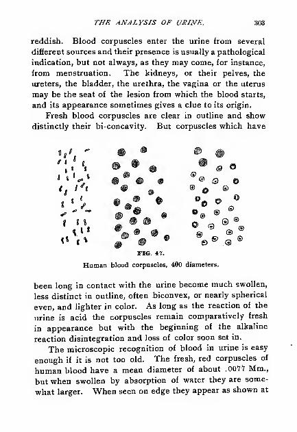
THE ANALYSIS OF URINE. 303
reddish. Blood corpuscles enter the urine from several
different sources and their presence is usually a pathological
indication, but not always, as they may come, for instance,
from menstruation. The kidneys, or their pelves, the
ureters, the bladder* the urethra, the vagina or the uterus
may be the seat of the lesion from which the blood starts,
and its appearance sometimes gives a clue to its origin.
Fresh blood corpuscles are clear in outline and show
distinctly their bi-concavity. But corpuscles which have
m • © #» • % ® @ o
@ © • ® Q ©• 9 ® O ®
• • • % © °
m • © »® ® ®
J ®@ % 8»a
• *• * • •• * o •@ 9 © ®FIG. 47.
Human blood corpuscles, 400 diameters.
been long in contact with the urine become much swollen,
less distinct in outline, often biconvex, or nearly spherical
even, and lighter in color. As long as the reaction of the
urine is acid the corpuscles remain comparatively fresh
in appearance but with the beginning of the alkaline
reaction disintegration and loss of color soon set in.
The microscopic recognition of blood in urine is easy
enough if it is not too old. The fresh, red corpuscles of
human blood have a mean diameter of about .OOV? Mm.,
but when swollen by absorption of water they are some-
what larger. When seen on edge they appear as shown at
1,.f 1

304 THE ANALYSIS OF URINE.
the left in the figure above. If presenting the fiat side to
the eye they appear as discs whose centers grow alternately
light and dark by changing the focus of the instrument.
In old urine, especially with alkaline reaction they appear
as granulated spheres, shown in the center of the figure.
In all cases the color is more or less yellowish. It is gen-
erally assumed that the paler washed out corpuscles come
from lesions higher up, from the pelvis, or kidney even,
while the brighter fresh blood points to a lesion nearer the
point of discharge, that is from the bladder or urethra.
This is pretty certain to be the case if the blood is dis-
charged but little mixed with the urine and settles rapidly
as a distinct mass.
Mucus and Pus Corpuscles.
These are white corpuscles somewhat larger than the
red blood corpuscles and spherical in outline. The term
leucocyte is frequently applied to these as well as to the so-
called white corpuscles of blood. Their size varies greatly
but the average diameter may be given as .009 Mm. All
these corpuscles present when fresh a slightly granular
appearance and occasionally show one or more nuclei.
The addition of a little acetic acid to the sediment brings
the nucleus out distinctly so that it may be seen under the
microscope as a characteristic appearance.
Mucus corpuscles in small number are normally present
in urine, but pus corpuscles enter the urine as a constituent
of pus itself which is an albuminous product discharged
from suppurating surfaces and not normal. In a former
chapter it was shown that the reactions of mucus and
albumin are distinct, but urine containing pus always
affords reactions for albumin. Pus in urine tends to form
a sediment at the bottom of the containing vessel, and maybe recognized by the following method :

THE ANALYSIS OF URINE. 305
Donne's Test. Pour the urine from the sediment and
add to the latter an equal volume of thick potassium
hydroxide solution, or a small piece of the solid potassa.
Stir with a glass rod. The strong alkali converts the pus
into a thick viscid mass closely resembling white of egg.
Sometimes this is so thick that the test-tube containing it
can be inverted without spilling it. In. alkaline urine this
glairy mass is sometimes spontaneously formed.
'.?•'
FIG. 48.
Pus corpuscles, 400 diameters.
The appearance of the mucus or pus corpuscles in
urine depends largely on the concentration of the latter.
In urine of low. specific gravity the corpuscles absorb water
and swell to larger size than normal, while in a highly con-
centrated urine they may give out water and become re-
duced in size and shrunken in appearance.
To recognize them under the microscope transfer a
few drops of the sediment to a slide and cover as usual.
If the nuclei are not distinct place a drop of diluted acetic
acid on the slide at the edge of the cover glass. Part of
the acid will flow under the cover and mix with the urine.
As it does this the clearing up of the corpuscles with ap-
pearance of the nuclei can be very easily followed. Urine

306 THE ANALYSIS OF URINE.
containing much pus is white and milky. The same ap-
pearance is often noticed with an excess of earthy phos-
phates, but the latter clear up with acids while the pus
does not.
Epithelium Cells.
Epithelium cells from different sources may appear
normally in the urine, and the light cloud which separates
from normal urine on standing consists chiefly of these
cells. When present in small amount this epithelium has
usually no clinical importance, as it easily finds its wayinto the urine from the bladder, vagina or urethra. An
FIG. 49.
Scaly and spherical epithelium.
abundance of cells from these organs would, however, be
considered pathological, pointing to a catarrhal condition.
Unfortunately, it is not possible in all cases to deter-
mine the source of the cells, as found in urine, partly be-
cause cells from different localities have frequently the
same general appearance, and partly because, owing to
immersion in the urine, they become greatly changed fromwhat they are in the tissue as shown by the microscopic

THE ANALYSIS OF URINE. 307
study of sections. It is customary to make three roughdivisions of the cells as found in the urine :
1. Spherical cells. 2. Columnar or conical cells. 3.
Flat or scaly cells.
The spherical cells are probably normally much flat-
tened but by absorption of water they become swollenand globular. These cells may be derived from several
sources, as from the uriniferous tubules or from the deeperlayers of the lining membrane of the pelvis of the kidney,or the bladder, or the male urethra.
These cells have a well-defined nucleus resembling that
of a pus cell. But they are much larger, and besides show
FIG. SO.
Conical epithelium.
the nucleus without addition of acid. In nephritis, or
other structural diseases of the kidney, these round cells
are found along with albumin, and their recognition is
then a matter of importance as indicating a breaking downof the tubular walls. Sometimes these cells form a variety
of tube cast, to be described later. But it must be remem-
bered that we cannot distinguish with certainty between
the cells from the tubules and those from the other locali-
ties mentioned.
Conical cells come generally from the pelvis of the kid-

308 THE ANALYSIS OF URINE.
ney, from the ureters and urethra. Some of these cells are
furnished with one or two processes, and are broad in
the middle and taper toward each end, while the others
are broad at the base and taper to a point.
The large flat cells come from the vagina or bladder,
and it is generally impossible to distinguish between them.
Sometimes they are very nearly circular, sometimes irregu-
larly polygonal in outline. Sometimes the vaginal epi-
thelium is found in layers of scales, which appear thicker
and tougher than the cells from the bladder, which occur
singly.
What was said about the decomposition of blood or pus
cells in urine obtains also for the various epithelium cells.
In acid urine they may maintain their distinct outlines
many days, but in alkaline secretion they soon undergo
disintegration, which makes their recognition practically
impossible. In general the greatest importance attaches
to the cells from the tubules of the kidney. The presence
of albumin in more than minute traces in the urine would
suggest that any smaller spherical cells present may have
had their origin in the kidney rather than in the bladder
or male urethra. In general it may be said that urine con-
taining large numbers of the smaller, round tubule cells
with albumin will also show casts.
Mucin Bands
Urine containing much mucus sometimes exhibits a de-
posit consisting of long threads or bands, curved and bent
in every direction. These bands are important because
they are sometimes confounded with the tube casts to be
described next. They can be produced in urine highly
charged with mucus by the addition of acids, and appear
therefore sometimes spontaneously when the urine becomesacid. These threads are sometimes covered with a fine

THE ANALYSIS OF URINE. 309
deposit of granular urates and these bear some resem-
blance to granular casts. In general, however, they are
relatively longer and narrower than the true casts of the
uriniferous tubules. The mucin threads can occur and
frequently do occur in urine entirely free from albumin,
while true tube casts are usually associated with albumin,
although not always, as will be explained below. The
1^:FIG. 51.
Mucin bands or threads.
length and shape of the mucin threads may generally be
relied upon to distinguish them from true casts.
Casts.
The structures properly termed casts are seldom found
in urine which does not contain albumin. They are
formed in the uriniferous tubules, and to a certain extent,
are "casts" of portions of the same. Their specific
gravity differs but little from that of the urine, for which
reason they remain long in suspension. It is therefore
necessary to allow the urine to stand some hours at rest,
over night or longer, before attempting an examination.
Casts of the uriniferous tubules rarely appear in normal

310 THE ANALYSIS OF URINE.
urine and their recognition is therefore a matter of the high-
est importance in diagnosis. Much has been written on
the subject of the origin of these bodies in the kidney and
several theories have been advanced to account for their
formation and chemical constitution. Most of this discus-
sion would be out of place in a work like the present deal-
ing mainly with questions of analysis, but enough will
be given to aid the student in his examinations. It must
be said that few subjects are more perplexing to the begin-
ner than that of their certain recognition, because of the
fact that some varieties are so transparent as to be almost
invisible, while others are closely resembled by formations
0)
*-'
FIG. 53.
Epithelium casts. To the right a bunch of urates or false cast.
of entirely different nature not pathological. With prac-
tice, however, these difficulties can be surmounted.
Most of the bodies termed casts are formed of organ-
ized structures or the remains of such, but another andrather common form consists of crystalline matter, usually
uric acid or fine granular urates.
These bunches of urates have no pathological signifi-
cance and are of frequent occurrence. Urine containing
them clears up by heat and the deposits themselves are

THE ANALYSIS OF URINE. 311
dissipated by weak alkali. While it is true that they re-
semble to some degree the so-called granular casts referred
to below there are certain well-defined points of difference.
The bunches of urates lack the coherence which can be ob-
served in the true casts, and besides the granulation is
finer and more clearly defined.
The fact that mucin bands occasionally appear cov-
ered with a precipitate of granular urates has been re-
ferred to. These aggregations are more compact than the
loose bunches of urates just mentioned and much longer
FIG. 53.
Blood casts and granular casts.
generally. They are also darker and therefore more easily
seen than are the casts proper or the urates.
The true casts are made up of matter in which evidence
of cell structure or transformation is visible. An accurate
classification of these bodies cannot yet be made, and, as
said, authors differ regarding the importance of several
forms and their origin. But for our purpose it will be suf-
ficient to make the following rough division, which accords
in the main with what is found in the text-books of urine
analysis:

312 THE ANALYSIS OF URINE.
J, Blood casts; 2, Epithelium casts; 3, Granular casts;
4, Fatty casts; 5, Waxy casts; 6, Hyaline casts.
What are termed blood casts consist of or contain co-
agulated blood recognized by the corpuscles. Plugs of
this coagulated matter are forced out from the tubules by
pressure from behind, and form one of the most character-
istic varieties of casts. They are generally very dark in
color, and easily distinguished from other matter. A repre-
sentation of blood casts is given in the preceding cut.
In epithelium casts the characteristic substance is the
lining epithelium of the tubule. Sometimes this lining
epithelium becomes detached in the form of a hollow cyl-
inder, the walls consisting of the united cells. Again, the
coagulated contents of the tubule in passing out may carry
the epithelium with it as a coating. In either case a grave
disorder of the kidneys is indicated, as acute nephritis, or
other disease in which a profound alteration of the internal
structure of the organ is involved.
What are termed granular casts, proper, appear in a
variety of forms, produced probably by the disintegration
of blood or epithelium casts.
There is no uniformity in the fineness of the granulation;
sometimes a high amplification is necessary to disclose the
structure. Occasionally blood corpuscles, epithelium, fat
globules and crystals can be detected in them, and whenderived from blood cast disintegration they usually have
a yellowish red color, which makes their recognition com-
paratively easy. In outline they are generally regular,
with rounded ends, one of which is somewhat pointed.
Frequently, however, they appear to be broken, the ends
showing irregular fracture.
Fatty casts contain oil drops produced by some variety
of fatty degeneration of the tissues of the kidney. These
oil drops may form coherent bunches, or they may be held

THE ANALYSIS OF URINE. 313
by patches of epithelium. It also happens that epithelium
or granular casts may be partially covered by oil drops.
The name fatty cast is applied to those in which the fat
globules predominate. Along with these globules the
microscope sometimes shows crystals of free fatty acids,
and probably also of soaps containing calcium and mag-
nesium.
Waxy casts consist of the peculiar matter produced by
amyloid degeneration of the kidney. They have a glisten-
ing waxlike or vitreous appearance, and refract light very
i'i%/
fi-j
FIG. 54.
Waxy and hyaline casts.
strongly. Sometimes they reach a great length, and they
frequently are found with blood corpuscles or oil drops on
the surface. They have been detected in several renal
disorders. Illustrations are given to the left above.
True hyaline casts are nearly transparent and hard to
see unless the illumination is very carefully managed. To
detect them it is often necessary to add a few drops of a
dilute solution of iodine in potassium iodide to the sedi-
ment. This imparts a slight color which renders them
visible.
The hyaline casts seem to be formed by the passage of

314 THE ANALYSIS OF URINE.
homogeneous matter from the tubules, leaving the epithe-
lium behind. A cast is rarely perfectly hyaline, as at least
an occasional blood corpuscle, fat globule or epithelium cell
will usually be found attached to it. Waxy casts may be
looked upon as a special form of hyaline casts. Very im-
perfect representations are given in the above cut.
In general, it must be said that the representation of
these casts on paper is a very difficult matter. Ordinarily
they are drawn and printed much too heavy and dark.
Hyaline casts do not necessarily indicate kidney disease,
although this is usually the case. They have been found in
urine free from albumin and under circumstances not con-
nected with renal disorders.
The preservation of sediments containing casts is un-
usually difficult because of the nature of the material to be
preserved. In urine of the slightest alkalinity their dis-
integration soon begins so that the outlines are rendered
indistinct, often making identification impossible.
For temporary preservation the addition of chloroform
renders as good service as anything else. Many other sedi-
ments can be permanently mounted and kept for future
comparison but with casts this can rarely be done.
Beginners are apt to overlook casts in their first examin-
ations. It must be remembered that some of them are
nearly transparent and unless brought into proper focus
they may not be seen at all. At the outset students usu-
ally employ too bright a light in looking for casts. While
no specific directions can be given regarding the inten-
sity of illumination best suited to the purpose, this may be
said that the light commonly found necessary in studying
ordinary histological slides is far too bright to use in the
search for casts.
Practice alone, first under the direction of the instruc-
tor, will indicate what is proper here.

THE ANALYSIS OF URINE. 315
Spermatozoa.
These minute bodies, as found in the semen of man,have a mean length of about .050 Mm. Nearly one-tenth
of this is in the head portion. When observed in recently
discharged semen they have a characteristic spontaneousmovement by which they are propelled forward rapidly.
This motion is soon lost if the semen is diluted with wateror similar liquid. Hence, as usually seen in urine they are
entirely motionless. They are found abundantly in the
urine of men after coitus or nocturnal emissions, and also
FIG. 55.
Human spermatozoa.
in spermatorrhoea, when their presence is continuous and
characteristic.
In the urine of women they are likewise found after con-
nection, and their detection here has often interest from
the medico-legal standpoint. The proof of rape can often
be established by the recognition of spermatozoa.
Although their motion is soon lost in foreign liquids the
substance itself of the spermatozoa is not readily destroyed.
In this respect they are more permanent, probably, than all
other organized structures found in the urine and can be
readily distinguished after many days or months even in
urine, or in sediments which have become dry.
For their recognition a power of 250 diameters is suffi-
cient, but 350 to 400 diameters is preferable. With this

316 THE ANALYSIS OF URINE.
higher amplification it is possible to readily distinguish
between spermatozoa and certain fungus growths bearing
some resemblance to them.
Cancer Tissue.
Fragments of cancerous and other morbid growths are
occasionally met with in the urine, and when certainly
recognized are an important aid in diagnosis.
The recognition of cancer cells along with the several
kinds of epithelium cells found in urine is difficult and lies
somewhat beyond the usual line of urine examinations.
FIG. 56.
Cancer cells which have been seen in urine.
Sometimes the identification is comparatively simple
because of the unusual shape of the cells, or when they are
present in great number, but this is not often the case.
The cut shows some of these cells figured by Beale.
Fungi.
The urine sometimes contains certain fungus growthsthe recognition of which is important. These may haveentered the urine after voiding or they may have comefrom the bladder.
Normal urine when passed is probably free from fungi
of all kinds, but in a short time certain organisms enter it

7WE ANALYSIS OF URINE. 317
from the air or from other sources and become active in
producing in it characteristic changes.
The three important groups of fungi, the schizomycetes or
bacteria, the hyphomycetes or moulds and the blastomycetes or
yeasts are represented in the organisms sometimes found in
the urine. The conditions under which they are found will
be briefly explained.
Of the bacteria the following have been observed :
Micrococcus Ureae. This is the exceedingly commonform found on urine undergoing alkaline fermentation by
which urea is converted into ammonium carbonate. It is
usually introduced from the air and multiplies very rapidly
under ordinary conditions. Nearly all old specimens of
urine, unless containing some active preservative are
found infected with this small organism. The micrococci
are minute spherical bodies belonging to the suborder
sphmrobacteria, and are found separate or in chains. Theyare the smallest of the organized forms occurring in urine
and appear under a power of 250 diameters but little more
than points. While generally finding their way into urine
after it has been voided they are occasionally present in
the bladder. It is usually held that under such circum-
stances they have been introduced by a dirty catheter or
sound, although cases are on record where this has not
been proven.
In the bladder they give rise to alkaline fermentation,
so that the voided urine may show ammonium carbonate
directly.
Streptococcus Pyogenes is a pathogenic form some-
times found in the urine in cases of infectious diseases.
Sarcinae. The genus sarcina is frequently classed with
the sphaerobacteria and several species have been found in

318 THE ANALYSIS OF URINE.
urine. The cells are larger than those of micrococcus
ureae, and are arranged in groups of two or four usually.
They are not pathogenic.
Bacilli. Several species of the genus bacillus are found
in urine in disease. The most important of these are the
typhoid bacillus, bacillus typhi abdominalis, the tubercle
bacillus, bacillus tuberculosis and the bacillus of glanders,
bacillus mallei. These bacilli occur in urine only during
the progress of the corresponding diseases and their detec-
tion is of the highest interest. A description of the
m >."«»%& \\\h
M«i«
*
tfS *
FIG. 57.
Micrococci and other bacteria.
methods to be followed for the certain demonstration of
these bodies is not within the scope of this book, but must
be looked for in the laboratory manuals of bacteriology.
Spirilla. Certain species of the genus spirillum have
been found in urine. The best known of these is the
spirillum of relapsing fever, spirillum Obermeieri. This is
only found rarely and as its habitat is the blood of relaps-
ing fever patients it must enter the urine through a haemor-
rhage into the kidney. Its form is that of a long, wavyspiral, which makes its detection somewhat easy.

THE ANALYSIS OF URINE. 319
Although not pathogenic it is well to call attention to
certain moulds which may sometimes be seen in urine. Thecommon blue-green mould, penicillium glaucum, is the best
known of these, and is occasionally found in urine along
with yeast cells. Another mould which has been found in
urine is the oidium lactis, commonly occurring in milk and
butter. It has been observed in fermenting diabetic urine.
Both of these fungi enter the urine after voiding. In
urine which has stood sometime in a cool place the penicil-
lium glaucum sometimes becomes covered with an incrusta-
tion of urates or minute crystals of uric acid.
QO(f
FIG. 58.
Yeast cells and common mould.
Finally we have yeast cells in urine and sometimes in
great numbers. Like other fungi they enter the urine
from the air and when not very abundant have no signifi-
cance. In great numbers the yeast cells suggest presence
of sugar. The ordinary yeast plant, saccharomyces cerevisice,
is shown, isolated and budding, in the accompanying
figure.
Other Parasites.
Besides fungi the urine in rare cases contains other
parasites of animal as well as vegetable nature. Some of

320 THE ANALYSIS OF URINE.
these enter it accidentally from the air and have no inter-
est, but a few are present in the urine when passed, and
are of importance. Among these are certain thread worms,
the eggs of worms and hooklets of a species of tape-
worm, the taenia echinoeoccus.
These forms can appear in the urine only through rup-
ture from some other organ, and while rare here are com-mon enough in Egypt and other tropical countries. Urine
containing eggs of worms, the worms themselves or frag-
ments usually contains blood or other evidence of rupture.

Chapter XVIII.
UNORGANIZED SEDIHENTS AND CALCULI.
Unorganized Sediments.
Uric Acid.
A MONG the more common of the unorganized sediments**• found in urine this must be mentioned first. As wasexplained in the last chapter uric acid occurs normally in
combination in all human urine.
Some time after its passage urine often undergoes what
has been spoken of as the acidfermentation by which a pre-
cipitate of urates and even free uric acid may appear. This
reaction is in no sense due to a ferment process in the ordi-
nary sense of the term, but is probably brought about by a
purely chemical double decomposition. Urine contains
acid sodium phosphate and neutral sodium urate and it has
been suggested that these react on each other according to
the following equation:
Na2C
6H
2N 4 3
+NaH 2P0 4 =NaC 5
H3N 4 3 + Na
gHP0 4
The precipitate of acid urate settles out and forms a
light reddish deposit. If the amount of acid phosphates
present is excessive the reaction may go still further, re-
sulting in the precipitation of free uric acid. The well char-
acterized crystals of uric acid are often found with the
sediment of fine urates. Sometimes this liberation and pre-
cipitation of the acid takes place in the bladder and the
urine as passed shows the crystals or "gravel." If they are
relatively large, which is sometimes the case, their passage
through the urethra may cause severe pain.

322 THE ANALYSIS OF URINE.
As the illustrations show uric acid occurs in a great
variety of forms. The rosettes and whetstone shaped crys-
tals are probably the most common, while long spiculated
forms are frequently seen. Pure uric acid is colorless but
as deposited from urine it is always reddish yellow, be-
cause of its property of carrying down coloring matters.

THE ANALYSIS OF URINE. 323
The crystals are often so large that their general form can
be seen by the naked eye; usually, however, they are
minute.
Uric acid crystals when once deposited are not readily
redissolved by heat, but they go into solution by the addi-
tion of alkali. If the urine contains extraneous matter, as
specks of dusts, bits of hair, cotton or wool fibers the crys-
tals are very apt to deposit on them.
Urates.
The common fine sediments of urine are usually urates
or amorphous phosphates. They can be most readily dis-
A
B
FIG. 61.
Common crystalline and granular urates.
tinguished by their behavior with acids and on application
of heat. Urates disappear on warming the urine contain-
ing them, while a phosphate sediment is rendered more
abundant. A urate sediment is little changed by acids,
while the phosphates dissolve completely if the urine is
made acid in reaction with hydrochloric or nitric acid. The
acid urates of sodium and ammonium are the must abun-
dant and are shown in the cut. Acid ammonium urate may

324 THE ANALYSIS OF URINE.
exist in urine which has become alkaline from the decom-
position of urea and formation of ammonium carbonate,
and may therefore be seen in company with the phosphate
sediments. The other urates dissolve in alkaline urine.
Like uric acid the urates appear in a great variety of forms,
and there is still some uncertainty about the composition
of some of their crystals which have been found in urine.
Leucin and Tyrosin.
These two substances are of rare occurrence in urine
and appear only under pathological conditions. Urine con-
taining them shows usually strong indications of the pres-
ence of biliary matters as they generally are found in con-
sequence of some grave disorder of the liver in which de-
struction of its tissue is involved. They have been most
frequently found, and associated, in acute yellow atrophy
of the liver and in severe cases of phosphorus poisoning.
In general they must be considered as products of disinte-
gration and are produced in the intestine in large quantity
by bacterial agency in the last stages of the digestion of
albuminoids, as was pointed out in an earlier chapter.
As both bodies are slightly soluble they may not be seen
directly, but only after partial concentration of the urine.
In pure condition leucin crystallizes in thin plates but
from urine it separates in spherical bunches made up of
fine plates or needles. These bunches are sometimes so
compact that it is hard to distinguish between them and
other substances, particularly lime soaps and oil drops.
Chemical tests must therefore be applied. If mercurous
nitrate is added to a leucin solution and the mixture is
warmed metallic mercury precipitates. This test can be
carried out only when the substance is abundant enough to
be purified by crystallization from hot water. Pure leucin
when strongly heated with nitric acid on platinum forms a

THE ANALYSIS OF URINE. 325
colorless residue, which when heated with potassium hy-
droxide leaves an oil-like drop that does not wet the plati-
num.
Tyrosin is usually seen in long needles, which some-times are bunched in the form of sheaves and more readily
recognized than is leucin. Tyrosin heated with nitric
acid on platinum turns orange yellow, and leaves a dark
residue which becomes reddish yellow by addition of
caustic alkali. Solutions containing tyrosin when treated
FIG. 63.
Leucin spheres, tyrosin needles and cystin plates.
hot with mercuric nitrate and potassium nitrite turn red
and finally throw down a red precipitate.
Leucin and tyrosin may be present in the urine yet not
show as a sediment. For their detection under these cir-
cumstances precipitate the urine with basic lead acetate
and filter. Separate the excess of lead from the filtrate by
hydrogen sulphide and filter again. Concentrate the fil-
trate on a water-bath to a syrup and treat it with a little
absolute alcohol to remove urea. Some leucin may dissolve

326 THE ANALYSIS OF URINE.
with the urea in this treatment. Next boil the residue with
60 to 70 per cent alcohol, filter, concentrate the filtrate to a
small volume and allow it to stand in a cool place for crys-
tallization. If crystals appear their form indicates whether
the\' consist of pure tyrosin or a mixture of tyrosin and
leucin. The latter, being more soluble in strong alcohol,
can be separated by washing with this liquid. The final
tyrosin residue can be used for the tests given above.
The alcoholic solutions may contain leucin, which can
be recognized after evaporation. Both leucin and tyrosin
decompose readily in urine undergoing putrefactive
changes; it is therefore necessary to apply the test to urine
as fresh as possible.
Cystin.
This is a rare sediment, although it is found constantly
in the urine of certain individuals. It crystallizes in thin
hexagonal plates, small ones sometimes resting upon or
overlapping large ones. The crystals are regular in form
but variable in size and readily recognized. A rare form
of uric acid crystallizes in a somewhat similar manner but
the two substances differ in their behavior toward am-
monia. To distinguish between them in the microscopic
test place a drop of ammonia water on the slide and allow
it to pass under the cover glass. Cystin dissolves, but,
unless heated, uric acid does not. When the ammoniaevaporates cystin reprecipitates.
Cystin is precipitated from urine by addition of acetic
acid. Mucin and uric acid may come down at the sametime. The precipitate is collected on a filter, washed with
water and finally dissolved in ammonia. By neutralizing
the ammoniacal filtrate with acetic acid and concentrating
a little, it comes down in the characteristic form suitable
for microscopic recognition.

THE ANALYSIS OF URINE.
Cholesterin.
327
This substance occurs occasionally in urine, and pos-
sibly only in cases of cystitis. It is recognized by its
characteristic crystalline form, large, thin plates, shownin the following cut. These plates are nearly transparent
but from their size cannot be overlooked.
FIG. 63.
Cholesterin plates and fat globules.
Fat Globules.
These are often seen in urine, but in most cases have
not been voided with it. They can come from several
extraneous sources, as from a catheter, from vessels in
which the urine is collected or sent for examination, from
admixed sputum, etc., which facts should be borne in mind.
Fat has been found in cases of fatty degeneration of the
kidney and more abundantly in chyluria where communi-
cation seems to be formed between the lymphatics and the
urinary tract by the invasion of the small thread wormsreferred to above.
Hippuric Acid.
This acid is found normally in human urine in small
amount. It may be found in large quantity after taking

328 THE ANALYSIS OF URINE.
benzoic acid and may even appear in crystalline form in the
sediment. It has no pathological importance, ordinarily.
Calcium Carbonate.
This is sometimes observed as a coarse, granular sedi-
ment which dissolves with effervescence in acetic acid. It
occasionally forms dumb-bell crystals, and is devoid of path-
ological importance.
Calcium Sulphate.
Crystals of this substance are rarely found in urine.
They form long colorless needles or narrow, thin plates.
Calcium Oxalate.
We have here one of the commoner of the crystalline
bodies observed in urine.
This may be found in neutral or alkaline urine, but
more commonly in that of acid reaction. It occurs nor-
mally and sometimes is very abundant, especially after the
consumption of vegetables containing oxalic acid.
Two principal forms of the crystals are found, the octa-
hedral and dumb-bell crystals.
The octahedra have one very short axis which gives
the crystals a flat appearance. When seen with the short
axis perpendicular to the plane of the cover glass, which is
the common position, they appear as squares crossed by
two bright lines. Sometimes they are seen on edge, and
then present a rhomb in section with one diameter very
much shorter than the other.
A form of triple phosphate bears a slight resemblance
to calcium oxalate, but it is soluble in acetic acid, while
the oxalate is not.
The dumb-bells are much less common than the octa-
hedra, and are found in several modified forms, as shownin one of the figures.

THE ANALYSIS OF URINE. 329
The clinical significance of the oxalate is not clearly
understood; It does not seem to be characteristic of any
disease even when occurring in quantity. It has been
found considerably increased in dyspeptic conditions, but
not always, and many of the statements found concerning
its significance seem to have been based on insufficient
observations.
Urine may contain a large amount of oxalic acid which
does not show as a sediment, but must be found by pre-
FIG. 64.
Calcium oxalate.
cipitation by calcium chloride in presence of ammonium
hydroxide. Acetic acid is then added in very slight excess
and the mixture is allowed to stand for precipitation.
The constant or prolonged excretion of large amounts
of oxalic acid is spoken of as oxaluria.
The Phosphates.
It was explained in Chapter XVI. that phosphates of
alkali and alkali-earth metals occur normally in the urine,
and a method was given for their measurement. As sedi-

330 THE ANALYSIS OF URINE.
ment we know several forms of calcium and magnesium
phosphates and the microscopic detection of these will be
here explained. In normal fresh urine of acid reaction these
phosphates are held in solution, but if the urine as passed
is alkaline it is often turbid from the presence of basic
phosphates held in suspension. Urine which has stood
long enough to undergo the alkaline fermentation always
contains phosphates in the sediment. Finally, it must be
remembered that a neutral or very slightly acid urine, con-
FIG. 65.
Triple phosphate.
taining ammonium salts in abundance, may also deposit
a crystalline precipitate of ammonium magnesium phos-
phate. The common phosphate sediments are those con-
sisting of ammonium magnesium phosphate (triple phos-
phate), basic magnesium phosphate, neutral calcium
phosphate and mixed amorphous phosphates of calcium
and magnesium.
Triple Phosphate. Of the crystalline phosphate de-
posits this is the most abundant and at the same time the
most characteristic.

THE ANALYSIS OF URINE. 331
The crystals are the largest found in urine, and from
their shape are sometimes spoken of as coffin lid crystals.
Ordinarily they are not found in perfectly fresh urine, but
after it has undergone the alkaline fermentation they are
generally present in profusion.
Basic Magnesium Phosphates. Crystals having the
composition Mg3(P0
4 ) 2.22H
3are sometimes found in
urine of nearly neutral reaction. They consist of thin,
FIG. 66.
Neutral calcium phosphate and amorphous phosphate.
transparent rhombic plates with angles of approximately
60° and 120°. If urine containing this sediment becomes
alkaline triple phosphate forms.
Neutral Calcium Phosphate. This has the compo-
sition CaHP0 4.2H
zO and is found in urine of neutral or
slightly acid reaction. It crystallizes frequently in rosettes
formed of wedge-shaped single crystals uniting at their
apices. The cut above shows some variations in the form.
Amorphous Phosphates. Finally we have the very
common, finely granular earthy phosphates in amorphous
condition. This sediment dissolves readily in weak acetic

332 THE ANALYSIS OF VRINE.
acid and is colorless. The common amorphous urate sedi-
ment is colored and does not dissolve in acetic acid. Onaddition of sodium carbonate or hydroxide to urine the pre-
cipitate which forms consists mainly of this phosphate.
These several phosphates can be produced artificially
and should be made for study and comparison. Theneutral (basic) magnesium phosphate can be made by dis-
solving 15 Gm. of crystallized common sodium phosphate
in 200 Cc. of water and mixing this with 3.7 Gm. of crystal-
lized magnesium sulphate in 2000 Cc. of water. Enoughsodium bicarbonate is added to give an amphoteric reac-
tion and then the mixture is allowed to stand a day or
more for precipitation.
Crystals of triple phosphate of peculiar form are often
obtained by adding ammonia to urine, and sometimes a
trace of ammonia is sufficient to throw down the crystals
of neutral calcium phosphate. The latter can also be ob-
tained by adding to a weak solution of crystallized sodium
phosphate a trace of acid and then a very little calcium
chloride solution.
Foreign Matters.
The sediment of urine often contains foreign sub-
stances which have become mixed with it accidentally.
The most common of these are hairs, woolen, cotton or
silk fibers, granules of starch, fat globules, dust and sand
granules, bits of woody fiber and remains of articles of
food. Some of these are represented in the cuts on the
following pages.
Urinary Calculi.
Calculi like the sediments just described are formed bythe precipitation of certain substances from the urine, but
in compact form. Occasionally a calculus consists of a
single substance, as calcium oxalate or cystin, but in the

THE ANALYSIS OF URINE. 333
great majority of cases a mixture of bodies is present,
these being deposited usually in layers around a nucleus
which serves as the foundation of the concretion. Calculi
are built up much as certain forms of crystals are by suc-
cessive depositions on a nucleus. Uric acid is a very
common nucleus on which may be deposited urates,
phosphates, organic matters, etc.
Calculi are sometimes distinguished as primary or sec-
ondary. Primary calculi may be traced to an alteration of
Z, linen fibers; H, hemp fibers; J, jute fibers; B, cotton fibers; S, silk
fibers; A, alpaca wool; E, fine wool; W, common wool.
the urine of such a nature that its reaction is constantly
acid. The foundation for the concretions in this case is
found in the kidney and they are built up of such sub-
stances as most easily deposit from acid urine. Secondary
calculi are generally formed in the bladder and have as
nuclei matters precipitated from alkaline urine, coagulated
blood or other organic substances. Sometimes fragments
introduced into the bladder from without serve as the foun-
dation for these secondary formations. Bits of catheters,
remains of bougies, and other things have been found as

334 THE ANALYSIS OF URINE.
the nuclei around which concretions have formed. Therecognition of the nucleus is a matter of the first impor-
tance as this gives a clew to the determining cause active
in the formation of the calculus.
In making an examination, then, of a calculus, it is
first cut in two by means of a very sharp thin saw. This
exposes the nucleus which may often be recognized by the
eye alone. If one of the halves be polished it is often
possible to discern distinctly the various layers grouped
around the center.
FIG. 68.
Left, pubic hair with spermatozoa; center, hair of woman's head;right, cat's fur.
In a large number of cases examined by Ultzmann
about 80 per cent were found to contain uric acid as the
nucleus.
Chemical Examination.
In the chemical examination of a calculus several
methods may be employed. We may begin by applying
certain preliminary tests designed to show the general na-
ture of the stone.
Heat Test. Reduce some of the calculus to a pow-der and heat to bright redness on platinum foil. Two cases
may arise: (a), the powder is completely consumed; (b),
the powder is only partially consumed or not at all.

THE ANALYSIS OF URINE. 335
Case (a.) If this is the result of the incineration the
following substances may be suspected:
Uric Acid, which may be recognized by dissolving a
little of the powder in weak alkali, precipitating by hydro-
chloric acid and examining the precipitate by the micro,
scope.
Ammonium Urate. This gives the above reaction
under the microscope, and is further recognized by the lib-
eration of ammonia when heated with a little pure sodiumhydroxide solution.
Cystin. Dissolve some of the powder in ammonia, fil-
ter if necessary and allow drops of the filtrate to evaporate
spontaneously on a slide. Cystin is then recognized by the
microscope as already explained. Cystin contains sulphur
which on burning on the platinum foil gives rise to a disa-
greeable sharp odor. If a little of the powder be heated
with a mixture of potassium nitrate and sodium carbonate
the sulphur is oxidized to sulphate, which may be recog-
nized by the usual tests.
Xanthin. This is a rare substance in calculi. Those
consisting wholly of xanthin are brown in color and take a
wax-like polish. A method of recognition will be given
below.
Organized Matter. Parts of blood cells, epithelium,
precipitated mucin, pus corpuscles and similar substances
may become entangled with the growing stone and even
form a large part of it. On burning, these bodies are rec-
ognized by the characteristic odor of nitrogenous matter.
Case (i.) When an incombustible residue is left on the
platinum foil the stone may contain the following constitu-
ents:

336 THE ANALYSIS OF URINE.
Calcium Oxalate. Stones of this substance are very
hard and break with a crystalline fracture. They are often
called "mulberry calculi." When the powder is heated it
decomposes, leaving carbonate, which may be recognized
by its effervescence with acids.
Calcium and Magnesium Phosphates. These leave
a residue in which the metals and phosphoric acid may be
detected by simple tests of qualitative analysis. The ig-
nited powder is soluble in hydrochloric acid without effer-
vescence. When ammonia is added to this solution in
quantity sufficient to give an alkaline reaction a precipitate
of triple phosphate or calcium phosphate appears, which
may be recognized by the microscope.
The above tests are generally sufficient to tell all that
is practically necessary about the calculus. If more de-
tailed information is desired a systematic analysis may be
made according to the following scheme.
Systematic Analysis. 1. Reduce the calculus to a
fine powder and pour over it some water and finally dilute
hydrochloric acid in a beaker. Warm gently half an hour,
or longer, on the water-bath. Then allow to cool and
filter.
2. Treatment of the residue. It seldom happens that
the calculus is completely soluble in the weak acid. Aresidue usually remains which may contain uric acid, xan-
thin, calcium sulphate, and remains of organized matter.
To prove the xanthin treat the residue with warm dilute
ammonia and filter. The filtrate contains the xanthin if
it is present. Acidify it with nitric acid and add a small
amount of silver nitrate solution. This produces a floccu-
lent precipitate which dissolves by warming, and crystal-
lizes on cooling in bunches of fine needles.
In the residue free from the xanthin look for calcium

THE ANALYSIS OF URINE. 337
sulphate by extracting with water and applying the usualtests. This solution may contain uric acid which is recog-nized by evaporation and crystallization after adding a lit-
tle hydrochloric acid. In the final residue some uric acidmay be also present. Dissolve in alkali, reprecipitate withhydrochloric acid, and examine any crystals which mayform under the microscope.
3. Treatment of the hydrochloric acid solution. Thismay contain calcium oxalate, cystin, the phosphates, andpossibly some xanthin. Look for the last in a small por-
tion of the solution. Make this portion alkaline with am-monia, add a few drops of calcium chloride solution, filter
if a precipitate forms and treat the filtrate with ammoni-acal silver nitrate solution. In presence of xanthin a floc-
culent precipitate forms.
Dilute the remaining and larger portion of the hydro-
chloric acid solution with twice its volume of water, addenough ammonia to give a strong alkaline reaction and
then acetic acid to restore a weak acid reaction. By this
treatment phosphates are held in solution while calcium
oxalate, if present, precipitates. Therefore allow the mix-
ture to stand half an hour and then filter off any precipi-
tate which appears. This precipitate may contain cystin
as well as calcium oxalate. Cystin may be dissolved
by pouring ammonia on the filter, and on evaporating the
ammoniacal solution is obtained in form suitable for mi-
croscopic examination.
The residue free from cystin is dried and heated to red-
ness on platinum foil. This treatment converts calcium
oxalate into carbonate. Place the foil in a beaker and add
some dilute acetic acid; an effervescence shows the car-
bonate. To the clear solution add next some ammoniumoxalate which gives a white precipitate of calcium oxalate,
if the latter metal is present.

338 THE ANALYSIS OF URINE.
We have next to look for the phosphates and bases in
the acetic acid solution obtained after filtering off cyst in
and calcium oxalate. More calcium may be present, in
excess of that combined as oxalate, which may be recog-
nized by adding a little solution of ammonium oxalate. If
a precipitate forms treat the whole of the liquid with the
ammonium oxalate, after warming on the water-bath, allow
to stand an hour and filter. Concentrate the filtrate in
platinum to a small volume, transfer to a large test-tube and
add enough ammonia to produce an alkaline reaction. If
a precipitate appears now it must consist of magnesiumphosphate, showing both magnesium and phosphoric acid
as present in the original. If no precipitate appears mag-
nesium is absent but phosphoric acid may still be present.
To find it divide the ammoniacal liquid into two portions.
To one add a few drops of magnesia mixture, and to the
other add nitric acid in slight excess and then ammoniummolybdate reagent. Both tests should yield the reactions
characteristic of phosphates, if present.
This procedure serves for the recognition of the im-
portant constituents of calculi. But ammonium, potassium
and sodium compounds are sometimes present and may be
recognized readily. To detect ammonium salts the original
calculus powder may be heated with pure potassium
hydroxide solution, or the hydrochloric acid solution of the
calculus may be neutralized and heated with the samesolution. The ammonia is recognized by the odor or byits reaction on moistened red litmus paper.
To recognize the alkali metals a solution of the
powdered calculus in hydrochloric acid is treated withpure ammonia and a little ammonium carbonate in excess.The precipitate formed is allowed to settle and filtered off.
The filtrate is then evaporated to dryness in a platinumdish and the residue strongly heated to drive off all am-

THE ANALYSIS OF URINE. 339
monium salts. What is now left contains sodium and
potassium if they were present in the original. Moisten
this final residue with water and a drop of hydrochloric
acid and test with a platinum wire in the flame of a Bun-
sen burner, using a deep blue glass when looking for
potassium. Only a very intense yellow color can be taken
as indicative of sodium.


APPENDIX.
Test Solutions and Tables.


APPENDIX. 343
TABLE OF ATOMIC WEIGHTS.
(ACCORDING TO MEYER AND SEUBERT.)
Aluminum[
AlAntimony SbArsenic i AsBarium BaBeryllium . .
.
BeBismuth BiBoron i BBromine BrCadmium CdCaesium
|Cs
Calcium CaCarbon CCerium
j
CeChlorine CIChromium CrCobalt
;
CoColumbium CbCopper CuDidymium DiErbium ErFluorine FGallium GaGermanium GeGold AuHydrogen
;
HIndium InIodine I
Iridium Ir
Iron\
FeLanthanum
!La
Leadj
PbLithium Li
Magnesium MgManganese I
MnMercury. I Hg
AtomicWeight.
27.04119.674.9136.9
9.03208.910.979.76111.5132.739.9111.97
139.935.3752.058.693.763.18142.0166.019.069.972.3196.71.0
113.6126.53192.555.88138.2206.4
7.0124.354.8199.8
Name.

344 APPENDIX.
List of General Reagents and Test Solutions.
Acid, sulphuric (strong). The commercial acid is sufficient for.
most purposes, where a strong acid is called for. Where the pure acid
is required it is mentioned in the text.
Acid, sulphuric (dilute). Add one part of the above acid to four
parts of distilled water, mix thoroughly, and allow to stand twenty-fourhours. Then siphon, or pour off the clear liquid, which is ready for use.
The strong acid must be poured into the water very slowly, and with
constant stirring, to avoid a too sudden elevation of temperature.
Acid, nitric (strong). The strong commercial acid can be employedin most cases where this acid is called for. A pure strong acid is also
employed, occasionally.
Acid, nitric (dilute). Where this acid is called for as a reagent it
should be made by mixing one part of the pure strong acid with four
parts of distilled water It should be free from traces of chlorine andsulphates.
Acid, hydrochloric (strong). The yellow commercial acid is large-
ly used in the laboratory in the preparation of other substances It is
seldom pure enough to be employed as a test reagent.
-A colorless acid, free from organic matter, iron and traces of sul-
phates, must be used when the pure strong acid is called for.
Acid, hydrochloric (dilute.) This is frequently used in liberat-
ing hydrogen sulphide, carbon dioxide, and hydrogen, and for other pur-poses, and need not be pure. Where the dilute acid is called for as areagent it must be made by mixing one part of the pure strong acidwith four parts of distilled water.
Acid, acetic. Mix one part of the pure "glacial " acid with fourparts of water.
Ammonia water. The strong solution is seldom used in analysis.
The solution usually employed is made by mixing one volume of thestronger ammonia water (containing about 28 per cent of the gas) withthree volumes of distilled water.
The solution should be free from carbonic acid, as presence of this
would interfere with several of the tests where it is employed.
Ammonium carbonate. Dissolve one part of the pure powderedcrystals in five parts of dilute ammonia water.
Ammonium chloride. Dissolve one part of the pure salt in tenparts of water.
Ammonium molybdate. A solution of this salt is chiefly used as atest for phosphoric acid, and should be prepared in this way : Dissolve3 Gm of the crystals in 20 Cc. of water, and pour this solution in 20Cc. of strong nitric acid. Warm the mixture to about 40° C. (notabove), and allow to settle.

APPENDIX. 345
As the reagent does not keep well, it should be made only in smallquantities.
Ammonium oxalate. Dissolve one part of the pure crystals intwenty parts of water.
Ammonium sulphide. Dilute a quantity o£ strong ammonia waterwith an equal volume of water. Take three-fifths of this mixture andsaturate it with hydrogen sulphide. Then add the remaining two-fifthsof the diluted ammonia water and mix thoroughly.
Barium chloride. Dissolve one part of the crystals in ten parts ofwater.
Calcium hydroxide. Slake pure white lijne and pour over it alarge excess of water. Allow to settle and throw away the clear liquid.Again add pure water, shake thoroughly, allow to settle as before, anddecant the clear liquid into bottles for use. These bottles must betightly stoppered. The portion rejected contains small amounts ofimpurities, possibly present in the lime.
Lead acetate. One part of the pure crystals to ten parts of water.It may be necessary to add a few drops of acetic acid to secure a clearsolution.
Lead acetate, basic. To make a liter of solution of this reagentweigh put 170 Gm. of lead acetate and 120 Gm. of yellow lead oxide.
Dissolve the lead acetate in 800 Cc. of boiling, distilled water, in aglass or porcelain vessel. Then add the oxide of lead and boil for halfan hour, occasionally adding enough hot distilled water to make up theloss by evaporation. Remove the heat, allow the liquid to cool, and addenough distilled water, previously boiled and cooled, to make the prod-uct measure 1,000 Cc. Finally, filter the liquid in a covered funnel.
Solution of basic lead acetate should be kept in well-stopperedbottles.
Magnesia mixture. Dissolve 100 Gm. of magnesium sulphate and100 Gm. of ammonium chloride in 800 Cc. of water, and add 100 Cc. of
strong ammonia water. Allow the mixture to stand twenty-four hours,
and filter.
Mercuric chloride. One part of the pure crystals to twenty parts
of water.
Millon's reagent. Dissolve one part of mercury in two parts of
strong nitric acid, by aid of heat finally, and after cooling dilute the
solution with twice its volume of water.
Potassium bichromate. Dissolve one part of the pure crystals in
ten parts of water. The dry powdered crystals are also used.
Potassium chromate. Dissolve one part of the crystals, free
from chlorine, in ten parts of water.
Potassium ferrocyanide. Dissolve one part of the crystals in
twenty parts of water.

346 APPENDIX.
Potassium hydroxide. Several solutions are employed in an-
alytical chemistry. For most purposes one containing ten per cent of
the "stick'' hydroxide is sufficient.
Sodium hydroxide. Dissolve one part of the best "stick" hydrox-ide in ten pans of water, allow to settle, and decant the clear solution.
This solution acts on glass bottles, and soon deposits a sediment.
Hence, a great deal of it should not be made at one time. The glass
stoppers of bottles containing sodium hydroxide, and many other sub-
stances, should be covered with a thin layer of paraffine, which prevents
their sticking fast.
Sodium hypochlorite. The "chlorinated soda" of the U. S. P. is
to be used here, and the solution may be made in this manner: Weighout 75 Gm. of good commercial "chloride of lime" and rub it up with
200 Cc. of water to a thin cream. Allow this to settle and pour the
liquid through a filter. Stir the residue with a second 200 Cc. of water,
pour the whole on the filter and wash the insoluble residue with 100 Cc.
of water, allowing this to mix with the 400 Cc. Now dissolve 150Gm. of sodium carbonate (crystals) in 300 Cc. of hot water andpour this into the other solution. Warm the mixed solution and stir it
well. Pour the mixture on a filter and when the liquid has run throughpour on water enough to bring the filtrate up to 1000 Cc. The solution
so obtained should be kept in the dark.
Special Reagents.
Solutions for Sugar Tests.
Fehling solution. This has been referred to at length in ChaptersII. and XII., and its preparation will now be given. In presence of
alkali copper solutions are reduced by dextrose approximately accordingto this proportion:
0(CuSO4.5H aO) : C.HhO,.1244.00 : 179.58.
from which it follows that 34.64 Mg. of the crystallized sulphate in so-
lution oxidizes 5 Mg. of dextrose in solution.
The Fehling solution proper consists of a mixture of copper sul-
phate, an alkali and a tartrate. Investigation has shown that asalkali sodium hydroxide is preferable and that Rochelle salt is the besttartrate for the purpose. It has also been found that the best results areobtained if the copper and tartrate are mixed just before needed for use.
Therefore, prepare solutions separately as follows:
1. Dissolve 69.28 Gm. of pure, recrystallized copper sulphate in dis-
tilled water to make 1 liter of solution. Much of the copper sulphatesold by druggists contains ferrous sulphate and is not suitable for the pur-pose.
2. Dissolve 100 Gm. of sodium hydroxide in sticks in 500 Cc. ofwater, heat to boiling and add gradually 350 Gm. of pure recrystallizedRochelle salt. Stir until all is dissolved. Allow the solution to stand 24hours in a covered vessel, then filter through asbestos into a liter flask
and add water enough to make the solution 1 liter. The sodium hydrox-ide for this purpose should be the grade designated ' precipitated by al-
cohol" and the Rochelle salt should be practically pure.

APPENDIX. 347
3. By mixing equal volumes of these two solutions the Fehling liquidis prepared, containing 34.04 Mg. of the copper sulphate in each cubiccentimeter. This mixture is made when required for use.
The Loewe solution. According to Loewe copper solutions pre-pared with glycerol are more stable than are those with a tartrate, whilethe oxidizing power of the copper hydroxide is practically the same.For quantitative tests Loewe prepares a solution by mixing
Crystallized copper sulphate 34.64 Gm.Pure glycerol 26.00 Gm.Sodium hydroxide solution, 1.34 Sp. Gr. 70.00 Cc.
with a small amount of water and heating to dissolve, after which thesolution is diluted to 1 liter
When used as a qualitative test this solution is diluted by addingglycerol. In place of the crystallized copper sulphate Loewe recom-mends to weigh out the corresponding amount of precipitated, washedand dried copper hydroxide, wliich can be kept indefinitely in perfectlystable form.
Pavy's solution. The use of the Pavy liquid as a sugar test wasexplained in Chapter XII. It is prepared as follows:
Dissolve 34.64 Gm. of crystallized copper sulphate, 170 Gm. of Ro-chelle salt and 170 Gm. of good stick potassium hydroxide in distilled
water to make 1 liter. Mix 120 Cc. of this solution with 400 Cc. of
strong ammonia water, Sp. Gr. 0.88, and dilute with distilled water to 1
liter. The oxidizing power of this solution is assumed to be just one-
tenth of that of the Fehling solution, which would follow if the reaction
takes place in the proportion, 6 (CuSCvoHaO) : CeHuOe .
Loewe-Pavy solution, as recommended by Dr. Purdy.This is made by dissolving
Crystallized copper sulphate 4.74 Gm.Potassium hydroxide 23. 50 '
'
Ammonia water, Sp. Gr. 0.90 450.00 Cc.
Glycerol 38.00 "
in enough water to make 1000 Cc.
Dissolve the copper sulphate and glycerol in 200 Cc. of the water.
In another 200 Cc. dissolve the alkali. Mix the two solutions, cool, addthe ammonia and make up to the 1000 Cc. Pure chemicals must be em-ployed
* According to Dr. Purdy 3.") Cc. of this solution oxidizes 20 Mg. of
dextrose.
Schmiedeberg's solution. Dissolve 34.64 Gm. of pure crystallized
copper sulphate in 200 Cc. of water and 16 Gm. of mannitol in 100 Cc.
of water. Mix the two solutions and add 480 Cc. of sodium hydroxidesolution, having a specific gravity of 1.145. Dilute to 1 liter. This solu-
tion is assumed to have the oxidizing power of the Fehling solution.
Knapp's solution. This is made by dissolving 10 Gm of dry mer-curic cyanide in 600 Cc. of distilled water. To this solution is added100 Cc. of a solution of sodium hydroxide, having a specific gravity of
1.145 and the mixture is diluted to 1 liter.
Sachsse's solution. Dissolve 18 Gm. of pure mercuric iodide and

348 APPENDIX.
25 Gm. of potassium iodide in water. Add a solution of 80 Gm. of goodpotassium hydroxide and dilute to 1 liter. These solutions are nearly
equal in oxidizing power. It has been shown that 50 Cc. of Sachsse's
solution is reduced by 168 Mg. of dextrose.
Solutions for Water Tests.
Nessler's reagent. Dissolve 13 Gm. of mercuric chloride and 35
Gm. of potassium iodide in 600 Cc. of water by aid of heat. When all
is dissolved add a little more mercuric chloride until a trifling red pre-
cipitate remains. Dissolve 160 Gm. of potassium hydroxide in 200 Cc.
of water, and when cold add this solution to the other. Allow the mix-
ture to stand twenty-four hours and pour the clear solution from the
brownish precipitate. Sensitize this liquid poured off by adding, a drop
at the time, solution of mercuric chloride as long as a clear solution is
left on shaking.
Alkaline permanganate solution. Dissolve 8 Gm. of potassium
permanganate crystals in 800 Cc. of water, add 200 Gm. of potassium
hydroxide in sticks and water to make 1200 Cc. Boil this solution downto 1000 Cc. in a porcelain dish, cool and pour into a bottle which close
with a paraffined glass stopper.
Other solutions used in water tests are described in Chapter VIII.
Indicators.
The indicators most commonly employed in the titration of acids
and alkalies are aqueous or alcoholic solutions of lilmtts, cochineal, phenolphthalein, methyl orange and rosolic acid. In addition to these certain
others are employed for special purposes, and among these there may bementioned, tongo red, btnzopurpurin, methyl violet and the iropccolins . Afew explanations will be given on the preparation of the first.
Litmus. Crude litmus comes in commerce in the form of smallblue cubes. These are powdered and extracted by hot water. Theaqueous solution is concentrated and acidified with acetic acid, after
which it is evaporated to a paste. Treat this with an excess of 85 percent alcohol which dissolves foreign matters but leaves the true color.
Throw the mixture on a filter and wash the residue with strong alcohol.
Then dissolve the precipitated color on the filter by means of hot waterand keep this aqueous solution for use in a bottle loosely stoppered as ac-
cess of air is necessary for its preservation.
A neutral litmus solution is used for certain purposes and may be pre-
pared by dividing the aqueous solution just described into two portions,
one of which is rendered faintly acid by nitric acid, while the other is
made alkaline by potassium hydroxide, added in drops of very dilute
solution. On mixing these two liquids the product will be found prac-tically neutral.
Cochineal. A solution is made by extracting 1 part of the crushedcochineal with 10 parts of weak alcohol. This indicator is valuable in
titrating in presence of carbonic acid or ammonia.

APPENDIX. 349
Phenol phthalein. Dissolve 1 part of the pure commercial prod-uct in 200 parts of 50 per cent alcohol. This indicator cannot be well
used with ammonia or in presence of free carbonic acid.
Methyl orange. Dissolve 1 part of the color in 1,000 parts of
distilled water. Although this solution is very weak a single drop is
sufficient for an ordinary titration; with more the change of color is less
characteristic or sharp The indicator is valuable in the titration of
carbonates or ammoniacal liquids. Carbonic acid does not act on it.
Rosolic acid. Dissolve 1 part in 500 parts of 50 per cent alcohol.
This is a sensitive indicator for the mineral acids.
Tables of Weights and Measures.
The Metric System.
1 Meter = 100 Centimeters (Cm.) = 1,000 Millimeters (Mm.1 Liter = 1,000 Cubic Centimeters (Cc.)
1 Kilogram = 1,000 Grams (Gm.)1 Gram = 1.000 Milligrams (Mg.)
American Weights and Measures.
1 Gallon1 Pint1 Fluidounce1 Fluidrachm
1 Avoirdupois pound1 Avoirdupois ounce
1 Apothecaries' ounce1 Apothecaries' drachm
8 Pints.
16 Fluidounces.8 Fluidrachms.60 Minims.16 Avoirdupois ounces.
437# Grains.
8 Drachms.60 Grains.
Equivalents.
1 Meter1 Liter
1 Liter
1 Cubic Centimeter
1 Kilogram1 Kilogram1 Gram1 U. S. fluidounce
1 Imperial fluidounce
1 U. S. Apoih. ounce
1 Avoird. ounce
1 Grain
39 37033.815
35.21916.231
32.15135.27415.432
29.57
28.3931.10328.34964.798
Inches.
U. S Fluidounces.Imp. Fluidounces.
U. S. Minims.U. S. Apoth. ounces.Avoirdupois ounces.Grains.Cc.
Cc.
Gm.Gm.Milligrams.
fj S Fluidounce of Water weighs 0.95 U. S. Apoth. ounce.
96 U S Fluidounces of Water weigh 100 Avoirdupois ounces.
1 Imperial Fluidounce of Water weighs 1 Avoirdupois ounce.
Determination of the Specific Gravity of Liquids.
The specific gravity of liquids may be found by several methods,
most accurately by means of the pycnometer or small weighing bottle.

350 APPENDIX.
For approximate tests specific gravity bulbs, illustrated by the urinom-
eter already described, are very commonly used.
A very convenient and accurate apparatus for these determinations
is the Mohr-Westphal balance, shown in the figure below. From the
outer end of the beam is suspended a small weight, A, usually in the
form of a thermometer, which hung in the air, holds the beam in equilib-
FIG. 69.
num. If the weight is immersed in pure water in the jar G, a counter-poise, B, must be hung on the end of the beam to restore the equilibrium.This weight B, when hung on the beam, is therefore able to hold thelatter in horizontal position when the liquid below has unit density.The beam is decimally divided and other specific gravities are found bythe use of the riders shown. C has the same weight as B, D=^^C, and-£=tV D- With the weights hung as in the figure the specific gravity ofthe liquid in the jar is 1.025. The apparatus gives a correct result for*some definite temperature only, usually 15° C.

APPENDIX. 351
Approximate Specific Gravity Tables.
Hydrochloric Acid.

352 APPENDIX.
Sulphuric Acid.
Sp. Gr.15°
4°


354 APPENDIX.
Ammonia Water.
Sp. Gr.15°
4°

APPENDIX. 355
Alcohol.
Per cent byvolume.

356 APPENDIX.
Volume and Specific Gravity of Water of Different
Temperatures.
(Calculations of Volkmann from results of Kopp, Hagen, Matthiesen,
Jolly and Pierre.)
Temperature C.
1
2
3
4
5
6
7
8
9
1011
12
131415
1617181920212223.
24,
2530,
Specific gravity, orweight of i Cc.
of water invacuo in grams.
Differences for i°,
Sp. Gr. and volume.
999889999399997
000009999999997
999829997499965999549994399930999159990099884
9984799827998079978599762997399971499577
0.00005.00004.00002.00001.00001.00002.00004.00005.00006.00008.00009.00011.00011.00013.00015.00015.00016.00018.00019.00020.00021.00022.00023.00023.00025.00027
Volumes of onegram of water
in Cc.
1.000121.000071.000031.000011.000001.000011.000031.000071.000121.000181.000261.000351.00046
•1.000571.000701.000851.001001.001161.001341.001531.001731.001941.002161.002381.002621.002871.00425

APPENDIX. 357
Specific Gravity and Volume of Mercury for Certain
Temperatures.
Temp.C.

358 APPENDIX.
The Relation Between the Barometric Pressure and Boiling
Point of Water.
(For Correction of Thermometers.)
Barometric

APPK.\ D1X. 359
Table Showing the Average Composition of Several Vegetableand Animal Food Stuffs.
WheatRyeBarleyOatsCornBuckwheat, with hullsRicePeas, ripe
Beans, ripe
Peas, greenBeans, greenPotatoesBeets
Fat steer
Medium steer
Fat sheepMedium sheepFat hogLean bogHorse flesh
Water.
13.6515.0613.7712.3713.1211.
13.1114.9914.7678.4484.0'
75.4887.71
Pro-teids.
12.3511.5211.1410.419.85
10.307.85
22.8524.276.355.431.951.09
Fat.
1.751.792.165.234.622.810.881.791.61
0.530.330.150.11
Carbo- Celluhydrate lose.
67.9167.8164.9357.7808.4155.8176.5252.3649.0112.007.35
20.699.26
2.532.015.3111.192.4916.430.635.437.091.872.080.750.98
Ash.
1.811.812.693.021.51
2.721.012.583.260.810.740.980.95
55.4272.2547.9175.9947.4072.5774.27
17.1920.9114.8017.1114.5420.2521.71
26.385.19
36.395.7737.346.812.55
1.081.170.851.330.721.101.01
animal.The values given for the animal products represent the whole

360 APPENDIX.
List of Apparatus Required in Performing the Experiments of
This Book.
The following list embraces the important apparatus called for in
the experiments of the Chemical Physiology and Urine Analysis. Whenthought necessary some of the pieces may be used in common by several
students. The apparatus can be purchased by students at the attached
prices, kindly furnished the author by Messrs. E. H. Sargent & Co., of
Chicago:
3 beakers, 100 Cc., each : 10
2 beakers, 250 Cc, each 18
2 beakers, 400 Cc, each 25
2 flasks, 100 Cc, each 15
2 flasks, 250 Cc, each 20
2 flasks, 500 Cc, each 25
1 rack and 10 6-inch test-tubes 75
1 Bunsen burner and rubber tube 75
1 porcelain dish, 6 inches 45
2 porcelain dishes, 3 inches, each 20
1 test-tube holder, wooden 10
1 test-tube brush, sponge end. 10
1 50 Cc. burette, g. s 2.00
1 50 Cc. pipette 40
1 25 Cc. pipette 35
1 5 Cc. pipette 15
1 100 Cc. graduate 50
1 100° thermometer 85
1 funnel tube 15
2 funnels, 70 Mm., each 15
1 horn spatula, 6 inches 15
1 retort stand with 3 rings 70
1 6-inch copper water-bath with rings 1.50
1 piece of wire gauze, 4 inches square 10
In addition to these items the student will need an assortment of
corks, glass tubing, rubber connections, files and a few other small arti-
cles.

INDEX.
Abnormal coloring matters 246
Absorption analysis 156-165
Acetic acid fermentation 55
Aceto-acetic acid 240
Acetone in urine 238
Acetonuria 238
Acid albumin 74
Air tests 142
Albumins 67
classification of 70
composition of 67
in urine 187
reactions for 68
Albumose 81 and 204
amount of 206
tests for 83
Alcohol tables 355
Alcoholic fermentation 54
Alkali albumin 73
Alkapton 251
Almen's test 250
Alpha-naphthol test 224
Ammonia water 354
Amorphous phosphates 331
urates 323
Amount of albumin 198
albumose 206
phosphates . 285
sugar 227
uric acid 257
Amyloid substance 89
Analysis of calculi 332-339
of meat extract 169-173
by the spectroscope. . . .156-165
Animal starch 57
Antialbumose 82
Antipeptone 82
Aqueous vapor tension 357
Atomic weights 343
Bacilli 318
Baryta solution 267
Beef extract 127-131
Bile 117
Bile acid 117 and 252
action on fats 119
pigments 118
colors in urine 246
Bilirubin 117
Biliverdin 117
Bismuth test 220
Biuret test 69 and 208
Blood 91-101
coagulation of 94
coloring matters 248
composition of 91 and 92
corpuscles 108 and 302
corpuscle counters. 109 and 110
spectra 105 and 106
tests 92
Boettger's bismuth test 220
Bone constituents Ill
Bright's disease 188
British gum 29
Bruecke's solution 221
Burettes 11
Burette apparatus 277
Butyric fermentation 56
Calcium carbonate 328
oxalate 328
phosphate 331
sulphate 328

362 INDEX.
Calculi 332
Cancer tissue 316
Carbohydrates 19-59
Casein 75
Casts 309-314
Centrifugal machine 300
Chlorides in urine 289
Chlorides in water 135
Cholesterin , 327
Chrysophanic acid 250
Classification of albumins 70
of sediments 302
Coagulated blood 94
proteids 80
Coagulation test 189
Colloids 16
Coloring matter in urine 242
Color of urine 185
Composition of bone Ill
of milk 121
Corpuscle counters 109
Crystallin 78
Crystalloids 16
Cystin 326
Determination of albumin 198
of chlorides 289
of nitrogen 165
of phosphates 285
of sugars 35
of urea 265
of uric acid 254
Derived albumins 73
Dextrin 29
Dextrose 30
tests for 31-33
Diabetes mellitus 182
Diacetic acid 240
Dialysis 15
Dialyzer 17
Diffusion of peptones 89
Digestion of proteids 82
Doremus' apparatus 281
Double iodide test 194
Egg albumin 71
Emulsions 62
Epithelium 306
Esbach's albuminometer. . . . .
.
200
Expired air 141
Extract of meat 127
of pancreas 28
Fats 59-66
crystallization of 63
crystals 64 and 65
in milk 122
origin of 59
saponification 60
Fatty acids 61
Fehling's solution 35
test 217
Fermentation of sugar 54
test 225
Ferrocyanide test 196
Fibrin 80
Fibrinogin 79
Fleischl's hamiometer 100
Flour 131
Foodstuffs 359
Foreign matters in urine 332
Fungi 316
Gastric juice 114
tests of 114
Globin 80
Globulins 77
Glycerol 62
Glycocholic acid 148
Glycocoll 146
Glycogen 57
Gmelin's test 246
Gowers' hasmoglobinometer. .
.
98

INDEX.
Gravimetric method 199
Guaiacum test 92
Gunning's method 160
Haemometer 100
Haeser's coefficient 181
Hsemin crystals 93
Haycraft's method 259
Heat test 189
Heller's test 247 and 249
Haemoglobin 94
amount of 97-101
Hasmoglobinometer 98
Hemipeptone 82
Hippuric acid 262
Hofmeister's test 208
Huefner's apparatus 274
Hydrochloric acid 351
Indican 244
Indicators 348 and 349
Inosite in urine 237
Introduction 1
Invert sugar 36
Kjeldahl process 165-169
Knapp's solution 232
Lactic fermentation 56
Lactose in urine 237
Laevulose in urine 236
Lardacein 89
Legal's test 239
Leucin 82
and tyrosin... 149 and 324-326
preparation of 149 and 140
Liberation of nitrogen 273
Lieben's test 239
Lieberkuehn's jelly 74
Liebig's method 265
Loewe's test 220
Magnesium phosphate 331
Maltose 36
Meal 131
Measuring apparatus 11-12
Meat extract 127 and 169
Mercuric nitrate solution 267
Mercuric-potassium iodide test 194
Mercury, specific gravity 357
Methaemoglobin 105
Micrococcus ureae 317
Milk 121
peptonization of 126
sugar 34
Millon's reagent 69
Myosin 79
Molisch's test 33
Moore's test 215
Mucin 211-212
bands 308-309
Mucus corpuscles 304-305
Murexid test 255
Native albumirs 79
Nicol's prism 40-44
Nitric acid 351
Nitric acid test 191
Nitrates and nitrites 139
Nitrogen, determination of 1 65
Normal colors 242
Oder of urine 184
Organic compounds 3
classification of 5
Organized sediments 301-302
Outline of urine tests 178
Oxidation tests 137
Pancreatic digestion 87
Paraglobulin 78
Pavy's solution 230
Pepsin, tests of 84
Peptic digestion 83
Peptones 81 and 206

364 INDEX.
Peptones, diffusion of 89
tests for 83
Peptonized milk 136
Peptonuria 206
Phenols in urine 251
Phenylhydrazinetest 223
Phosphates 329
in urine 282
Picric acid test 197
Pigments of bile 118
Polarimetry 233
Polariscopes 39
Polarization of light 40
methods 39-53
Potassium hydroxide 353
Preliminary tests 17?
Preparation of urea 143
Preservation of sediments 299
Proteids 67-90
in milk 124
separation of 169
Pus corpuscles 304
Reaction of blood 93
of urine 1 83
Reagents 344-348
Rennet 125
Saccharoses 33
Sachsse's solution 232
Salicylic acid 251
Saliva -* . . 113
Salkowski-Ludwig method 256
Santonin 250
Schmiedeberg's test 220
Sediment of urine 298
Serum albumin 71 and 188
globulin 202
Silver nitrate solution. .291 and 294
Sodium chloride solution 292
Sodium hydroxide 353
Special problems 143
Specific gravity 17 *nd 34j>
of urine
tables351-357
Specific rotation
Spectra of blood 105
Spectro-photometer 150
Spectro-photometry 150 165
Spectroscope 1"1
Spermatozoa """
Squibb's apparatus 279
Standard solutions 8
Starch 19-29
and acids 22
and malt extract 26
and pancreatic extract.... 27
and saliva 25
preparation 19
properties 20
Struve's test 249
Stutzer's method 169
Sugars. 29-58
determination of 35
in milk 123
in urine 213
reactions 226
Sulphocyanate solution 294
Sulphuric acid 352Syntonin 77
Synthesis '. 146
Tanret's solution 194
Tension of aqueous vapor. .... 357
Test solutions 344-348
Tests,
Almen's 250alpha-naphthol .' 224bismuth
; . 220biuret 69 and 208Boettger's 220coagulation 189congo red ijg

INDEX. 365
Tests,
Donne's '305
double iodide 194
emerald green 115
Esbach's 200
Fehling 31 and 217
fermentation 225
ferrocyanide 196
Gmelin's 118 and 246
guaiacum 92
Haycraft's 259
Heller's 247 and 249
Hofmeister's 208
Knapp's 232
Legal' s 239
Lieben's 239
Loewe's 220
methyl violet 116
Molisch's 33
Moore's 215
murexid 255
Nessler 136
oxidation 137
Pavy's 230
permanganate 138
Pettenkofer's 117
phenyl hydrazine ... 33 and 223
phloroglucin and vanillin.
.
116
picric acid 197
polarization 39
Sachsse's 23S
Schmiedeberg's 220
specific gravity 179
Struve's 249
sulphate 203
Tanret's 194
Trommer's 30 and 215
tropaeolin 116
Trousseau's . ..' 247
Weyl's 130
xantho-proteic 69
Thermometers 13
correction 358
Triple phosphate 330
Trommer's test 215
Trousseau's test 247
Tyrosin 82 and 149
preparation of 149 and 150
Unorganized sediment. .301 and 321
Uranium solution 286
Urates 323
Urea 263
preparation of 143-145
solution 268
Uric acid 245 and 321
synthesis of 14G
Urinary calculi 332
Urine, aceto-acetic acid in 240
acetone in 238
albumins in 187
albumose in 204
alkapton in 251
analysis of 177
bile acids in 252
biliary colors in 246
blood colors in 248
blood corpuscles in 302
calcium oxalate in 328
cancer tissue in 316
casts in 309
chlorides in 289
cholesterin in 327
chrysophanic acid in 250
color 185
colors in 242
epithelium in 306
fungi in 816
hippuric acid in 262
indican in 244
lactose in 237
lsevulose in 236

36C INDEX.
Urine,
leucin and tyrosin in 324
mucin in 211
mucin bands in 308
mucus and pus in 304
oder 184
peptone in 206
phenols in 251
phosphates in 282 and 329
reaction of 183
salicylic acid in 251
santonin in 250
sediment of 298
serum globulin in 202
specific gravity of 179
spermatozoa in 315
sugar in 213
urea in 263
uric acid in 254 and 321
urobilin in 242
Urine,
urohaematin in 244
urophain in 244
Urinometer 180
Urobilin 242
Urohaematin 244
Urophain 244
Vitellin 78
Volhard's method. 293
Volume method 200
Water 134
analysis of 135-144
boiling point 358
specific gravity of 358
Weights and measures 349
Weyl's reaction 130
Xanthoproteic reaction 69
Yeast 132