Laboratory Manual of Biochemistry
28
University of Baghdad College of Sciences Chemistry Department Laboratory Manual of Biochemistry For 2 year Second cours Biology By Assistant teacher Shaema Sadoon Fadil
Transcript of Laboratory Manual of Biochemistry

University of BaghdadCollege of Sciences
Chemistry Department
Laboratory Manual ofBiochemistry
For 2 year
Second cours
Biology
By Assistant teacher Shaema Sadoon Fadil

Assis. Teach. shaema sadoon Biochemistry Lab. Exp.1
Qualitative evaluation of proteins
Proteins are polymers of about 20 different types of amino
acids which connected with each other by “peptide bonds".
( H-N-CO- )
: Functions
1. Enzymes: all enzymes are proteins and these acts as biocatalysts in
the cells
2. They are the constituents of bio membranes (carriers, transporters or
Translators).
3. Antibodies
Some of proteins acts as hormones, such as insulin.4
Transport proteins like hemoglobin and ferritin.5
Structural proteins, e.g: hair and nail.6
Storage proteins, e.g: casein in milk.7
The proteins have four structures:
1-primary structure
2-secondary structure
tertiary structure 3-
4-quaternary structure
The proteins have bonds:
peptide bonds ♦
Sulfur bonds R-S-R ♦
Hydrogen bond ♦

Van der waals ♦
♦ Ionic bonds
Dipol-dipol♦
Exp.Name: The biuret test for peptide bonds
Alkaline copper sulphare reacts with compounds containing two or more
peptide bonds to give aviolet coloured comphex. The deoth of the
coloue obtained is a measure of the number of peptide bonds present in
the protein.the name of the test comes from the compound biuret
which gives a typical reaction.
Copper coordination complex
♦The reaction is not absolutely specific for peptide bonds ,
since any compound containing two carbonyl groups linked
through nitrogen or carbon atoms will give a positive result.
CO-C-CO or CO-N-CO
Method :
10 drops + 5 drops + 3 drops → violet complex
40%NaOH CuSO4 Albumin
Assis. Teach. shaema sadoonBiochemistry Lab. Exp.2
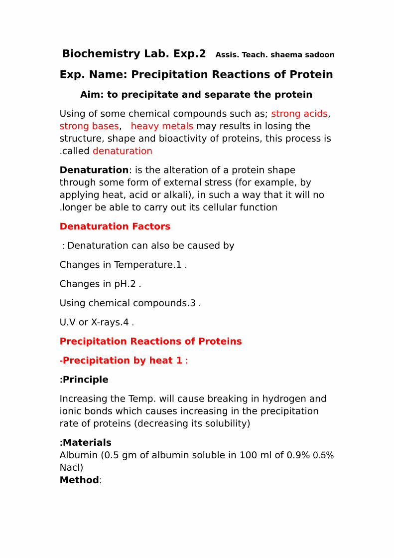
Exp. Name: Precipitation Reactions of Protein
Aim: to precipitate and separate the protein
Using of some chemical compounds such as; strong acids, strong bases, heavy metals may results in losing the structure, shape and bioactivity of proteins, this process is called denaturation.
Denaturation: is the alteration of a protein shape through some form of external stress (for example, by applying heat, acid or alkali), in such a way that it will no longer be able to carry out its cellular function.
Denaturation Factors
Denaturation can also be caused by:
.Changes in Temperature.1
.Changes in pH.2
.Using chemical compounds.3
.U.V or X-rays.4
Precipitation Reactions of Proteins
:Precipitation by heat 1-
Principle:
Increasing the Temp. will cause breaking in hydrogen and ionic bonds which causes increasing in the precipitation rate of proteins (decreasing its solubility)
Materials:0.5% Albumin (0.5 gm of albumin soluble in 100 ml of 0.9%
Nacl): Method

drops of Albumine 10→ يترسب البروتين اويتعكر ) In boiling water bath (∆ 10 min
♦ When albumin solution is heated, white coagulum is obtained because albumin is denatured by heat (Albumin is a coagulable protein).
2 -Precipitation by extreme pH:
Principle:changes in the pH can affect the chemistry of the amino acids and their residues.
Method: → drops + 5drops 10 → تعكر المحلولdrops 5→يذوب الراسب
NaOH(1N) Albumin HCl(1N)3 -precipitation by heavy metals:
Principle: At PH 7 and above proteins are usually negatively charged,the positively charged metal ion neutralizes this charge and the protein coms out of solution. Precipitation by heavy metals is,therefore,most effective at neutral to slightly alkaline PH values,although the solution must not be too alkalin otherwise there is a risk of precipitation of metal hydroxides.Note : The precipitate is frequently soluble in excess of the heavy metal solution since the excess ions confer a stabilizing positive charge on the particles .

♦Heavy metals: copper sulphate,lead acetate ,mercuric nitrate,HgCl2 ,CdSO4 , ZnSO4.
♦The heavy metals have high molecular weight and carry a positive charge.
)PH>7(♦The charge of the protein in alkalin medium is negative
Method:
→drops + 4-5drops 10 (راسب ابيض) تعكر المحلولAgNO3 Albumin
4 -Precipitation by acidic reagents:
Principle:These compounds carry a large negative charge, which neutralize positively charged proteins to form an insoluble salt.
♦Acidic reagents:sulphosalicylic acid,picric acid,tannic acid, trichloroacetic acid(TCA)
) PH<7(♦The charge of the protein in acidic medium is positive

Method:
→drops + 4-5drops 10 (راسب ابيض) تعكر المحلولTCA Albumin
Assis. Teach. shaema sadoonBiochemistry Lab. Exp.3
Exp. Name: Precipitation Reactions of Protein
5 -Precipitation by mineral acids:
Principle:These compounds carry a large negative charge, which neutralize positively charged proteins to form an insoluble salt.
♦Mineral acid :conc.HCl,conc.H2SO4,conc.HNO3
)PH<7(♦The charge of the protein in acidic medium is positive
Method: →drops + 4-5drops 10 (راسب ابيض) تعكر المحلول
HCl Albumin 6 -Precipitation by organic solvents
Principle:By using organic solvents such as; ethanol, acetone or ether, the protein will be precipitated as a result for the

hydrogen bonds formed.
:Method
→drops + 4-5drops 10 (راسب ابيض) تعكر المحلولethanol Albumin
7 -Precipitation by mineral salts (salting in and salting out):
Principle:The increasing in protein solubility when low concentrations of salt is added to a protein solution, is called “salting in”, While the decreasing in protein solubility (increasing precipitation) when high concentration of salt is added to a protein solution is called ''salting out''. Saturated ammonium sulphate{(NH4)2SO4}, saturated magnesium sulphate {MgSO4},saturated sodium chloride{NaCl} will causes precipitation of protein (salting out), while 0.9% sodium chloride NaCl will dissolve the protein (salting in).
♦The degree of protein precipitation with salts depends on the nature of the protein,its molecular weight, salt concentration and type.
:Method
salting in(10 رائق) محلول drops + 4-5 drops→ 0.9%NaCl Albumin
)salting out(10 (راسب ابيض) تعكر المحلول drops + 4-5 drops→ NaCl Albumin
8 -Precipitation at the Isoelectric point:
Principle:

♦ The Isoelectric point (PI) is the pH at which the protein carries no net electrical charge (positive charges= negative charges ).
♦ The proteins precipitate at their PI.
. Different proteins can be separated according to their PI values♦

Assis. Teach. shaema sadoon Biochemistry Lab. Exp.4
Exp. Name: Quantitative test for proteins
Aim: Biuret quantitative test
Principle:
Compounds containing two or more peptide bonds form a characteristic
purple color when treated with dilute alkaline copper sulphate .The
color is due to coordination complex of copper and four nitrogen atoms
and is related to the familiar blue color obtained with copper sulphate
and ammonia.
Materials:
♦ Stock standard protein (10 mg/ml)
♦ Biuret reagent
♦ Sodium chloride 0.9%
Method:
1- In a five clean and dry test tubes prepare (2ml) of the following
protein concentrations (0.0 , 1, 2, 4 , 6, 8 mg/ml) using sodium
chloride (0.9%) for the dilutions.
2- In another test tubes add (2ml) of a prorein solution with
unknown concentration.
3- Add 3ml biuret reagent to each test tube.
4- Mix well and warm at 37 C˚ for 10 mins .
5- Cool and read the absorbance at 540 nm .
6- Plot the known protein concentrations (as mg/ml) on the a-axis
versus the absorbance .Then calculate the concentration of the
unknown sample.
Note:
♦ Stock solution: is a concentrated solution or standard solution
that will be diluted to some lower concentrations for actual use.

♦ Blank solution: is a solution that containing no analyte that
would be measured.
The absorbance will be measured for the colored solution.♦
absorbed ♦ Absorbance: is a measure of the quantity of light
densityby a colored solution, it is also known as" optical density"
♦ Biuret test consists of (CuSO4 +NaOH).
♦ In order to study the quantitative test for proteins
, we shall prepare different concentrations of protein such as
“Albumin” from its stock solution
♦ By using dilution law we will prepare the following concentrations
)10 mg/mlStock standard protein ( .of Albumin

Total vol.
V2 Vol.of NaCl
0.9%
V1 Vol.of stock
Conc.of solution
Tube no.
2 ml(blank) 2 ml 0 ml 0 1
2 ml 1.8 ml 0.2 ml 1 2
2 ml 1.6 ml 0.4 ml 2 3
2 ml 1.2 ml 0.8 ml 4 4
2 ml 0.8 ml 1.2 ml 6 5
2 ml 0.4 ml 1.6 ml 8 6
2 ml(un) 0 2 ml of un. Un. 7
Add 3ml biuret reagent to each test tube
Mix well and warm at 37 C˚ for 10 mins
Cool and read the absorbance at 540 nm
A-AB A Conc. mg/ml Tube no. 0(Blank) 1
1 2 2 3 4 4 6 5
8 6 Un. 7
Note:
Conc. = concentration of protein
A = absorbance
AB = absorbance of blank
Assis. Teach. shaema sadoon Biochemistry Lab. Exp.5
Enzymes
♦ Enzymes are proteins molecules that act as biocatalysts which
accelerate the biochemical reactions in the cells .
♦ Enzymes differ from most other catalysts by being much more Specific.
There are two types of enzymes : ♦
.Endoenzymes; the enzymes that acts in the cells.1
.Exoenzymes; the enzymes that acts out of the cells.2
♦ Each enzyme needs a molecule upon which the enzyme act on and it is
Called suustrate.
♦ Enzymes may require non-protein organic compounds to do
Their function, these organic compound are called coenzymes or prosthetic groups.
Because enzymes are proteins and they have all the properties ♦
of proteins, they extremely unstable and may be denatured more or less
easily by changes in environment.
, Since the denaturation alters the biological properties of proteins ♦
the activity of enzymes is influenced markedly by many environmental
variables such as; Temperature, pH, the concentration of enzyme and
substrate, and the presence of inhibitors .
. “active site ” ♦Enzymes have a specific site on its surfaces which called
♦ The active site represent the site by which the substrate binds with the
enzyme.
♦The enzyme (E) and substrate (S) combine with each other to form an
unstable enzyme-substrate complex (ES) for the formation of product
(P).
♦Some enzymes need metal ions which serves as an activators
Mg⁺² is the common one
♦ Some enzymes are inhibited by various substances such as heavy
metal ions (Hg²⁺),(Pb²⁺),(Ag⁺).

The factors that affect on the enzymatic reactions:
1- Concentration of substrate: with a given quantity of enzyme the
reaction velocity increases with increasing substrate conc. until alimiting
value is reached “ enzyme saturated with substrate''.
2- Concentration of enzyme: in the presence of excess substrate the
velocity of reaction is proportional to the conc. of enzyme .
3- pH: Generally, an enzyme will be active over a relatively narrow range
of pH. The maximum value is referred as the “optimum pH''.
4-Temperature: the point at the accelerating effect of temp. increase is
balance by the denaturation of the enzyme is called the “optimum
Temperature''.
5-Concentration of products : the Accumulation of the product may
retard the enzymatic activity . This may be caused by the combination of
product with the active site, thus removing enzyme from the reaction. In
the living system, this type of inhibition is generally prevented by a quick
removal of products formed.
EXP name : Enzymatic activity of α – Amylase
1- Prepare six test tubes as followes
6 5 4 3 2 1 Contents
4.5 ml 3 ml 3 ml 3 ml 3 ml 3 ml 0.4%Starch
-- -- -- -- 1.5 ml 1 ml H2O
-- -- -- 1 ml -- -- 1% Na2CO3
-- -- 1 ml -- -- -- 1% NaCl -- 1 ml -- -- -- -- Na2C2O4 -- 0.5 ml 0.5 ml 0.5 ml -- 0.5 ml α – Amylase
2-Incubate all tubes at 37 ˚C for 15 mins ,stop the enzymatic reaction by
placing the tubes in a boiling water bath for 5 mins.then cool.
3- Transfer a drop of content of each tube onto a white tile and add a
drop of iodine solution to each aliquot and notice the blue color
complex.
4- Add 0.5 ml Benedict̛s qualitative reagent to each test tube and heat in
a boiling water bath for 5 mins .Notice the red cuprous oxide precipitate
in some of the tubes.

Assis. Teach. shaema sadoon Biochemistry Lab. Exp.6
. Exp. Name: The effect of substrate concentration on enzyme activity
Exp. Aim:
1-To find the effect of substrate concentration on the rate of the enzymatic reaction.
2-To find Km and Vmax by using michaelis- menten equation and line weaver Burk
equation.
Concentration of substrate
As we mentioned before, with a given quantity of enzyme the reaction velocity
increases with increasing substrate conc. until a limiting value is reached “ enzyme
saturated with substrate''.
■In order to study the effect of substrate concentration on the velocity of the
enzymatic reaction, we shall prepare different concentrations of substrate such as
“starch” from its stock solution.
■ Stock solution: is a concentrated solution or standard solution that will be diluted
to some lower concentrations for actual use.
■ Blank solution: is a solution that containing no analyte that would be measured.
■ By using dilution law we will prepare the following concentrations of starch.
# To prepare these different concentrations of starch solution,we will use the
dilution Law ( N1V1=N2V2)
1) first conc. 0.05% starch in 2ml → the final volume
N1V1=N2V2 → 1% хV1= 0.05 х 2ml → V1=0.1ml of 1% starch solution

Let all tubes
Boiling For 3 mint.
then Read the
absorbance at
540 nm
DNS(ml) NaOH(ml) Incubate All tubes
at 37 C˚
For 10 min. then
complete other
addition
α-amylase (ml)
NaCl(ml) H2O Vol.(ml)
Strach Vol.(ml)
Starch Conc.
Tube No.
0.5 0.5 0.5 0.5 1.9 0.1 0.05 % 1
0.5 0.5 0.5 0.5 1.8 0.2 0.1 % 2
0.5 0.5 0.5 0.5 1.6 0.4 0.2 % 3
0.5 0.5 0.5 0.5 1.2 0.8 0.4 % 4
0.5 0.5 0.5 0.5 0.4 1.6 0.8 % 5
0.5 0.5 0.5 0.5 0 2 1 % 6
0.5 0.5 0.5 0.5 2 0 0 (Blank) 7
The role of the used materials:
Starch: substrate ♦
H2O: for dilution purpose ♦
NaCl: activator for the α-amylase enzyme ♦
♦ α-Amylase: Enzyme
NaOH: to stop the enzymatic reaction ♦
oxidizing agent: Dinitrosalcylic acid (DNS) 3,5 ♦
♦ In the presence of α- amylase the starch will be converted to glucose which will be
oxidized by DNS and give us a colored solution.
♦ The absorbance will be measured for the colored solution.
♦ Absorbance: is a measure of the quantity of light absorbed by a colored solution, it
is also known as optical density. A-AB A Starch Conc.= [S] Tube No.
0.05% 1
0.1 % 2
0.2 % 3
0.4 % 4
0.8 % 5
1 % 6
0 (Blank) 7
where [S] = the conc. of substrate
A = absorbance
AB = absorbance of blank
Michaelis-Menten Equation

♦ Km: is the substrate concentration at which the enzymatic reaction velocity equal
half of its maximum velocity. This constant is related to the affinity of the enzyme for
the substrate.
So, if an enzyme has a small Km , it achieves maximal catalytic efficiency (Vmax ) at a
low substrate concentration.
Higher Km = lower the affinity = higher [S] required to reach 1⁄2 Vmax.
♦ Vmax: is the maximum velocity that the enzymatic reaction reaches when an
enzyme saturated with the substrate As [S] is first increased, the initial rate or
velocity (V0) increases with increasing substrate concentration. In the continuous
increasing of [S] ,
V0 increases less and less. Finally, V0 doesn’t increase anymore and velocity reaches
its maximum (Vmax), At this point, the enzyme is saturated with substrate, S.
Line weaver - Burk equation
♦ We can calculate Km and Vmax by using Line weaver – Burk equation.
♦ Line weaver - Burk equation is the reciprocal for Michaelis- Menten Equation.

A-AB 1/ A-AB A 1/[S] Starch Conc.= [S] Tube
No.
0.05% 1
0.1 % 2
0.2 % 3
0.4 % 4
0.8 % 5
1 % 6
0 (Blank) 7

Assis. Teach. shaema sadoon Biochemistry Lab. Exp.7
Exp. Name :
The effect of pH on enzyme activity.
Exp. Aim:
To find the effect of changes in pH on the activity of the enzyme.
♦ The structure of the enzyme has a great influence on the activity of
the enzyme. In other words, changes in the structure of the enzyme
affect the rate of chemical reactions.
♦ When the pH value of the reaction medium changes, the shape and
structure of the enzyme will change. For example, pH can affect the
ionization state of acidic or basic amino acids.
♦ There are carboxyl functional groups on the side chain of acidic amino
acids, as well as, there are amine-containing functional groups in the
side chain of basic amino acids.
♦ If the ionized state of amino acids in the protein is changed, the ionic
bonds that maintain the three-dimensional shape of the protein will
change. This may lead to changes in protein function or inactivation of
enzymes, in other words the denaturation will occur.
♦ The changing in pH not only affects the activity of the enzyme, but
also affects the charge and shape of the substrate, so that the substrate
cannot bind to the active site, or cannot be catalyzed to form a product.
♦ If the level of pH changes significantly, the enzyme and substrate
may be denatured. In this case, the enzyme and the substrate do not
recognize each other, so there will be no reaction.
♦ All enzymes have an ideal pH value, which is called optimal PH.
♦ Optimum pH is the point where the enzyme have the highest
value of activity.

Under the optimum pH conditions, each enzyme showed the
maximum activity, and when the pH value deviates from the ideal
conditions, the activity of the enzyme slows down and then stops.
♦ For example, the optimum pH of an enzyme that works in the
acidic environment of the human stomach is lower than that of an
enzyme that works in a neutral environment of human blood.
♦The enzyme has an active site at which the substrate bind to and the
shape of the active site will change with the change of pH value.
Method :
Tube 6 (blank) Tube (1→5) Materials
2 ml 2 ml Starch
1 ml 1 ml Buffer PH(3.6 ,4.4, 5.2 ,7 , 7.4)
Incubation 3min at 37C˚
----- 0.1 ml α- amylase
Incubation at 37C˚ for 15 min 0.5 ml 0.5 ml NaOH
0.5 ml 0.5 ml DNS
Boiling for 3min ,then read the Absorbance at (540 nm)
A-AB A PH Tube No. 3.6 1 4.4 2 5.2 3 7 4 7.4 5 Blank 6

Assis. Teach. shaema sadoon Biochemistry Lab. Exp.8
Exp. Name :
The effect Temperature on enzyme activity.
Exp. Aim:
To find the effect of changes in Temperature on the activity of the
enzyme.
♦ Collisions between all molecules increase as temperature increases.
This is due to the increase in velocity and kinetic energy that follows
temperature increases.
♦ Enzyme activity increases as temperature increases, and in turn
increases the rate of the reaction. This also means activity decreases at
colder temperatures.
♦ All enzymes have a range of temperatures when they are active, but
there are certain temperatures where they work optimally.
♦ The reaction rate increases with temperature to a maximum
level, then abruptly declines with further increase of temperature.
Because most enzymes rapidly become denatured at temperatures
above 40°C, most enzyme determinations are carried out somewhat
below that temperature.
Some enzymes lose their activity when frozen. ♦
♦ The conditions under which a particular enzyme is most active are
called the optimum conditions. When an enzyme is most active the
rate of the biological reaction it catalyzes is highest.
♦ The optimum temperature is the temperature at which the enzyme
reaches its maximum activity (Temp. at which the enzyme is more
active).

♦ As the temperature is increased enzyme activity increases to a
maximum value at the optimum temperature (around 37oC for most
human enzymes). As the temperature is increased above the optimum
temperature enzyme activity decreases.
♦ At low temperatures enzyme activity is low because the enzyme and
substrate molecules have less kinetic energy so there are fewer
collisions between them.
♦ At the optimum temperature, the kinetic energy in the substrate
and enzyme molecules is ideal for the maximum number of collisions.
♦ At high temperatures the shape of the enzyme is altered so that it is
no longer complementary to its specific substrate. This effect can be
permanent and irreversible and is called denaturation.
Method :
Tube 6 (blank) Tube (1→5) Materials
2ml 2ml Starch (0.5%) 0.5ml 0.5ml NaCl (0.9%)
1ml 0.5ml H2O
---- 0.5ml α- amylase
)˚C 100, 60, 37, 25, 4Incubation for 10min at different temp.(
0.5ml 0.5ml NaOH
0.5ml 0.5ml DNS
Boiling for 3min ,then read the Absorbance at (540nm)

A-AB A Temp. Tube No. 4 1 25 2 37 3 60 4 100 5 Blank 6

Assis. Teach. shaema sadoon Biochemistry Lab. Exp.9
EXP Name: Assay of Urease Enzyme Activity
Urease is a hydrolytic enzyme that catalyzes the urea into carbon dioxide
and ammonia .The enzyme commission number is 3.5.1.5. This reaction
follows
(NH2)2CO + H2O → CO2 + 2NH3
Every organism decomposes nucleic acids and proteins, generating nitrogenous waste because nucleic acids and proteins contain nitrogen. Mammals, amphibians and some invertebrates excrete nitrogenous waste as urea , which is produced in the liver .Urea is an especially good compound for disposing of nitrogen because it is water – soluble and less toxic than ammonia – the excretory produce of fish, for example. Human urine contains 2% urea.
Many species of bacteria produce urease , including Helicobacter
pylori, the bacterium responsible for stomach ulcers. Bydoing
this,H.pylori raises the PH of the gastric juice from about PH 3 to PH7
,the optimal PH for its growth.
Principle:
The method is based on the Berthelot reaction . Alkaline phenol and
sodium hypochlorite react with ammonia to form indophenol blue that
is proportional to the ammonia concentration present .The blue color
formed is intensified by the use of sodium nitropusside as a catalyst.

Reagents:
1- Enzyme substrate (25 mM urea in 100 mM phosphate buffer PH6.8):
-Add 13.121 g of Na2HPO4.7H2O and 7.044 g of NaH2PO4 to 800 ml of
distilled water in a suitable container. Adjust solution to PH 6.8 using HCl
or NaCl .Add distilled water until volume is 1L.
- Dissolve 1.50 g of urea in1L of 0.1M phosphate buffer PH 6.8.
2- Urease enzyme from beans: Grind 5 g of dry beans and add 5ml of
100 mM phosphate buffer PH 6.8.
3- Reagent A : contained 10 g phenol and 50 mg of sodium nirtoprusside
in 500ml of distilled water.
4- Reagent B: contained of 5.0 g sodium hydroxide and 8.4 ml of sodium
hypochlorite in 500 ml of distilled water.
Procedure:
6 5 4 3 2 1 No.of tube Final
volume Volume of buffer (ml)
Volume of urea (ml)
Final urea concentration (Mm)
Recor the
intensity of the
color at 630nm ג
To develop
the color,
incubate at 37˚C
for 5 minutes
Add 2 ml of
Reagent A +
2ml of Reagent
B
Add 0.1 of urease
extract and
incubate at 37˚C for 10
minutes
Pre incubation
at 37˚C
for 5 minutes
2 2 0 0.00 1
2 1.6 0.4 5 2
2 1.2 0.8 10 3
2 0.8 1.2 15 4
2 0.4 1.6 20 5
2 0 2 25 6
A-AB A Final urea
concentration (Mm)
Tube No.
0.00 1
5 2
10 3
15 4
20 5
25 6
0 (Blank) 7


Assis. Teach. shaema sadoon Biochemistry Lab. Exp.10
EXP Name: Estimation of ascorbic acid in lemon juice
Ascorbic is found in fruit, particularly citrus fruits , and vegetables . a
quantitatively significant dietary is ascorbate added to other foods as a
preservative .It cannot by synthesized by man ,other primates ,or the
guinea pig.
Ascorbate can be reversibly oxidized in biological systems to
dehydroascorbate and ,although its functions in man are not certain ,it
probably acts as a hydrogen carrier .It seems to be necessary for normal
collagen formation.
Principle:
Vitamin C (L-ascorbic acid) gets oxidized to its dehydro form by air
especially at alkaline PH. However, it is stable in an acidic solution .
Therefore ,vitamin C is extracted in metaphosphoric acid or in a mixture
of metaphosphoric and dilute acetic acid or by oxalic acid and acetic
acid. Its estimation in the extract is carried out by titrating it against 2,6-
dichlorphenol indophenol solution. Oxidized form of this dye is blue in
colour in an alkaline medium and red in an acidic medium . Reduced
form of the dye ,on the other hand, is colourless and its termed its leuco
form .The redox reaction accuring during the titration is:

Reagents:
1- Lemons, orange juice ,green papper ,black currant juice.
2- 2,6- dichlorophenol indophenol solution: Dissolve 52 mg of sodium
salt of the dye and 42 mg of sodium bicarbonate in water . Make up the
final volume to 500 ml.
3-Standard vitamin C solution: Dissolve 20 mg of vitamin C in 1L distilled
water.
4- Acetic acid glacial.
Procedure:
2.5 ml (standard solution) + 0.5 ml Acidic acid glacial ----→ V standard ( Titration with dye)
2.5 ml (lemon juice) + 0.5 ml Acidic acid glacial ----→ V test ( Titration with dye)
2.5 ml (distilled water) + 0.5 ml Acidic acid glacial ----→ V blank ( Titration with dye)
Calculation :
[Vitamin C ] mg/100ml ={( V test – V blank)/(V standard – V blank)} х
concentration of standard/ Vol. test) х 100 )
V standard = The volume of dye consumed by titration with standard
solution.
V test = The volume of dye consumed by titration with lemon juice.
V blank= The volume of dye consumed by titration with distilled water.
Vol. test = The volume of orange juice used to measure the
concentration of the vitamin C.