Laboratory Diagnosis of Viral Diseases - Fudan...
80
Laboratory Diagnosis of Viral Diseases LONG JIAN-ER, Ph.D. Lab of Medical Microbiology, Department of Medical Microbiology and Parasitology, Shang Medical College of Fudan University, 138 Yixueyuan R., Shanghai 200032, PR China Email: [email protected]; Tel.: +86-21-54237867
Transcript of Laboratory Diagnosis of Viral Diseases - Fudan...

Laboratory Diagnosis of Viral Diseases
LONG JIAN-ER, Ph.D.
Lab of Medical Microbiology, Department of Medical Microbiology and Parasitology, Shang Medical College of Fudan University, 138 Yixueyuan R., Shanghai 200032, PR ChinaEmail: [email protected]; Tel.: +86-21-54237867
Koch’s Postulates
1. Organism present only in diseased individuals
2. Organism cultivated in pure culture from diseased individual
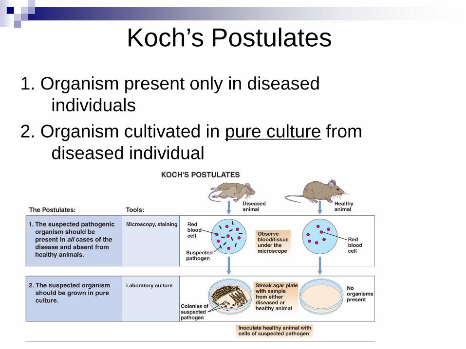
3. Organism causes disease when injected into healthy individuals
4. Organism re-isolated from infected individual from point 3.
Koch’s Postulates

River’s Postulates T.M. River, 1937 Modified from Koch’s Postulates (proof of bacterial
diseases)
1. Isolate virus from diseased hosts.2. Cultivation of virus in host cells.3. Proof of filterability.4. Production of a comparable disease when the
cultivated virus is used to infect experimental animals.5. Re-isolation of the same virus from the infected
experimental animal.6. Detection of a specific immune response to the virus.

Virus infected patients
Collected specimensCheck the infected cells under light microscope
Observe virus particles under electronic microscope
Virus culture and isolation
Serology test antibody test
Detection of viral antigen or genome
Identify viral propagationAntigen Genome Neutralization test
Hemagglutination inhibition testEIA/ELISA
Western BlotImmunofluoresence
EIA/ELISAWestern Blot
ImmunoelectrophesisRadio immune assay
PCRSouthern blotNorthern blot
Dot blotIn situ hybridization
CPEHemadsorption
HemagglutinationVirus interferenceNeutralization test
Plaque assayTCID50 assay
Procedures for laboratory viral diagnosis
Viral Diagnostics in the Clinical Laboratory
Over 70% of all infectious disease cases seen by a physician are due to viral infections.
Quality of patient specimens and their transport to the laboratory is important.
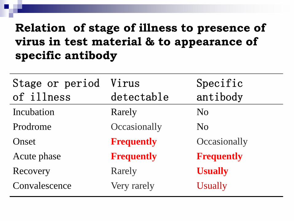
Relation of stage of illness to presence of virus in test material & to appearance of specific antibody
Stage or period of illness
Virus detectable
Specific antibody
Incubation Rarely No Prodrome Occasionally No Onset Frequently OccasionallyAcute phase Frequently Frequently Recovery Rarely Usually Convalescence Very rarely Usually
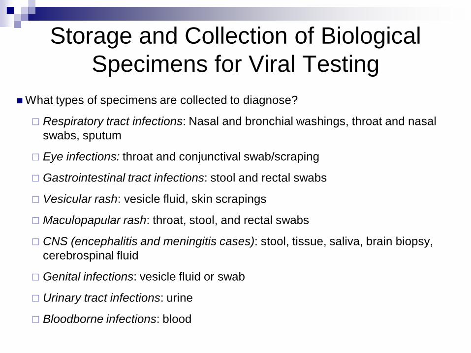
Storage and Collection of Biological Specimens for Viral Testing
What types of specimens are collected to diagnose?
Respiratory tract infections: Nasal and bronchial washings, throat and nasal swabs, sputum
Eye infections: throat and conjunctival swab/scraping
Gastrointestinal tract infections: stool and rectal swabs
Vesicular rash: vesicle fluid, skin scrapings
Maculopapular rash: throat, stool, and rectal swabs
CNS (encephalitis and meningitis cases): stool, tissue, saliva, brain biopsy, cerebrospinal fluid
Genital infections: vesicle fluid or swab
Urinary tract infections: urine
Bloodborne infections: blood
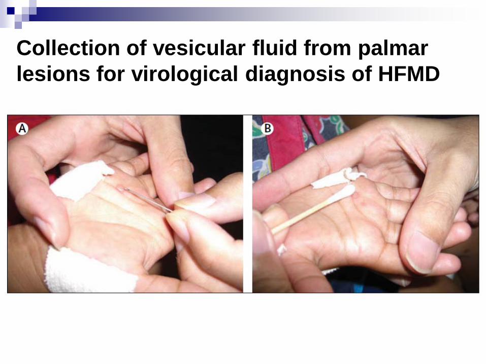
Collection of vesicular fluid from palmar lesions for virological diagnosis of HFMD

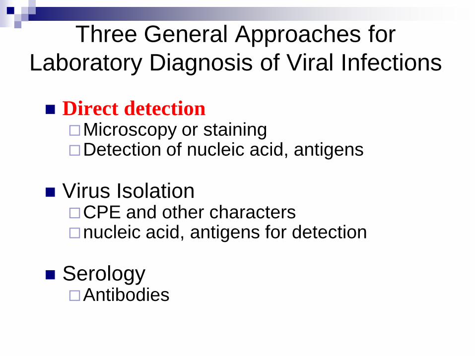
Three General Approaches for Laboratory Diagnosis of Viral Infections
Direct detectionMicroscopy or stainingDetection of nucleic acid, antigens
Virus Isolation (Indirect Examination)CPE and other charactersnucleic acid, antigens for detection
SerologyAntibodies
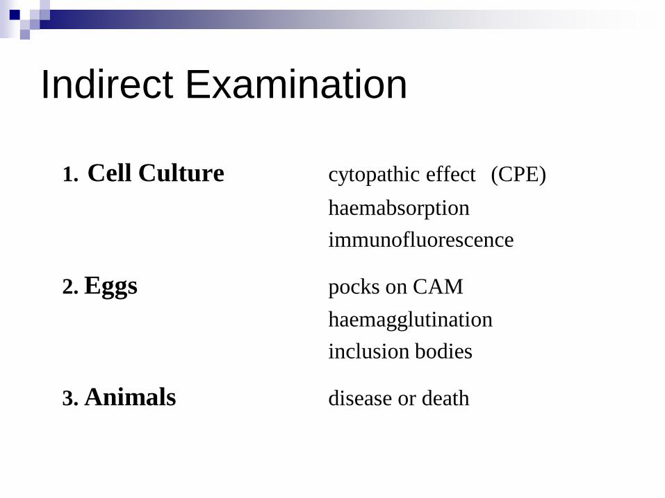
Indirect Examination
1. Cell Culture cytopathic effect (CPE)haemabsorptionimmunofluorescence
2. Eggs pocks on CAMhaemagglutinationinclusion bodies
3. Animals disease or death
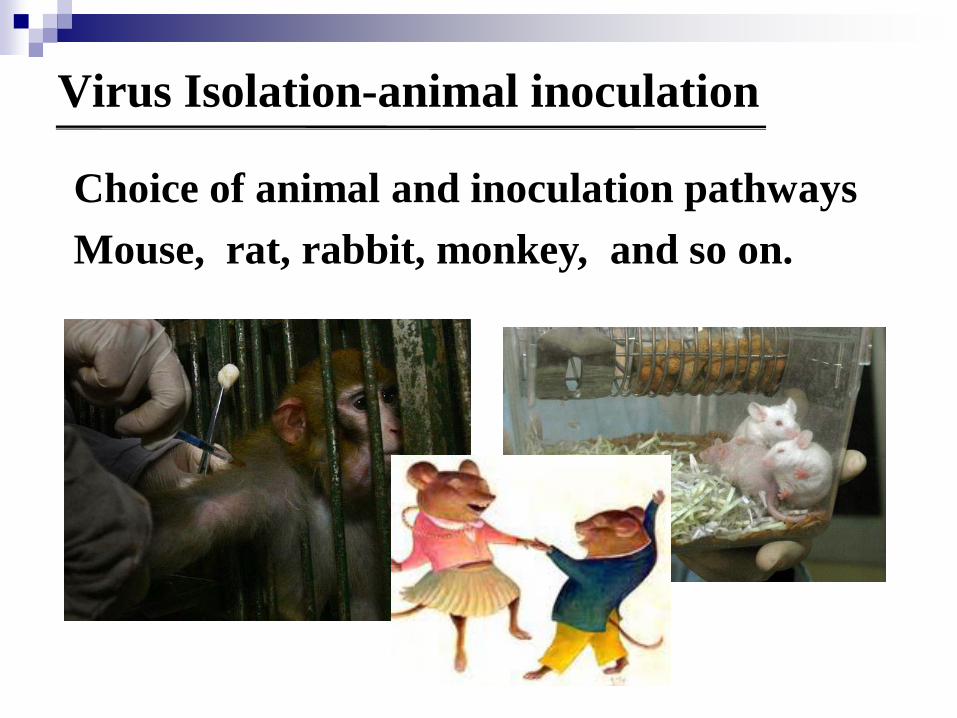
Choice of animal and inoculation pathwaysMouse, rat, rabbit, monkey, and so on.
Virus Isolation-animal inoculation
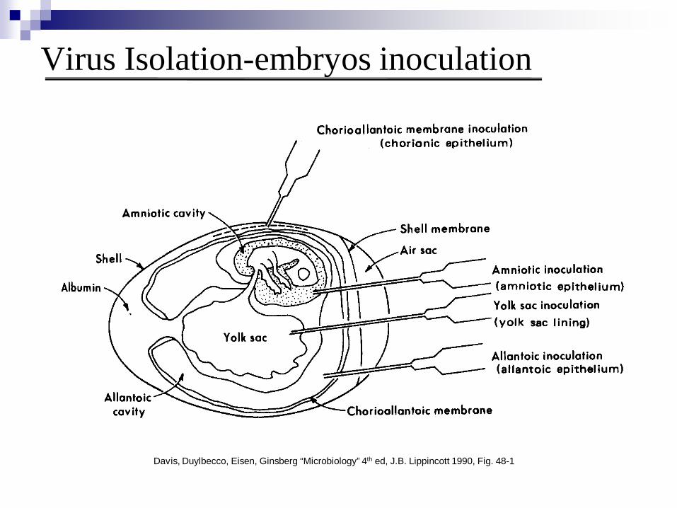
Davis, Duylbecco, Eisen, Ginsberg “Microbiology” 4th ed, J.B. Lippincott 1990, Fig. 48-1
Virus Isolation-embryos inoculation

d3 d5 d7
d10 d12 d16

9-11 days embryos taken out after virus infection
Virus propagation and vaccine production in enterprise industry

Virus Isolation-cell cultures
Cell Cultures are most widely used for virus isolation, there are 3 types of cell cultures:
1. Primary cells - Monkey Kidney2. Semi-continuous cells - Human embryonic kidney and skin fibroblasts3. Continuous cells - HeLa, Vero, Hep2, LLC-MK2, MDCK
Primary cell culture are widely acknowledged as the best cell culturesystems available since they support the widest range of viruses. However,they are very expensive and it is often difficult to obtain a reliable supply.Continuous cells are the most easy to handle but the range of virusessupported is often limited.
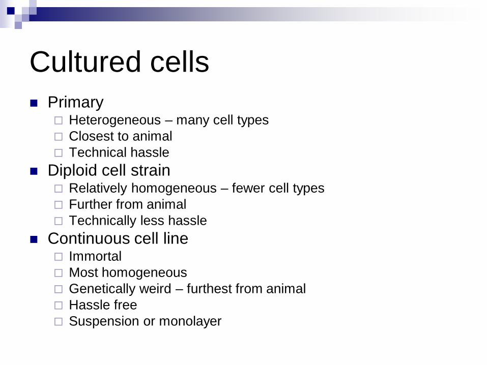
Cultured cells Primary
Heterogeneous – many cell types Closest to animal Technical hassle
Diploid cell strain Relatively homogeneous – fewer cell types Further from animal Technically less hassle
Continuous cell line Immortal Most homogeneous Genetically weird – furthest from animal Hassle free Suspension or monolayer

Laminar Flow Hoods
Virologist’s facility
Laminar vertical flow hoods
Contains HEPA filter
Removes 99.97% of
particles of 0.3μM or higher
Figure 5-3: Vertical laminar flow hood.

Primary cell culture
+ enzymes
time

Subculture
enzymes
time

Growth of cells in culture. A primary culture is defined as the original plating of cells from a tissue, grown to a confluent monolayer, without subculturing. A cell strain (solid line) is defined as a euploid population of cells subcultivated more than once in vitro, lacking the property of indefinite serial passage. Cell strains ultimately undergo degeneration and death, also called crisis or senescence. A cell line (dashed line) is an aneuploid population of cells that can be grown in culture indefinitely. Spontaneous transformation or alteration of a cell strain to an immortal cell line can occur at any time during cultivation of the cell strain. The time in culture and corresponding number of subcultivations or passages are shown on the abscissas. The ordinate shows the total number of cells that would accumulate if all were retained in culture. (From Fields Virology, 4th ed, Knipe & Howley, eds, Lippincott Williams & Wilkins, 2001 Fig. 2-2.)
Cell culture

Cytopathic Effects Visible results of viral infection Cell death by
Multiplying viruses Inhibition of DNA, RNA or protein synthesis Effects on permeability of membrane
Cytopathic effects (CPEs) of infected cells can be observed with inverted light microscopes
Rounding/detachment from plastic flask Syncytia/fusion
Fusion of cells Shrinkage Increased refractiability Aggregation Loss of adherence Cell lysis/death
Common observations of CPEs Inclusion body formation
Intracellular virus parts (replication or assembly) Hemadsorption assays

Cultured cell morphologies
Epithelial-like(human lung carcinoma, A549)
Fibroblast like(baby hamster kidney, BHK)
Fields Virology, 4th ed, Knipe & Howley, eds, Lippincott Williams & Wilkins, 2001, Fig. 2-3
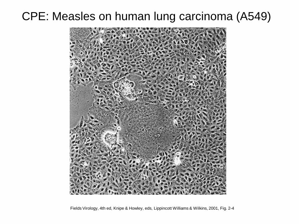
CPE: Measles on human lung carcinoma (A549)
Fields Virology, 4th ed, Knipe & Howley, eds, Lippincott Williams & Wilkins, 2001, Fig. 2-4

CPE: vaccinia on monkey kidney (BSC40)
Low multiplicity of infection (moi)single plaque
High moi, 48 hr
Fields Virology, 4th ed, Knipe & Howley, eds, Lippincott Williams & Wilkins, 2001, Fig. 2-4
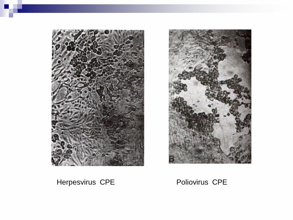
Herpesvirus CPE Poliovirus CPE

From Medical Microbiology, 5th ed., Murray, Rosenthal & Pfaller, Mosby Inc., 2005, Fig. 51-3.
Cytopathic Effect- inclusion bodies
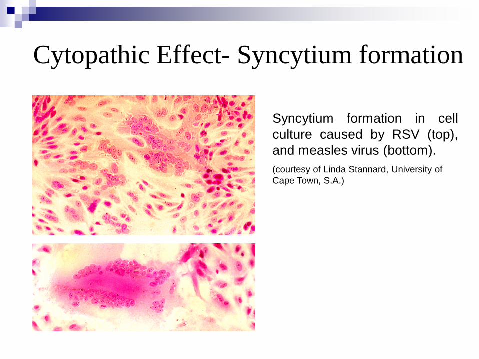
Cytopathic Effect- Syncytium formation
Syncytium formation in cellculture caused by RSV (top),and measles virus (bottom).(courtesy of Linda Stannard, University of Cape Town, S.A.)

SARS virus syncytia
measles virus

From Medical Microbiology, 5th ed., Murray, Rosenthal & Pfaller, Mosby Inc., 2005, Fig. 51-5.
Hemadsorption
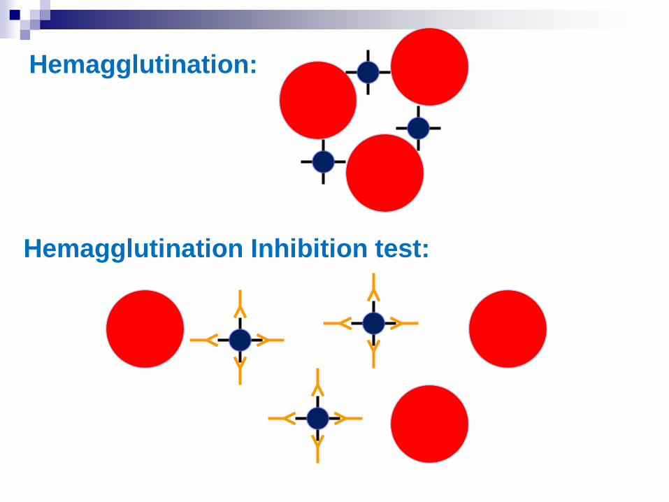
Hemagglutination:
Hemagglutination Inhibition test:

Hemadsorption Test
Some viruses agglutinate RBCsMumps, measles, influenza Hemagglutination
Clumps RBCs

rubella virus + cell CPE -enterovirus + cell CPE +
rubella virus
+ enteroviruscellCPE - CPE -
Interference +-
Virus infected cells-interference
Without significant CPE but interference other virus infection
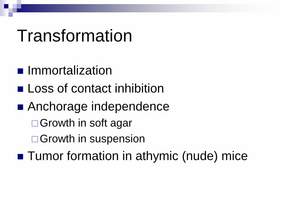
Transformation
Immortalization Loss of contact inhibition Anchorage independenceGrowth in soft agarGrowth in suspension
Tumor formation in athymic (nude) mice

Common Methods
Four methods:Quantitative assays
Plaque assays TCID50
Hemmagglutination assaysTransformation assays
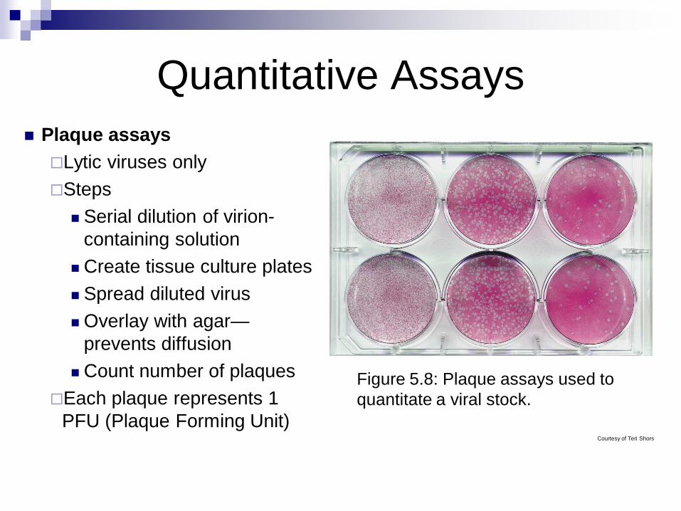
Quantitative Assays Plaque assaysLytic viruses onlySteps Serial dilution of virion-
containing solutionCreate tissue culture plates Spread diluted virusOverlay with agar—
prevents diffusionCount number of plaques
Each plaque represents 1 PFU (Plaque Forming Unit)
Figure 5.8: Plaque assays used to quantitate a viral stock.
Courtesy of Teri Shors
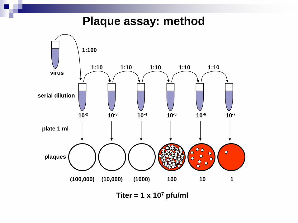
1:100
1:10 1:101:101:101:10
10-2 10-3 10-4 10-5 10-6 10-7
virus
serial dilution
plate 1 ml
plaques
100 10 1(1000)(100,000) (10,000)
Titer = 1 x 10⁷ pfu/ml
Plaque assay: method
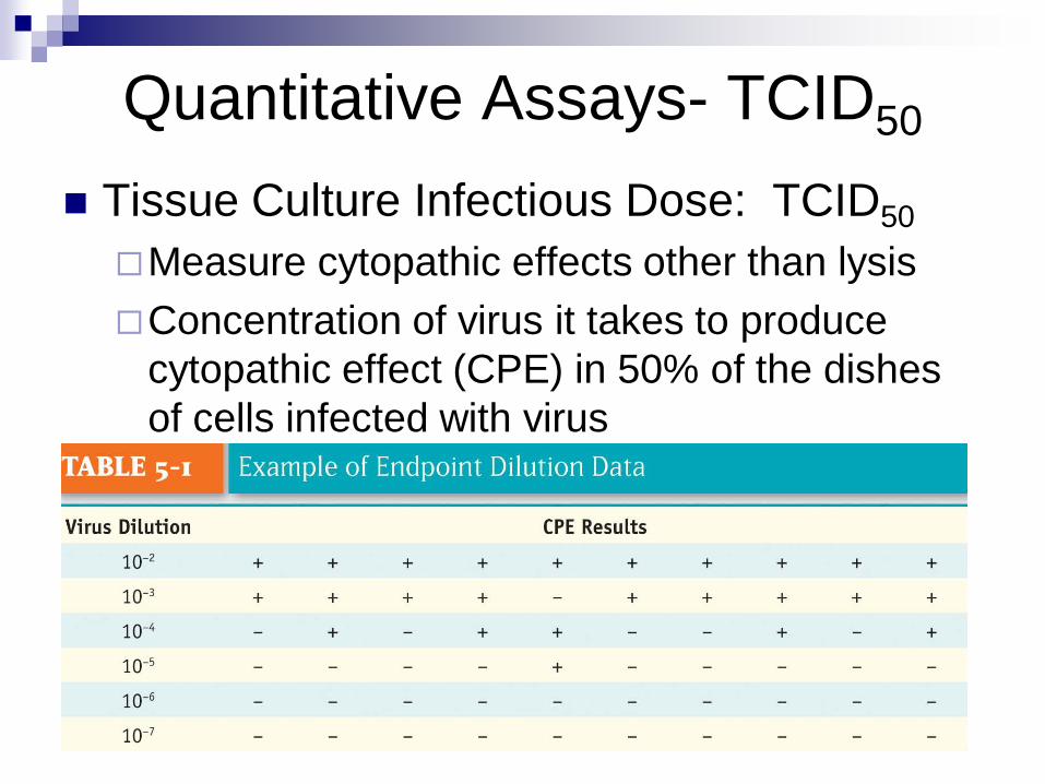
Quantitative Assays- TCID50
Tissue Culture Infectious Dose: TCID50Measure cytopathic effects other than lysisConcentration of virus it takes to produce
cytopathic effect (CPE) in 50% of the dishes of cells infected with virus

= infected
= uninfected
Endpoint titration
Five replicate wells of cells are infected with one ml of each of four different virus dilutions, incubated, and scored for infection by looking for CPE. In this example, the final titer is 106.3 TCID50per ml. (TCID = tissue culture infective dose)
10-5 10-6 10-7 10-85
repl
icat
e sa
mpl
ings
1
2
3
4
5
dilution

Hemagglutination test: method
Titer = 32 HA units/ml
1:8
1:2 1:21:21:21:2
8 16 32 64 128 256
virus
serial dilution
mix with red blood cells
side view
top view

Hemagglutination assay. Seven different samples of influenza virus, numbered 1 through 7 at the left, were serially diluted as indicated at the top, mixed with chicken red blood cells (RBC), and incubated on ice for 1 to 2 hours. Wells in the bottom row contain no virus. Agglutinated RBCs coat wells evenly, in contrast to nonagglutinated cells, which form a distinct button at the bottom of the well. The HA titer, shown at the right, is the last dilution that shows complete hemagglutination activity. (From Fields Virology, 4th ed, Knipe & Howley, eds, Lippincott Williams & Wilkins, 2001, Fig. 2-8)
Hemagglutination assay: influenza virus

Method Amount (per ml)
Direct electron microscope count 1010 EM particles
Quantal infectivity assay in eggs 109 egg ID50
Quantal infectivity assay by plaque formation 108 pfu
Hemagglutination assay 103 HA units
Comparison of quantitative methods
Fields Virology, 4th ed, Knipe & Howley, eds, Lippincott Williams & Wilkins, 2001, Table 2-4
Three General Approaches for Laboratory Diagnosis of Viral Infections
Direct detectionMicroscopy or stainingDetection of nucleic acid, antigens
Virus IsolationCPE and other charactersnucleic acid, antigens for detection
SerologyAntibodies
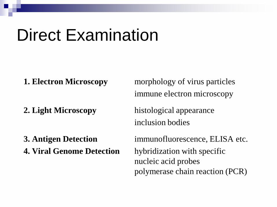
Direct Examination
1. Electron Microscopy morphology of virus particles immune electron microscopy
2. Light Microscopy histological appearanceinclusion bodies
3. Antigen Detection immunofluorescence, ELISA etc.4. Viral Genome Detection hybridization with specific
nucleic acid probes polymerase chain reaction (PCR)
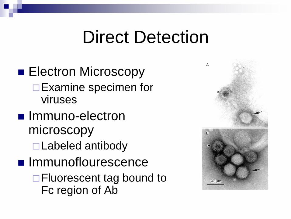
Direct Detection
Electron MicroscopyExamine specimen for
viruses Immuno-electron
microscopyLabeled antibody
ImmunoflourescenceFluorescent tag bound to
Fc region of Ab

Electronmicrographs
Adenovirus Rotavirus
(courtesy of Linda Stannard, University of Cape Town, S.A.)
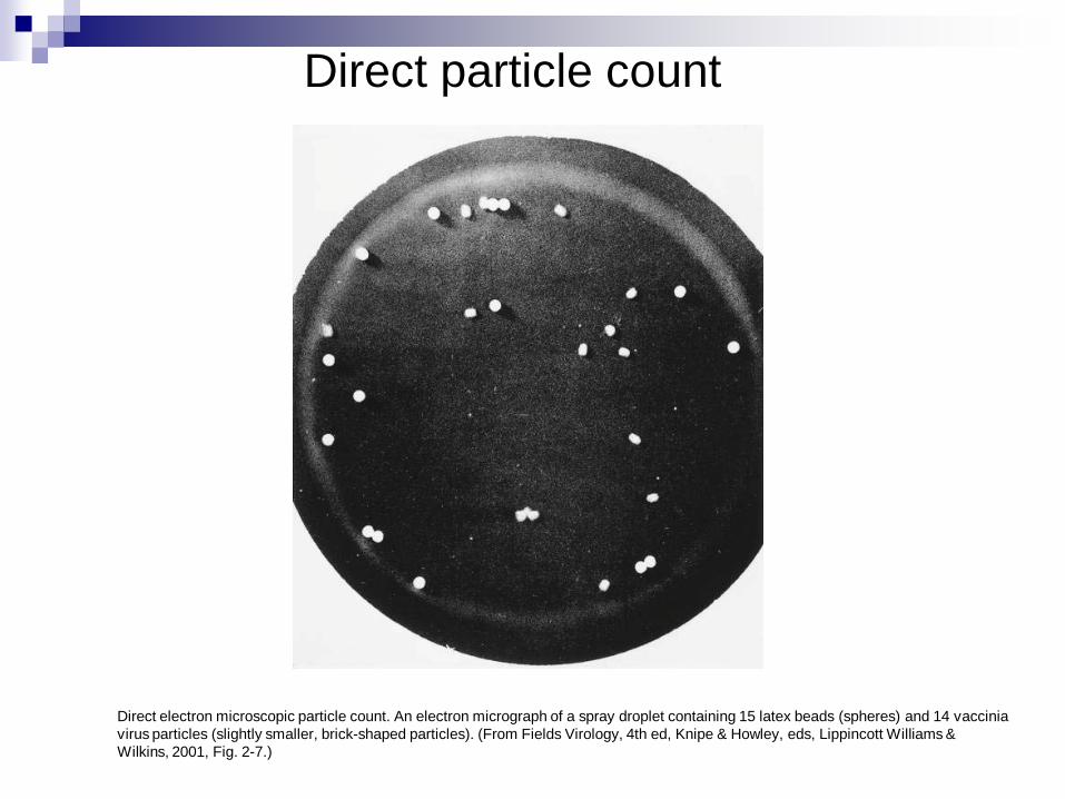
Direct electron microscopic particle count. An electron micrograph of a spray droplet containing 15 latex beads (spheres) and 14 vaccinia virus particles (slightly smaller, brick-shaped particles). (From Fields Virology, 4th ed, Knipe & Howley, eds, Lippincott Williams & Wilkins, 2001, Fig. 2-7.)
Direct particle count

Rapid Diagnosis Based on the Detection of Viral Antigens
Nasopharyngeal Aspirate RSVInfluenza A and BParainfluenzaAdenovirus
Faeces RotavirusesAdenovirusesAstrovirus
Skin HSVVZV
Blood CMV (pp65 antigenaemia test)
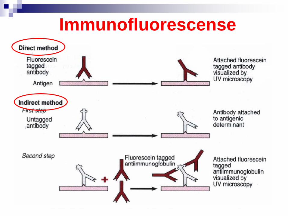
Immunofluorescense
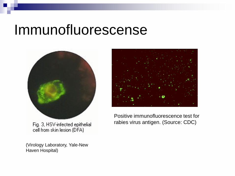
Immunofluorescense
Positive immunofluorescence test for rabies virus antigen. (Source: CDC)
(Virology Laboratory, Yale-New Haven Hospital)

CMV pp65 antigenaemia test
(Virology Laboratory, Yale-New Haven Hospital)

Advantages and Disadvantages
Advantages
Result available quickly, usually within a few hours.
Potential Problems
Often very much reduced sensitivity compared to cell culture,can be as low as 20%. Specificity often poor as well.
Requires good specimens.
The procedures involved are often tedious and time-consuming and thus expensive in terms of laboratory time.
Methods for Rapid Diagnosis of Viral genome
1. Polymerase Chain Reaction
(PCR);
2. Molecular hybridization
(Southern or Northern Blot);

Each cycle doubles the copy number of the target

PCR machine
Real-time PCR machine
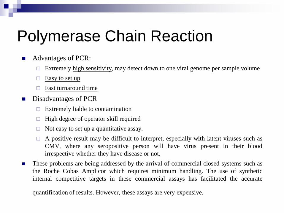
Polymerase Chain Reaction Advantages of PCR:
Extremely high sensitivity, may detect down to one viral genome per sample volume Easy to set up Fast turnaround time
Disadvantages of PCR Extremely liable to contamination High degree of operator skill required Not easy to set up a quantitative assay. A positive result may be difficult to interpret, especially with latent viruses such as
CMV, where any seropositive person will have virus present in their bloodirrespective whether they have disease or not.
These problems are being addressed by the arrival of commercial closed systems such asthe Roche Cobas Amplicor which requires minimum handling. The use of syntheticinternal competitive targets in these commercial assays has facilitated the accurate
quantification of results. However, these assays are very expensive.

Simple and rapid detection of human EV 71 by RT-LAMP

Nucleic acid molecular hybridizationSpecimen DNA
Hybrid membrane
Digested by endonuclease
Running agarose gel
Denatured into ssDNA
Hybridized with ssDNA*(probe)
Radioactivity or labeled enzyme to develop color

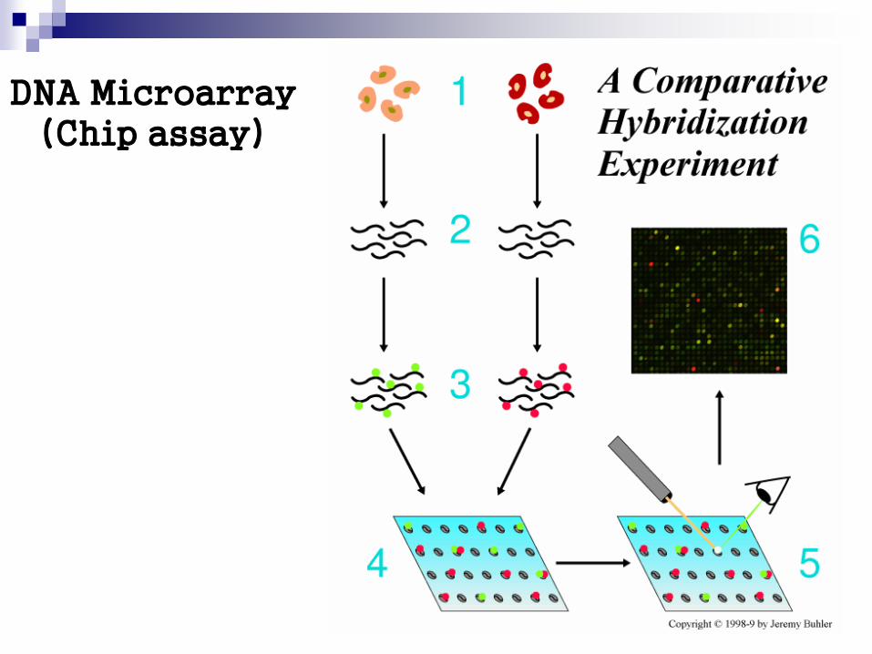
DNA Microarray (Chip assay)

in situ hybridization

Dot Blot Hybridization
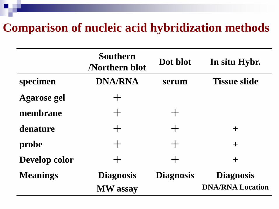
Southern /Northern blot Dot blot In situ Hybr.
specimen DNA/RNA serum Tissue slide
Agarose gel +
membrane + +
denature + + +
probe + + +
Develop color + + +
Meanings Diagnosis MW assay
Diagnosis Diagnosis DNA/RNA Location
Comparison of nucleic acid hybridization methods
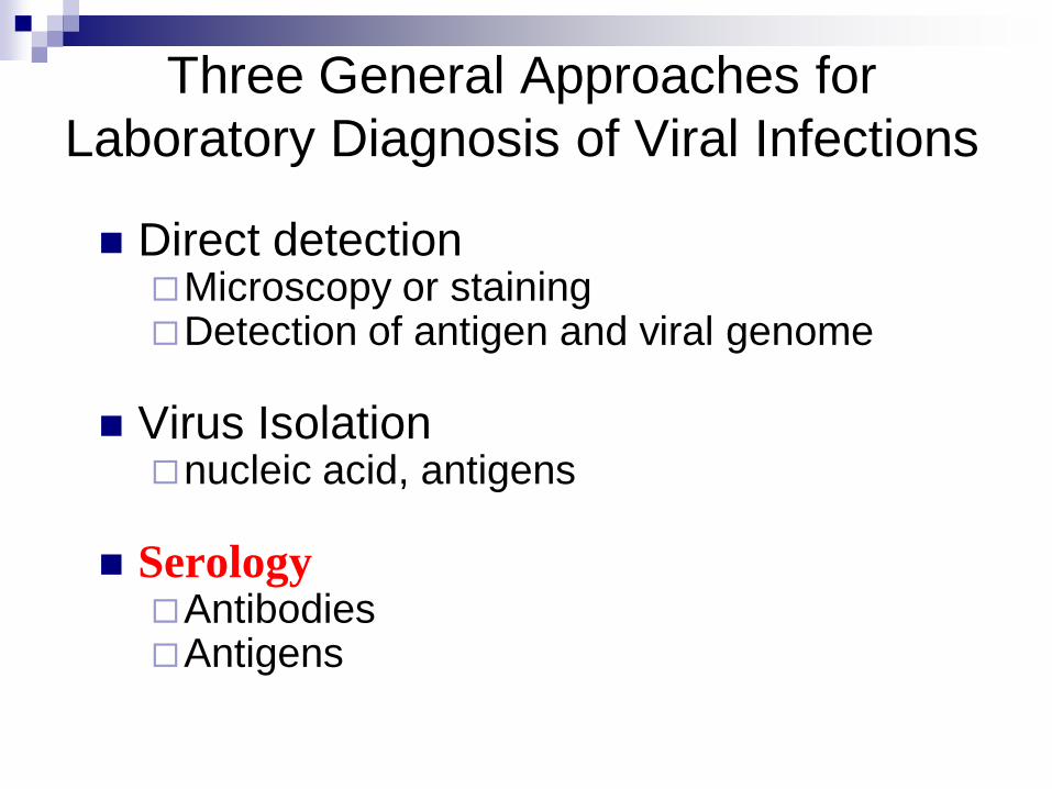
Three General Approaches for Laboratory Diagnosis of Viral Infections
Direct detectionMicroscopy or stainingDetection of antigen and viral genome
Virus Isolationnucleic acid, antigens
SerologyAntibodiesAntigens
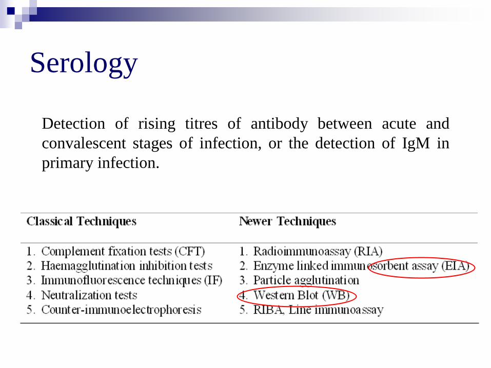
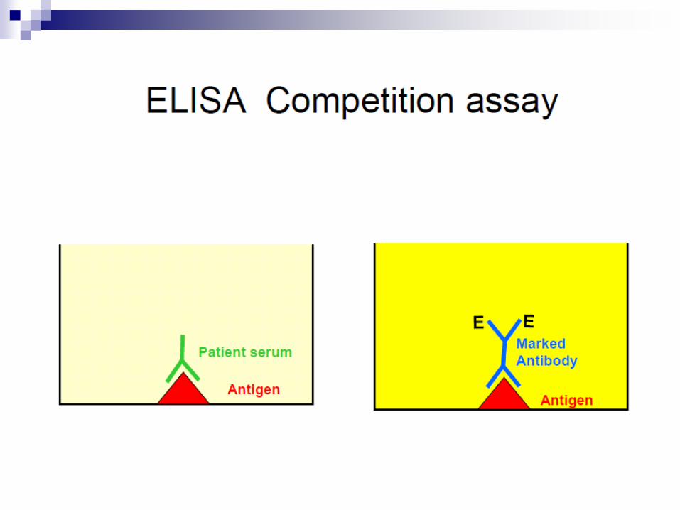
Serology
Detection of rising titres of antibody between acute andconvalescent stages of infection, or the detection of IgM inprimary infection.

Viral Serology Indirect
Primary and secondary responses to viral infections IgM (1st exposure) IgG (2nd exposure)
Figure 5.18: Primary (1 degree) and secondary (2 degree) antibody responses toward a viral pathogen.

Viral Serology
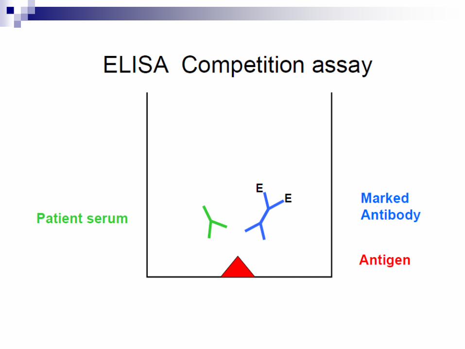
Enzyme-Linked Immunosorbent Assays (ELISAs) Enzyme reacts with substrate to produce colored
product Very sensitive
HIV test If positive twice, Western Blotting is performed next
Could detect viral antigens or antibodies

ELISA Procedures
Modified from Specter, S. C., R. L. Hodinka and S. A. Young. Clinical Virology Manual, Third Edition . ASM Press, 2000.
Antibodies coated

© Hank Morgan/Science Photo Library/Photo Researchers, Inc.
Figure 5.20: HIV ELISA test.
Viral Serology
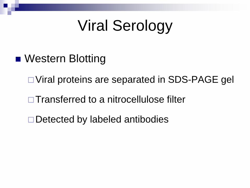
Western Blotting
Viral proteins are separated in SDS-PAGE gel
Transferred to a nitrocellulose filter
Detected by labeled antibodies
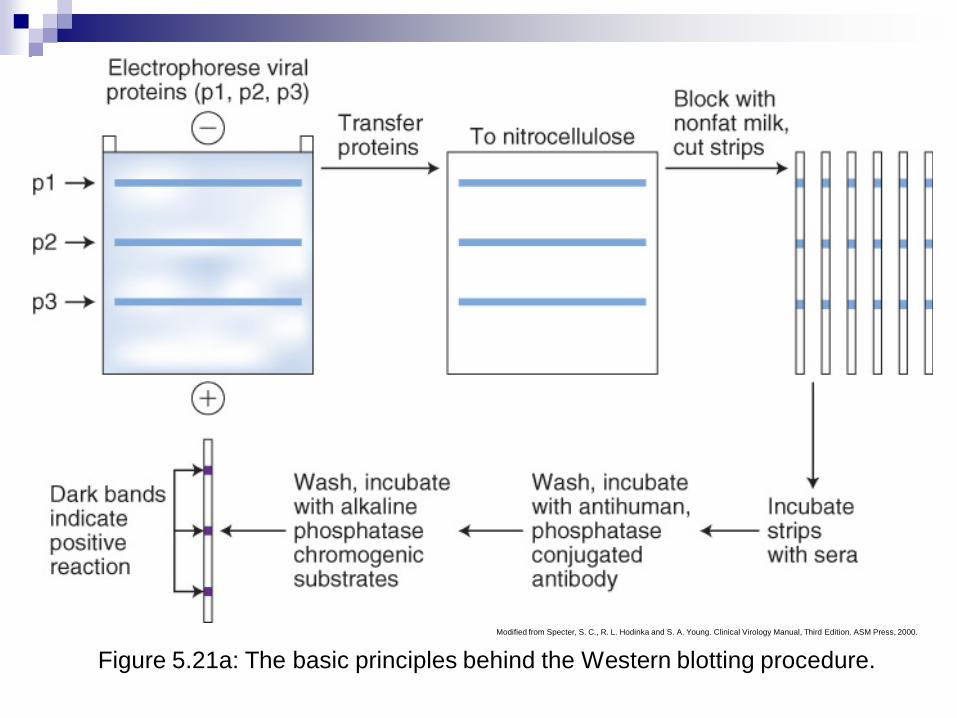
Figure 5.21a: The basic principles behind the Western blotting procedure. Modified from Specter, S. C., R. L. Hodinka and S. A. Young. Clinical Virology Manual, Third Edition. ASM Press, 2000.

From Medical Microbiology, 5th ed., Murray, Rosenthal & Pfaller, Mosby Inc., 2005, Fig. 51-7.
Antibody detection: western blot
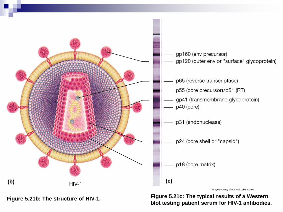
Figure 5.21b: The structure of HIV-1.
Image courtesy of Bio-Rad Laboratories
Figure 5.21c: The typical results of a Western blot testing patient serum for HIV-1 antibodies.
(c)(b)

From Medical Microbiology, 5th ed., Murray, Rosenthal & Pfaller, Mosby Inc., 2005, Fig. 51-6.
Antigen, antibody detected by neutralization and hemagglutination or inhibition assay
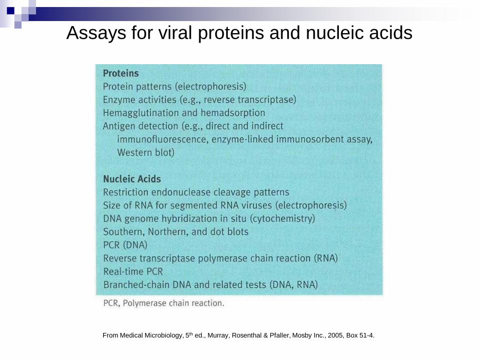
From Medical Microbiology, 5th ed., Murray, Rosenthal & Pfaller, Mosby Inc., 2005, Box 51-4.
Assays for viral proteins and nucleic acids
Summary
4 main clinical diagnostic techniques Culture, serology, antigen detection, nucleic acid
detectionVirus culture
Cultured cell types Cytopathic effect Not all viruses can be cultured
Virus quantitation Biological Physical
Basic serological techniques

Virus infected patients
Collected specimensCheck the infected cells under light microscope
Observe virus particles under electronic microscope
Virus culture and isolation
Serology test antibody test
Detection of viral antigen or genome
Identify viral propagationAntigen Genome Neutralization test
Hemagglutination inhibition testEIA/ELISA
Western BlotImmunofluoresence
EIA/ELISAWestern Blot
ImmunoelectrophesisRadio immune assay
PCRSouthern blotNorthern blot
Dot blotIn situ hybridization
CPEHemadsorption
HemagglutinationVirus interferenceNeutralization test
Plaque assayTCID50 assay
Procedures for laboratory viral diagnosis

Terms & Questions 1.Primary cell, continuous cell, cell strain.2.Molecular hybridzation, viral molecular
diagnosis.3.Neutralization test, hemagglutination inhibition
test, TCID50,ID50,LD50,PFU,MOI.4.CPE, inclusion body, syncytia, virus interference.5. How to determine a patient with HIV infection?