JIPBS · 2Microbiology laboratory analyst in HIKMA Pharma pharmaceutical company, Egypt. Mostafa...
12
©JIPBS, All rights reserved Journal of Innovations in Pharmaceuticals and Biological Sciences www.jipbs.com ISSN: 2349-2759 Abstract In the view of many recalls of different pharmaceutical dosage forms withdrawn from the market as reported worldwide, a continuous microbiological environmental monitoring (EM) programs for bioburden in clean area should be established to identify their potential impact on the manufactured medicinal products. One of the most important criteria of observations is the surface microbe – herein focus on bacterial ecology - cleanliness monitoring. Surfaces can be direct or indirect source of the contamination of the final medicinal products. Random surface samples using contact plates were taken aseptically from the newly established pharmaceutical plant as a part from total EM and assessment program in clean rooms for drug manufacturing. After incubation of the samples a process of isolation and identification was conducted using Gram-stain and miniaturized BBL™ Crystal™ enteric/non- fermenter (E/NF) and Gram-positive (GP) miniaturized biochemical identification system. Data were interpreted and analyzed, showing that identified bacteria were belonging to Staphylococcaceae, Bacillaceae, Actinomycetaceae, Dermacoccaceae and Micrococcaceae contribution to more than 60% from the total samples. Only 8% of the total samples (2% Gram-positive bacilli and 6% Gram-negative rods) were not identified by the commercial identification system. However, there was different colonial morphology observed for the same identified bacteria, which are represented, by Arcanobacterium pyogenes, Bacillus circulans, B. cereus, Micrococcus lylae and Kytococcus sedentarius from different surface samples. A simple quantitative risk index was applied showing significant health risk in the manufacturing environment of some bacterial families over others in order different from their ecological distribution in the pharmaceutical facility. Key words: Environmental monitoring; contact plates; clean room; BBL™ Crystal™; quantitative risk index. *Corresponding author: Mostafa Essam Eissa, Microbiological Quality Control Department, Hikma Pharma Pharmaceutical Company, Egypt. 1. Introduction Microbial contamination costs companies thousands to millions of dollars annually through the equipment damage, production downtime, product contamination, JIPBS Research article Assessment of the Risk in Pharmaceutical Facility to the Human Health Based on the Ecological Surface Quality of Bacteria in the Clean Area Mostafa Essam Eissa* 1 , Ahmed Saber Nouby 2 1 Microbiological quality control section head in HIKMA Pharma pharmaceutical company, Egypt. 2 Microbiology laboratory analyst in HIKMA Pharma pharmaceutical company, Egypt.
Transcript of JIPBS · 2Microbiology laboratory analyst in HIKMA Pharma pharmaceutical company, Egypt. Mostafa...

©JIPBS, All rights reserved
Journal of Innovations in Pharmaceuticals and Biological Sciences
www.jipbs.com
ISSN: 2349-2759
Abstract In the view of many recalls of different pharmaceutical dosage forms withdrawn from the market as reported worldwide, a continuous microbiological environmental monitoring (EM) programs for bioburden in clean area should be established to identify their potential impact on the manufactured medicinal products. One of the most important criteria of observations is the surface microbe – herein focus on bacterial ecology - cleanliness monitoring. Surfaces can be direct or indirect source of the contamination of the final medicinal products. Random surface samples using contact plates were taken aseptically from the newly established pharmaceutical plant as a part from total EM and assessment program in clean rooms for drug manufacturing. After incubation of the samples a process of isolation and identification was conducted using Gram-stain and miniaturized BBL™ Crystal™ enteric/non- fermenter (E/NF) and Gram-positive (GP) miniaturized biochemical identification system. Data were interpreted and analyzed, showing that identified bacteria were belonging to Staphylococcaceae, Bacillaceae, Actinomycetaceae, Dermacoccaceae and Micrococcaceae contribution to more than 60% from the total samples. Only 8% of the total samples (2% Gram-positive bacilli and 6% Gram-negative rods) were not identified by the commercial identification system. However, there was different colonial morphology observed for the same identified bacteria, which are represented, by Arcanobacterium pyogenes, Bacillus circulans, B. cereus, Micrococcus lylae and Kytococcus sedentarius from different surface samples. A simple quantitative risk index was applied showing significant health risk in the manufacturing environment of some bacterial families over others in order different from their ecological distribution in the pharmaceutical facility.
Key words: Environmental monitoring; contact plates; clean room; BBL™ Crystal™; quantitative risk index.
*Corresponding author: Mostafa Essam Eissa, Microbiological Quality Control Department, Hikma Pharma Pharmaceutical Company, Egypt.
1. Introduction
Microbial contamination costs companies thousands to millions of dollars annually
through the equipment damage, production downtime, product contamination,
JIPBS
Research article
Assessment of the Risk in Pharmaceutical Facility to the Human Health Based on the Ecological Surface Quality of Bacteria in the Clean Area Mostafa Essam Eissa*1, Ahmed Saber Nouby2 1Microbiological quality control section head in HIKMA Pharma pharmaceutical company, Egypt. 2Microbiology laboratory analyst in HIKMA Pharma pharmaceutical company, Egypt.
Mostafa Essam Eissa et al., JIPBS, Vol 2 (4), 608-619, 2015
609
investigations, and energy losses. The scope of the majority of reputable companies; nowadays, is focused on understanding the sources of contaminants [1]. Clean rooms are essential in aseptic pharmaceutical and food production. Screening bacteria isolates and identifying them is part of good manufacturing practice, and will aid in finding a more effective disinfection method [2]. Monitoring the environment for microorganisms is an important control function because it is important in achieving product with compendial requirements [3]. As reported in the Pakistan Journal of Scientific and Industrial Research [4], various types of tablets, both coated and non-coated, were found to be contaminated with bacteria such as Klebsiella aerogenes, Bacillus cereus, Pseudomonas aeruginosa and Staphylococcus aureus. According to the Center for Drug Evaluation and Research (CDER) reports to the nation on drug safety and quality, there were 401 prescriptions and 101 over-the counter (OTC) drug recalls in the fiscal year of 2005. Out of the top 10 reasons for the recalls in 2005 by the FDA microbial contamination of non sterile products was listed as number three in addition to miscellaneous current good manufacturing practices (cGMP) deviations encountered during pharmaceutical drug manufacturing. A few examples from many recalls of medicinal products from 2004 to 2008 – issued by the FDA Safety Information and Adverse Event Reporting System (AERS) - included but not limited to Bacillus cereus, Serratia marcescens, Corynebacterium species, Staphylococcus aureus, Pseudomonas aeruginosa, Pseudomonas fluorescens/putida and Enterobacter cloacae [5]. Even in the aseptic manufacturing industry, Sterile drug
products may be contaminated via their pharmaceutical ingredients, process water, packaging components, manufacturing environment, processing equipment, and manufacturing operators [6]. The current study aimed to determine the bacterial ecological distribution profile of the clean area surfaces in the newly established pharmaceutical plant and to establish quantitative risk assessment technique to verify the quality of bacterial Bioburden that could impact drug manufacturing processes. This method would allow the determination of the sources of contamination to take control over it. 2. Materials and Methods All the nutrient media and chemicals were purchased from OXOID (Basingstoke, Hampshire) and Sigma-Alrich (St. Louis, MO 63103), respectively. Plastic 9 mm, sterile plates were purchased from Sterilin Limited (solaar house, 19 mercers row, Cambridge, UK). Isolates were obtained from the microbiology laboratory in the quality control department after incubation in Series BD 115 Incubators with natural convection (BINDER GmbH, Im Mittleren, Ösch 5, 78532 Tuttlingen, Germany). The bacterial colonies were isolated and identified using miniaturized biochemical identification kits BBL™ Crystal™ enteric/non -fermenter (E/NF) and Gram-positive (GP) Identification System and Gram-stain reagents purchased from BD (Becton Dickinson Microbiology Systems, Cockeysville, Md.). The investigated bacteria were identified using techniques as described in the instruction manual of the manufacturer of the identification system. All media were sterilized by autoclaving in validated steam sterilizer (FEDEGARI FOB3, Fedegari Autoclavi SpA, SS 235 km 8, 27010 Albuzzano (PV), Italy). All

Mostafa Essam Eissa et al., JIPBS, Vol 2 (4), 608-619, 2015
610
microbial processes were made under validated and calibrated biological safety cabinet (Jouan MSC 9 Class II A2 BioSafety Cabinet, Thermo Fisher Scientific Inc. 81 Wyman Street, Waltham, MA, USA 02451). All used media were checked for their sterility and growth promotion (GP) properties as described in [7] prior to their use. Illustrations of generated data and calculations were performed using Microsoft Office Excel 2007. Pareto charts were constructed using Minitab® v17.1.0. Quantitative risk assessment was constructed for selected bacterial families based on Failure Mode and Effects Analysis (FMEA) method as described by Sandle [8] but with modification. FMEA schemes vary in their approach, scoring and categorization. All approaches share in common a numerical approach to each of the following categories: i) Severity ii) Occurrence iii) Detection. Where: i) Severity (S) is the impact of the presence of specific microbes on the quality of the manufacturing processes ii) Occurrence (O) is the frequency of the presence of specific bacterial family (based on past experience) iii) Detection (D) is based on the monitoring systems in place and on how likely a contamination can be detected. Based on the ordinary risk score (RS) equation:
RS= S.O.D…………………..…………. Eq. (1)
However, the above method is based on a scoring system, which is subjective in nature. In order to adopt FMEA for quantitative environmental microbiological hazard evaluation, the following modification of the equation (1) was adopted in logarithmic scale for convenience of interpretation of the risk values obtained:
QR= log10 [(C. Fr) +1] / log10 (ID +1)…… Eq. (2)
Where: QR= Quantitative Risk which value increases
with risk, hazard and vice versa. C= Total microbial count obtained from
standard microbiological techniques +1 (Nominator) = Correction factor to
accommodate situations with microbial count= zero.
Fr= Frequency of detection as a fraction from total surface samples.
ID= Minimum Infective Dose of specific representative bacteria of the family.
+1 (Denominator) = Correction factor to accommodate those microorganisms with ID= 1.
By analogy, the nominator represented the frequency (O) and (1 / log10 (ID +1)) as the severity (S). Detectability factor was omitted in the last equation because it has no variable influence in the current study. During the initial phase of microbiological environmental monitoring (EM), there is not sufficient amount of data to allow trending and to generate control charts for the total microbial count (C) with control limit (CL) and an upper control limit (UCL). So the value taken would be the action limit of the area class C to calculate the risk at its maximum value of C= 25 CFU/25 cm2 for each bacterial family [9]. It should be noted that the risk value should be monitored to the microbiological hazard that could be transferred to patient through pharmaceutical product. However, direct measurement through incorporation of the product parameters would be hard to accomplish because it is difficult to calculate the transfer rate or factor from surface to drug unless extensive, prolonged study could be performed to correlate surface to medicinal product contamination and its route and mechanism which must be

Mostafa Essam Eissa et al., JIPBS, Vol 2 (4), 608-619, 2015
611
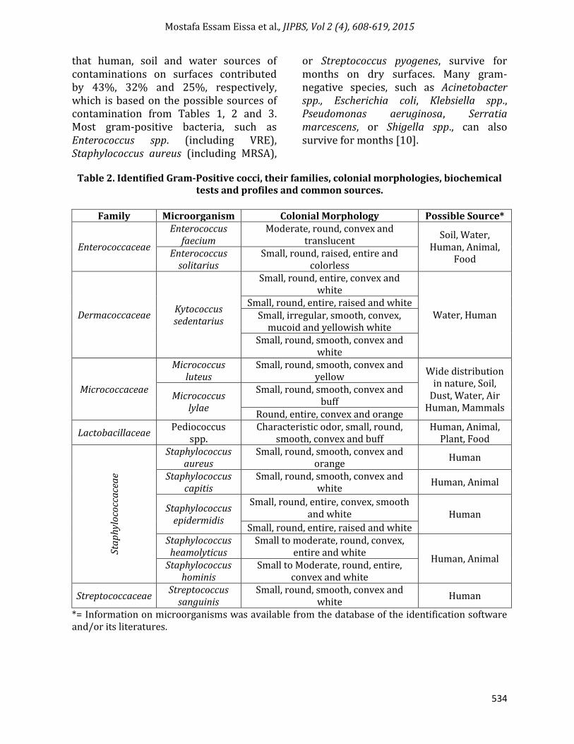
specific for each area, product and process combination. 3. Results and Discussion Table 1, 2 and 3 shows the identified bacteria, their families, colonial morphologies, biochemical reactions (required by the miniaturized biochemical
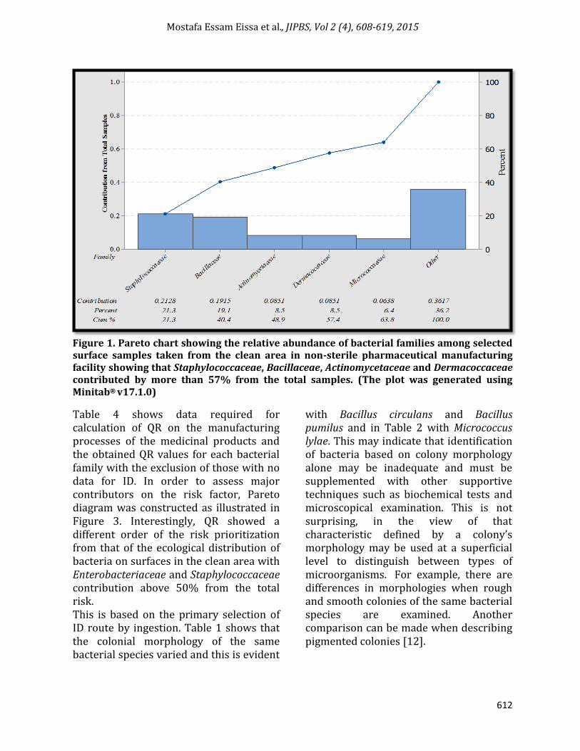
identification system for E/NF and GP kits) and their possible source. Bacteria from families Staphylococcaceae, Bacillaceae, Actinomycetaceae, Dermacoccaceae and Micrococcaceae contributed to about 64% of the total samples. This finding is illustrated by Pareto chart in Figure 1.
Table 1. Identified Gram-Negative bacilli, their families, colonial morphologies, biochemical
tests and profiles and common sources.
Family Microorganism Colonial Morphology Oxidase /Indole
Possible Source*
Moraxella- ceae
Acinetobacter lwoffii
Moderate to large, wrinkled and white
N/N Soil, Water,
Human
Enterobac-teriaceae
Klebsiella pneumoniae ssp.
Ozaenae**
Small to moderate, round, entire, raised and transparent
N/N Soil, Human,
Animal
Klebsiella pneumoniae ssp. rhinosclerometis
Large, round, entire, convex and white
N/N
NA Gram-Negative
Rods
Small, round, entire and transparent
N/N
NA
Large, round, entire, raised and translucent buff
N/N
Small, round, entire, flat and buff N/N
Pseudomon-adaceae
Pseudomonas putida
Large, entire, convex and creamy P/N Soil, Water
Sphingomo-nadaceae
Sphingomonas paucimobilis
Large, irregular, smooth, flat and reddish orange
N/N Soil, Water,
Plant *= Information on microorganisms was available from the database of the identification software and/or its literatures. **= Identification system gave an alternative ID of a slightly lower value of confidence factor (c.f.) to Serratia fonticola which is similar to Klebsiella pneumoniae in habitat and belongs to the same family. N= Negative result. P= Positive result. If unidentified microorganisms were excluded, Gram-positive organisms would be contributed by 84% (cocci 52% and bacilli 32%) from the total detected bacteria on surfaces while Gram-negative rods gave 16% of the total samples. Bacteria widely distributed in clean rooms are mainly a group of Gram positive strains, showing high resistance to
selected disinfectants as demonstrated by Wu and Liu [2]. Their study also showed that predominant contaminant bacteria in the clean rooms of pharmaceutical facility were a group of Gram positive bacteria: either spore-forming Bacillus species or non-sporulating Staphylococcus species and Microbacterium species. This is demonstrated in Figure 2 which shows
Mostafa Essam Eissa et al., JIPBS, Vol 2 (4), 608-619, 2015
534
that human, soil and water sources of contaminations on surfaces contributed by 43%, 32% and 25%, respectively, which is based on the possible sources of contamination from Tables 1, 2 and 3. Most gram-positive bacteria, such as Enterococcus spp. (including VRE), Staphylococcus aureus (including MRSA),
or Streptococcus pyogenes, survive for months on dry surfaces. Many gram-negative species, such as Acinetobacter spp., Escherichia coli, Klebsiella spp., Pseudomonas aeruginosa, Serratia marcescens, or Shigella spp., can also survive for months [10].
Table 2. Identified Gram-Positive cocci, their families, colonial morphologies, biochemical
tests and profiles and common sources.
Family Microorganism Colonial Morphology Possible Source*
Enterococcaceae
Enterococcus faecium
Moderate, round, convex and translucent
Soil, Water, Human, Animal,
Food Enterococcus
solitarius Small, round, raised, entire and
colorless
Dermacoccaceae Kytococcus sedentarius
Small, round, entire, convex and white
Water, Human Small, round, entire, raised and white
Small, irregular, smooth, convex, mucoid and yellowish white
Small, round, smooth, convex and white
Micrococcaceae
Micrococcus luteus
Small, round, smooth, convex and yellow
Wide distribution in nature, Soil,
Dust, Water, Air Human, Mammals
Micrococcus lylae
Small, round, smooth, convex and buff
Round, entire, convex and orange
Lactobacillaceae Pediococcus
spp. Characteristic odor, small, round,
smooth, convex and buff Human, Animal,
Plant, Food
Sta
ph
ylo
cocc
ace
ae
Staphylococcus aureus
Small, round, smooth, convex and orange
Human
Staphylococcus capitis
Small, round, smooth, convex and white
Human, Animal
Staphylococcus epidermidis
Small, round, entire, convex, smooth and white Human
Small, round, entire, raised and white Staphylococcus heamolyticus
Small to moderate, round, convex, entire and white
Human, Animal Staphylococcus
hominis Small to Moderate, round, entire,
convex and white
Streptococcaceae Streptococcus
sanguinis Small, round, smooth, convex and
white Human
*= Information on microorganisms was available from the database of the identification software and/or its literatures.
Mostafa Essam Eissa et al., JIPBS, Vol 2 (4), 608-619, 2015
613
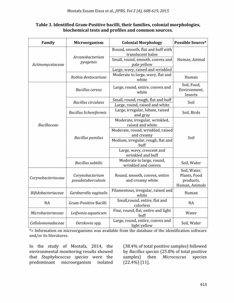
Table 3. Identified Gram-Positive bacilli, their families, colonial morphologies, biochemical tests and profiles and common sources.
Family Microorganism Colonial Morphology Possible Source*
Actinomycetaceae
Arcanobacterium pyogenes
Round, smooth, flat and buff with translucent halos
Human, Animal Small, round, smooth, convex and pale yellow
Large, wavy, raised and wrinkled
Rothia dentocariosa Moderate to large, wavy, flat and
white Human
Bacillaceae
Bacillus cereus Large, round, entire, convex and
white
Soil, Food, Environment,
Insects
Bacillus circulans Small, round, rough, flat and buff
Soil Large, round, raised and white
Bacillus licheniformis Large, irregular, lobate, raised
and gray Soil, Birds
Bacillus pumilus
Moderate, irregular, wrinkled, raised and white
Soil
Moderate, round, wrinkled, raised and creamy
Medium, irregular, rough, flat and buff
Large, wavy, crescent and wrinkled and buff
Bacillus subtilis Moderate to large, round,
wrinkled and convex Soil, Water
Corynebacteriaceae Corynobacterium
pseudotuberculosis Round, smooth, convex, entire
and creamy white
Soil, Water, Plants, Food
products, Human, Animals
Bifidobacteriaceae Gardnerella vaginalis Filamentous, irregular, raised and
white Human
NA Gram-Positive Bacilli Small,round, entire, flat and
colorless NA
Microbacteriaceae Leifsonia aquaticum Fine, round, flat, entire and light
buff Water
Cellulomonadaceae Oerskovia spp. Large, round, entire, convex and
light yellow Soil, Water
*= Information on microorganisms was available from the database of the identification software and/or its literatures. In the study of Mostafa, 2014, the environmental monitoring results showed that Staphylococcus species were the predominant microorganism isolated
(38.4% of total positive samples) followed by Bacillus species (25.8% of total positive samples) then Micrococcus species (22.4%) [11].
Mostafa Essam Eissa et al., JIPBS, Vol 2 (4), 608-619, 2015
612
Figure 1. Pareto chart showing the relative abundance of bacterial families among selected surface samples taken from the clean area in non-sterile pharmaceutical manufacturing facility showing that Staphylococcaceae, Bacillaceae, Actinomycetaceae and Dermacoccaceae contributed by more than 57% from the total samples. (The plot was generated using Minitab® v17.1.0)
Table 4 shows data required for calculation of QR on the manufacturing processes of the medicinal products and the obtained QR values for each bacterial family with the exclusion of those with no data for ID. In order to assess major contributors on the risk factor, Pareto diagram was constructed as illustrated in Figure 3. Interestingly, QR showed a different order of the risk prioritization from that of the ecological distribution of bacteria on surfaces in the clean area with Enterobacteriaceae and Staphylococcaceae contribution above 50% from the total risk. This is based on the primary selection of ID route by ingestion. Table 1 shows that the colonial morphology of the same bacterial species varied and this is evident
with Bacillus circulans and Bacillus pumilus and in Table 2 with Micrococcus lylae. This may indicate that identification of bacteria based on colony morphology alone may be inadequate and must be supplemented with other supportive techniques such as biochemical tests and microscopical examination. This is not surprising, in the view of that characteristic defined by a colony’s morphology may be used at a superficial level to distinguish between types of microorganisms. For example, there are differences in morphologies when rough and smooth colonies of the same bacterial species are examined. Another comparison can be made when describing pigmented colonies [12].

Mostafa Essam Eissa et al., JIPBS, Vol 2 (4), 608-619, 2015
615
Figure 2. 3-D Stacked Bar diagram showing the contribution of each type of bacteria from each of the major sources of the contamination in the clean rooms based on the frequency of detection from surface samples. (The graph was generated using Microsoft Office Excel 2007).
Figure 3. Pareto chart showing the QR of medicinally significant bacterial families among selected surface samples taken from the clean area in non-sterile pharmaceutical manufacturing and testing facilities showing that Enterobacteriaceae, Staphylococcaceae and Bacillaceae contributed by more than 65% from the total calculated risk. (The plot was generated using Minitab® v17.1.0)
An initial extensive identification program of EM isolates was an essential step as the most likely microflora found in pharmaceutical manufacturing can be hard to come by a definite source. The most commonly occurring
microorganisms come from human skin (either commensurable or transient) are Gram-positive microorganisms which include the following: Staphylococcus aureus, Micrococcus species and Bacillus species. Whereas those associated with

Mostafa Essam Eissa et al., JIPBS, Vol 2 (4), 608-619, 2015
616
eyes, ears and mucus include Gram-negative microorganisms, which can arise on rare occasions directly from the operator and include Pseudomonas aeruginosa [13]. Strains of Staphylococcus
aureus, members of Enterobacteriaceae and Pseudomonas aeruginosa were isolated from pharmaceutical effluent water [14].
Table 4. QR calculated for medically significant bacterial families, excluding those
unidentified and non-clinically significant bacteria.
Sr.
Bacterial Family Relative Abundance
Infective Dose* [15,16,17,18,19]
QR
1 Actinomycetaceae 0.09 5.6x10⁹ (Oth.) 0.051 2 Bacillaceae 0.19 1.0x10⁶ (Ing.), 2.0x10⁴ (Skn.), 8.0x10³ (Inh.) 0.127 3 Bifidobacteriaceae 0.02 2.0x10¹⁰ (Oth.) 0.018 4 Corynebacteriaceae 0.02 3.6x10⁷ (Oth.) 0.025 5 Enterobacteriaceae 0.04 1.0x101 (Ing.) 0.302 6 Enterococcaceae 0.04 1.0x10⁷ (Ing.) 0.045 7 Microbacteriaceae** 0.02 3.6x10⁷ (Oth.) 0.025 8 Moraxellaceae 0.02 1.0x10⁶ (Oth.) 0.031 9 Pseudomonadaceae 0.02 1.0x10³ (Skn), 1.0x10¹⁰ (Ing.) 0.062
10 Sphingomonadaceae 0.02 1.0x10⁶ (Ing.) 0.031 11 Staphylococcaceae 0.21 1.0x10⁵ (Oth.) 0.160 12 Streptococcaceae 0.02 1.0x10³ (Ing.) 0.062
*= In the absence of data on the infective doses (ID) of the human, those of the experimental animals were used till reliable data on the infectious doses of man could be determined. The lowest ID value was chosen if literatures provide a range of values. **= L. aquaticum was originally classified as ‘Corynebacterium aquaticum’ on the basis of its morphological and physiological characteristics [20], so the approach of giving the same ID of Corynebacteriaceae to Microbacteriaceae was selected. N.B. For those microorganisms with ID for more than one route of administration, ID per ingestion route (Ing.) was chosen primarily over skin (Skn.) and inhalation (Inh.) route. Bacteria with ID value per other (Oth.) routes of infection was used where no other data were available for the infective dose per oral route.
The current risk highlights the importance of covering the gap between the increasing number of pathogenic microorganisms and the available data about their infectiveness properties and its doses. The FDA Center for Food Safety and Applied Nutrition (CFSAN) has published a handbook on food-borne pathogenic bacteria referred to as the “Bad Bug Book”. Listed in this handbook is the following known pathogenic, bacteria, including: Salmonella spp., Staphylococcus aureus, Yersinia enterocolitica, Yersinia pseudotuberculosis, Shigella spp.,
Streptococcus spp. Bacillus spp., Enterococcus spp., Klebsiella pneumoniae, Pseudomonas spp., Serratia marcescens and Staphylococcus epidermidis Microbiology laboratory analyst in HIKMA
Pharma pharmaceutical company, Egypt. It seems that with the increase in the number of people with weak immune systems and microorganisms that have developed resistance to antimicrobials, the list of organisms of concern continues to grow. Therefore, it behooves the pharmaceutical microbiologist to stay abreast of the latest publications on the
Mostafa Essam Eissa et al., JIPBS, Vol 2 (4), 608-619, 2015
617
topic of objectionable organisms, to include published product recalls listed in trade publications such as The Gold Sheet [5]. The QR applied herein showed agreement with that required by USP<62>, 2014 with the exception of that Bacillaceae was ranked at the 3rd highest priority in the hazard of the product, while it is not listed by Pharmacopeia in the test of specified microorganisms [22]. This is not strange in view of what is listed in the “Bad Bug Book”. On the other hand, Pseudomonadaceae - in the present study – ranked 11th in the tested bacterial families when the oral route is considered which indicated that it is of low risk priority when considering manufacturing oral products. This is in agreement with Martinez, 2002 conclusion [23]. However, when considering the dermal route the risk significantly increased, indicating a rise in the possible hazard that could be delivered to patients through skin. Conclusion The current applied methodology for assessment and control of the microbiological surface cleanliness in the clean rooms in the non-sterile pharmaceutical manufacturing provides a guide for improvement and minimization of microbial hazard delivered to the customers through medicinal products in order to provide safe drug to the patients. This risk may work in complementation with similar risk studies performed for air, pharmaceutical water and other inputs that influence the quality of the manufactured products. When sufficient data are generated from an EM program of the clean area, the risk, study can be further broken into specific parts of each product, process and area. The limitations of the current QR are affected by
inadequacy of data about infective doses for each microorganism and the difficulty of calculating of the transfer factor of contamination from surface to the product either directly or indirectly. The current procedure of QR can be used as a milestone for microbiological risk control in other industries and fields that affect customer health such as food, cosmetics, hospitals and other healthcare facilities. Acknowledgement This work was supported partially financially by HIKMA Pharma pharmaceutical company – 2nd Industrial zone - 6th of October city. Reference and writing style review was performed by Dr. Engy Refaat Rashed. Data gathering and collection was performed by the microbiology laboratory team of the quality control department. References 1. Eissa ME, Abd El Naby M, Beshir MM:
Bacterial vs. fungal spore resistance to peroxygen biocide on inanimate surfaces. Bulletin of Faculty of Pharmacy Cairo University 2014; 52(2):219–224. http://doi.org/10. 1016/j.bfopcu.2014.06.003
2. Wu GF, Liu XH: Characterization of predominant bacteria isolates from clean rooms in a pharmaceutical production unit. J. Zhejiang Univ. Sci. B. 2007; 8(9):666–672.
3. Ljungqvist B, Reinmüller B: Interaction between air movements and the dispersion of contaminants: clean zones with unidirectional air flow. J. Parenter. Sci. Technol. 1993; 47(2):60-69.
4. Obuekwe IF, Ogbimi AO, Obuekwe CO: Microbial Contamination of Pharmaceutical Products in a Tropical

Mostafa Essam Eissa et al., JIPBS, Vol 2 (4), 608-619, 2015
618
Environment. Pakistan J. Sci. Ind. Res. 2002; 45(5):341–344.
5. Clontz L: Microbial limit and bioburden tests: validation approaches and global requirements. 2nd Ed. New York: CRC Press; 2008.
6. USP Chapter<1072>: Disinfectants and Antiseptics. United States Pharmacopeia 38/National Formulary 33, Baltimore, MD, USA; 2015.
7. USP Chapter<61>: Microbiological examination of non-sterile products: microbial enumeration tests. United States Pharmacopeia 38/National Formulary 33, Baltimore, MD, USA; 2015.
8. Sandle T: The use of risk assessment tools for microbiological assessment of clean room environments [Internet], GMP Guru: Insight Systems Inc 2010 [uploaded on 2010 Sep 27; cited 2015 Apr 25]. Available from: http://www.insightcgmp.com/gmp-guru/risk-assessment-tools.pdf.
9. World Health Organization (WHO): Environmental Monitoring of Clean Rooms in Vaccine Manufacturing Facilities: Points to consider for manufacturers of human vaccines. Geneva: Switzerland; 2012, pp 23.
10. Kramer A, Schwebke I, Kampf G: How long do nosocomial pathogens persist on inanimate surfaces? A systematic review. BMC Infect Dis. 2006; 6:130-137.
11. Eissa ME: Studies of Microbial Resistance against Some Disinfectants: Microbial Distribution & Biocidal Resistance in Pharmaceutical Manufacturing Facility. 1st Ed. Saarbrücken: LAP Lamber Academic Publishing; 2014, pp 14-53.
12. Breakwell D, Woolverton C, MacDonald B, Smith K, Robison R: Colony Morphology Protocol
[Internet]. ASM Microbe Library: American Society for Microbiology; 2007 Sep 29 [updated 2013 July 22; cited 2015 Jan 8]. Available from: http://www.microbelibrary.org/component/resource/laboratory-test/3136 -colony-morphology-protocol.
13. Jiminez L: Microorganisms in the Environment and their relevance to Pharmaceutical Processes, in: L. Jiminez (Ed.), Microbial Contamination Control in the Pharmaceutical Industry. New York: MarcelDekker; 2004, pp 8-9.
14. Lateef A: The microbiology of a pharmaceutical effluent and its public health implications. World J. Microbiol. Biotechnol. 2004; 20 (2):167-171.
15. Jost BH, Songer JG, Billington SJ: An Arcanobacterium(Actinomyces) pyogenes Mutant Deficient in Production of the Pore-Forming Cytolysin Pyolysin Has Reduced Virulence. Infect. Immun. 1999; 67(4):1723-1728
16. Bergogne-Berezin E, Towner KJ: Acinetobacter spp. as nosocomial pathogens: microbiological, clinical, and epidemiological features. Clin Microbiol. 1996; 9 (2):148-165.
17. Burrel DH: Experimental induction of caseusnlymphadenitis in sheep by intralymphatic inoculation of Corynobacterium bovis. Research in Veterinary Science. 1978; 24:269-276.
18. Leggett HC, Cornwallis CK, West SA: Mechanisms of Pathogenesis, Infective Dose and Virulence in Human Parasites. PLoS Pathog. 2012. doi: 10.1371/journal.ppat.1002512. 8(2): 1002512.
19. Madhaiyan M, Poonguzhali S, Lee J, Senthilkumar M, Lee KC, Sundaram S: Leifsonia soli sp. nov., a yellow-pigmented actinobacterium isolated
Mostafa Essam Eissa et al., JIPBS, Vol 2 (4), 608-619, 2015
619
from teak rhizosphere soil. Int J Syst Evol Microbiol 2010. doi: 10.1099/ijs.0.014373-0.1322-1327.
20. Gama JA, Abby SS, Vieira-Silva S, Dionisio F, Rocha EPC: Immune Subversion and Quorum-Sensing Shape the Variation in Infectious Dose among Bacterial Pathogens. PLoS Pathog 2012. doi: 10.1371/journal.ppat.1002503. e1002503-e1002503.
21. Cundell AM: Review of the Media Selection and Incubation Conditions for the Compendial Sterility and Microbial Limit Tests. Pharmacopeial Forum 2002; 28 (6):2034-2041.
22. USP Chapter<62>: Microbiological examination of non-sterile products: test for specified microorganisms. United States Pharmacopeia 38/National Formulary 33, Baltimore, MD, USA; 2015.
23. Martinez JE: Microbial Bioburden on Oral Solid Dosage Forms [Internet]. Pharmaceutical Technology; 2002 Feb [cited 2015 May 8]. Available from: http://images.alfresco.advanstar.com/alfresco_images/pharma/2014/08/22/618a9b92-e86b-4302-9c1f-f289091c4b4b/article-9672.pdf. 58-70.