Lab: Using filters to build an electrocardiograph (ECG or...
6
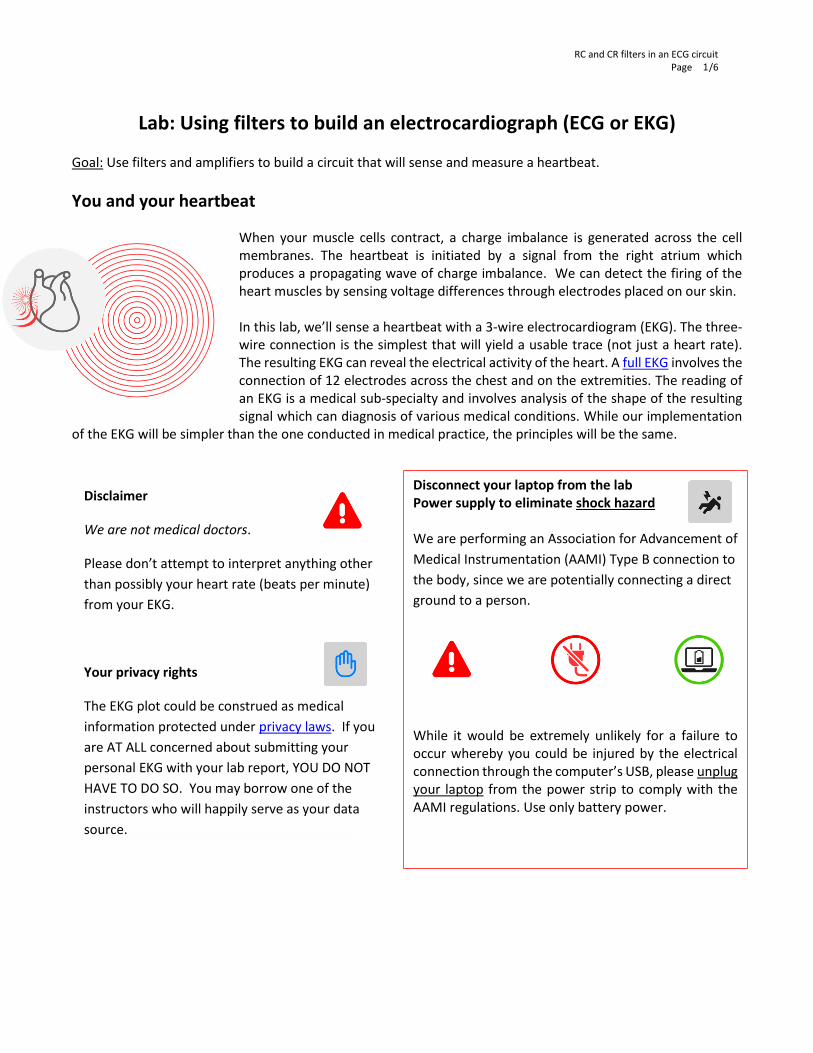
RC and CR filters in an ECG circuit Page /6 1 Lab: Using filters to build an electrocardiograph (ECG or EKG) Goal: Use filters and amplifiers to build a circuit that will sense and measure a heartbeat. You and your heartbeat When your muscle cells contract, a charge imbalance is generated across the cell membranes. The heartbeat is initiated by a signal from the right atrium which produces a propagating wave of charge imbalance. We can detect the firing of the heart muscles by sensing voltage differences through electrodes placed on our skin. In this lab, we’ll sense a heartbeat with a 3-wire electrocardiogram (EKG). The three- wire connection is the simplest that will yield a usable trace (not just a heart rate). The resulting EKG can reveal the electrical activity of the heart. A full EKG involves the connection of 12 electrodes across the chest and on the extremities. The reading of an EKG is a medical sub-specialty and involves analysis of the shape of the resulting signal which can diagnosis of various medical conditions. While our implementation of the EKG will be simpler than the one conducted in medical practice, the principles will be the same. Disclaimer We are not medical doctors. Please don’t attempt to interpret anything other than possibly your heart rate (beats per minute) from your EKG. Your privacy rights The EKG plot could be construed as medical information protected under privacy laws. If you are AT ALL concerned about submitting your personal EKG with your lab report, YOU DO NOT HAVE TO DO SO. You may borrow one of the instructors who will happily serve as your data source. Disconnect your laptop from the lab Power supply to eliminate shock hazard We are performing an Association for Advancement of Medical Instrumentation (AAMI) Type B connection to the body, since we are potentially connecting a direct ground to a person. While it would be extremely unlikely for a failure to occur whereby you could be injured by the electrical connection through the computer’s USB, please unplug your laptop from the power strip to comply with the AAMI regulations. Use only battery power.
Transcript of Lab: Using filters to build an electrocardiograph (ECG or...
RC and CR filters in an ECG circuit Page /6
1
Lab: Using filters to build an electrocardiograph (ECG or EKG) Goal: Use filters and amplifiers to build a circuit that will sense and measure a heartbeat.
You and your heartbeat
When your muscle cells contract, a charge imbalance is generated across the cell membranes. The heartbeat is initiated by a signal from the right atrium which produces a propagating wave of charge imbalance. We can detect the firing of the heart muscles by sensing voltage differences through electrodes placed on our skin. In this lab, we’ll sense a heartbeat with a 3-wire electrocardiogram (EKG). The three-wire connection is the simplest that will yield a usable trace (not just a heart rate). The resulting EKG can reveal the electrical activity of the heart. A full EKG involves the connection of 12 electrodes across the chest and on the extremities. The reading of an EKG is a medical sub-specialty and involves analysis of the shape of the resulting signal which can diagnosis of various medical conditions. While our implementation
of the EKG will be simpler than the one conducted in medical practice, the principles will be the same.
Disclaimer
We are not medical doctors.
Please don’t attempt to interpret anything other
than possibly your heart rate (beats per minute)
from your EKG.
Your privacy rights
The EKG plot could be construed as medical
information protected under privacy laws. If you
are AT ALL concerned about submitting your
personal EKG with your lab report, YOU DO NOT
HAVE TO DO SO. You may borrow one of the
instructors who will happily serve as your data
source.
Disconnect your laptop from the lab Power supply to eliminate shock hazard We are performing an Association for Advancement of
Medical Instrumentation (AAMI) Type B connection to
the body, since we are potentially connecting a direct
ground to a person.
While it would be extremely unlikely for a failure to occur whereby you could be injured by the electrical connection through the computer’s USB, please unplug your laptop from the power strip to comply with the AAMI regulations. Use only battery power.

RC and CR filters in an ECG circuit Page /6
2
Figure 1: First section of the EKG circuit.
The circuit in Figure 1 uses the same instrumentation amplifier chip you used in the strain gauge lab. The connection AD623 connection diagram is shown below.
The resistor between pins 1 and 8 sets the gain of the amplifier,
𝐺 =𝑉𝑜𝑢𝑡
𝑉𝑖𝑛= 1 +
100 𝑘Ω
𝑅𝐺 ,
as described in the AD623 data sheet. The chip provides an output which is simply,
𝑉𝑜𝑢𝑡 = 𝑉𝑟𝑒𝑓 + 𝐺(𝑉𝑖𝑛+ − 𝑉𝑖𝑛−).
What is output voltage, 𝑉𝑜𝑢𝑡, when 𝑉𝑖𝑛+ = 𝑉𝑖𝑛−?
How is the output affected by 𝑽𝒓𝒆𝒇?
Because of setting 𝑉𝑟𝑒𝑓 to + _____ Volts, our
0-5 V power can amplify the signal when (𝑉𝑖𝑛+ − 𝑉𝑖𝑛−)> 0 and (𝑉𝑖𝑛+ − 𝑉𝑖𝑛−)<0.
If we used the ground as the reference, then we would only be able to see anything when 𝑉𝑖𝑛+ > 𝑉𝑖𝑛−; we would lose the signal when (𝑉𝑖𝑛+ − 𝑉𝑖𝑛−)<0.
1. Build the circuit shown
There are two filters in this circuit. Draw a dotted line
around each and identify what type of filter it is. What
are the characteristic frequencies of each filter?
What are we using for 𝑉𝑟𝑒𝑓 in Figure 1?

RC and CR filters in an ECG circuit Page /6
3
The EKG schematic is broken into two sections: Figure 1 and 2. BUILD SLOWLY AND TEST AS YOU ADD FEATURES.
DO NOT GO ON TO FIGURE 2 UNTIL YOU BELIEVE FIGURE 1 IS WORKING.
We will use the WaveGen feature of the Analog Discovery to simulate a heartbeat and test the circuit as you go.
For this testing, you do not need to collect data for your lab report. Simply diagnose whether the circuit is acting as you expect. Make sure that you understand what the circuit is doing to different test frequencies and connect that expectation to your observations. Keep the amplitude of the WaveGen fixed, try adjusting the frequency to values greater than a heartbeat, less than a heartbeat, and equal to heartbeat. Monitor the circuit output. Remember the ultimate goal will be to preserve relevant frequencies and remove frequencies associated with noise and other artifacts not associated with the EKG.
2. Test as you build Figure 1.
Ensure the Discovery and circuit share a ground. Use Wavegen 1 as the left wrist. Use a wire connected to Ground as the right wrist.
Use Channel 1+ as a probe at points in your circuit
to see if you are getting the signal you expect.
Where should you
connect Channel 1-?
Connect Discovery
Set up WaveGen as a sinusoidal signal
What would be a good choice
for a simulated heartbeat
frequency in Hz?
Note the low amplitude and
offset of the sine wave.
►Run scope and test Figure 1
RC and CR filters in an ECG circuit Page /6
4
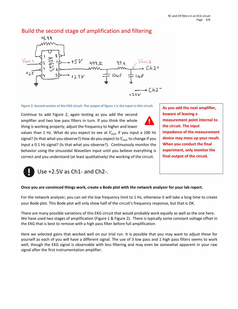
Figure 2: Second section of the EKG circuit. The output of figure 1 is the input to this circuit.
Continue to add Figure 2, again testing as you add the second
amplifier and two low pass filters in turn. If you think the whole
thing is working properly, adjust the frequency to higher and lower
values than 1 Hz. What do you expect to see at 𝑉𝑜𝑢𝑡 if you input a 100 Hz
signal? (Is that what you observe?) How do you expect to 𝑉𝑜𝑢𝑡 to change if you
input a 0.1 Hz signal? (Is that what you observe?). Continuously monitor the
behavior using the sinusoidal WaveGen input until you believe everything is
correct and you understand (at least qualitatively) the working of the circuit.
Once you are convinced things work, create a Bode plot with the network analyzer for your lab report.
For the network analyzer, you can set the low frequency limit to 1 Hz, otherwise it will take a long time to create
your Bode plot. This Bode plot will only show half of the circuit’s frequency response, but that is OK.
There are many possible variations of this EKG circuit that would probably work equally as well as the one here. We have used two stages of amplification (Figure 1 & Figure 2). There is typically some constant voltage offset in the EKG that is best to remove with a high pass filter before full amplification. Here we selected gains that worked well on our trial run. It is possible that you may want to adjust these for yourself as each of you will have a different signal. The use of 3 low pass and 1 high pass filters seems to work well, though the EKG signal is observable with less filtering and may even be somewhat apparent in your raw signal after the first instrumentation amplifier.
As you add the next amplifier,
beware of leaving a
measurement point internal to
the circuit. The input
impedance of the measurement
device may mess up your result.
When you conduct the final
experiment, only monitor the
final output of the circuit.
Build the second stage of amplification and filtering
Use +2.5V as Ch1- and Ch2-.

RC and CR filters in an ECG circuit Page /6
5
Now that you are convinced things work, you will need:
Pad placement:
inside of the wrists to the AD623 through the 100K resistor;
ground electrode can be on the back of your hand. Any electrode placement on the arms will work. The measurement will need to go across the body (i.e. left and right arms). The wrists, inside forearms, or close to your shoulder all work – depending on what you are comfortable with and whether you are wearing short or long sleeves today. Closer to your heart will typically yield a larger signal. Maintaining good skin contact is important. (The 100 K resistor is for safety to isolate you from the power sources. This extra resistor is to comply with medical
electronics standards; the risk of shock through the computers USB power supply is exceedingly low. )
Make sure to save some data before you close down for the day. See Figure 3 for a sample of what a “good” trace
looks like. Also, take a picture of your circuit before you disassemble.
3. Collect heartbeat data from a person
3 3 1 Willing data
source – you or
someone else
caution Shock
hazard
Disconnect AC power
& earth ground
Use battery
power only
Connect electrodes to circuit

RC and CR filters in an ECG circuit Page /6
6
Deliverables
In your lab report you should include:
1. A Bode plot of the final circuit.
2. A clean EKG trace.
3. A picture of your beautiful, clean circuit with no long loopy wires.
4. You will also want to provide a basic description of the circuit function; i.e. answer how many filters are
used? What types? What are the characteristic frequencies? What is the overall amplification of the final
circuit?
Now that our circuits are becoming more complex you will need to provide a little more interpretation and analysis
than in some of the first few lab reports for this course.
Figure 3: Sample EKG trace.