Lab discussion leaders: 02 April
38
1 1 1. Circulation (Ch 23) http://eebweb.arizona.edu/eeb_course_websites.htm Lecture 26 24 March 2008 Vertebrate Physiology ECOL 437 (MCB/VetSci 437) Univ. of Arizona, spring 2008 Kevin Bonine & Kevin Oh 14-11, Vander 2001 2 Housekeeping, 24 March 2008 Upcoming Readings Mon 24 Mar: Ch 23 Wed 26 Mar: Ch 23 LAB Wed 26 Mar: no reading Fri 28 Mar: Ch 23 Mon 31 Mar: Ch 24 Lab discussion leaders: 02 April 1pm – Vangie & Christina 3pm – Prasun & Ajay Lab discussion leaders: 09 April 1pm – none 3pm – Nina
Transcript of Lab discussion leaders: 02 April

1
1
1. Circulation (Ch 23)
http://eebweb.arizona.edu/eeb_course_websites.htm
Lecture 2624 March 2008
Vertebrate PhysiologyECOL 437 (MCB/VetSci 437)Univ. of Arizona, spring 2008
Kevin Bonine & Kevin Oh
14-11, Vander 2001
2
Housekeeping, 24 March 2008
Upcoming ReadingsMon 24 Mar: Ch 23Wed 26 Mar: Ch 23 LAB Wed 26 Mar: no readingFri 28 Mar: Ch 23Mon 31 Mar: Ch 24
Lab discussion leaders: 02 April1pm – Vangie & Christina3pm – Prasun & Ajay
Lab discussion leaders: 09 April1pm – none3pm – Nina

2
3
Vertebrate
Circulation
4
1. Circulation 2. Heart Muscle3. Heart Function4. Diving Response
12-10 Randall et al.

3
5
(Eckert, 12-4)To
Lungs
From body
To BodyviaAORTA
FromLungs
Mammalian Heart
Chordae tendinae
6
Vertebrate Circulation (too big for diffusion!)
Divided into Central and Peripheral
Heart is main propulsive organ
Arterial system-distributes blood-regulates pressure
Capillaries-transfer between blood and tissues
Venous system-return blood to heart-storage reservoir
Focus on Mammalian Circulation with some exceptions

4
7Knut Schmidt_Nielsen 1997
Gravityand BP
8
Circulatory Roles and Components
Valves control direction of blood flow
Smooth muscle controls diameter of peripheral vessels, thereby altering resistance and flow to different tissues
Sherwood 1997

5
9
Vander 2001
Circulatory Roles and Components
-Gases (CO2, O2)-Nutrients-Waste-Hormones-Antibodies-Salts-etc.
-TemperatureRegulation
RBCs
-Blood volume 5-10% of body volume
10
gills simple (and linear):1. Blood goes to gills2. O2-rich blood goes to tissues3. O2-poor blood goes to heart4. Blood gets pumped back to gills
lungs more complex because get 2 circuits in parallel:1. Pulmonary circuit (lower pressure)2. Systemic circuit (higher pressure)
Development of Terrestrial Circulatory System:

6
11
Fish Circulation through gills
(Eckert, 12-16)
12
Addition of lungs more complicated (Eckert, 12-16)
Watervs.air

7
13
Two parallel closed circuits:
1. Pulmonary(lower press.)
2. Systemic
(Eckert, 12-3) Note venousreservoir
Mammalian Circulation
14
9-3, Sherwood 1997
Tissue Beds in Parallel, not Series
All cells within 2-3 cells of a capillaryCan control amount of flow to each tissue independently

8
15
In addition to Heart,
Blood also moved via1. Elastic recoil of arteries2. Squeezing of vessels during body movement3. Peristaltic contractions of smooth muscle in vessels
16
(Eckert, 12-4)ToLungs
From body
To BodyviaAORTA
FromLungs
Mammalian Heart
Chordae tendinae

9
17
14-14,
Vander 2001
Mammalian HeartNo valves as Enter
Atria
18
Amphibians and Reptiles (except crocodilians) with3 chambers (= one ventricle, two atria)
- incomplete ventricular septum
- BUT separate rich and poor blood
- AND alter pressure in systemic and pulmonary
- able to alter flow to systemic or pulmonary circuit
Non-Mammalian Heart Examples:

10
19
Amphibians:
only vertebrates where O2 poor blood to skin(as well as to lungs)
adults with paired pulmocutaneous arteriesdivide into two branches1. Pulmonary2. Cutaneous (to flanks and dorsum)
skin provides 20-90% O2 uptake30-100% CO2 release
Cardiovascular System
20
FROG Heart
conus arteriosusw/ spiral valve
trabeculae(create channels)
role of Tb and HR
CardiovascularSystem
Pough et al., 2001Fig 6-8
Gets poor
Gets rich
11
21
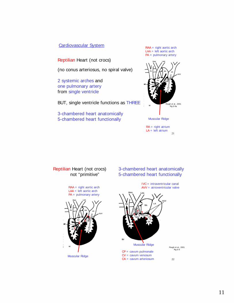
Reptilian Heart (not crocs)
(no conus arteriosus, no spiral valve)
2 systemic arches and one pulmonary arteryfrom single ventricle
BUT, single ventricle functions as THREE
3-chambered heart anatomically5-chambered heart functionally
Cardiovascular System RAA = right aortic archLAA = left aortic archPA = pulmonary artery
Muscular Ridge
Pough et al., 2001Fig 6-9a
RA = right atriumLA = left atrium
22
Pough et al., 2001Fig 6-9
Reptilian Heart (not crocs) not “primitive”
RAA = right aortic archLAA = left aortic archPA = pulmonary artery
Muscular RidgeCP = cavum pulmonaleCV = cavum venosumCA = cavum arteriosum
IVC = intraventricular canalAVV = atrioventricular valve
1
22
1 4
2
55
6
77
3-chambered heart anatomically5-chambered heart functionally
Muscular Ridge
3

12
23
R to LO2 poor to systemic via aortic arches(short delay between valves opening)
L to RO2 rich to pulmonary artery(longer delay between valves opening)
Reptilian and Amphibian Circulation
Cardiac Shunts (in 3-chambered heart)
1. temperature regulation2. breath holding (diving, turtle in shell, inflated lizards)3. stabilize O2 content of blood when breathe intermittently
24
Mammalian fetus:
Ductus arteriosus (R -> L shunt, lung bypass)-pulmonary artery to systemic arch-when lung inflate resistance down (pulm)-when lose placental circ. resistance up (syst)-closes at birth
Foramen ovale (interatrial shunt R -> L)-hole in wall between atria-closes at birth
13
25
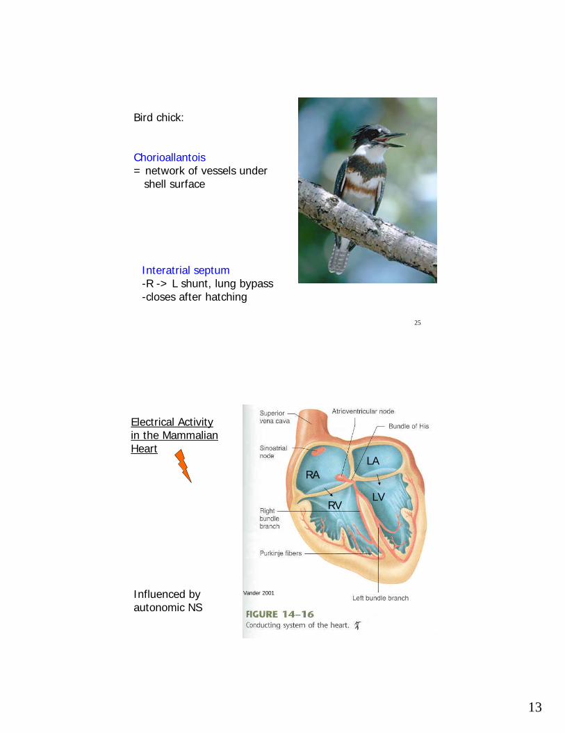
Bird chick:
Chorioallantois= network of vessels under
shell surface
Interatrial septum-R -> L shunt, lung bypass-closes after hatching
26
Vander 2001
Electrical Activity in the Mammalian Heart
Influenced by autonomic NS
RALA
LVRV

14
27
Sherwood 1997
Cardiac Cells electronically linked by Gap Junctions
(except from atrial to ventricular cells…)
28
Vander 2001
Electrical Activity in the Mammalian Heart

15
29
Recall AP and refractory period differences…
(Eckert, 12-7)
30
Vander 2001
Types of Cardiac Cells:
A. Contractile
B. Conducting
~ autorhythmic
~ fast-conducting
SA nodeAV node
InternodalInteratrialBundle of HisPurkinjeEtc.

16
31
Types of Cardiac Cells:
A. Contractile
B. Conducting
- 1° autorhythmic
-1° fast-conducting
SA nodeAV node
Pacemakers:
-Normally HR driven by SA node
-Others are Latentpacemakers
-Called Ectopicpacemaker when node other than SA driving HR
InternodalInteratrialBundle of HisPurkinjeEtc.
32
Sherwood 1997

17
33
Sherwood 1997
~ SA node ~ latent rate
Sherwood 1997
34
Sherwood 1997
SA
AV
other
The
Hea
rt R
ate
Trai
n
oops

18
35
9-11, Sherwood 1997
Autorhythmic Cardiac Muscle (e.g. SA node)
~Transient Ca2+
channels
K+, Na+
Which way would you
alter channel permeabilitiesto speed or slow HR??
36
Sherwood 1997
Vander 2001Contractile Cardiac MuscleCa2+ current maintains plateau

19
37
Cardiac Muscle (the other striated muscle)
-Small muscle fiber cells with only one nucleus
-Individual fibers are connected to neighbors electronically via gap junctions
-Two types of fibers:1. Contractile (similar to skeletal muscle)2. Conducting (including pacemaker cells)
Do not contract, but transmit electrical signal
-Cardiac contraction myogenic (arises within heart)Can be influenced by autonomic nervous system(alpha, beta adrenoreceptors increase [Ca2+])
-Long-lasting AP with long plateau phase, and longrefractory period - why?
38
Cardiac Muscle (the other striated muscle)
-Intracellular calcium from SR and across plasma membrane (unlike in skeletal)
-Dihydropyridine receptors in T-tubules arevoltage-activated calcium channels
-Ryanodine receptors then release more calcium from SR into the cytoplasm (calcium-induced calcium release)
-During relaxation, Calcium pumped actively back into SR and out across plasma membrane
20
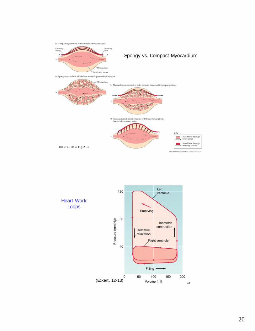
39Hill et al. 2004, Fig. 23.3
Spongy vs. Compact Myocardium
40(Eckert, 12-13)
Heart Work Loops

21
41
(Eckert, 12-4)To
Lungs
From body
To BodyviaAORTA
FromLungs
Mammalian Heart
Chordae tendinae
42
(Eckert, 12-8)
(Q,R,S masks atrial repolarization)

22
43
(Eckert, 12-8)
P
Q, R, S
T
4414-25, Vander 2001
WiggersDiagram
Valves open/close where pressure curves cross
760 mmHg = 1 atm= 9.8 m blood
1:2

23
45
Sherwood 1997
Atrial Kick
Heart filled ~same with increased HR
46
Sherwood 1997
Vander 2001
Frank-Starling Curve
Systole = Ventricular Emptying
Diastole = Ventricular Filling (rest)

24
47
Cardiac Output:
CO = cardiac output (ml/min from 1 ventricle)
SV = stroke volume (ml/beat from 1 ventricle)
= EDV – ESV (end-diastolic – end-systolic volume)
HR = heart rate (beats/min)
CO = HR x SV
- Heart can utilize different types of energy sources (unlike brain)
MABP = CO x TPRMABP = DP + 1/3(SP-DP)
48
Knut Schmidt-Nielsen 1997
Exercise
OxygenConsumptionX 20
Cardiac Output 6x

25
49
Cardiac Output Control
Sympathetic speeds heart rateand increases contractility
1. Norepinephrine binds to beta1 adrenergic receptors
2. Increases cAMP levels and phosphorylation
3. Activates cation channels (Na+) and increases HR
4. Epi and Norepi activate alpha and beta1adrenoreceptors which increase contractility and rate of signal conduction across heart
50Vander 2001
How increase contractility?
More Ca2+

26
51
HR control
Parasympathetic vs. Sympathetic
(Eckert, 12-5)
52
HR control
Parasympathetic slows heart rate
-Innervate Atria (Vagus nerve = Xth cranial nerve)
-Cholinergic (ACh)
-Alter SA node pacemaker potential by ⇑ K+ permeability
⇓ Ca2+ permeability
Parasympathetic innervation of AV node slows passage of signal between atria and ventricles

27
53
Peripheral Circulation
- Endothelium lining vessels- Middle layer with smooth muscle (esp. arteries)- Outer fibrous layer
Capillaries with ~ only Endothelium
54
(Eckert,12-26)

28
55
Peripheral Circulation
Compliance vs. Elasticity
~ Veins vs. Arteries
Volume Reservoir vs. Pressure Reservoir
56
Volume Reservoir vs. Pressure Reservoir
14-34, Vander 2001
(Eckert, 12-27)~Constant P and Q at Capillaries!

29
57
Venous System
- low pressure (11 mm Hg or less)
- thin walled veins with less muscle
- more compliant and less elastic
- valves
- blood moved by skeletal muscle (and smooth)
- breathing creates vacuum (low pressure) in chest to aid blood flow to heart
58
Microcirculation
- endothelium in capillaries is permeable
1. continuous
2. Fenestrated (kidney, gut)
3. Sinusoidal (liver, bone)
Less permeable
- Movement across walls, between walls, in vesicles
More permeable
- Bulk Flow…

30
59
Bulk Flow…
Fluid Pressurevs.Osmotic Pressure
(Eckert,12-38)
Filtration > Uptake
Lymph System to return excess fluid
Faster than diffusion
60
Bulk Flow… - Edema
- No RBCs; therefore not red
Lymph System
- Starvation- Lungs- Kidneys
- Drains interstitial spaces- has valves and smooth musculature- empties into thoracic duct at vena cavae- transport system for large hormones and fats into
blood stream- filariasis, elephantiasis- Reptiles and Amphibians with lymph hearts

31
61
Circulatory System Regulation
1. Feed Brain and Heart First2. Next Feed Tissues in Need3. Maintain volume, prevent edema, etc.
BaroreceptorsChemoreceptorsMechanoreceptorsThermoreceptors
Info. integrated at Medullary Cardiovascular Centermedulla oblongata and pons
Depressor Center Parasympathetic Effectors
Pressor Center Sympathetic Effectors
62
Circulatory System Regulation
Baroreceptors increase AP firing rate when BP increases
Sensed at carotid sinus, aortic arch, subclavian, common carotid, pulmonary
Usually leads to Sympathetic suppression to decrease BP
(Eckert, 12-43)

32
63
Circulatory System Regulation
Arterial Chemoreceptors in carotid and aortic bodies(More details when discuss ventilation)
e.g., low O2, high CO2, low pH leads to bradycardia and peripheral vasoconstriction(diving and not inflating lungs)
What about when not diving?
64
Circulatory System Regulation
Cardiac Mechanoreceptors and Chemoreceptors
Alter heart rate AND blood volume
e.g., ANP (Atrial Natruiretic Peptide) released in response to stretch
- leads to increased Na+ excretionand therefore greater urine output
33
65
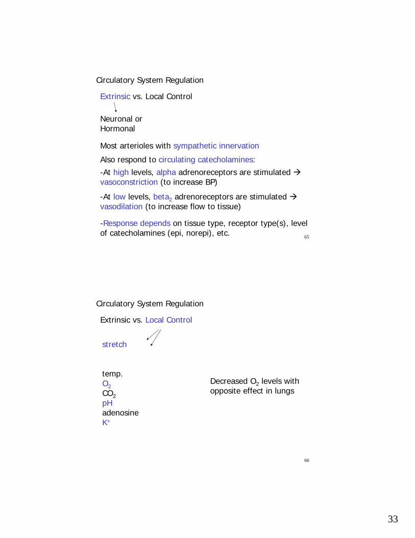
Circulatory System Regulation
Extrinsic vs. Local Control
Neuronal or Hormonal
Most arterioles with sympathetic innervation
Also respond to circulating catecholamines:-At high levels, alpha adrenoreceptors are stimulated vasoconstriction (to increase BP)
-At low levels, beta2 adrenoreceptors are stimulated vasodilation (to increase flow to tissue)
-Response depends on tissue type, receptor type(s), level of catecholamines (epi, norepi), etc.
66
Circulatory System Regulation
Extrinsic vs. Local Control
stretch
temp.O2CO2pHadenosineK+
Decreased O2 levels with opposite effect in lungs

34
67
Circulatory System Regulation
Extrinsic vs. Local Control
(Eckert, 12-45)
-Vasodilation-Relaxation
-Viagra acts by blocking breakdown of cGMP
NO (nitric oxide)
Released fromvascular endothelium:
68
Circulatory System Regulation
Extrinsic vs. Local Control
-Vasodilation
Histamine
Released in response to injury of connective tissue and leukocytes:

35
69
(Hill et al., 2004Fig 23.11)
70
Hemodynamics in Vessels
Vander 2001
14-11, Vander 2001
Flow depends primarily on pressure gradient and resistance

36
71
Hemodynamics
- Poiseuille’s Law:
Flow rate
8Lη
Q = (P1 – P2)πr4
Pressure Gradient
radius4
length viscosity
Use to approximate flow
Small change in radius large change in flow rate
72
Hemodynamics
- From Poiseuille’s Law:
Resistance
Q
R = (P1 – P2)
Pressure Gradient
radius4
Flow rate
viscosity
Small change in radius large change resistance
= 8Lη
πr4
length
Modifiable if vessel distensible under pressure

37
73
(Eckert, 12-25)
Summed resistance reduces pressure…
74
(Eckert, 12-23)Total Flow Rate same all along Circulatory System
River
Lake
River

38
75(Eckert, 12-24)
Shapes of curves slightly different because of RBCs (viscosity)
76
Why does blood in the lower extremities of aquatic organisms not pool as it may do in legs of
humans, giraffes, etc.?
FISH:
Blood tends to pool in tail b/c inertia and compression waves when swimming
-Veins in middle of body-Accessory caudal (tail) heart in some species