lab diagnosis TB ممتاز
45
description
Â
Transcript of lab diagnosis TB ممتاز


Introduction 1Introduction 1
Laboratory DiagnosisLaboratory DiagnosisTuberculosisTuberculosis
Dr. Kiarash Ghazvini Department for bacteriology and virology,
Mashhad University of medical Sciences

long, slender, straight or curved, acid fast bacilli
Mycobacterium tuberculosis
slow growers, obligate aerobes, intracellular bacterium
structure composed of high molecular weight acidic waxes,
mycolic acid, cord factor
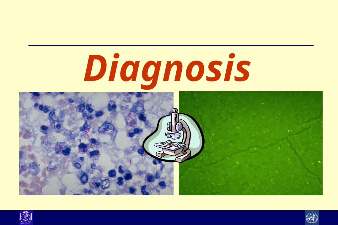
Diagnosis
EpidemiologEpidemiology 2y 2
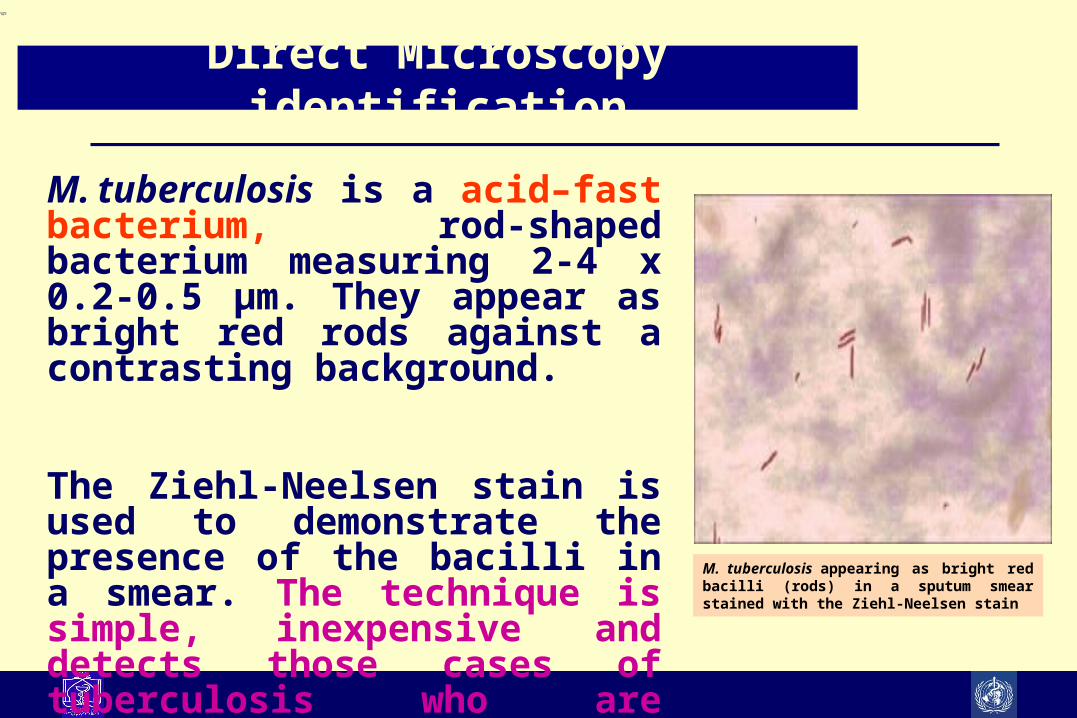
M. tuberculosis is a acid–fast bacterium, rod-shaped bacterium measuring 2-4 x 0.2-0.5 μm. They appear as bright red rods against a contrasting background.
The Ziehl-Neelsen stain is used to demonstrate the presence of the bacilli in a smear. The technique is simple, inexpensive and detects those cases of tuberculosis who are infectious.
Direct Microscopy identification
M. tuberculosis appearing as bright red bacilli (rods) in a sputum smear stained with the Ziehl-Neelsen stain


Reporting on AFB MicroscopyReporting on AFB Microscopy
Number of bacilli seen Result reported
None per 100 oil immersion fields Negative
1-9 per 100 oil immersion fields Scanty, reportexact number
10-99 per 100 oil immersion fields 1+
1-10 per oil immersion field 2+
> 10 per oil immersion field 3+
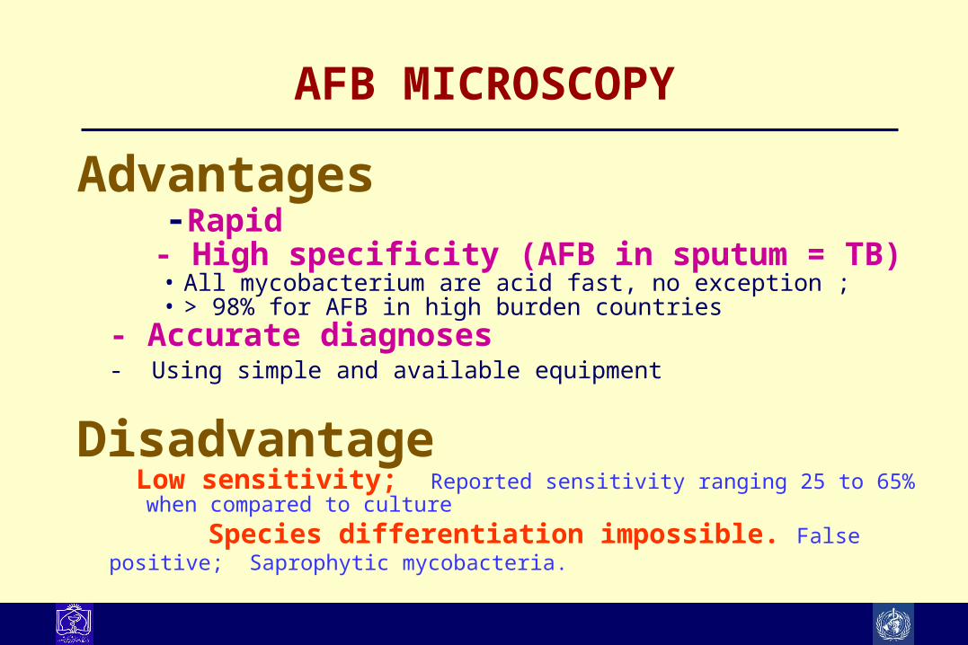
AFB MICROSCOPY
Advantages -Rapid - High specificity (AFB in sputum = TB)
• All mycobacterium are acid fast, no exception ; • > 98% for AFB in high burden countries
- Accurate diagnoses- Using simple and available equipment
Disadvantage Low sensitivity; Reported sensitivity ranging 25 to 65% when compared to culture
Species differentiation impossible. False positive; Saprophytic mycobacteria.

81%93%
100%
0%
50%
100%
First Second Third
Cum
ulat
ive
Pos
itivi
tyThree sputum smears are optimal
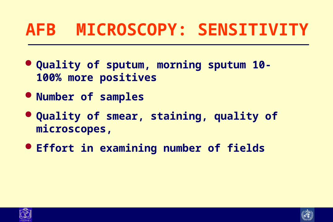
AFB MICROSCOPY: SENSITIVITY
Quality of sputum, morning sputum 10-100% more positives Number of samples Quality of smear, staining, quality of microscopes, Effort in examining number of fields
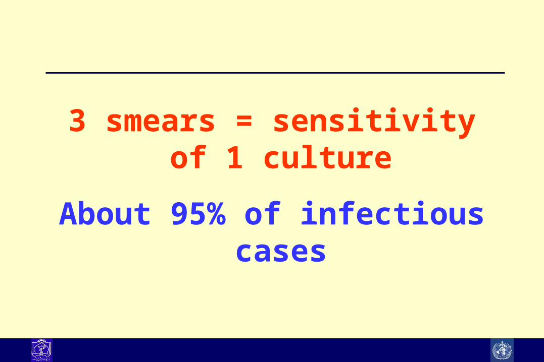
3 smears = sensitivity of 1 culture
About 95% of infectious cases

Smear-positive patients are 4-20 times more infectiousUntreated, a smear-positive patient may infect 10-15 persons/yearSmear-positive patients are much more likely to die if untreated
Rouillon A. Tubercle 1976;57:275-99
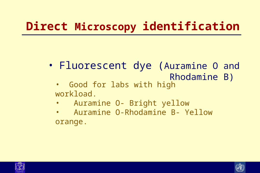
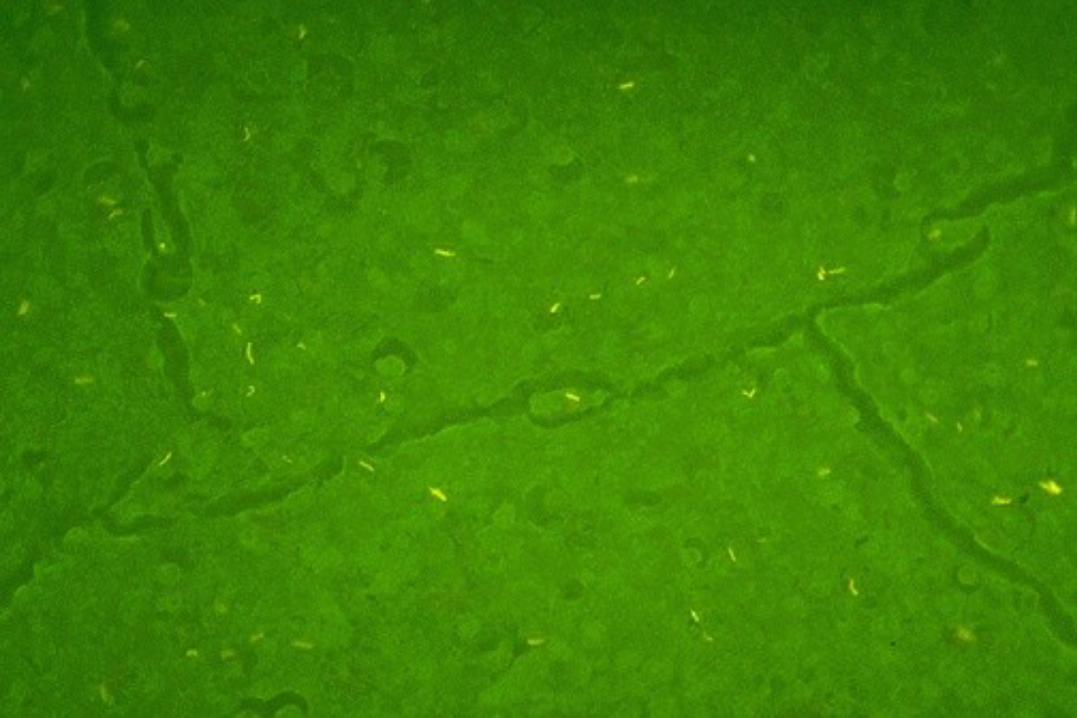
Direct Microscopy identification
• Fluorescent dye (Auramine O and Rhodamine B)
• Good for labs with high workload. • Auramine O- Bright yellow • Auramine O-Rhodamine B- Yellow orange.

Appropriate collection techniquesAppropriate collection techniques
–Time• Sputum -3 consecutive samples.
– Early morning complete sputum.• Urine -3 or more consecutive samples
– 24 h pooled ?
–Volume – Pleural, peritoneal fluids– Cerebrospinal fluid


CultureCultureEpidemioEpidemio
logy 3logy 3
M. tuberculosis grows in Lowenstein Jensen medium, which contains inhibitors to keep contaminants from outgrowing the organism.
Because of its slow growth, it takes 4-6 weeks before small buff-coloured colonies are visible on the medium.
Typical small, buff coloured colonies of M. tuberculosis on Lowenstein Jensen medium
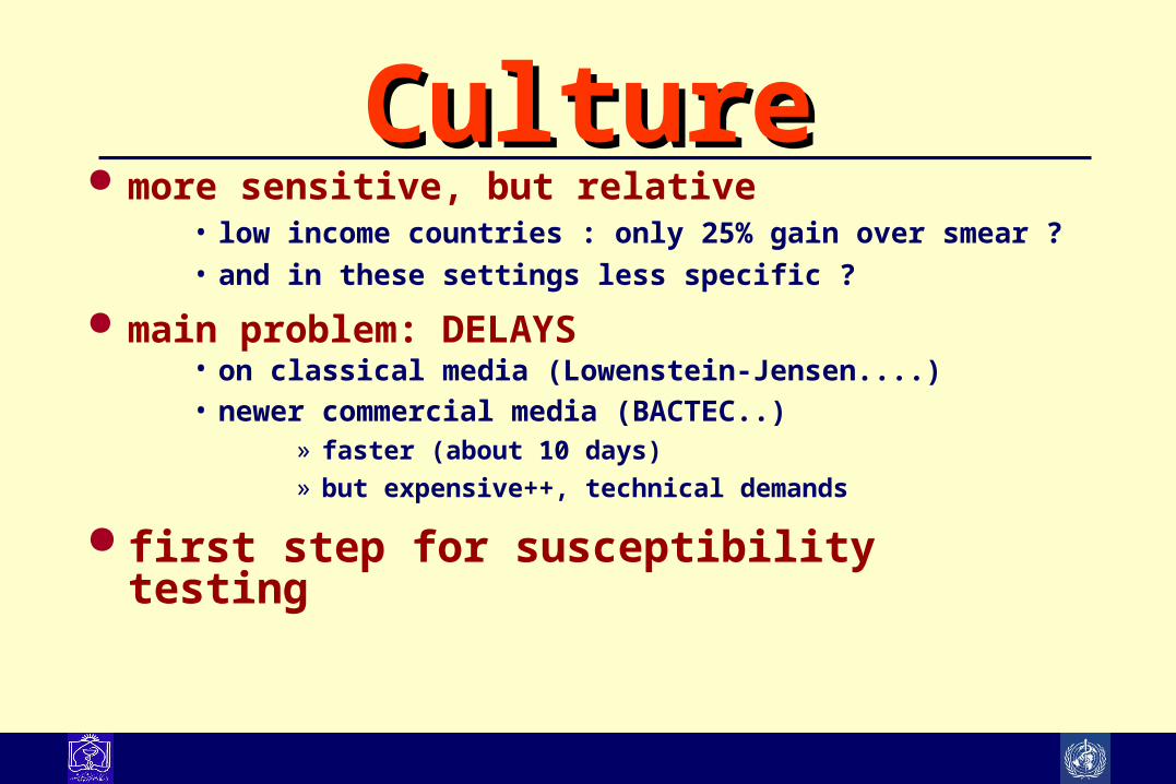
CultureCulture more sensitive, but relative
• low income countries : only 25% gain over smear ?• and in these settings less specific ?
main problem: DELAYS• on classical media (Lowenstein-Jensen....)• newer commercial media (BACTEC..)
» faster (about 10 days)» but expensive++, technical demands
first step for susceptibility testing
Other Methods for Other Methods for Laboratory Diagnosis of Laboratory Diagnosis of
Tuberculosis
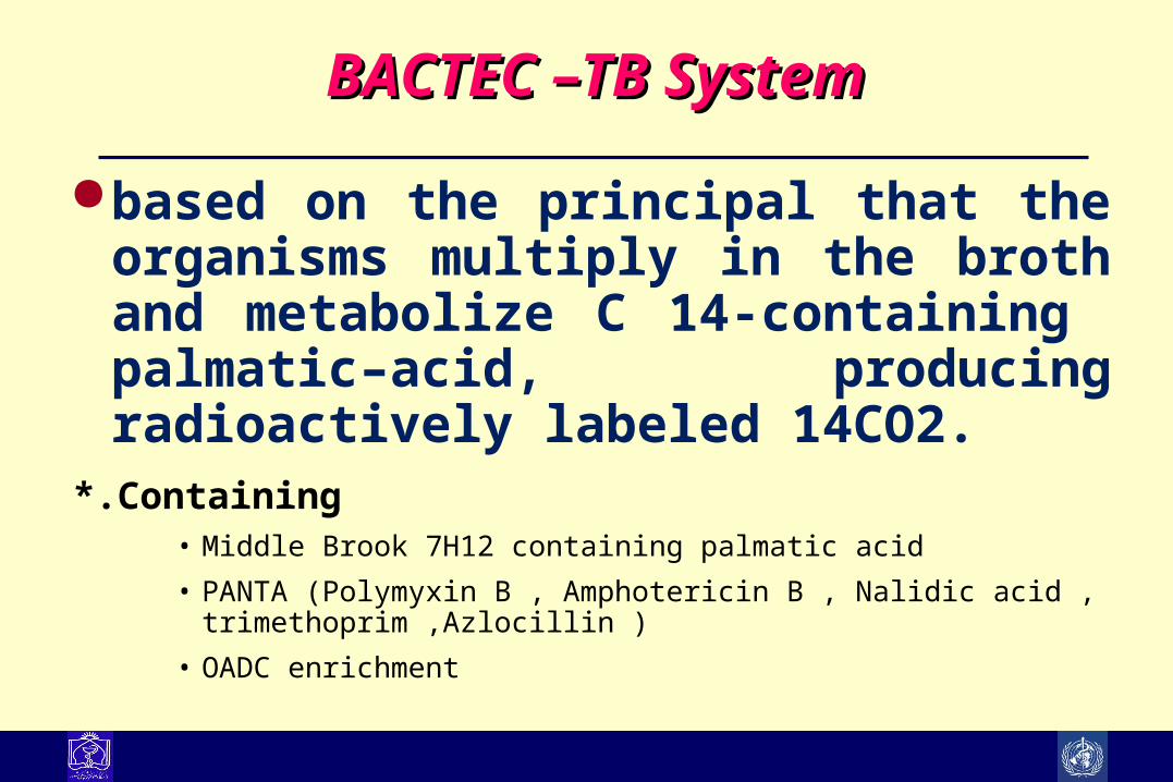
BACTEC –TB SystemBACTEC –TB System
based on the principal that the organisms multiply in the broth and metabolize C 14-containing palmatic–acid, producing radioactively labeled 14CO2.
*.Containing• Middle Brook 7H12 containing palmatic acid • PANTA (Polymyxin B , Amphotericin B , Nalidic acid ,
trimethoprim ,Azlocillin ) • OADC enrichment

Mycobacterium Growth Indicator Tubes Mycobacterium Growth Indicator Tubes (MGIT)(MGIT)
A fluorescent compound is embedded in silicone on the bottom of 16 x 100 mm
round-bottom tubes.
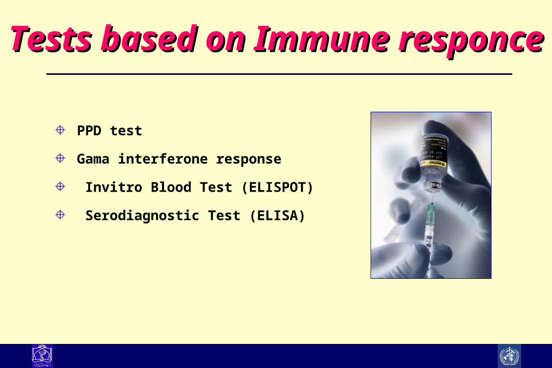
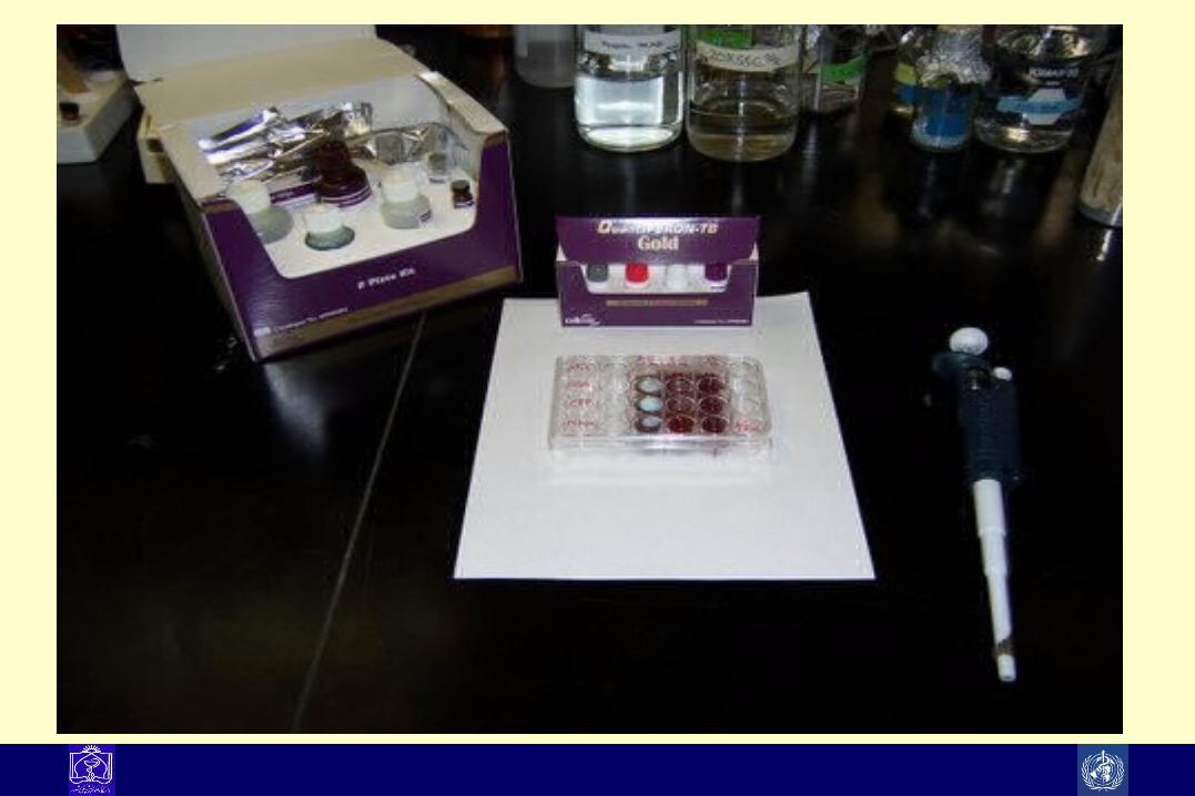
Tests based on Immune responceTests based on Immune responce
PPD test
Gama interferone response
Invitro Blood Test (ELISPOT)
Serodiagnostic Test (ELISA)

Blood Tests for TBBlood Tests for TB New blood tests examine immune response to TB antigens Draw blood from patient, expose white cells to TB antigens,
look for signs of immune response Two most common methods are ELISA and ELISPOT

Lancet 356: 1099 2000

Blood Tests for TBBlood Tests for TB One-time blood draw Less inter-reader variability than PPD Looks at response to 2 TB-specific antigens: ESAT-6 and
CFP-10 Antigens are not found in BCG, most non-TB mycobacteria ELISA and ELISPOT technologies commercially available
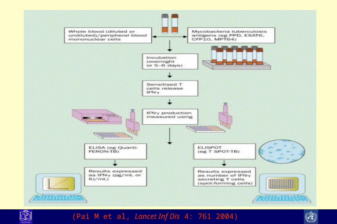
(Pai M et al, Lancet Inf Dis 4: 761 2004)
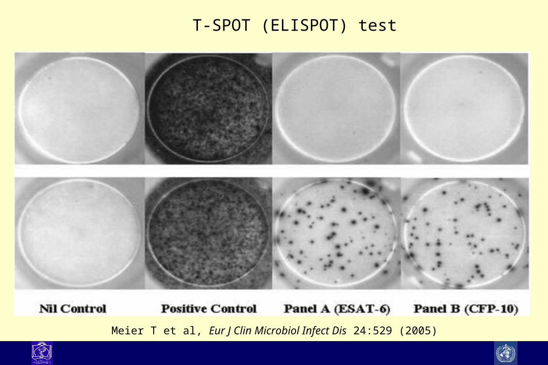
Meier T et al, Eur J Clin Microbiol Infect Dis 24:529 (2005)
T-SPOT (ELISPOT) test

TSTTST Blood testsBlood tests
Test itself cheap Clearly correlated with
development of TB Can be done anywhere Reliability fair Cross-reacts with BCG,
NTM
More expensive Unclear correlation with
development of TB Blood must be sent quickly to
lab Reliable Does not cross-react with
BCG, most NTM
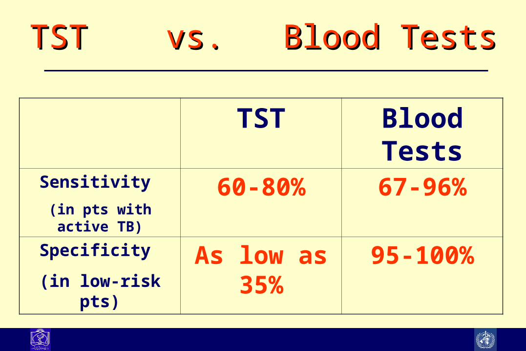
TST vs. Blood TestsTST vs. Blood Tests
TST Blood TestsSensitivity
(in pts with active TB)60-80% 67-96%
Specificity
(in low-risk pts)As low as 35% 95-100%

SummarySummary
Have great potential to reduce false-positive results Likely more sensitive for active TB, but clinical utility in this
setting less clear Logistical and cost barriers to implementation Ideally will replace TST in many settings
Adenosine deaminase (ADA)Adenosine deaminase (ADA) Adenosine deaminase (ADA) and interferon gamma studied
for dx of extrapulmonary TB Both are markers of immune response to TB
Not that specific

Immune dx of TBImmune dx of TB ADA and interferon gamma may be useful in endemic areas In low-incidence areas, specificity and sensitivity are not
good enough for routine use More specific markers needed
PCR polymerase chain amplification
1) Diagnose tuberculosis rapidly by identifying DNA from M. tuberculosis in clinical samples.
2) Determine rapidly whether acid-fast organisms identified by microscopic examination in clinical specimens are M.tuberculosis
3) Identify the presence of genetic modifications known to be associated with resistance to some anti mycobacterial agents.
4) Determine whether or not isolates of M.tuberculosis from different patients have a common origin in the context of
epidemiological studies.


Molecular methodMolecular method
Sensitivity and Specificity of PCRSensitivity and Specificity of PCR
Incase of smear and culture positive the sensitivity is ranging 80% to 90% with specificity of 97%-99%.
Incase of smear negative and culture positive the sensitivity is ranging 60% to 80% with specificity of 97%-
99%.
Disadvantages:– Identification of the target sequence of DNA doesn't imply organism viability.– Contamination of samples by product from previous PCR experiments
NAA Summary NAA is useful to distinguish TB from NTM in smear +
specimens Less sensitive in smear – specimens Clinical judgement must always take priority Relatively expensive tests; need data to support cost-
effectiveness

Real time PCRReal time PCR
Rapid analysis (typically under 90 min)
No post-amplification processing (no gels or autoradiographs)
Automated (data collection and analysis)
Objective (controls & standards can be built-in)
Precise, sensitive and reproducible

TB/HIV
MDR/XDR-TB