Lab 9 Histology and Gross Anatomy of the Nervous System.
39
Lab 9 Histology and Gross Anatomy of the Nervous System
-
Upload
diana-perry -
Category
Documents
-
view
228 -
download
3
Transcript of Lab 9 Histology and Gross Anatomy of the Nervous System.

Lab 9
Histology and Gross Anatomy of the Nervous System

Lab 9 Activities1. Identify neuron, pyramidal cell,
Purkinje cell, and peripheral nerve slides
2. Identify boldfaced structures on brain models
3. Identify specified parts in lab manual on sheep brain dissection
4. Identify cranial nerves (by name + number I - XII)

Parts of a Neuron Soma Dendrites Axon

Cell Body (Soma)
Contains the usual cellular organelles Primary site of synthesis & metabolism Cell membrane contains receptors to respond to stimuli Cell membrane transmits graded potentials Most cell bodies are located within the CNS
clusters of neuron cell bodies in the CNS are called nucleinuclei clusters of neuron cell bodies in the PNS are called gangliaganglia

Dendrites Short, tapering, highly
branched processes Not myelinated Contain some cell organelles Cell membrane contains
receptors to respond to stimuli
Transmit graded potentials towards the soma

Axons (nerve fiber) Originate at a cone-shaped region –
the axon hillock A single process that transmits
action potentials away from the soma
May be long (1 meter) or short (1 mm)
May branch (=collaterals) Collaterals end at
axonal terminals synapse with neurons or effector cells
(muscles or glands)

The Myelin Sheath Lipid-rich, covering of axons
Oligodendrocytes in CNS Neurilemmocytes in PNS
Dendrites are never myelinated Protects & electrically insulates Increases speed of nerve impulses
10-150x faster than unmyelinated fibers

STRUCTURAL CLASSIFICATIONS
Neurons
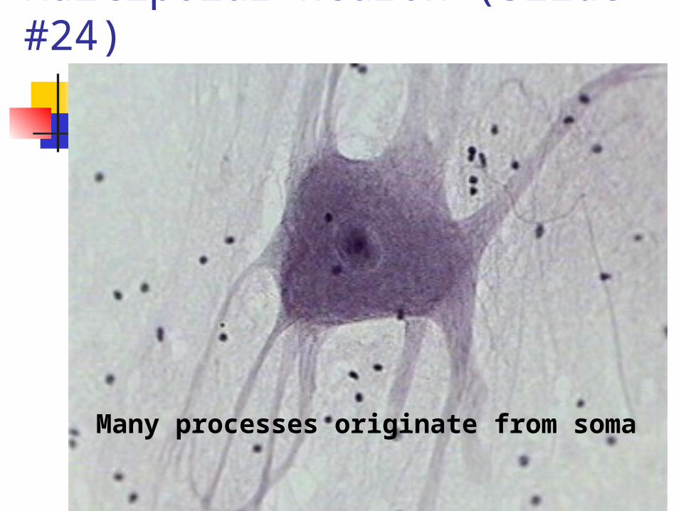
Multipolar Neuron (slide #24)
Many processes originate from soma

Bipolar Neuron
Two processes originate from soma

Unipolar Neuron
A single process originates from soma
SPECIFIC TYPESNeurons
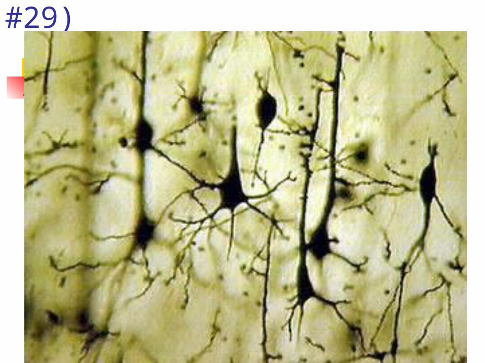
Pyramidal Cells (slide #29)

Pyramidal Cells

Pyramidal Cells

Purkinje Cells (slide #30)

Purkinje Cells of Cerebellum

Purkinje Cells

Purkinje Cells cytoskeleton stain

Peripheral Nerve
• Connective tissue elements:• Epineurium
(wraps nerve)• Perineurium
(wraps fascicle)
• Endoneurium (wraps axon & myelin sheath)

Nerve, cross-section (slide #28)
Epineurium
Perineurium
Endoneurium
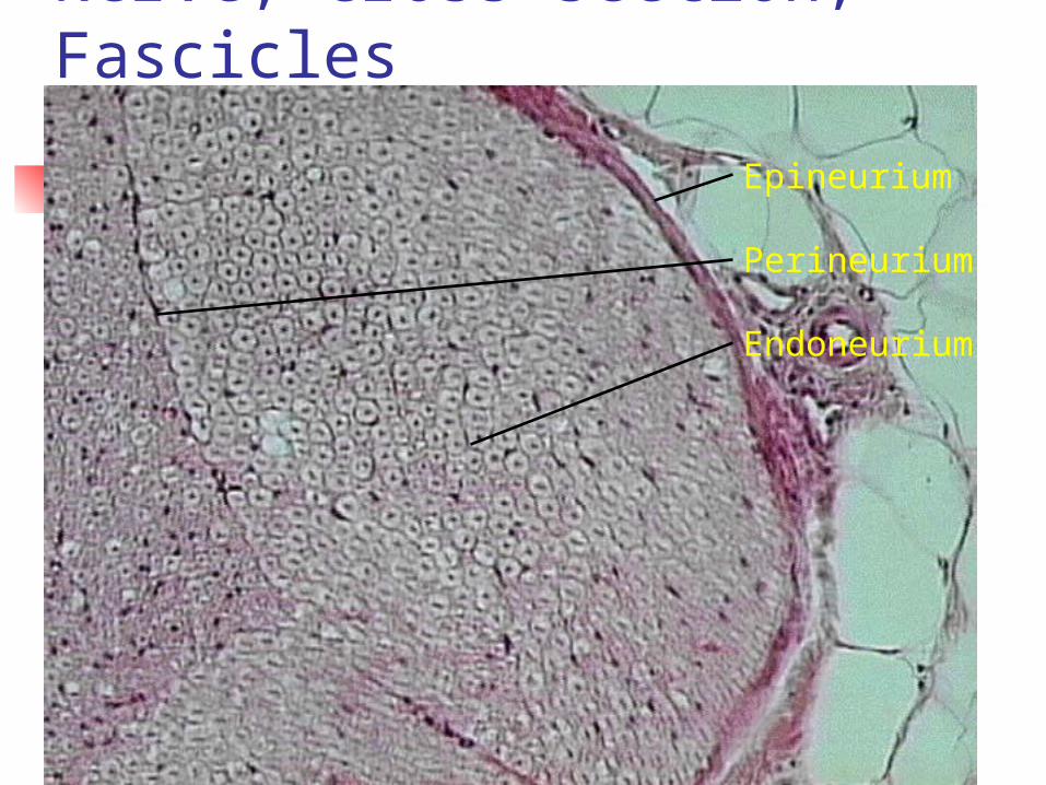
Nerve, cross section, Fascicles
Epineurium
Perineurium
Endoneurium

Nerve, cross section, Myelinated Fibers

Myelinated Axons, cross-section

Myelinated Axons, cross-section

Nerve, Long Section, 40X (slide # 28)

Nerve, long section, 100X

Neurofibrilar Node Nerve, 400X

Neurofibrilar Node, 1000X
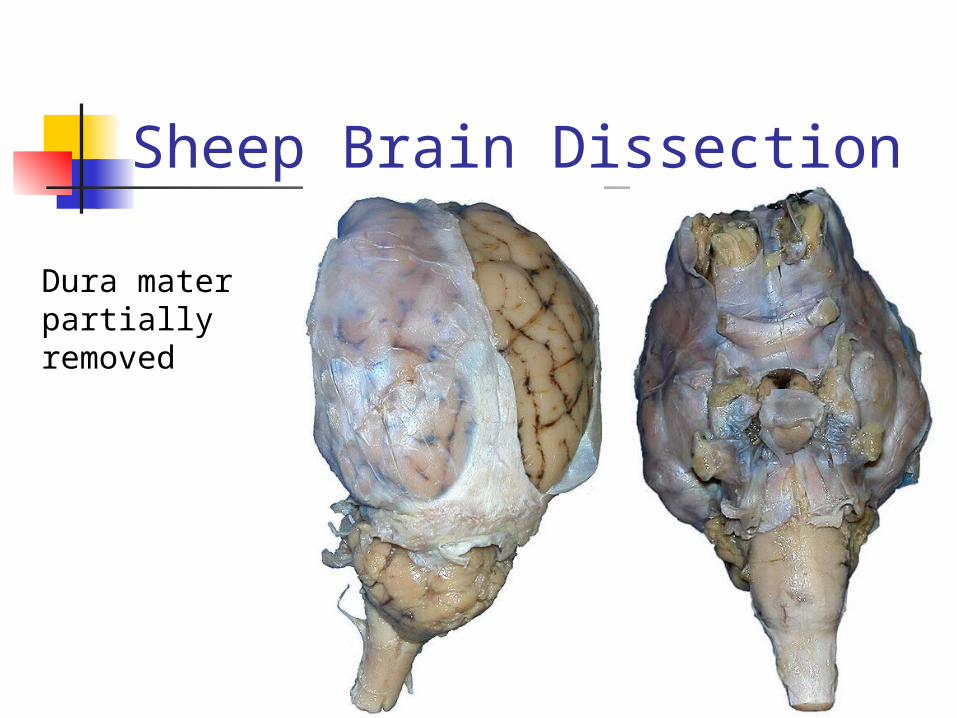
Sheep Brain Dissection
Dura mater partially removed

Sheep Brain Dissection
andandpia materpia mater

Sheep Brain Dissection
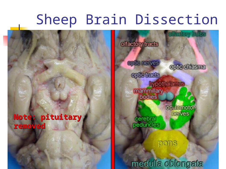
Sheep Brain Dissection
Note: pituitary Note: pituitary removedremoved

Sheep Brain Dissection
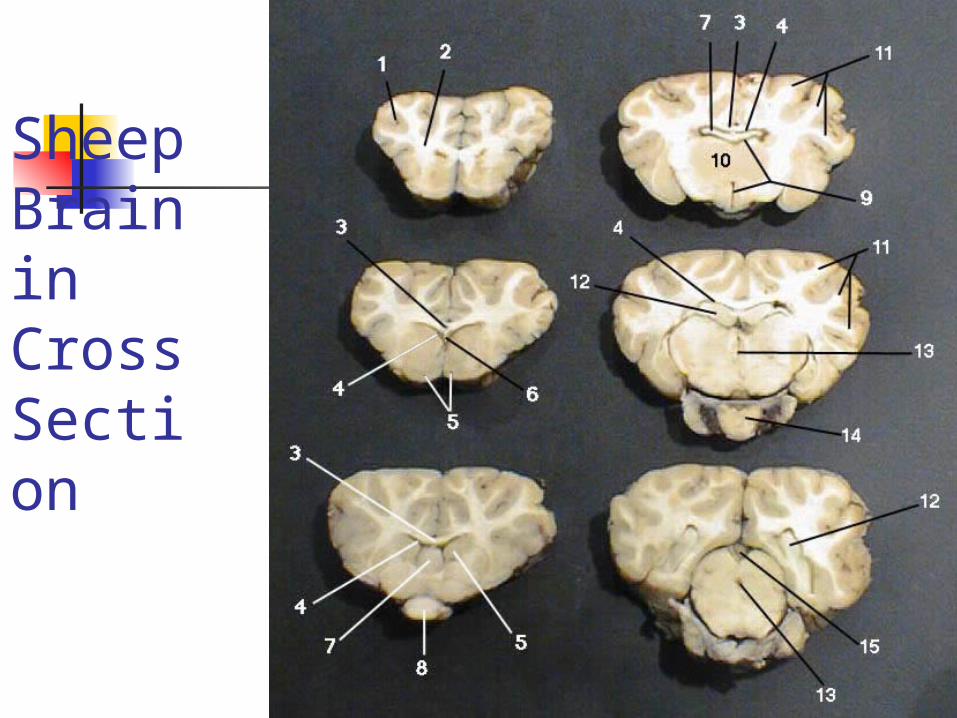
Sheep Brain in Cross Section

Key to Sheep Brain in Cross Section
1. Gray Matter 2. White Matter 3. Corpus Callosum 4. Lateral Ventricle 5. Caudate Nucleus 6. Septum
Pellucidum 7. Fornix 8. Optic Chiasm
9. Third Ventricle 10. Thalamus 11. Corona Radiata 12. Hippocampus 13. Cerebral
Aqueduct 14. Pituitary Gland 15. Pineal Gland

Human Cranial Nerves

Human Cranial Nerves

End of Lab 9 Presentation