LAB 5 - Bluegrass Community and Technical College · 2019-08-08 · Glomerulus (2) medulla . 53 B....
17
49 LAB 5 ANATOMY OF THE RESPIRATORY, DIGESTIVE, AND URINARY SYSTEMS Assignments: Due before lab: Quiz: Complete the cardiovascular case studies (page 48). Label diagrams of respiratory, digestive and urinary models (pages 55-65). Due next lab: Lab Exam I – covers labs 1 through 4, the Interactive Physiology CD ROM exercises, and the cardiovascular case studies. Summer Classes: In the summer, Labs 5 & 7 are combined. The quiz covers the Respiratory IP exercises on pages 69-73. Objectives: Identify anatomical structures on models of the Respiratory, Digestive, Urinary Systems Identify slides of the Respiratory, Digestive, and Urinary Systems and various indicated structures Identify organs of the Respiratory, Digestive, and Urinary Systems in the fetal pig
Transcript of LAB 5 - Bluegrass Community and Technical College · 2019-08-08 · Glomerulus (2) medulla . 53 B....

49
LAB 5
ANATOMY OF THE RESPIRATORY, DIGESTIVE, AND URINARY SYSTEMS
Assignments: Due before lab: Quiz: Complete the cardiovascular case studies (page 48). Label diagrams of respiratory, digestive and urinary models (pages 55-65). Due next lab: Lab Exam I – covers labs 1 through 4, the Interactive Physiology CD ROM exercises, and the cardiovascular case studies. Summer Classes: In the summer, Labs 5 & 7 are combined. The quiz covers the Respiratory IP exercises on pages 69-73.
Objectives: Identify anatomical structures on models of the Respiratory, Digestive, Urinary Systems Identify slides of the Respiratory, Digestive, and Urinary Systems and various indicated structures Identify organs of the Respiratory, Digestive, and Urinary Systems in the fetal pig

50
A. Slides / Histology
Respiratory 1. Trachea (microscope or computer) Identify: hyaline cartilage ring (1) pseudostratified ciliated columnar epithelium (2) goblet cells (5) 2. Lung (computer) Identify: alveoli simple squamous epithelium (1) simple cuboidal epithelium (2)
51
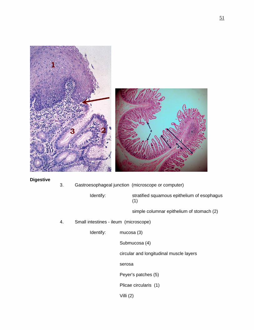
Digestive 3. Gastroesophageal junction (microscope or computer) Identify: stratified squamous epithelium of esophagus
(1)
simple columnar epithelium of stomach (2)
4. Small intestines - ileum (microscope) Identify: mucosa (3) Submucosa (4)
circular and longitudinal muscle layers
serosa Peyer's patches (5) Plicae circularis (1) Villi (2)
52
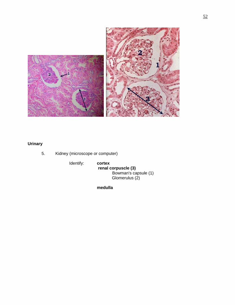
Urinary 5. Kidney (microscope or computer) Identify: cortex
renal corpuscle (3) Bowman's capsule (1)
Glomerulus (2) medulla
53
B. Fetal Pig Anatomy RESPIRATORY Rostrum (snout) Trachea External nares (nostrils) Lungs Larynx Diaphragm DIGESTIVE Esophagus Stomach Liver Gall Bladder (Pancreas - if present) Small intestine Rectum Large intestine: spiral colon Anus URINARY Right + left kidney Right + left ureter Urinary bladder Urethra C. Pig Kidney parts to know: hilum ureter renal capsule renal cortex renal medulla renal pyramids renal pelvis (renal artery) (renal vein)
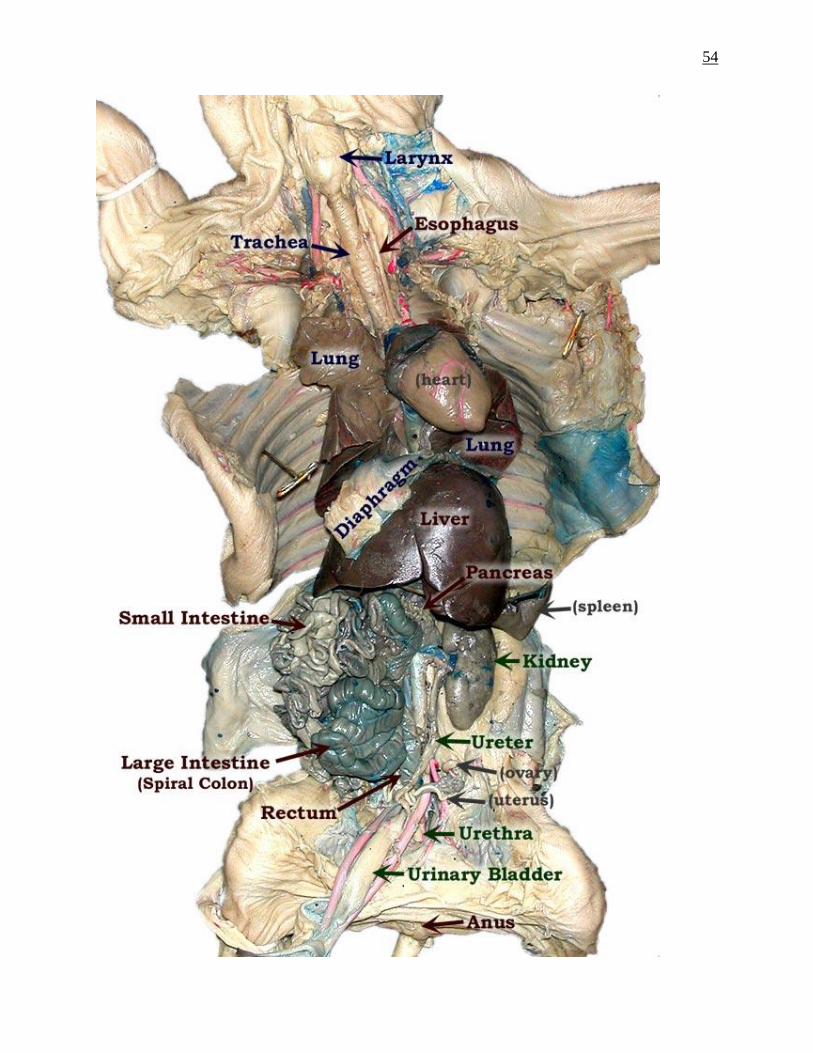
54
55
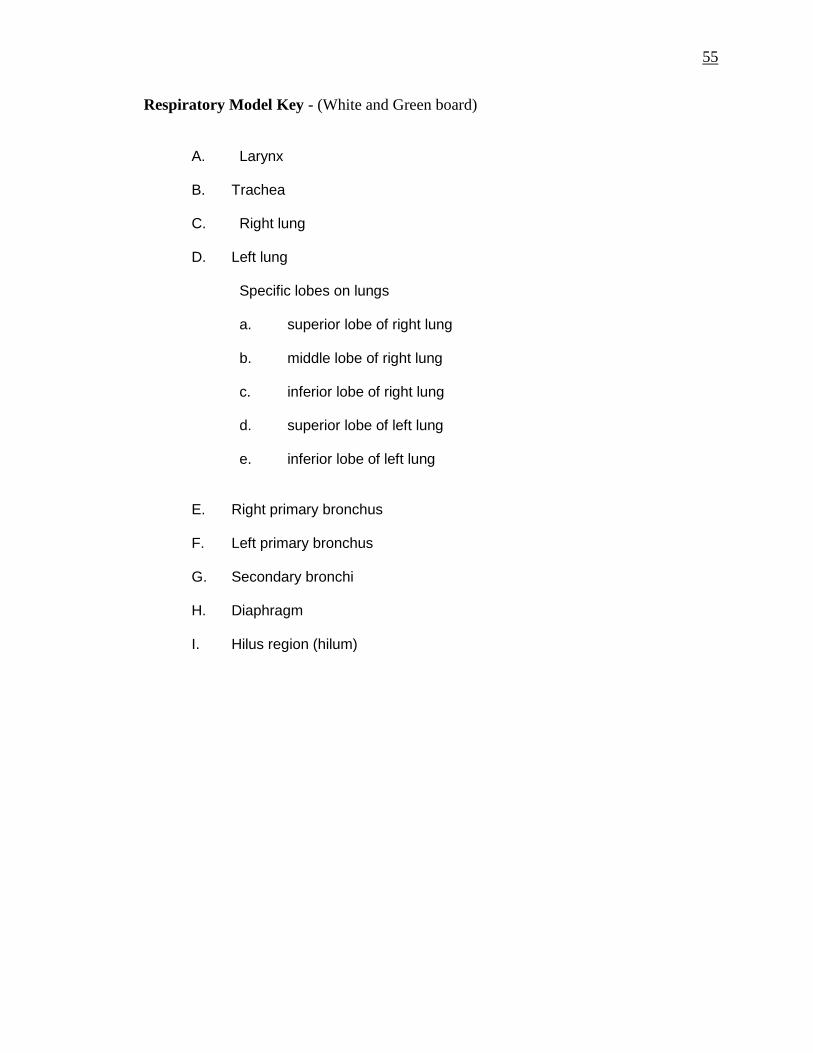
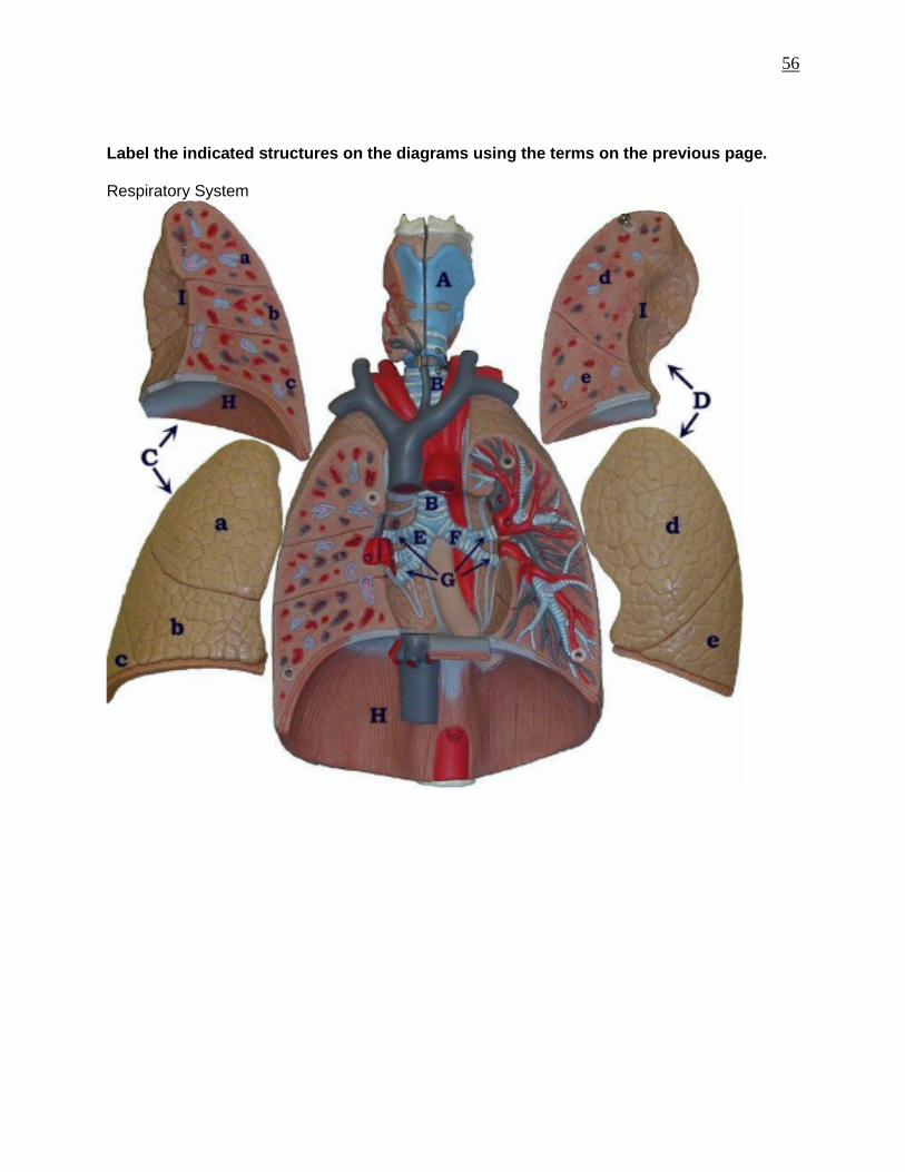
Respiratory Model Key - (White and Green board) A. Larynx B. Trachea C. Right lung D. Left lung Specific lobes on lungs a. superior lobe of right lung b. middle lobe of right lung c. inferior lobe of right lung d. superior lobe of left lung e. inferior lobe of left lung E. Right primary bronchus F. Left primary bronchus G. Secondary bronchi H. Diaphragm I. Hilus region (hilum)
56
Label the indicated structures on the diagrams using the terms on the previous page. Respiratory System

57
F. Stomach Model Key 2. Esophagus 3. Cardiac sphincter 3A. Cardiac region 4. Fundic region 5. Body region 6. Pyloric region 6A. Pyloric sphincter 15. Pancreas 16. Pancreatic duct G. Liver Model Key a. Right lobe b. Left lobe c. Right hepatic duct d. Left hepatic duct 18. Common hepatic duct 19. Cystic duct 20. Gallbladder

58
Identify all the structures listed on the previous page. (Stomach Model)
Identify all the structures listed on the previous page (Stomach Model key – Pancreas)

59
Identify all the structures listed on the Liver Model key.
60
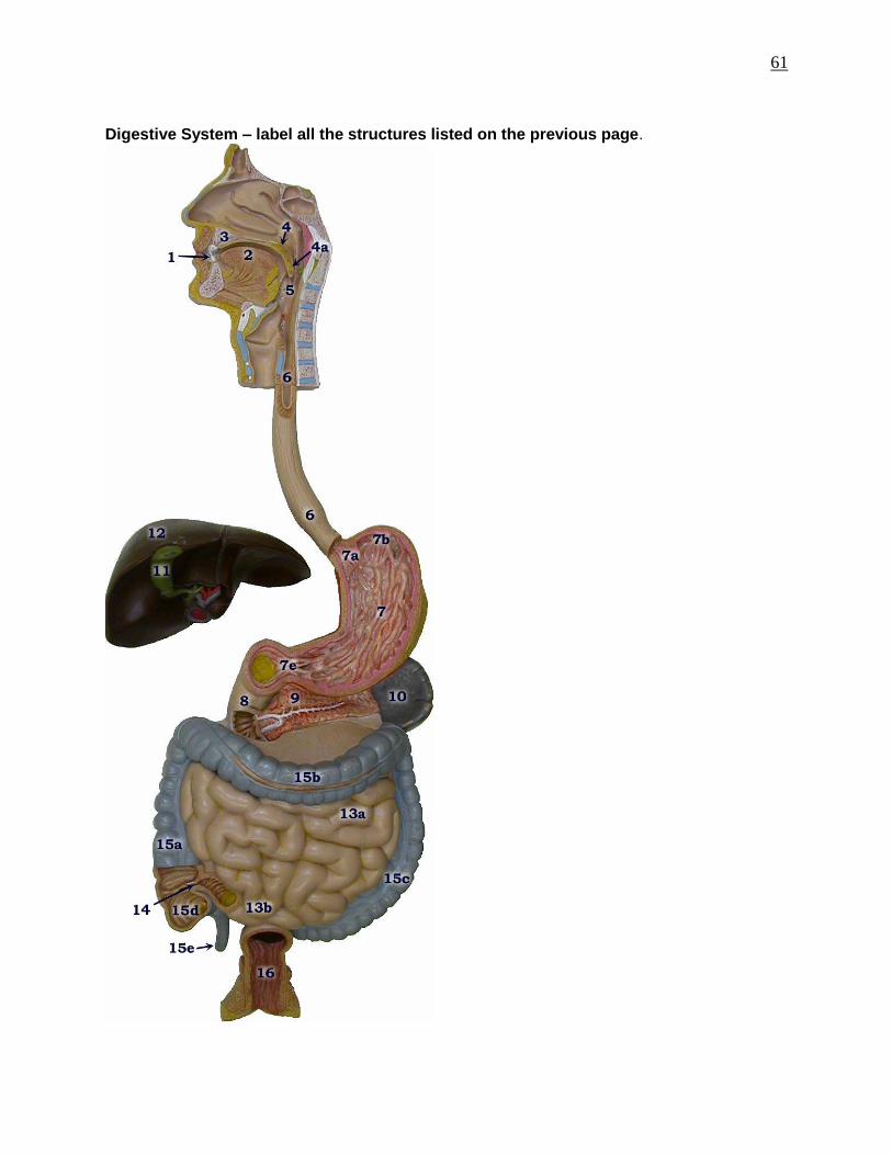
H. Digestive System Model Key 1. Tooth 2. Tongue 3. Hard palate 4. Soft palate a. Uvula 5. Pharynx 6. Esophagus 7. Stomach a. Cardiac sphincter b. Fundus e. Pyloric sphincter 8. Duodenum (first part of small intestine) 9. Pancreas (pancreatic duct - white lines through center of pancreas) 10. Spleen 11. Gallbladder 12. Liver 13. Small Intestine (8. Duodenum) a. Jejunum b. Ileum 14. Ileocecal junction 15. Large intestine (Colon) a. Ascending colon b. Transverse colon c. Descending colon d. Cecum e. Vermiform appendix 16. Rectum
61
Digestive System – label all the structures listed on the previous page.
62
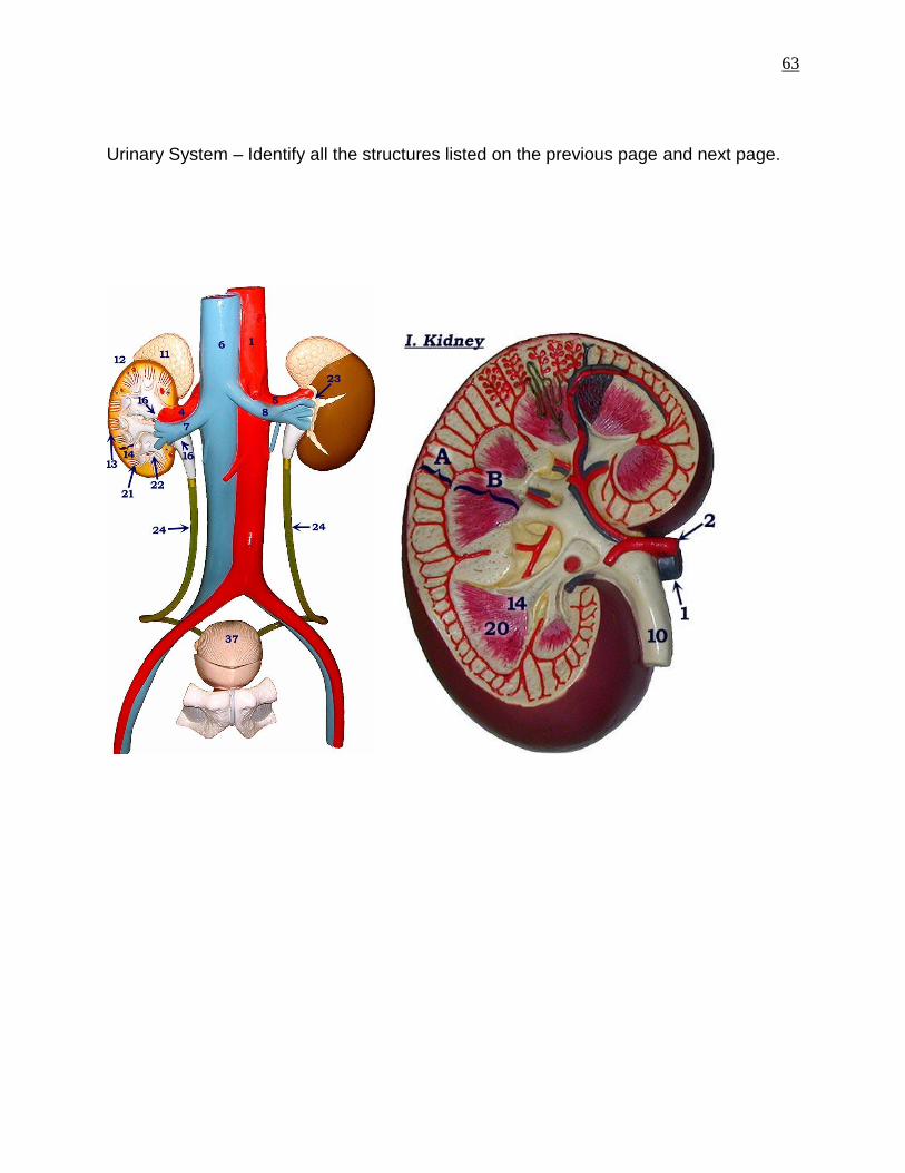
I. Urinary Model Key 1. Abdominal aorta 4. Right renal artery 5. Left renal artery 6. Inferior vena cava 7. Right renal vein 8. Left renal vein 11. Right adrenal gland 12. Right kidney 13. Renal cortex 14. Renal medulla 16. Renal pelvis 21. Renal pyramid 22. Renal papilla 23. Renal hilum (hilus)
24. Ureters (yellow tubing) 37. Bladder
63
Urinary System – Identify all the structures listed on the previous page and next page.
64
J. Urinary model key - 3 parts Part I. Large Kidney (enlarged 3 times) A. Renal cortex B. Renal medulla 1. Renal vein 2. Renal artery 10. Ureter 14. Renal Papilla 20. Renal pyramid Part II. Large nephron A. Renal Cortex 1. Renal corpuscle 2. Proximal convoluted tubule c. Loop of Henle 4. Distal convoluted tubule 5. Collecting duct 8. Glomerulus Part III. Large renal corpuscle (enlarged 700 times) 1. Afferent arteriole 2. Glomerulus (tuft of capillaries) 3. Efferent arteriole 5. Bowman's Capsule

65
Identify all the structures listed on the previous page.