Lab 3: Actionscript User Interface Lab: GUI Lab Sep. 11 th, 2012.
Upload
farinbarbieCategory
view
12download
2
'". . {
. ~.,~ -~
_ ...."'~ ..~~.,... ••.•...•.._....j..,. ~~. . ~t·,
lysis QuestionsExplain how mitosis leads to two daughter cells, each of which is diploid and genetically
entical to the original cell. What activities are going on in the cell during interphase?
- .(': V\ (). ';",,-\.e c-~Vv>,£-,e ~p 'AVe' e (') \ ,;s ~&"eJ {.),\,. c\..<;PTY\Q.~'"<s:"" s::J hI At sy ,,\; c~-e s ~\- ",e\~ e C~S"b~ ''0, ~,{Y\ S
n==(')s,()VV'\e;::, c~"'cl ('eO~~D\e<;. 'CR (')('rI. e 1:"1 ~e
'" ps, 0~ -The ~p ce\\. \;s:oY'O fY\e-\-n~\(),se o\o.\-e :fue\
< ~e, c''y'Sc.'N'O.~:J. s. 'i3. p ae ~~ ",rl 'fYInVP \0 ~e
"" P6 .•~e C\S'~'''\(,)\ CP'\ A-os=m$.-\\.ND\(-fev'1~(,o. d~O\aA..o \s ) o-~~ c ~o <"\,,)<2..""\'5, ,e:>c..C\.l'S" • c \
How does mitosis differ in plant and animal cells? How does plant mitosis accommodate arigid, inflexible cell wall?
J ~~o.'S:.,0$ 'm ""'\ ~e." ~\ 6.....'i\.-\':::' \\0. ~ ~£ '" ";) a\ 'M. §' ci~-\.ffi'S.J
\.\LY\ Os£' cA-- 3\1ID \\~s. o-\- ~h.y:n;!> os=- S-OC)\.s.\Y\ o.,Vlm\
,. {?' vn,\-o:;,\S (»('C'tlC-S o..YW w~,~ V\e.", CR\\S "',e ~'N\eJ ~~~\(),XA- (4"\\ w~\-::. n-;-\g s= fY\'~ $"<" o.,'P ~o<:,,'(y\(?al b~
e;S ~Q,,\- "6(" Ot' \Q\-e ~E' ODW, '0, c..e\\ Q.,V\ol ei~es-
. What is the role of the centrosome (the area surrounding the centrioles)? Is it necessary formitosis? Defend your answer.
~"5 ~e. CR '('\.\--<;C)~'<V\ ~c.. ~ ~e ?n e" ci~ -\\e.\1C'\.e\I S ) The A Q \Jf'-\e« M e (\ m~\o\\c, 50~ '\ oJ \ ('
ProcedureIt is hard to imagine that you can estimate how much time a cell spends in each phase of celldivision from a slide of dead cells, yet this is precisely what you will do in this part of the lab.Since you are working with a prepared slide, you cannot get any information about how longit takes a cell to divide. What you can determine is how many cells are in each phase. Fromthis, you can infer the percentage of time each cell spends in each phase.
1. Observe every cell in one high-power field of view and determine which phase of thecell cycle it is in, This is best done in pairs. The partner observing the slide calls out thephase of each cell while the other partner records. Then switch so the recorder becomes theobserver and vice versa. Count at least two full fields of view. If you have not counted atleast 200 cells, then count a third field of view.
2. Record your data in Table 3.1.
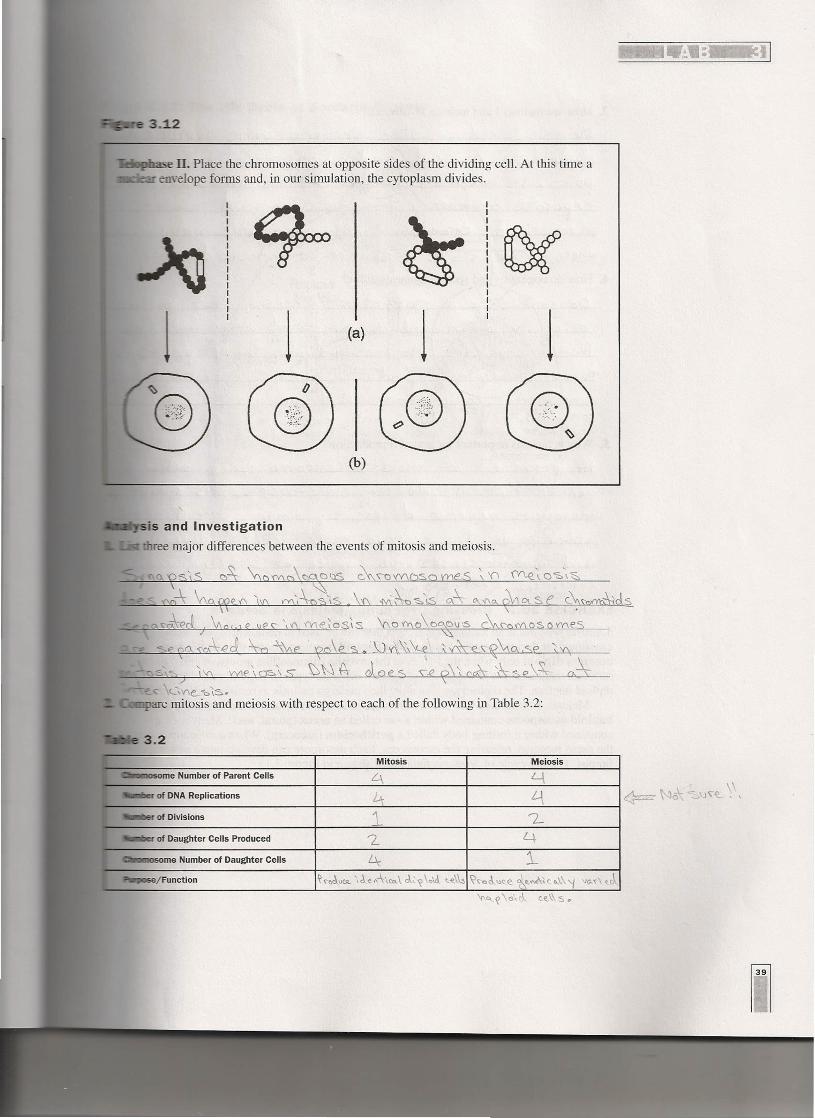
__.-e 3.12
.••• [lkilse II. Place the chromosomes at opposite sides of the dividing cell. At this time a_i"'IIi>- •.,. envelope forms and, in our simulation, the cytoplasm divides.
1 1 1 1(a)
• ..,'5is and Investigationthree major differences between the events of mitosis and meiosis.
(b)
\
'00-\ {\CAWe.;(\ IV\ (Y\~\n<\s. \1/\ :!Vi~'§>'\S 0,\ 0\,:(\0. ~cx So e 0S'~ds
I
<',€'l{o.'S'C\ .(OJ, \0 ~e '!(1"I\p S. .·\)IO\\\~ ;-ds::R,~~o""e ,\1'\ ,
c~J
c- <,~v-e, "i::.'i=:'.are mitosis and meiosis with respect to each of the following in Table 3.2:
me Number of Parent Cells
Mitosis Meiosis
of DNA Replications 41
of Daughter Cells Produced
CII_osome Number of Daughter Cells

3. How are meiosis I and meiosis II different?
he ~a =;:,\ S \" Cf' d.~\C' e .<... -\\of' c:...~DJ'<V'-Q ?, 0 'IY1f (1 \I M bee 'COY})
(1~pOld -\0 \no'(2 o\d O,V\cl ~ ~Ck'A~S -t"'e "'<'l'!'V)Q\0'(s{\q",
~a \cs ...\ Y\ l{Y\e.\Q 'S c::., ::ss:. --\~ S'\ 5 \e (' c'Y\'CQ '(Y\Q\ '\ d'S
- \
f\\sn ,'" aD($;'f\e_~"\s c1\.o \L\'fIe s,l S d:\If~de,s ~e.. c'I-\o ~ o.s. N) o~--s'M e c e \\ U'O€ '\)\1 0\\ 't f c(') cl \J c.....'1\0,. rl V\ q \ c.c-c).t C.~ \\ o..d -\w (')
I a <
:'>MOI.\\ ~r:s~ ~('Le:s. htr- w'·\\ ;L7'>~{\\-eCl.'o-..e<.\o,~~", Qd'\
oo';\~"'\ N\ c.e\\ ...?~cl\Jc..es b''''\~o"'~ a...c.....\-~ e. ~ C~~ ~Y\\~\..-e..~~~:I\C-\.~o .~~'A~\S -\Vt~ oc""'~. ~v~"d..s o~ ~ e...<:''-;I'I c€,,~\'S.
5. Why lS meIOSISImportant for sexual reproduction? ~
J\\ cro«"'-c.. Q.e"S. C?s '(Y\~,a -s,\s &,oW\t'lSOWl<' <. e.x.c..~(U\~~
9lcS:()£~C'- '{'n~5"O\:::::' J J\'f\~ CroSS-ave,~ ••~IlS '(Y\~\p';:,\-s
'",~~",s.es ~V\e~<, ~"e. <\\<J~~ o,MoYlb.. F~e..-n\ c:.e.\\S
16\Y\\('''>A Co,.'\--.J €s t\w. e >N\\-,\'\.f D YV1 ove 0, ((\.e_:~c... <1\ \! 5:;s;=-.~'1
(f) '" -\-\ (\ IJ as, e,\10 \ v\-, 0 '<\...
EXERCISE 38.2: Crossing Over during Meiosis'in Sordaria
Sordaria fimicola is an ascomycete fungus that can be used to demonstrate the results ofcrossing over during meiosis. Sordaria is a haploid organism for most of its life cycle. Itbecomes diploid only when the fusion of the mycelia (filamentlike groups of cells) of twodifferent strains results in the fusion of the two different types of haploid nuclei to form adiploid nucleus. The diploid nucleus must then undergo meiosis to resume its haploid state.
Meiosis, followed by one mitotic division, in Sordaria results in the formation of eighthaploid ascospores contained within a sac called an ascus (plural, asci). Many asci arecontained within a fruiting body called a perithecium (ascocarp). When ascospores are maturethe ascus ruptures, releasing the ascospores. Each ascospore can develop into a new haploidfungus. The life cycle of Sordaria fimicola is shown in Figure 3.13.

Analysis of Results1. Using your data in Table 3.3, determine the distance between the gene for spore color and
the centromere. Calculate the percentage of crossove!:!:'by dividing the number of crossoverasci (2:2:2:2 or 2:4:2) by the total number of asci X 100. To calculate the map distance,divide the percentage of crossover asci by 2. The percentage of crossover asci is dividedby 2 because only half of the spores in each ascus are the result of a crossover event(Figure 3.15). Record your results in Table 3.3.
2. Draw a pair of chromosomes in MI and Mil and show how you would get a 2:4:2arrangement of ascospores by crossing over. (Hint: refer to Figure 3.15).

• .:.11: :
3. Calculate the percentage of cells in each phase, and record in Table 3.1.
Consider that it takes, on average, 24 hours (or 1,440 minutes) for onion root tip cells tocomplete the cell cycle. You can calculate the amount of time spent in each phase of the cellcycle from the percentage of cells in that stage.
Percentage of cells in stage x 1,440 minutes = minutes of cell cycle spent in stage
Table 3.1
SCA.M?\e.. \" \ Me..Number of Cells Percent of Total Time in
Cells Counted Each Stage; V1. E CAe:. '" c.. e.\.\
Field l. Field 2 Field 3 Total
2\ t-\~ L\ ~\V) Interphase 2,3 j) ~3 ~3 ~3 o.~ '2-Z '2.:' 'Z. ~b.~ °/0 \ \ \-., \L\-~"'\I<1
l A~ n...'lV': () Prophase ~:::.r '1L 24 L,3 2[; 3 27. \b3 ~L bOlo +'t-., 'bM:,
!.:..6.....yV\;Y\ Metaphase~ 3 5 2- Lt :> 3' 2A: 4.~ /0 ~'"I' \5 M'"
VV"\', v'\ Anaphase :r ..• 1- ~. '6 era 2\., \' }'S(V1~'bo 1- ? 1> 4- '\ ~"\
\\ VV\'-v"\Telophase 3 L ~ b 5 "+ 5 '::>1- b. y /0 'Z..~'\3 S.,..,
Total Cells Counted "C)OQ
Questions1. If your observations had not been restricted to the area of the root tip that is actively
dividing, how would your results have been different?3,\i.gS-p !J>.IOI0A '-A.C>,.\Je ~e@", 0.. S\-evue C f€ ~"C'<",(\\-Q;c)"e... (,p\ "
-\oO,-\- !b.)€:es. ~'Q\l~ {Y\\\.o s,'s } Thy:=> '~.g C'~;\.Y\ C)~\co -\ 0-,\ c.. ~\\ C CH) "'-tR j (,b,\ 0\)\ rl "'-0. \J e.. '0e£... -o. oS m Ci \ \ f' '\ ,
2. Based on the data in Table 3.1, what can you infer about the relative length of time an onionroot tip cell spends in each stage of cell division?
~ 0 (\\Ci\ a>rA- -\: ~ ce'\ "5-"e 1\6S~: \O~:6 \~X)~cl -\\'01\.£ \v'\ \.-A~'~""Qse, T ~Y\rl ±\DQ \R!{\~% o~ -\(~~
d..-e..~g>Do 7",P Q,'f:, C~\\ ~ s ~\)."ou*- £ C\ c...""" f,e egd \(\9 4-0I.5e0.."" ~\- n'Q.,('~e;;, h r,~""().,,~ ) w",,"e.'C2. ~\- .:::.~e()d5 ~€.. <.,'y,C:'\-PS-e"<l.~~ o~ -\-~.rAe...
3. Draw and label a pie chart of the onion root tip cell cycle using the data from Table 3.1.
Title: ~'Q Reo-\: \\ c...~\\(; c
\e\c"~c,,,3e.. to.'-\ "/0f\'(\C»o~""Cl.S-e... "'\~ % 7
M CL~\, ~(),~e. G., t "/0-:t=2========~----I