L. EL ASSASSE, S. BOUTACHALI, F. AMRAOUI, T. AMIL, S. CHAOUIR, A. DARBI Radiology service, Military...
15
HEAD AND NECK: HN18
-
Upload
basil-lucas -
Category
Documents
-
view
217 -
download
0
Transcript of L. EL ASSASSE, S. BOUTACHALI, F. AMRAOUI, T. AMIL, S. CHAOUIR, A. DARBI Radiology service, Military...

HEAD AND NECK: HN18
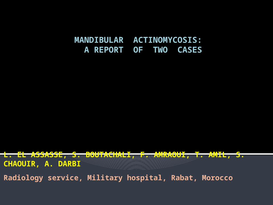
MANDIBULAR ACTINOMYCOSIS: A REPORT OF TWO CASES
L. EL ASSASSE, S. BOUTACHALI, F. AMRAOUI, T. AMIL, S. CHAOUIR, A. DARBI
Radiology service, Military hospital, Rabat, Morocco

Introduction:
The mandibular actinomycosis is a rare condition, usually secondary to tooth extraction, mucous wound or mandibular fracture.
We report in this work two cases of mandibular actinomycosis explored by orthopantomogram and facial CT and whose diagnosis is confirmed by histology.
Observation and results:
These two patients aged 26 and 60 years, both having a history as a dental extraction with delayed mucosal healing, having presented a mandibular pain with perimandibular tumefaction.
Radiological aspects were not specific with uni or multigeodic osteolysis.
Observation and results:
A wide excision was indicated for them with empiric antibiotic therapy.
Pathological examination of surgical
specimens revealed actinomycotic osteitis.
Observation and results:
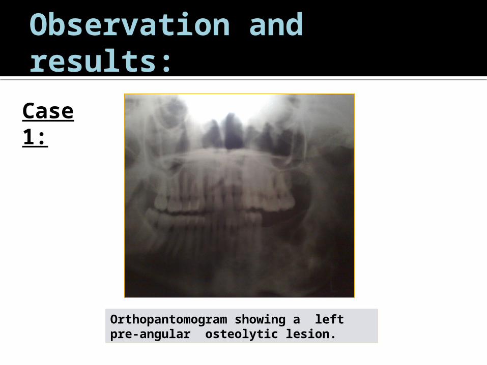
Case 1:
Orthopantomogram showing a left pre-angular osteolytic lesion.

Observation and results:
Case 1:
Facial CT showing well defined multilocular osteolytic lesion in the horizontal branch of left mandible.
Observation and results:
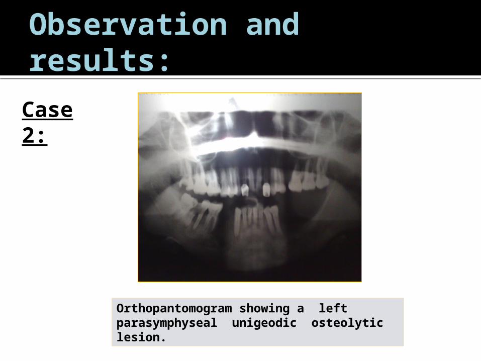
Case 2:
Orthopantomogram showing a left parasymphyseal unigeodic osteolytic lesion.

Discussion:
The cervicofacial actinomycosis is a rare infectious disease whose incidence is estimated at 5 per 100,000 people.
It is most frequent in adults between 20 and 60 years with sex ratio 3 males to 1 female .
It is caused by germs long regarded as intermediate between fungi and bacteria but which proved to be true bacteria: Actinobacteria or actinomycetes .
Involvement of the head and neck region is the most frequent (50 to 75% of cases), thoracic and abdominal locations are rarer.

Discussion:
Contributing factors sometimes found are poor oral hygiene, oral trauma or surgery , dental procedures and salivary lithiasis.
Chronic tonsillitis, mastoiditis, and otitis are also important risk factors for actinomycosis.

Discussion:
The radiological signs of actinomycotic osteitis are not specific.
Involvement may be osteolytic (uni or multigeodic) or osteoblastic.
The severe forms of mandibular actinomycosis can develop into the skull base or cervical spine.

Discussion:
Diagnosis is histological.
Treatment consists of prolonged antibiotic therapy and surgical debridement.
Prognosis depends on early diagnosis and treatment.

Conclusion:
The diagnosis of actinomycosis should be considered in patient with recurrent and chronic suppuration.
The appearance on imaging is not specific and the diagnosis of certainty is histological.
References:
Chobaut.J.C, Maniere.C. et coll. L'actinomycose en ORL. A propos d'un cas localisé aux fosses nasales. Ann. Oto-laryngol. Chir. Cervicofac. (Paris). 1994, 111 :292-294.
Simony.J, Puissant.A. et coll. Un cas d'actinomycose cervicale évoluant
depuis trois ans sans traitement. Ann. Dermatol. Venereol. 1986 ; 113 : 555 – 558.
Del Rosario.N, Rickman.L. Letters. Cervico-facial actinomycosis. Arch.
Otolaryngol. Head neck. Surg. 1987 ;113 :777-778.
Gorlin.R.J, Goldman.H.M. (Eds) Thomas oral pathology. St louis, mosby, 1970.
Sodagar.R, Kohoute.E. Actinomycosis of tongue as pseudotumor.
Laryngoscop. 1972 ; 81 : 2149 – 2152. 6) Antoine.G.A, Antoine.J.A. Cervicofacial actinomycosis. Ear nose Throat. J. 1986 ; 65 :483-485.
References:
Walker.R.S, Middelkamp.J.N. et coll. Mandibular osteomyelitis caused by actinomyces israelii. Oral. Surg. 1981 ; 51 : 243-244.
Stenhous.D. Intraoral actinomycosis. Report of five
cases. Oral. Surg. 1975 ; 34 : 547-552. Vannier.J.D, Schaison.G. et coll. Actinomycosis
osteomyelitis of the skull and atlas with lace dissemintion. Eur. J. pediatr. 1986 ; 145 :316-318.