Krew cz. 2 - kzf.amp.edu.pl DDS Program/blood dds 2018.pdfbilirubin into the urobilinogen, most of...
81
Blood cells Magdalena Gibas –Dorna MD, PhD Dept. of Physiology, PUMS
Transcript of Krew cz. 2 - kzf.amp.edu.pl DDS Program/blood dds 2018.pdfbilirubin into the urobilinogen, most of...

Blood cells
Magdalena Gibas –Dorna MD, PhD
Dept. of Physiology, PUMS

Blood composition
55% plasma 45% cells
(99% RBCs) < 1% WBCs i PLTs

Composition of the blood
The blood is a mixture of cells, fluid, proteins and metabolites.
Blood has four major elements:
- red blood cells (transport oxygen from the lungs to organs and peripheral sites; water-base buffer)
- white blood cells (have a defensive role in destroying invading organisms e.g. bacteria and viruses)
- platelets (the first line of defence against damage to blood vessels)
- plasma (the proteinaceous substance in which the other three elements circulate)

Functions of the blood
Delivers nutrients from the digestive system to all parts of
the body
Transports oxygen from the lungs to all parts of the body
Transports carbon dioxide from all parts of the body to
the lungs
Transports waste products from cells to the external
environment mainly via the kidneys
Transports hormones from the endocrine system to target
cells or organs within the body.

Functions of the blood
Through continuous exchange of it's components with tissue fluids promotes fluid and electrolyte balance
Defends the body against attack from foreign organisms via the white blood cells and antibodies
Defends the body against injury or infection via the inflammatory response
Prevents serious hemorrhage by the clotting process
Maintains the body's temperature
by circulating heat

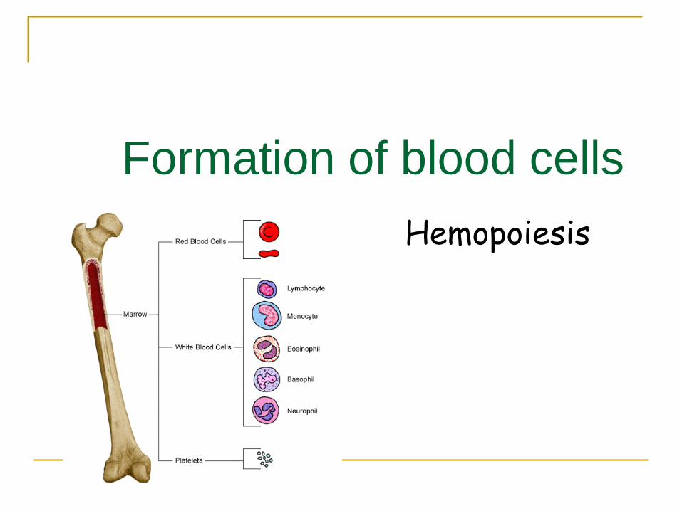
Formation of blood cells
Hemopoiesis

Hemopoietic cells (those which produce blood) first appear in the yolk sac of the 2-week embryo.
By 8 weeks, blood making has become established
in the liver of the embryo, and
by 12-16 weeks the liver has become the major site of blood cell formation. It remains an active hemopoietic site until a few weeks before birth.
The spleen is also active during this period, particularly in the production of lymphoid cells, and
the fetal thymus is a transient site for some lymphocytes.

Development of Marrow
The highly cellular bone marrow becomes an active blood making site from about 20 weeks gestation and gradually increases its activity until it becomes the major site of production about 10 weeks later.
At birth, active blood making red marrow occupies the entire capacity of the bones and continues to do so for first 2-3 years after birth

Development of Marrow
The red marrow is then very gradually replaced by inactive,
fatty, yellow, lymphoid marrow.
The yellow marrow begins to develop in the shafts of the
long bones and continues until (by 20-22 years) red marrow
is present only in the upper ends of the femur and humerus
and in the flat bones
total amount of active red marrow is nearly identical in the
child and the adult

Bone Marrow
Bone marrow is composed
of 2 compartments:
a. Extravascular
b. Intravascular
The central venous sinusoid has a permeable basement membrane
Red cells squeeze into the sinusoidal lumen, leaving their nuclei behind in the cellular matrix.
Mature blood cells (from bone marrow) are attracted to the site of migration by chemotactic factors

Red Marrow Function
About two-thirds of its mass functions in white
cell production (leucopoiesis), and one-third in
red cell production (erythropoiesis).
However there are approximately 700 times as
many red cells as white cells in peripheral
blood.

Distribution of active marrow LOCATION % of TOTAL MARROW
Pelvis 40
Vertebrae 28
Cranium-mandible 13
Ribs 8
Sternum 2
Ends of long bones 8

Erythropoiesis
The pluripotential stem cell is defined as the precursor
cell from which all erythrocytes, leukocytes, and megakaryocytes are formed (i.e. all blood cells have a common cell line of origin)
These stem cells are very rare (only about one in 10,000 bone marrow
cells)
produce, by mitosis, two kinds of progeny: more stem cells
cells that begin to differentiate along the paths leading to the various kinds of blood cells


The process of erythrocyte development are
characterised by:
the gradual appearance of hemoglobin and disappearance of ribonucleic acid (RNA) in the cell,
the progressive degeneration of the cell's nucleus which is eventually extruded from the cell,
the gradual loss of
cytoplasmic organelles,
for example mitochondria,
a gradual reduction in
cell size

Reticulocytes Between 2 and 6% of a new-
born baby's circulating red
cells are reticulocytes, but this
reduces to less than 2% of
RBCs in the healthy adult.
Reticulocyte count increases
in conditions in which rapid
erythropoiesis occurs
A reticulocyte normally takes
2-4 days to mature into an
erythrocyte.

Normally normoblastic renewal amounts to 12-25% of all nucleated cells in the bone marrow.
Percentage of erythroblasts increases with their maturity(most of normoblasts; less proerythroblasts)
requirement – renewal increases to 30%-50%, and even more, there is also increase in percentage of immature cells – normoblastic reaction with shift to the left (normoblasts in blood stream ).
Normoblstic reaction of bone marrow

Red Blood Cells the biconcave shape increases the
cell's surface area and facilitates diffusion of O2 and CO2 into or out of the cell
the lack of nuclei and organelles
contribute to increased Hb content
and gas-carrying capacity
normal erythrocytes must be very flexible. They become deformed when flowing through capillaries and narrow pores (slits) in the spleen
Red blood cell flexibility
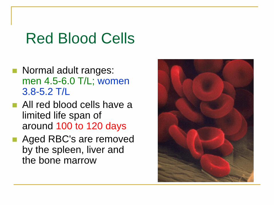
Normal adult ranges: men 4.5-6.0 T/L; women 3.8-5.2 T/L
All red blood cells have a limited life span of around 100 to 120 days
Aged RBC's are removed by the spleen, liver and the bone marrow
Red Blood Cells

The Metabolism of Hemoglobin
Amino Acids
Transferrin
Fe3+ Storage
Globin Hydrolyzed
RBC’s
Heme
Reduction
Macrophage
NADPH + H
Biliverdin
Bilirubin
Serum
albumin
Bound
Bilirubin
Liver cell
Bilirubin Glucuronic
acid
Bile salt
Biliary
duct system
Duodenum
Pancreas
Bile containing
bilirubin
Urobilinogen Bacteria
via
bloodstream conversion
Urobilin
Kidney
Urine
Intestinal tract
Bacterial oxidation Stercobilin
Feces
Fe3+
• Spleen, liver, and marrow
macrophages recognize and ingest
old RBCs
• Heme is converted to bilirubin;
• Bound bilirubin is transported to the
liver, where it is converted into bile
salt
• Small intstine bacteria convert
bilirubin into the urobilinogen, most of
which is eliminated in the feces in the
form of stercobilin
• Some urobilinogen is absorbed from
the intestine and excreted with urine,
where it becomes oxidized to urobilin

Anemias
Abnormally low oxygen-carrying capacity of
the blood resulting from deficiency in the
number of :
- RBC,
- Hb,
- or both
Anemia is considered to be present if Hb
is less than 12g/dL (norm: men 13-17g/dL,
women 12-16 g/dL)
Hb molecule

Acute blood loss
hemorrhagic anemia (normocytic,
normochromic).
B12 or/and folate deficiency
megaloblastic anemia (macrocytic,
hyperchromic, immature RBC).
Fe deficiency microcytic, hypochromic
anemia
Hemolytic anemia increased rate of RBCs
destruction (normocytic)
Genetic abnormality/chemical exposure
aplastic anemia (lack of RBC production)

Signs and symptoms
of anaemia
CNS
Debilitating fatigue
Dizziness, vertigo
Depression
Impaired cognitive function
Immune system
Impaired T cell and
macrophage function
Cardiorespiratory system
Exertional dyspnoea
Tachycardia, palpitations
Cardiac enlargement, hypertrophy
Increased pulse pressure,
systolic ejection murmur
Risk of life-threatening cardiac
failure
Gastro-intestinal system
Anorexia
Nausea
Genital tract
Menstrual problems
Loss of libido
Vascular system
Low skin temperature
Pallor of skin, mucous
membranes and conjunctivae
Adapted from Ludwig H, Fritz E. Semin Oncol. 1998;25(suppl 7):2-6.


How many Hb molecules in one
RBC?
More than 250 milion molecules!!!

RBC indexes
MCHC - mean corpuscular hemoglobin
concentration (norm: 34%/RBC)
MCV - mean corpuscular volume (norm: 78-
95fL)
MCH - mean corpuscular hemoglobin index
- Hb mass within RBC (norm: 29pg/RBC)

Typical blood cells parameters

Regulation of erythropoiesis Hormones and
lymphokines (erythropoietin, BPA interleukins, ACTH, TSH, thyroid hormones, glucocorticoids, testosteron etc.)
vitamins (B12, folic acid, B6)
metals (Fe, Co, Cu, Mn, Zn)

Erythropoietin
(EPO)
Erythropoietin is
synthesized in the
fibroblasts of the kidney
cortex and is released into
the blood in response to
hypoxia in the renal arterial
blood supply.
Erythropoietin is a
glycoprotein. It is
inactivated by the liver and
excreted in the urine.
About 10% of EPO is
synthesized in other
tissues

Hypoxia
Oxygen-carrying
capacity of
blood
Reduces
Increases
Increases
Red blood
cell count
Stimulates
Erythropoiesis
in
Red bone marrow
Kidneys
Stimulates
Secrete into blood
Erythropoietin
Low blood O2
kidneys
release
erythropoietin
increased
erythropoiesis

Hormones affecting erythropoiesis
adrenocorticotrophic hormone (ACTH),
human growth hormone (GH)
thyroid-stimulating hormone (TSH),
thyroid hormones (T3, T4),
adrenal cortical steroids (cortisol),
all promote erythropoietin formation

Dietary requirements for sufficient red
blood cell production Dietary element Role in red blood cell production
Protein Required to make red blood cell proteins and
also for the globin part of hemoglobin
Vitamin B6 It plays role in heme synthesis
Vitamin B12 and folic acid Needed for DNA synthesis and are essential in
the process of red blood cell formation
Vitamin C
Required for folate metabolism and also
facilitates the absorption of iron. Extremely
low levels of Vitamin C are needed before any
problems occur. Anemia caused by lack of
Vitamin C (scurvy) is now extremely rare
Iron Required for the heme part of hemoglobin
Copper and Cobalt (heme synthesis,
EPO)
There are some reports suggesting that these two
minerals are essential for the production of red
blood cells in other animals but not in humans

Iron Turnover
• Iron absorption is greatest in the duodenum and decreases progressively as one moves distally down the intestine.
• Iron is much more readily absorbed in its ferrous (Fe2+) form than in its ferric (Fe3+) form.
• Vitamin C can increase iron absorption by serving as a reducing agent to maintain iron as Fe2+.
ferric ferrous
Vit C ferric

Iron in organism
65-75% in Hb
About 13% in ferritin
About 12 % in hemosiderin
About 5% in mioglobin
1% in enzymes (catalase,
cytochromes)
1% in transferrin
Spleen, iron in macrophages

Normal vs. low iron

Vitamin B12 and folic acid


Megaloblastic anemias When vitamin B12 or folate is deficient, DNA synthesis is interrupted.
This leads to megaloblastic changes (macrocytosis). There is often
erythroid hyperplasia in the marrow but most of these immature cells die
before reaching maturity.
The lack of DNA synthesis affects the neutrophils leading to nuclear
hypersegmentation.
Often a mild pancytopenia is seen but thrombocytopenia can be severe.
Hemorrhagic spots

Neurologic symptoms may include:
Impaired perception of deep touch and vibration
Present Babinski reflex
Paresthesias (feeling of "pins and needles")
Dementia in severe cases
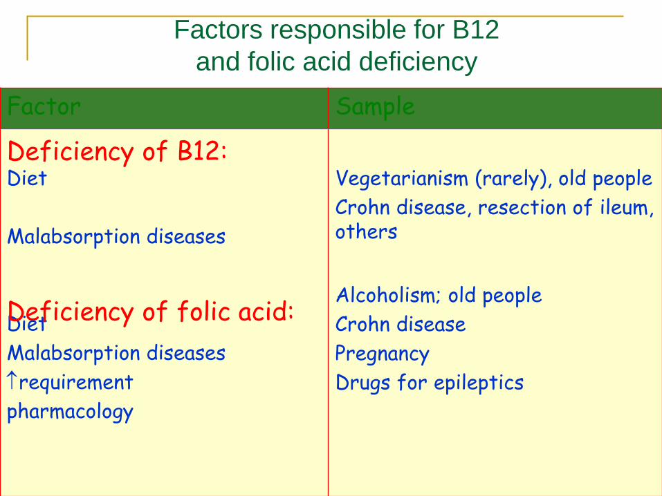
Factors responsible for B12
and folic acid deficiency
Factor Sample
Diet
Malabsorption diseases
Diet
Malabsorption diseases
requirement
pharmacology
Vegetarianism (rarely), old people
Crohn disease, resection of ileum, others
Alcoholism; old people
Crohn disease
Pregnancy
Drugs for epileptics
Deficiency of folic acid:
Deficiency of B12:

Polycythaemia (excess red blood cell
production)
RBCs > 6.0 T/L, HCT and HGB
Reasons:
- primary (cancer of myeloid tissue)
- secondary increase in EPO sythesis (high altitude, chronic lung disease, smoking)
- pathological increase of EPO
(kidney dieases, liver tumors)

Granulopoiesis

Factors affecting granulopoiesis
Sympathetic system increases granulopoiesis
ACTH and glucocorticoids increase count of neutrophils
and decrease count of eosinophils, lymphocytes and
basophils
Thyroid hormones, pituitary hormones, adrenal
hormones and estrogens – increase granulopoiesis
Positive feedback – products of WBCs degradation

Normal neutrophil renewal in bone marrow amounts to 60-70%
of all nucleated cells in the bone marrow. Percentage of
neutrophils increases with their maturity(most of mature
neutrophils; less myeloblasts)

The RBC's in the background appear normal. The important finding here is
the presence of many PMN's. An elevated WBC count with mainly
neutrophils suggests inflammation or infection. A very high WBC count
(>40G/L) that is not a leukemia is known as a "leukemoid reaction". This
reaction can be distinguished from leukemia by the presence of large
amounts of leukocyte alkaline phosphatase (LAP) in the neutrophils.
leukemia
Leukemoid reaction

Here are very large, immature myeloblasts typical for acute
myelogenous leukemia (AML) that is most prevalent in young adults.

Granulocytes
In the circulation, about 50% of
granulocytes adhere closely to the
internal surface of the blood
vessels. These are called
marginal cells and are not
normally included in the white cell
count.
The other half circulate
(circulating cells) in the blood and
exchange with the marginal
population.
Within hours granulocytes
may leave the circulation in
response to specific
requirements for these cells
in the tissues.
They may survive in the
tissues for 4 or 5 days, or
less, depending on the
conditions they meet.

There are five main types of
white blood cells (Schilling’s count):
· neutrophils 45-65 %
· eosinophils 1-4 %
· basophils 0.5-1 %
· lymphocytes 20-40 %
· monocytes 3-8 %
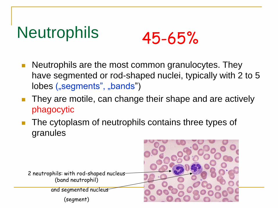
Neutrophils
Neutrophils are the most common granulocytes. They
have segmented or rod-shaped nuclei, typically with 2 to 5
lobes („segments”, „bands”)
They are motile, can change their shape and are actively
phagocytic
The cytoplasm of neutrophils contains three types of
granules
45-65%
2 neutrophils: with rod-shaped nucleus (band neutrophil)
and segmented nucleus
(segment)

Neutrophils
Primary granules are non-
specific and contain
lysosomal enzymes, and
some lysozyme. The
granules are similar to
lysosomes.
The enzymes (MPO)
produce hydrogen
peroxide

Oxygen-depedent killing
Respiratory (oxidative) burst
Molecular oxygen reduced to a range of
intermediates:
- superoxide anion
- hydrogen peroxide
- hypochlorite anions
- singlet oxygen
- hydroxyl radicals
they are powerful anti-bacterial agents

Oxygen-independent killing
Lysozyme destroys
bacterial cell walls
Cationic proteins cause
pH to fall
Acid hydrolase enzymes
degrade carbochydrates,
proteins, lipids, and
nucleic acids
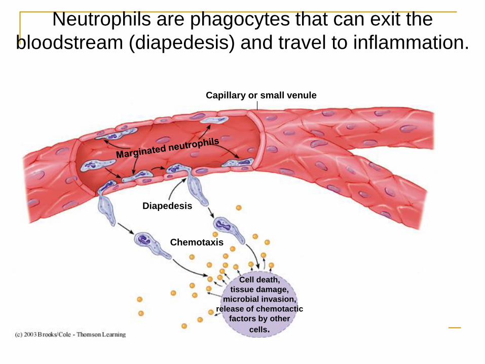
Capillary or small venule
Diapedesis
Chemotaxis
Cell death,
tissue damage,
microbial invasion,
release of chemotactic
factors by other
cells.
Neutrophils are phagocytes that can exit the
bloodstream (diapedesis) and travel to inflammation.

Phagocytosis

Phagocytosis
2. Attachment of the
bacterium to the long
membrane evaginations,
called pseudopodia.
opsonization

Phagocytosis
3. Ingestion of the
bacterium forming a
"phagosome," which
moves toward the
lysosome.

Phagocytosis
4. Fusion of the
lysosome and
phagosome, releasing
lysosomal enzymes
into the phagosome.

Phagocytosis
5. Digestion of the
ingested material.

Phagocytosis
6. Release of digestion
products from the
cell

Bacteria
Nucleus
Neutrophil
Phagolysosome
Lysosomes
containing
digestive
enzymes
1. Ingestion of bacteria by neutrophil. 2. Phagosome forms around bacteria. 3. Degranulation of lysosomes to form digestive vacuole (phagolysosome). 4. Bacterial lysis by digestive enzymes. 5. Dispersement of phagosome in cytosol. 6. Neutrophil lysis.
Neutrophils get to an infection early in large
numbers, ingest microbes, die, and damage tissue
Formation of
phagosome

Types of phagocytic cells
NEUTROPHILS (polymorfonuclear)
- most common/active
- first to side of injury
- short lived (4-5days)
EOSINOPHILS
- allergic responses
- parasitic worms
MONOCYTES
- develop into macrophages
Wandering MACROPHAGES
- travel as monocytes
- chemotaxis during inflammation
Fixed MACROPHAGES
- lymph nodes, spleen, most organs (e.g. skin, brain, liver, kidneys)
- long lived (months to years)
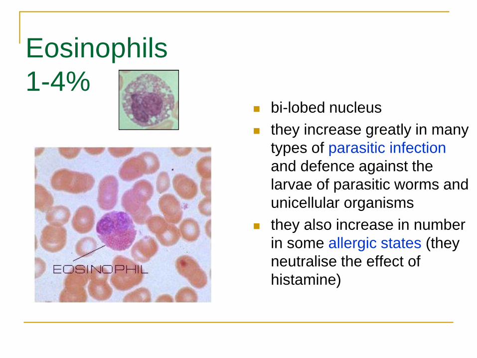
Eosinophils
1-4% bi-lobed nucleus
they increase greatly in many
types of parasitic infection
and defence against the
larvae of parasitic worms and
unicellular organisms
they also increase in number
in some allergic states (they
neutralise the effect of
histamine)

Eosinophils - properties
The lysosomes contain oxidase, peroxidase and
phosphatases
Eosinophils exhibit chemotaxis;
They respond to eosinophilic chemotactic factors
released by basophils
Their attraction depends on the presence of
antibodies specific to foreign proteins (phagocytosis
of Ag-Ab complexes)

Eosinophils The granules of eosinophils contain a substance called MBP (major basic protein) which is toxic to many parasitic larvae.
Eosinophils also have surface receptors for the antibody: immunoglobulin E (IgE).
These receptors are not found in neutrophils and again this is thought to reflect their role in parasitic infection.
eosinophil with a bilobed nucleus and reddish granules in the cytoplasm. Just underneath it is a small lymphocyte

Function of eosinophils -
summary
they regulate allergic reactions
they defence against parasitic
infections
they participate in antigen
presentation (for antibodies
synthesis)
they play role in hemostasis
(plasminogen)

Basophils
0-1% characterised by their large cytoplasmic granules, and very little cytoplasm
actually become mast cells on leaving the blood and entering surrounding tissues
both basophils and mast cells have highly specific receptors for IgE produced in response to various allergens
basophils are not phagocytic cells !!!

Basophils
Response to specific allergens
is rapid and results
degranulation and release of
histamine and other agents
(among them SRS-A, heparin).
The reaction known as
immediate hypersensitivity.
fever, some forms of asthma,
urticaria (nettle rash) and most
seriously anaphylactic shock.

Allergen degradation
Degradation of allergen by macrophages, presentation to T-cells and B-cells, and production of IgE which causes histamine basophil release

Basophil funtion - summary
facilitate cell migration
to the site of
inflammation
participate in allergic
reactions
modulate blood clotting
and lipid profile (via
heparin)

Monocytes
3-8%
the largest cell type seen in blood smears
nuclei are not multilobular like granulocytes, but may be U-shaped or deeply indented (S-shaped)
Monocytes are actively phagocytic
Monocytes can migrate out of the bloodstream and become tissue macrophages
they form part of a cell network known as the monocyte-macrophage system


Monocytes
Tissue macrophages (sometimes
called histiocytes) respond more
slowly than neutrophils to chemotactic
stimuli
They ingest and destroy bacteria,
dead cells, iron and foreign matter
They also function as modulators of
the immune response by processing
antigen structure and facilitating the
concentration of antigen at the
lymphocyte's surface (antigen
presentation)

Identify the segmented neutrophil, band neutrophil, lymphocyte, monocyte, eosinophil,
basophil, and platelet in the image below:

Platelets
At any one time, about two-thirds of the body's platelets are circulating in the blood and one-third are pooled in the spleen.
the life span of platelets is between
1 and 2 weeks
if not consumed in the process of blood clotting, they are destroyed by macrophages in the liver and spleen

Mature lymphocyte
9-14 micrometer
Nucleus round but may
be slightly indented or
eccentric deep purplish
blue and is composed
of dense chromatin
aggregates
Cytoplasm is light blue
present as thin rim
around the nucleus or
may be quite abundant
depending on size
Relative count 30-40%;
Life span of a week to a few months (memory cells – years)
Lymphoid tissue

Absolute lymphocyte count
Absolute lymphocyte count (ALC) is usually used to determine
ranges of normal for lymphocytes.
The ALC is higher in neonates and young children (up to 8000
cells/microL) but in those > 12 years of age is normally up to
4000 cells/microL.
Lymphopenia is usually defined as < 1000-1500 cells/microL
in children and < 3000 cells/microL in adults .
Lymphocytosis > 4000 cells/microL in adults and > 9000
cells/microL in children
Normal leukocyte count is 4.5-11.0 x 1000/mm2 with about
22-44% overall being lymphocytes.

Acquired Immunity: Lymphocytes
B cell and T cells
lymphopoiesis

Lymphocytes – general
classification
Two types of lymphocytes
T-Cells (Thymus derived)
Natural Killer Cells (similar to T cells, lack antigen-
specific receptors, lack CD8)
CD4+ T-Cells (helper cells)
CD8+ T-Cells (cytotoxic cells)
B-Cells (Bone Marrow derived)
Memory cells (both B and T cells)
CD = „cluster of differentiation”

Dendritic cells are APCs in the skin, mucosa and lymphoid tissues.

Comparing B cells with T cells