Know Your KNEE Emily Delello Salene Sheridan. The knee joint is 3 joints in1 The Tibiofemoral joint...
50
Know Your KNEE Emily Delello Salene Sheridan
-
Upload
adelia-bryant -
Category
Documents
-
view
217 -
download
1
Transcript of Know Your KNEE Emily Delello Salene Sheridan. The knee joint is 3 joints in1 The Tibiofemoral joint...

Know Your KNEEEmily DelelloSalene Sheridan

The knee joint is 3 joints in1
The Tibiofemoral joint is a hinge joint permitting flextion and extension.
The Femoralpatellar joint is a plane joint where the patella glides across the distal end of the femur during knee flextion.
Structurally it is a bicondylar joint allowing some rotation when the knee is partially flexed and when the knee is extending.

Femur

Tibia

Fibula

Patella
Left and Right???

Patella

MUSCLES Of the Leg

Rectus Femoris
Origin: Anterior Inferior iliac spine
Insertion : Tibial Tuberosity
Action: Hip flexion, Knee extension Innervation: Femoral Nerve Vascular supply: Lateral circumflex artery

Lateral Circumflex Femoral Artery


Vastus intermedialis
Origin: Anterior femur Insertion: Tibial tuberosity via patellar tendon
Action: Knee extension
Innervation: Femoral nerve
Vascular supply: Lateral circumflex femoral artery
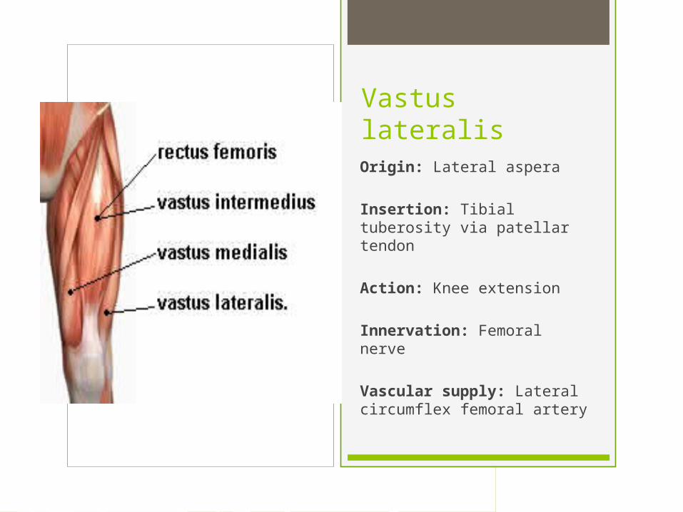
Vastus lateralis Origin: Lateral aspera Insertion: Tibial tuberosity via patellar tendon
Action: Knee extension
Innervation: Femoral nerve
Vascular supply: Lateral circumflex femoral artery

Vastus Medialis Origin: Linea aspera Insertion: Tibial tuberosity via patellar tendon
Action: Knee extension
Innervation: Femoral nerve
Vascular supply: Circumflex femoral artery

Vastus Lateralis
Vastus Medialis

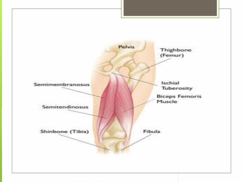
Bicep Femoris Origin: Long head of the Ischial tuberosity. Short head the lateral lip of linea aspera
Insertion: Fibular head
Action: Long head, extends hip and flexes knee. Short head, flexes knee
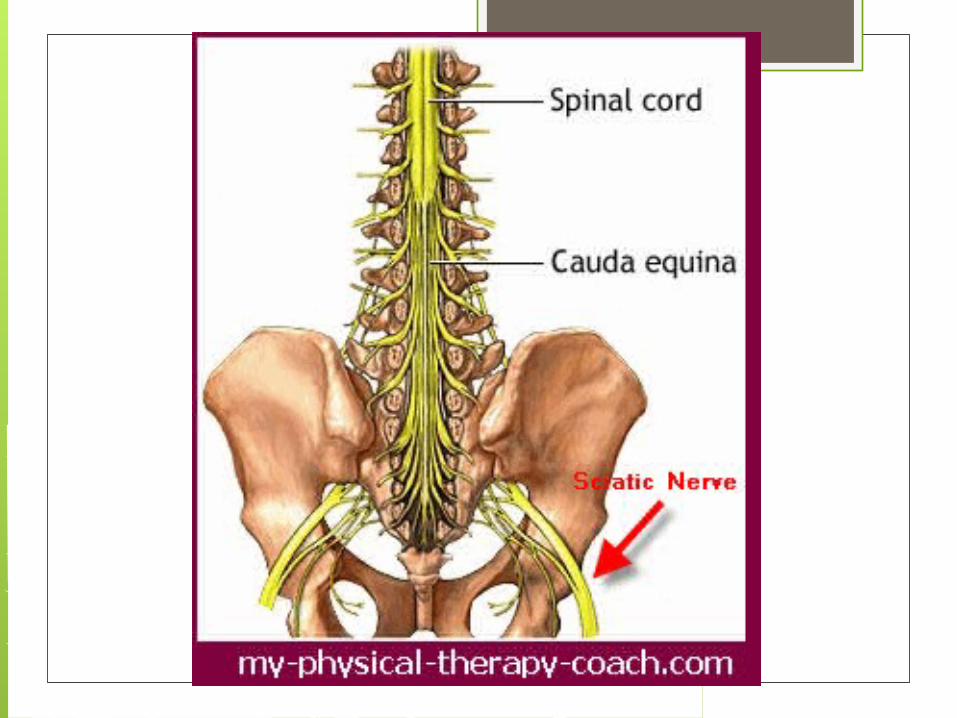
Innervation: Long head, sciatic nerve. Short head, common peroneal nerve
Vascular supply: Inferior gluteal artery


SemimembranousOrigin: Ischial Tuberosity Insertion: Posterior surface of medial condyle of tibia
Action: Extend hip and flex knee
Innervation: Sciatic nerve
Vascular supply: Inferior gluteal artery


Semimembranosus Semitendinosus

Semitendoninosus Origin: Ischial tuberosity Insertion: Anteromedial surface of Proximal tibia
Action: Extend hip and flex knee
Innervation: Sciatic nerve
Vascular supply: Deep femoral


PopliteusOrigin: Lateral condyle of femur
Insertion: Posteriorly on medial condyle of tibia
Action: Initiates knee flexion
Innervation: Tibial nerve
Vascular Supply: Popliteal artery

gastrocnemiusOrigin: Medial and lateral condyles of femur Insertion: Posterior calcaneus
Action: Knee flextion, ankle plantar flextion
Innervation: Tibial nerve
Vascular supply: Popliteal artery


Medial Head of Gastrocnemius
Lateral Head

Ligaments A ligament is a tough band of fibrous tissue that connects bone to bone or bone to cartilage and supports and strengthens joints.

KEY 1= Quadriceps femoris tendon 2=Patellar Ligament 3= Oblique popliteal ligament 4=arcuate popliteal ligament 5=Tibial collateral ligament 6=Fibular collateral ligament 7=Anterior cruciate ligament 8=Posterior cruciate ligament 9=Transverse ligament

1
2

4
4

3
5
6
7
8


1
2

2

9
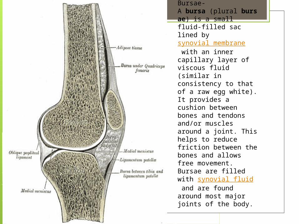
Bursae-A bursa (plural bursae) is a small fluid-filled sac lined by synovial membrane with an inner capillary layer of viscous fluid (similar in consistency to that of a raw egg white). It provides a cushion between bones and tendons and/or muscles around a joint. This helps to reduce friction between the bones and allows free movement. Bursae are filled with synovial fluid and are found around most major joints of the body.

Bursae 1=Prepatellar Bursa 2=Deep Infrapatellar Bursa 3=Suprapatellar Bursa 4=Subcutaneous Infrapatellar Bursa

1
2
3

4

Cartilage
1.Firm, whitish, flexible connective tissue found in various forms in the larynx, in the external ear, and in the articulating surfaces of...2.A particular structure made of this tissue.


THE END!!!!!!!