
Knee OA with septic arthritis - msr.my
15
Knee OA with septic arthritis • Peri-articular osteopenia **Also osteomyelitis of the femur • Subtle periosteal reaction in femoral diaphysis
Transcript of Knee OA with septic arthritis - msr.my

Knee OA with septic arthritis
• Peri-articular osteopenia **Also osteomyelitis of the femur
• Subtle periosteal reaction in femoral diaphysis


Modified from Jacobson JA, Girish G, Jiang Y, Resnick D. Radiographic evaluation of arthritis: inflammatory conditions. Radiology. 2008;248(2):378-89

Osteoarthritis
is commonly bilateral and nearly sym-metric in distribution (Fig 9). It is im-portant to closely evaluate the lateralaspect of the fifth metatarsal head, be-cause this is often the first site of a boneerosion in the foot and, at times, suchinvolvement occurs prior to hand orwrist involvement (Fig 10). Becauserheumatoid arthritis is a disease thataffects synovium diffusely, other sites ofinvolvement include tendon sheaths and
bursae such as the retrocalcaneal bursa.Loss of the normal radiolucent trianglebetween the posterosuperior margin ofthe calcaneus and the adjacent Achillestendon suggests the presence of bursalfluid, with subjacent calcaneal erosionsindicating inflammation (Fig 11).
Other peripheral joints may also beinvolved in rheumatoid arthritis, withsimilar findings. Joint involvement in-cludes the knees (Fig 12), the hips
(Fig 13), and the sacroiliac and glenohu-meral joints, with involvement of thelast of these often associated with ahigh-riding humeral head related to alarge rotator cuff tear. Spinal involve-ment is also possible. At the C1-C2 ar-ticulation, the odontoid process may beeroded, and the anterior atlantodens in-terval may be abnormally widened (!3mm in adults), especially with neck flex-ion (Fig 14) (5).
Figure 3
Figure 3: (a) Illustration of synovial joint shows joint fluid (f) and articular cartilage (c). (b) Illustration and (c) radiograph show inflammatory arthritis, synovitis, andpannus (P) causing cartilage destruction. Marginal erosions (arrows) are seen where subchondral bone plate is exposed to intraarticular synovitis. f " Fluid.
Figure 4
Figure 4: Osteoarthritis. Posteroanterior radio-graph shows interphalangeal joint space narrow-ing, subchondral sclerosis, and osteophyte forma-tion (arrows).
Figure 5
Figure 5: Septic arthritis.(a) Posteroanterior and(b) oblique radiographs show jointspace narrowing (arrows), os-teopenia, soft-tissue swelling, anda bone erosion (arrowhead).
REVIEW FOR RESIDENTS: Radiographic Evaluation of Arthritis Jacobson et al
Radiology: Volume 248: Number 2—August 2008 381
• non-uniform joint space narrowing
• subchondral sclerosis
• osteophyte formation (arrows)

Osteoarthritis ò 1st CMC joint; STT joint
scaphoid trapezoid trapezium
ò Interphalangeal joint; sparing MCP joint
ò Erosive OA same distribution – seagull or gull-wing deformities
ò CPPD – atypical distribution for OA – MCPJ, radioscaphoid and the capitate-lunate joint (stair-step pattern) +/- chondrocalcinosis. Hook-like osteophytes at 2/3rd MC head
Evaluation of the Hand Film 57
FIGURE 2-39. Osteoarthritis.
Soft tissue change: Distortion around DIPs
Subluxations: Laterally at second DIPs
Mineralization: Normal
Calcification: None
Joint spaces: Nonuniform loss—best seen at second DIPs
Erosions: None
Bone production: Osteophytes at DIPs and PIPs; subchondral sclerosis—greater multangular, distal navicular, and base of first metacarpal
Distribution: DIPs, PIPs; first carpometacarpal joint and greater multangular-navicular joint

CPPD
52 Approach to Radiographic Changes Observed in a Specif ic Joint
FIGURE 2-33. Narrowing of the radiocarpal joints with subchon-dral sclerosis involving the radius and lunate in a posttraumatic osteoarthritis. There is an old fracture of the radial styloid.
FIGURE 2-34. Narrowing of the radionavicular joint and the capitate-lunate joint with subchon-dral sclerosis surrounding these articulations in a patient with CPPD crystal deposition disease.
1. radioscaphoid joint
2. capitate-lunate joint
² narrowing ² subchondral
sclerosis
1
2

Modified from Jacobson JA, Girish G, Jiang Y, Resnick D. Radiographic evaluation of arthritis: inflammatory conditions. Radiology. 2008;248(2):378-89

54 Approach to Radiographic Changes Observed in a Specif ic Joint
COMMON ARTHROPATHIES OF THE HAND—A RADIOGRAPHIC SUMMARY
Seven radiographs are presented here (Figs. 2-36 to 2-42) illustrating the common arthropathies involving the hand and summarizing all the individual features discussed in this chapter.
FIGURE 2-36. Early rheumatoid arthritis.
Soft tissue change: Symmetrical swelling around MCP joints and wrist
Subluxations: None
Mineralization: Juxta-articular osteoporosis
Calcification: None
Joint spaces: Maintained
Erosions: Early aggressive (arrows)
Bone production: None
Distribution: PIPs, MCPs, and pancarpal
Early Rheumatoid Arthritis

Evaluation of the Hand Film 55
FIGURE 2-37. Late rheumatoid arthritis.
Soft tissue change: Atrophy
Subluxations: MCP joints (proximal phalanges subluxed ulnarly and palmarly)
Mineralization: Diffuse osteoporosis
Calcification: None
Joint spaces: Uniform loss—PIPs, MCPs, and pancarpal
Erosions: Large aggressive
Bone production: None
Distribution: PIPs, MCPs, and pancarpal
Late Rheumatoid Arthritis
Evaluation of the Hand Film 55
FIGURE 2-37. Late rheumatoid arthritis.
Soft tissue change: Atrophy
Subluxations: MCP joints (proximal phalanges subluxed ulnarly and palmarly)
Mineralization: Diffuse osteoporosis
Calcification: None
Joint spaces: Uniform loss—PIPs, MCPs, and pancarpal
Erosions: Large aggressive
Bone production: None
Distribution: PIPs, MCPs, and pancarpal

56 Approach to Radiographic Changes Observed in a Specif ic Joint
FIGURE 2-38. Psoriasis.
Soft tissue change: Fusiform digit swelling—first, second, and fourth digits
Subluxations: None
Mineralization: Normal
Calcification: None
Joint spaces: Destroyed fourth DIP, first MCP-IP, and second DIP
Erosions: Large, aggressive; pencil-in-cup erosion of fourth DIP and IP joint of thumb
Bone production: Solid periosteal new bone formation fourth and fifth prox-imal phalanges (arrows); fluffy new bone around thumb
Distribution: MCPs, PIPs, and DIPs, but in a 1st, 2nd and 4th ray distri-bution. The 3rd and 5th ray are minimally involved.
Sausage-finger Pencil-in-cup

56 Approach to Radiographic Changes Observed in a Specif ic Joint
FIGURE 2-38. Psoriasis.
Soft tissue change: Fusiform digit swelling—first, second, and fourth digits
Subluxations: None
Mineralization: Normal
Calcification: None
Joint spaces: Destroyed fourth DIP, first MCP-IP, and second DIP
Erosions: Large, aggressive; pencil-in-cup erosion of fourth DIP and IP joint of thumb
Bone production: Solid periosteal new bone formation fourth and fifth prox-imal phalanges (arrows); fluffy new bone around thumb
Distribution: MCPs, PIPs, and DIPs, but in a 1st, 2nd and 4th ray distri-bution. The 3rd and 5th ray are minimally involved.
Psoariasis
Evaluation of the Hand Film 57
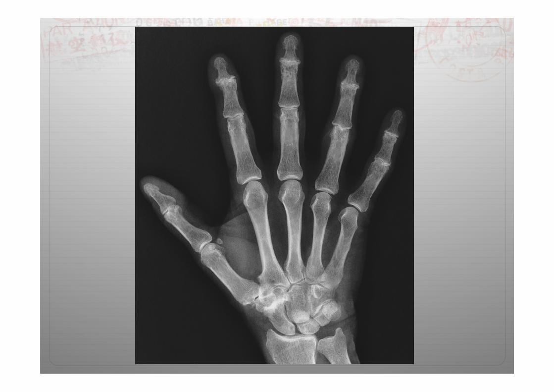
FIGURE 2-39. Osteoarthritis.
Soft tissue change: Distortion around DIPs
Subluxations: Laterally at second DIPs
Mineralization: Normal
Calcification: None
Joint spaces: Nonuniform loss—best seen at second DIPs
Erosions: None
Bone production: Osteophytes at DIPs and PIPs; subchondral sclerosis—greater multangular, distal navicular, and base of first metacarpal
Distribution: DIPs, PIPs; first carpometacarpal joint and greater multangular-navicular joint

Evaluation of the Hand Film 57
FIGURE 2-39. Osteoarthritis.
Soft tissue change: Distortion around DIPs
Subluxations: Laterally at second DIPs
Mineralization: Normal
Calcification: None
Joint spaces: Nonuniform loss—best seen at second DIPs
Erosions: None
Bone production: Osteophytes at DIPs and PIPs; subchondral sclerosis—greater multangular, distal navicular, and base of first metacarpal
Distribution: DIPs, PIPs; first carpometacarpal joint and greater multangular-navicular joint
Osteoarthritis

58 Approach to Radiographic Changes Observed in a Specif ic Joint
FIGURE 2-40. Erosive osteoarthritis.
Soft tissue change: Swelling at the third and fourth PIPs
Subluxations: Laterally at third and fourth PIPs
Mineralization: Normal
Calcification: None
Joint spaces: Nonuniform loss—best seen at third PIP and first IP
Erosions: Central erosions—combined with osteophytes to produce “seagull” appearance
Bone production: Osteophytes at PIPs, DIPs, first carpometacarpal joint and greater multangular-navicular joint; subchondral sclerosis at second to fourth DIPs, PIPs and IP joint of thumb; ankylosis fifth DIP
Distribution: PIPs and DIP; first carpometacarpal and greater multangular-navicular joint


