Kinetics of Mn3+–Oxalate Formation and Decay in Reactions Catalyzed by Manganese Peroxidase...
8
Kinetics of Mn 31 ±Oxalate Formation and Decay in Reactions Catalyzed by Manganese Peroxidase of Ceriporiopsis subvermispora Ulises Urzu ´ a,* Philip J. Kersten,² and Rafael Vicun ˜ a* ,1 *Departamento de Gene ´tica Molecular y Microbiologı ´a, Pontificia Universidad Cato ´lica de Chile, Casilla 114-D, Santiago, Chile; and ²Forest Products Laboratory, USDA-Forest Service, One Gifford Pinchot Drive, Madison, Wisconsin 53705 Received July 24, 1998, and in revised form August 21, 1998 The kinetics of Mn 31 – oxalate formation and decay were investigated in reactions catalyzed by manga- nese peroxidase (MnP) from the basiomycete Ceripo- riopsis subvermispora in the absence of externally added hydrogen peroxide. A characteristic lag ob- served in the formation of this complex was shortened by glyoxylate or catalytic amounts of Mn 31 or hydro- gen peroxide. MnP titers had a minor effect on this lag and did not influence the decay rate of the complex. In contrast, Mn 21 and oxalate drastically affected maxi- mal concentrations of the Mn 31 – oxalate complex formed, the decay of which was accelerated at high Mn 21 levels. The highest concentration of complex was obtained at pH 4.0, whereas an inverse relation- ship was found between the pH of the reaction and the decay rate of the complex with MnP present. In the absence of MnP, the best fit for the decay kinetics of the complex gave an order of 3/2 at concentrations in the range of 30 –100 mM, with a k obs 5 0.012 min 21 M 20.5 at pH 4.0. The rate constant increases at lower pH values and decreases at high oxalate concentrations. The physiological relevance of these findings is dis- cussed. © 1998 Academic Press Key Words: manganese peroxidase; Mn 31 – oxalate; superoxide; hydrogen peroxide; Ceriporiopsis subvermispora. Manganese peroxidase (MnP) 2 is secreted by white-rot fungi and is thought to play an important role in the degradation of lignin during wood decay. MnP catalyzes the oxidation of Mn 21 to Mn 31 , which in turn can oxidize phenolic substrates including lignin substructures (1). The kinetics of MnP from Phanerochaete chrysosporium has been well charac- terized (2– 4). We have studied the ligninolytic sys- tem of another white-rot fungus, Ceriporiopsis sub- vermispora. This fungus selectively decays the lignin of wood and might be useful in softening wood chips prior to mechanical pulping (5). C. subvermispora secretes several isoenzymes of MnP and laccase, with isoelectric points that vary with the composi- tion of the growth medium (6, 7). In contrast to P. chrysosporium, no lignin peroxidase has been iden- tified from C. subvermispora. We recently published attempts to identify physio- logical sources of H 2 O 2 required for extracellular MnP activity in C. subvermispora (8). Efforts failed to find oxidases such as those secreted by other wood-decay fungi. We therefore studied the possibility that hydro- gen peroxide might be generated by MnP through the manganese-dependent oxidation of organic acids se- creted by the fungus. Indeed, it has been described that MnP from P. chrysosporium oxidizes oxalate, produc- ing CO 2 and formate radical (9). The latter reacts with oxygen to give a second molecule of CO 2 and superox- ide. Superoxide then is reduced by Mn 21 to give hydro- gen peroxide and Mn 31 (10), both of which further accelerate MnP-catalyzed reactions. Similarly, glyoxy- late is oxidized by MnP with the production of formate and formate radical from the organic acid (11). Both oxalate and glyoxylate were found in the extracellular fluid of C. subvermispora grown in chemically defined media (8). In cultures containing low nitrogen, a cor- relation between MnP titers and mineralization of ox- alate was observed. In vitro assays confirmed the Mn 21 -dependent oxidation of [ 14 C]oxalate by MnP and 1 To whom correspondence should be addressed. Fax: 56-2- 2225515. E-mail: [email protected]. 2 Abbreviations used: MnP, manganese peroxidase; SOD, superox- ide dismutase. 0003-9861/98 $25.00 215 Copyright © 1998 by Academic Press All rights of reproduction in any form reserved. ARCHIVES OF BIOCHEMISTRY AND BIOPHYSICS Vol. 360, No. 2, December 15, pp. 215–222, 1998 Article No. BB980952
-
Upload
ulises-urzua -
Category
Documents
-
view
212 -
download
0
Transcript of Kinetics of Mn3+–Oxalate Formation and Decay in Reactions Catalyzed by Manganese Peroxidase...

Kinetics of Mn31–Oxalate Formation and Decay inReactions Catalyzed by Manganese Peroxidaseof Ceriporiopsis subvermispora
Ulises Urzua,* Philip J. Kersten,† and Rafael Vicuna*,1
*Departamento de Genetica Molecular y Microbiologıa, Pontificia Universidad Catolica de Chile, Casilla 114-D, Santiago,Chile; and †Forest Products Laboratory, USDA-Forest Service, One Gifford Pinchot Drive, Madison, Wisconsin 53705
Received July 24, 1998, and in revised form August 21, 1998
The kinetics of Mn31–oxalate formation and decaywere investigated in reactions catalyzed by manga-nese peroxidase (MnP) from the basiomycete Ceripo-riopsis subvermispora in the absence of externallyadded hydrogen peroxide. A characteristic lag ob-served in the formation of this complex was shortenedby glyoxylate or catalytic amounts of Mn31 or hydro-gen peroxide. MnP titers had a minor effect on this lagand did not influence the decay rate of the complex. Incontrast, Mn21 and oxalate drastically affected maxi-mal concentrations of the Mn31–oxalate complexformed, the decay of which was accelerated at highMn21 levels. The highest concentration of complexwas obtained at pH 4.0, whereas an inverse relation-ship was found between the pH of the reaction and thedecay rate of the complex with MnP present. In theabsence of MnP, the best fit for the decay kinetics ofthe complex gave an order of 3/2 at concentrations inthe range of 30–100 mM, with a kobs 5 0.012 min21 M20.5
at pH 4.0. The rate constant increases at lower pHvalues and decreases at high oxalate concentrations.The physiological relevance of these findings is dis-cussed. © 1998 Academic Press
Key Words: manganese peroxidase; Mn31–oxalate;superoxide; hydrogen peroxide; Ceriporiopsissubvermispora.
Manganese peroxidase (MnP)2 is secreted bywhite-rot fungi and is thought to play an importantrole in the degradation of lignin during wood decay.
MnP catalyzes the oxidation of Mn21 to Mn31, whichin turn can oxidize phenolic substrates includinglignin substructures (1). The kinetics of MnP fromPhanerochaete chrysosporium has been well charac-terized (2– 4). We have studied the ligninolytic sys-tem of another white-rot fungus, Ceriporiopsis sub-vermispora. This fungus selectively decays the ligninof wood and might be useful in softening wood chipsprior to mechanical pulping (5). C. subvermisporasecretes several isoenzymes of MnP and laccase,with isoelectric points that vary with the composi-tion of the growth medium (6, 7). In contrast to P.chrysosporium, no lignin peroxidase has been iden-tified from C. subvermispora.
We recently published attempts to identify physio-logical sources of H2O2 required for extracellular MnPactivity in C. subvermispora (8). Efforts failed to findoxidases such as those secreted by other wood-decayfungi. We therefore studied the possibility that hydro-gen peroxide might be generated by MnP through themanganese-dependent oxidation of organic acids se-creted by the fungus. Indeed, it has been described thatMnP from P. chrysosporium oxidizes oxalate, produc-ing CO2 and formate radical (9). The latter reacts withoxygen to give a second molecule of CO2 and superox-ide. Superoxide then is reduced by Mn21 to give hydro-gen peroxide and Mn31 (10), both of which furtheraccelerate MnP-catalyzed reactions. Similarly, glyoxy-late is oxidized by MnP with the production of formateand formate radical from the organic acid (11). Bothoxalate and glyoxylate were found in the extracellularfluid of C. subvermispora grown in chemically definedmedia (8). In cultures containing low nitrogen, a cor-relation between MnP titers and mineralization of ox-alate was observed. In vitro assays confirmed theMn21-dependent oxidation of [14C]oxalate by MnP and
1 To whom correspondence should be addressed. Fax: 56-2-2225515. E-mail: [email protected].
2 Abbreviations used: MnP, manganese peroxidase; SOD, superox-ide dismutase.
0003-9861/98 $25.00 215Copyright © 1998 by Academic PressAll rights of reproduction in any form reserved.
ARCHIVES OF BIOCHEMISTRY AND BIOPHYSICS
Vol. 360, No. 2, December 15, pp. 215–222, 1998Article No. BB980952

that this reaction is stimulated by physiological con-centrations of glyoxylate.
It seems probable that the mechanism used to sup-port MnP activity of C. subvermispora by oxidation ofsimple organic acids is also shared by P. chrysosporiumand other white-rot fungi. Organic acids such as ox-alate (2, 3, 12, 13), malonate (3), and glyoxylate (8) aresecreted by fungi producing MnP. It is proposed thatorganic acids also play an important role in MnP-cat-alyzed reactions by facilitating the release of Mn31
from the active site of the enzyme and stabilizing themetal ion by chelation (2–4).
The purpose of this work was to characterize theMn31–oxalate complex, a key intermediate in the se-ries of reactions leading to the formation of hydrogenperoxide as a result of oxalate oxidation by MnP. Ac-cordingly, we hereby report the kinetics of Mn31–ox-alate in reactions catalyzed by MnP from C. subvermis-pora in the absence of externally added hydrogen per-oxide and test the effects that parameters such as MnPtiter, pH, and the concentrations of Mn21 and oxalatemight have. The fluctuation of these variables in cul-ture could have a profound effect on the activity of theenzyme. These reactions are followed without the ad-dition of a phenolic substrate so that the primary oxi-dizing agent generated by MnP, Mn31–oxalate, couldbe monitored directly.
MATERIALS AND METHODS
Chemicals. Oxalic acid, hydrogen peroxide, succinic acid, andmanganese sulfate were obtained from Merck (Darmstadt, Germa-ny). Glyoxylic acid was purchased from Sigma (St. Louis, MO). Bo-vine liver catalase and bovine blood superoxide dismutase wereobtained from Boehringer Mannheim (Germany). Manganic acetatewas from Aldrich (Milwaukee, WI). Stock solutions of Mn31 wereprepared fresh each day by dissolving manganic acetate in 96%methanol to a final concentration of 15 mM (14). Hydrogen peroxideconcentration was determined by redox titration with potassiumpermanganate and sulfuric acid as described (15).
Manganese peroxidase production and purification. C. subver-mispora strain FP-105752 (Center for Forest Mycology Research ofthe Forest Product Laboratory, Madison, WI) was used for enzymeproduction with culture conditions and enzyme purification as pre-viously described (8). MnP was routinely assayed with vanillylac-etone as substrate (16) and a unit of activity was defined as theamount of enzyme required to oxidize 1.0 mmol of substrate perminute.
Mn31–oxalate formation assay. The formation of Mn31–oxalatecomplex was continuously measured as an increase in absorbance at270 nm (4). The amount of Mn31 formed was calculated using an e270
5 5600 M21 cm21. This extinction coefficient was determined byadding known amounts of manganic acetate to a solution containing1.0 mM oxalate, 0.1 mM MnSO4, and 50 mM sodium succinate, pH4.0. In this system, Mn21 interference was negligible. Mn31–oxalatecomplex formed enzymatically was measured in the same solution asabove (1 ml) using 0.02 units of MnP instead of manganic acetate.Reactions were initiated by addition of manganese sulfate and theywere followed at 30°C for 3 h in a Shimadzu UV160 spectrophotom-eter.
RESULTS
In our previous studies (8), we determined that themost active mineralization of oxalate in C. subvermis-pora cultures is observed when oxalate and glyoxylatelevels are approximately 1 and 0.25 mM, respectively.Accordingly, these concentrations were used for pre-liminary characterizations of the in vitro oxidation ofthese organic acids (8) and for the kinetic studies ofMn31–oxalate formation in MnP-catalyzed reactions(Fig. 1). A distinct lag in the formation of Mn31–ox-alate is observed with MnP and oxalate and this lag isshortened by including 0.25 mM glyoxylate (Fig. 1A).As expected, a decrease in the lag is much more pro-
FIG. 1. Mn31–oxalate kinetics in standard assay with MnP. Theformation of the complex in the reaction mixture described underMaterials and Methods was monitored at 270 nm (F). Parallel reac-tions contained, in addition, 0.25 mM glyoxylate (D), 5 mM H2O2 (Œ),10 mM Mn31 acetate (h), 4 mg SOD (■), 4 mg of catalase (✧), or lackedMn21 (E).
216 URZUA, KERSTEN, AND VICUNA

nounced upon addition of 10 mM Mn31 or 5 mM H2O2(Fig. 1B), the latter being the most effective. In con-trast, the lag increases in the presence of superoxidedismutase or catalase (Fig. 1B). This lag in the forma-tion of Mn31–oxalate, the activating effects of Mn31
and H2O2, and the inhibitory effects of SOD and cata-lase, are all consistent with the oxidation of oxalate asdetermined by CO2 evolution (8). A notable character-istic of the kinetic traces in Fig. 1 is that they all peakat approximately 60 mM Mn31–oxalate and then decaywith similar rates.
The titers of MnP were systematically changed todetermine the effects on peak concentrations of Mn31–oxalate formed and the subsequent decay of the com-plex in reactions containing 1 mM oxalate (Fig. 2). Themaximal levels of Mn31–oxalate were not proportionalto MnP titers but instead reached values of decreasingresponse at higher levels of MnP. Neither was there adirect relationship between Mn31–oxalate decay ratesand MnP titers. A likely contributing factor is thelimiting amount of Mn21 in the reaction as the level ofMn31–oxalate approaches 0.1 mM (the initial concen-tration of MnSO4). The effect of initial MnSO4 concen-trations was determined in the presence of 1 mM ox-alate and 0.02 units of MnP (Fig. 3). With 0.04 mMMnSO4, the peak level of Mn31–oxalate is about halfthat observed with the standard level of 0.1 mMMnSO4 and the subsequent decay rate is less. WithMnSO4 concentrations higher than 0.1 mM, the Mn31–
oxalate peak levels increased but were not proportionalto the added MnSO4. The decay of the Mn31–oxalate,after maximal complex accumulation, was significantlyfaster at the higher concentrations of MnSO4.
Oxalate concentrations dramatically affected Mn31–oxalate kinetics: the rate of Mn31–oxalate formation,the peak levels achieved, and the subsequent decayprofiles were distinctive (Fig. 4). At 0.5 mM oxalate,the kinetic trace was similar to that using standardconditions (1 mM oxalate) except that the peak level ofMn31–oxalate was about half and the lag in its forma-tion was longer. With 2.5 mM initial oxalate, the rate ofMn31–oxalate formation was slightly faster than withstandard reaction conditions and the peak level wassignificantly higher (about 50%). Furthermore, thesubsequent decay of the Mn31–oxalate appeared to bebiphasic with an increased rate of decay after 160 min.At 10 mM oxalate, the rate of Mn31–oxalate formationwas slower, the maximum concentration slightly less,and the subsequent decay rate faster when comparedto the standard conditions at 1 mM initial oxalate. At20 mM oxalate, the asymptotic behavior seemed to becompletely abolished with only a very slow increase inabsorbance observed over the 180-min time course.
The influence of pH on Mn31–oxalate kinetics withMnP was also determined (Fig. 5). The highest concen-tration and sustained level of Mn31–oxalate was ob-served at pH 4.0 with standard initial oxalate andMnSO4 concentrations. At pH values above 4.0, the
FIG. 3. Effect of initial concentrations of Mn21 on Mn31–oxalatekinetics with MnP. Concentrations of Mn21 are (mM): 0.04 (F), 0.1(h), 0.4 (D), and 1.0 (E).
FIG. 2. Effect of MnP titer on Mn31–oxalate kinetics. The complexwas formed in reaction mixtures containing different amounts ofenzyme. MnP titers are (units): 0.01 (■), 0.02 (h), 0.04 (D), and 0.08(F).
217REACTIONS CATALYZED BY MANGANESE PEROXIDASE OF Ceriporiopsis subvermispora
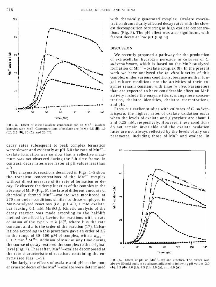
decay rates subsequent to peak complex formationwere slower and evidently at pH 6.0 the rate of Mn31–oxalate formation was so slow that a reflective maxi-mum was not observed during the 3-h time frame. Incontrast, decay rates were faster at pH values less than4.0.
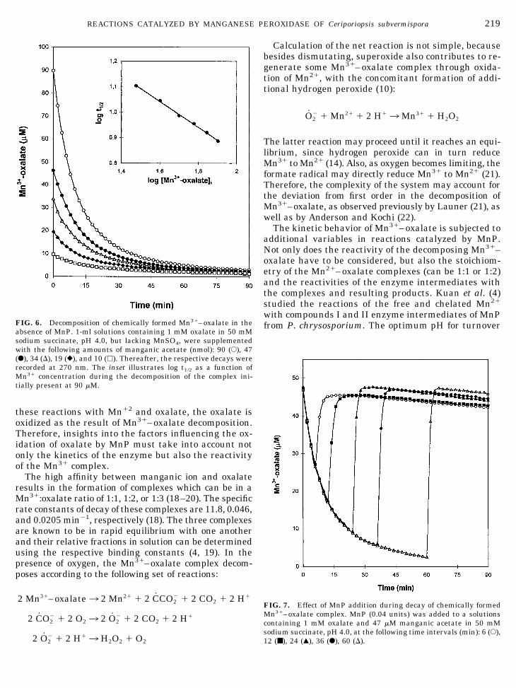
The enzymatic reactions described in Figs. 1–5 showthe transient concentrations of the Mn31 complexwithout direct measure of its rate of formation or de-cay. To observe the decay kinetics of the complex in theabsence of MnP (Fig. 6), the fate of different amounts ofchemically formed Mn31–oxalate was monitored at270 nm under conditions similar to those employed inMnP-catalyzed reactions (i.e., pH 4.0, 1 mM oxalate,but lacking 0.1 mM MnSO4). Kinetic analysis of thedecay reaction was made according to the half-lifemethod described by Levine for reactions with a rateequation of the type v 5 k [S]n, where k is the rateconstant and n is the order of the reaction (17). Calcu-lations according to this procedure gave an order of 3/2in the range of 30–100 mM of complex, with a kobs 50.012 min-1 M-0.5. Addition of MnP at any time duringthe course of decay restored the complex to the originallevel (Fig. 7). Thereafter, Mn31–oxalate decomposed atthe rate characteristic of reactions containing the en-zyme (see Figs. 1–5).
Similarly, the effects of oxalate and pH on the non-enzymatic decay of the Mn31–oxalate were determined
with chemically generated complex. Oxalate concen-tration dramatically affected decay rates with the slow-est decomposition occurring at high oxalate concentra-tions (Fig. 8). The pH effect was also significant, withfastest decay at low pH (Fig. 9).
DISCUSSION
We recently proposed a pathway for the productionof extracellular hydrogen peroxide in cultures of C.subvermispora, which is based on the MnP-catalyzedformation of Mn31–oxalate complex (8). In the presentwork we have analyzed the in vitro kinetics of thiscomplex under various conditions, because neither fun-gal culture conditions nor the activities of their en-zymes remain constant with time in vivo. Parametersthat are expected to have considerable effect on MnPactivity include the enzyme titers, manganese concen-tration, chelator identities, chelator concentrations,and pH.
From our earlier studies with cultures of C. subver-mispora, the highest rates of oxalate oxidation occurwhen the levels of oxalate and glyoxylate are about 1and 0.25 mM, respectively. However, these conditionsdo not remain invariable and the oxalate oxidationrates are not always reflected by the levels of any oneparameter, including those of MnP and oxalate. In
FIG. 5. Effect of pH on Mn31–oxalate kinetics. The buffer wasalways 50 mM sodium succinate, adjusted to following pH values: 3.0(}), 3.5 (F), 4.0 (h), 4.5 (E), 5.0 (D), and 6.0 (Œ).
FIG. 4. Effect of initial oxalate concentrations on Mn31–oxalatekinetics with MnP. Concentrations of oxalate are (mM): 0.5 (■), 1.0(h), 2.5 (F), 10 (D), and 20 (E).
218 URZUA, KERSTEN, AND VICUNA
these reactions with Mn12 and oxalate, the oxalate isoxidized as the result of Mn31–oxalate decomposition.Therefore, insights into the factors influencing the ox-idation of oxalate by MnP must take into account notonly the kinetics of the enzyme but also the reactivityof the Mn31 complex.
The high affinity between manganic ion and oxalateresults in the formation of complexes which can be in aMn31:oxalate ratio of 1:1, 1:2, or 1:3 (18–20). The specificrate constants of decay of these complexes are 11.8, 0.046,and 0.0205 min21, respectively (18). The three complexesare known to be in rapid equilibrium with one anotherand their relative fractions in solution can be determinedusing the respective binding constants (4, 19). In thepresence of oxygen, the Mn31–oxalate complex decom-poses according to the following set of reactions:
2 Mn31–oxalate3 2 Mn21 1 2 CCO22 1 2 CO2 1 2 H1
2 CO22 1 2 O23 2 O2
2 1 2 CO2 1 2 H1
2 O22 1 2 H13 H2O2 1 O2
Calculation of the net reaction is not simple, becausebesides dismutating, superoxide also contributes to re-generate some Mn31–oxalate complex through oxida-tion of Mn21, with the concomitant formation of addi-tional hydrogen peroxide (10):
O22 1 Mn21 1 2 H13Mn31 1 H2O2
The latter reaction may proceed until it reaches an equi-librium, since hydrogen peroxide can in turn reduceMn31 to Mn21 (14). Also, as oxygen becomes limiting, theformate radical may directly reduce Mn31 to Mn21 (21).Therefore, the complexity of the system may account forthe deviation from first order in the decomposition ofMn31–oxalate, as observed previously by Launer (21), aswell as by Anderson and Kochi (22).
The kinetic behavior of Mn31–oxalate is subjected toadditional variables in reactions catalyzed by MnP.Not only does the reactivity of the decomposing Mn31–oxalate have to be considered, but also the stoichiom-etry of the Mn21–oxalate complexes (can be 1:1 or 1:2)and the reactivities of the enzyme intermediates withthe complexes and resulting products. Kuan et al. (4)studied the reactions of the free and chelated Mn21
with compounds I and II enzyme intermediates of MnPfrom P. chrysosporium. The optimum pH for turnover
FIG. 7. Effect of MnP addition during decay of chemically formedMn31–oxalate complex. MnP (0.04 units) was added to a solutionscontaining 1 mM oxalate and 47 mM manganic acetate in 50 mMsodium succinate, pH 4.0, at the following time intervals (min): 6 (E),12 (■), 24 (Œ), 36 (F), 60 (D).
FIG. 6. Decomposition of chemically formed Mn31–oxalate in theabsence of MnP. 1-ml solutions containing 1 mM oxalate in 50 mMsodium succinate, pH 4.0, but lacking MnSO4, were supplementedwith the following amounts of manganic acetate (nmol): 90 (E), 47(F), 34 (D), 19 (}), and 10 (h). Thereafter, the respective decays wererecorded at 270 nm. The inset illustrates log t1/2 as a function ofMn31 concentration during the decomposition of the complex ini-tially present at 90 mM.
219REACTIONS CATALYZED BY MANGANESE PEROXIDASE OF Ceriporiopsis subvermispora

is pH 4.5 and at this pH the reaction with compound Iis too fast to study. Although Kuan et al. initiallyconcluded that the 1:1 Mn21–oxalate complex is thepreferred substrate for the oxidation with compound II,it has been recently confirmed that Mn21–oxalate doesnot bind to MnP (23). The latter report is in agreementwith previous observations by Wariishi et al. (3), whohad indicated that the free (hexa-aquo) Mn21 is readilyoxidized. These authors found no relationship betweenstimulation of enzyme activity and chelator size, indi-cating that the substrate is free Mn21 rather than thecomplex. Kishi et al. (2) suggest that the significantstimulation of MnP activity by oxalate is due to itschelation with Mn31, thereby facilitating the removalof the ion from the enzyme active site. Maximal stim-ulation of compound II reduction is observed at about 2mM oxalate (2, 4).
The kinetic traces in Fig. 1 are in good qualitativeagreement with our previous experiments (8) whereadditions of H2O2, glyoxylate, or Mn31 were tested fortheir effects on the lag on CO2 evolution from oxalate.Interestingly, the levels of peak Mn31–oxalate concen-trations are similar in all cases (approximately 60 mM).At this point of inflection, the rate of Mn31–oxalateproduction is equal to the rate of its decomposition. Therate of Mn31–oxalate decomposition at this concentra-
FIG. 8. Effect of oxalate on the decay of chemically formed complex. In these experiments, the complex was formed by adding 50 nmol ofMn31–acetate to 1 ml solutions containing the indicated concentration of oxalate and 100 mM sodium succinate, pH 4.0, as buffer. For eachreaction, the kobs was calculated according to the half-life method described by Levine (17). The dotted line indicates the reciprocal of thefraction (a) of trioxalate Mn13 complex at different concentrations of oxalate (see Discussion).
FIG. 9. Effect of pH on the decay of Mn31–oxalate. The 50 mMsodium succinate buffer was adjusted to the indicated pH valuesprior to forming the complex. Its decay was thereafter monitored at270 nm and values of kobs were obtained as indicated in the legend toFig. 8.
220 URZUA, KERSTEN, AND VICUNA
tion can be estimated from the decay traces of Fig. 6and is approximately 5.6 mM min21 (i.e., 0.012 3 603/2)and therefore it represents an estimate of MnP activityin terms of Mn31–oxalate produced.
However, the maximal concentration of Mn31–ox-alate observed in reactions is not always proportionalto MnP titers (Fig. 2). This nonlinear response could bedue, at least in part, to oxygen limitation. Anotherplausible explanation is that the theoretical limit forMn31–oxalate formation is 100 mM, the initial concen-tration of MnSO4 supplied to the reactions. Thereforethe maximal Mn31–oxalate would be expected toasymptotically approach 100 mM with increasing MnP.How close the Mn31–oxalate levels approach the the-oretical limit will be affected by the Km of MnP for thedepleting levels of Mn21 and by the rate of nonenzy-matic decay of the complex, which is faster at higherconcentrations. The limiting effect of manganese canbe better appreciated in the kinetic traces of Fig. 7, inwhich addition of MnP restores the level of complex tojust about the concentration of Mn31 added initially tothe reaction.
Available Mn21 in reactions can have additional ef-fects besides the maximal concentration of Mn31–ox-alate produced (Fig. 3). In reactions where the initialoxalate concentration was 1 mM and the concentrationof MnSO4 was varied, the maximal level of Mn31–oxalate produced was observed with 1 mM MnSO4.Curiously, the rate of complex decomposition at thehigher levels of MnSO4 appear exceptionally high afterthe inflection point. This possibly could be due to thecatalase activity of MnP destroying H2O2 when un-chelated Mn21 is present (24). This would effectivelysubterfuge the H2O2-dependent oxidations of MnP, in-cluding the oxidation of Mn21 to Mn31. A second plau-sible reason for the rapid decay of Mn31–oxalate athigher concentrations of Mn21 is that oxygen might belimiting. For example, at 0.4 mM MnSO4, Mn31–ox-alate reaches a maximum near 100 mM. At this con-centration, the Mn31–oxalate complex is expected todecompose at approximately 12 mM min21 (0.012 31003/2) and to consume oxygen at approximately halfthat rate. Given that the concentration of O2 in aque-ous solution is 240 mM (25), it is reasonably assumedthat the oxygen is near depletion after about 40 min.The return to Mn31–oxalate levels around 40 mMmight be a reflection of rate-limiting diffusion of oxy-gen into the solution.
Oxalate concentration also has a dramatic effect onmaximal Mn31–oxalate levels reached in reactionswith MnP and 0.1 mM MnSO4 (Fig. 4). Since free Mn21
(hexa-aquo) is the reported substrate for MnP (3, 23),the lack of correlation between oxalate concentrationand the peak Mn31–oxalate levels obtained above 2.5mM oxalate could be due to an increase in the fractionof the mono and dioxalate complexes of Mn21 (26). On
the other hand, an interpretation of the results shownin Fig. 4 must take into consideration the stability ofthe manganic complexes to spontaneous decay (Fig. 8).Examples to illustrate this argument are the kineticresponses at 1.0 and 2.5 mM oxalate. It would be pre-dicted that if there is a slower decay of the complex at2.5 mM and if there is equal MnP activity at the twooxalate concentrations, then the maximal Mn31–ox-alate concentration is expected to be higher with 2.5mM. This is what is observed (Fig. 4). On the otherhand, higher concentrations of oxalate favor the forma-tion of di- and trioxalate complexes, which are morestable than the monooxalate complex (18). The relativefraction of each of the three Mn13 complexes withdivalent oxalate was calculated (based on initial con-ditions) and possible correlations with kobs were tested.As it might be predicted by the relative stability of thetrioxalate complex, the rate of decay decreases as theconcentration of the latter increases. This is confirmedby the correlation observed between the kobs and thereciprocal of the fraction of trioxalate complex (Fig. 8).In these experiments, the concentration of monoox-alate complex is negligible. However, this predictivetheory based on increased stability of the Mn31–ox-alate at higher oxalate concentrations totally fails at 10and 20 mM oxalate. Our results suggest that there is aMnP response to higher concentrations of oxalate thatis not readily explained by the presteady-state kineticsof MnP with oxalate complexes (4) or by the stability ofthe complexes. This might also provide insight to whyWariishi et al. (3) found no relationship between stim-ulation of enzyme activity and chelator size whentested at 50 mM.
Maximal levels of complex formation were observedat pH 4.0 with MnP (Fig. 5). This pH value is close tothe pKa2 of oxalate and is slightly less than the pHoptimum for the MnP-catalyzed oxidation of vanillyla-cetone (27). We have shown that chemically generatedcomplex is more stable when both carboxyls are ionized(Fig. 9), which is consistent with previous results usingdifferent oxalate concentrations (18). The increasedstability of the complex at higher pH most likely de-creases its reactivity with peroxidase substrates. How-ever, we show here that a similar pH optimum is ob-served even without an aromatic substrate present.
The results presented here support our recently pro-posed model for extracellular hydrogen peroxide pro-duction by C. subvermispora that is based on the MnP-dependent oxidation of carboxylic acids secreted by thefungus (8). Indeed, we have confirmed both the forma-tion and the decay of the Mn31–oxalate complex underconditions mimicking those of the cultures. As men-tioned above, the optimum pH for complex formation isin the same range as for MnP activity and for degra-dation of synthetic lignin (27). The decay of the Mn31–oxalate at this pH occurs at a rate that appears com-
221REACTIONS CATALYZED BY MANGANESE PEROXIDASE OF Ceriporiopsis subvermispora

patible with the maintenance of a sustained level ofcomplex. Glyoxylate stimulates the formation of thecomplex, providing an explanation for the increasedlevels of mineralization of 14C-labeled oxalate observedin vitro when glyoxylate is present (8). Finally, oxalatewas supplied in our experiments at defined initial con-centrations which is in contrast to the continuous se-cretion of this metabolite during wood decay by thefungus. Oxalate stabilizes the complex and extracellu-lar concentrations of this organic acid above criticallevels would hinder the steady production of the H2O2required by MnP. This requirement is at least fulfilledin liquid cultures where the concentration of oxalatereaches a maximum of 2.5 mM (8).
ACKNOWLEDGMENTS
This work was financed by Grants from NSF/CONICYT INT9414084, FONDECYT 1971239, FONDECYT 2960010, and USDA/NRICGP 94-37103-1022.
REFERENCES
1. Glenn, J. K., Akileswaran, L., and Gold, M. H. (1986) Arch.Biochem. Biophys. 251, 688–696.
2. Kishi, K., Wariishi, H., Marquez, L., Dunford, H. B., and Gold,M. H. (1994) Biochemistry 33, 8694–8701.
3. Wariishi, H., Valli, K., and Gold, M. H. (1992) J. Biol. Chem. 267,23688–23695.
4. Kuan, I. C., Johnson, K. A., and Tien, M. (1993) J. Biol. Chem.268, 20064–20070.
5. Akhtar, M. (1994) Holzforschung 48, 199–202.6. Lobos, S., Larraın, J., Salas, L., Cullen, D., and Vicuna, R. (1994)
Microbiology 14, 2691–2698.7. Salas, C., Lobos, S., Larraın, J., Cullen, D., and Vicuna, R. (1995)
Biotechnol. Appl. Biochem. 21, 323–333.8. Urzua, U., Kersten, P. J., and Vicuna, R. (1998) Appl. Environ.
Microbiol. 64, 68–73.
9. Khindaria, A., Grover, T. A., and Aust, S. D. (1994) Arch. Bio-chem. Biophys. 314, 301–306.
10. Archibald, F. S., and Fridovich, I. (1982) Arch. Biochem. Biophys.214, 452–463.
11. Kuan, I. C., and Tien, M. (1993) Arch. Biochem. Biophys. 302,447–454.
12. Dutton, M. V., Evans, C. S., Atkey, P. T., and Wood, D. A. (1993)Appl. Microbiol. Biotechnol. 39, 5–10.
13. Kuan, I. C., and Tien, M. (1993) Proc. Natl. Acad. Sci. USA 90,1242–1246.
14. Aitken, M. D., and Irvine, R. (1990) Arch. Biochem. Biophys. 276,405–414.
15. Bernt, E., and Bergmeyer, H. U. (1974) in Methods of EnzymaticAnalysis (Bergmeyer, H. U., Ed.), Vol. 3, pp. 2246–2248, Aca-demic Press, New York.
16. Paszczynski, A., Huynh, V. B., and Crawford, R. (1985) FEMSMicrobiol. Lett. 29, 37–41.
17. Levine, I. N. (1978) Physical Biochemistry. McGraw–Hill, NewYork.
18. Taube, H. (1948) J. Am. Chem. Soc. 70, 1216–1220.19. Taube, H. (1948) J. Am. Chem. Soc. 70, 3928–3935.20. Cartledge, G. H., and Eriks, W. P. (1936) J. Am. Chem. Soc. 58,
2061–2065.21. Launer, H. F. (1933) J. Am. Chem. Soc. 55, 865–874.22. Anderson, J. M., and Kochi, J. K. (1970) J. Am. Chem. Soc. 92,
2450–2460.23. Banci, L., Bertini, I., Dal Pozzo, L., Del Conte, R., and Tien, M.
(1988) Biochemistry 37, 9009–9015.24. Timofeevski, S. L., and Aust, S. D. (1997) Biochem. Biophys. Res.
Commun. 3, 645–649.25. Cooper, T. (1977) The Tools of Biochemistry, p. 32, Wiley, New
York.26. Meites, L. (Ed). (1963) Handbook of Analytical Chemistry,
McGraw–Hill, New York.27. Tapia, J., and Vicuna, R. (1995) Appl. Environ. Microbiol. 61,
2476–2481.
222 URZUA, KERSTEN, AND VICUNA


![Reduction of Oxalate Levels in Tomato Fruit and … of Oxalate Levels in Tomato Fruit and Consequent Metabolic Remodeling Following Overexpression of a Fungal Oxalate Decarboxylase1[W]](https://static.fdocuments.in/doc/165x107/5af8e5787f8b9aff288c704b/reduction-of-oxalate-levels-in-tomato-fruit-and-of-oxalate-levels-in-tomato.jpg)