Kharkiv National Medical University Department of...
82
MENINGEAL SYNDROME IN CLINIC OF INFECTIOUS DISEASES Kharkiv National Medical University Department of Infectious Diseases D.Sc., Ass. Prof. Bondarenko A.V. Х А Р Ь К О В С К И Й • Н А Ц И О Н А Л Ь Н Ы Й • М Е Д И Ц И Н С К И Й • У Н И В Е Р С И Т Е Т
-
Upload
trinhkhanh -
Category
Documents
-
view
222 -
download
0
Transcript of Kharkiv National Medical University Department of...
MENINGEAL SYNDROME IN CLINIC OF INFECTIOUS DISEASES
Kharkiv National Medical University Department of Infectious Diseases
D.Sc., Ass. Prof. Bondarenko A.V.
ХАРЬКОВСКИЙ• НАЦ
ИОНАЛЬНЫЙ • МЕДИЦИНС
КИЙ
•УНИВЕРСИТЕТ

The brain and spinal cord are covered by connective tissue layers collectively called the meninges which form the blood-brain barrier: 1 – the pia mater 2 – the arachnoid mater 3 – the dura mater

• MENINGITIS – inflammation of meninges (inflammatory response is generally confined to arachnoid, subarachnoid space and pia mater – i.e. LEPTOMENINGITIS)
• Leptomeningitis - inflammation of the pia mater and arachnoid.
• Arachnoiditis - inflammation of the arachnoid membrane.
• Pachymeningitis - inflammation of the dura mater.
• Encephalitis - inflammation of the brain parenchyma (diffuse and/or focal).
• Myelitis - inflammation of the spinal cord parenchyma.
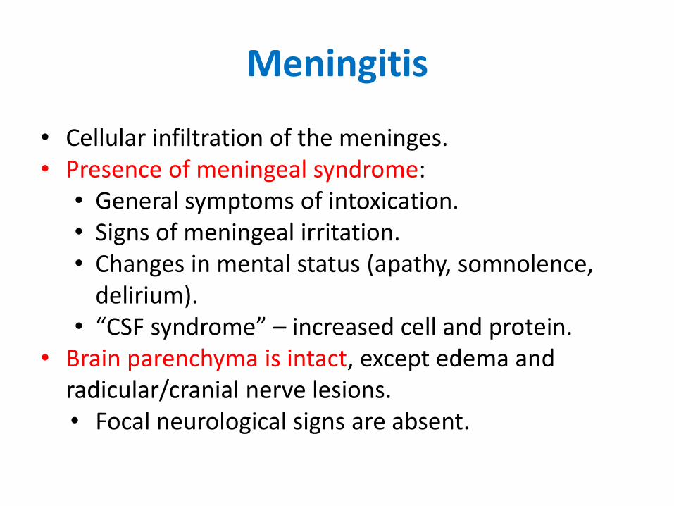
Meningitis
• Cellular infiltration of the meninges. • Presence of meningeal syndrome: • General symptoms of intoxication. • Signs of meningeal irritation. • Changes in mental status (apathy, somnolence,
delirium). • “CSF syndrome” – increased cell and protein.
• Brain parenchyma is intact, except edema and radicular/cranial nerve lesions. • Focal neurological signs are absent.

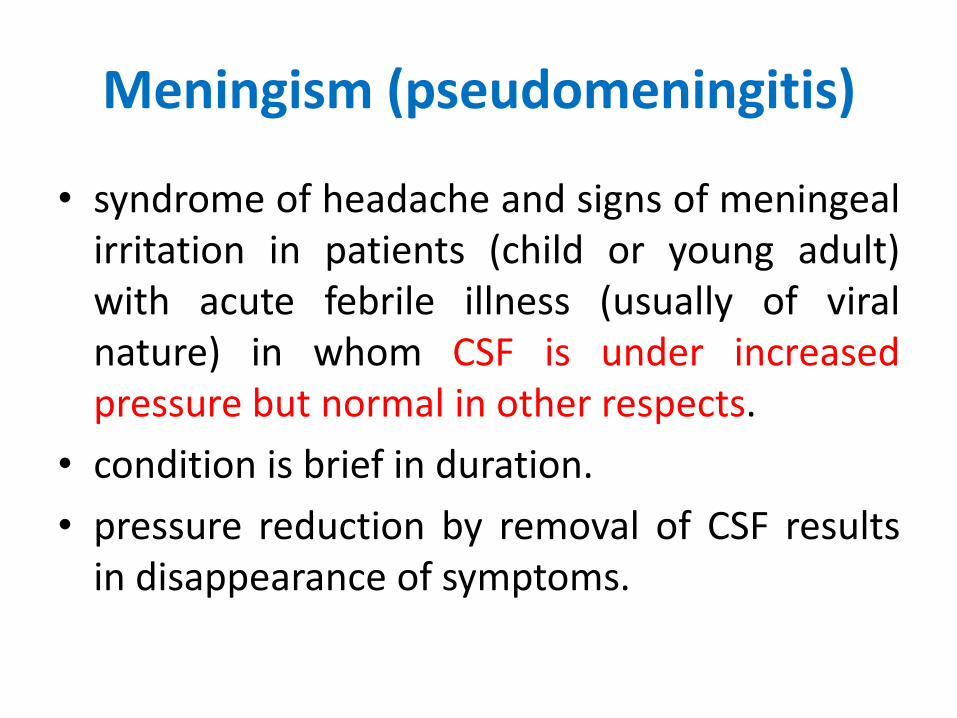
Meningism (pseudomeningitis)
• syndrome of headache and signs of meningeal irritation in patients (child or young adult) with acute febrile illness (usually of viral nature) in whom CSF is under increased pressure but normal in other respects.
• condition is brief in duration.
• pressure reduction by removal of CSF results in disappearance of symptoms.
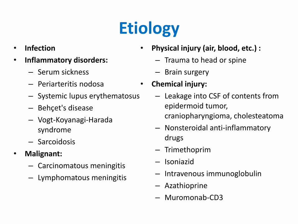
Etiology • Infection
• Inflammatory disorders:
– Serum sickness
– Periarteritis nodosa
– Systemic lupus erythematosus
– Behçet's disease
– Vogt-Koyanagi-Harada syndrome
– Sarcoidosis
• Malignant:
– Carcinomatous meningitis
– Lymphomatous meningitis
• Physical injury (air, blood, etc.) :
– Trauma to head or spine
– Brain surgery
• Chemical injury:
– Leakage into CSF of contents from epidermoid tumor, craniopharyngioma, cholesteatoma
– Nonsteroidal anti-inflammatory drugs
– Trimethoprim
– Isoniazid
– Intravenous immunoglobulin
– Azathioprine
– Muromonab-CD3
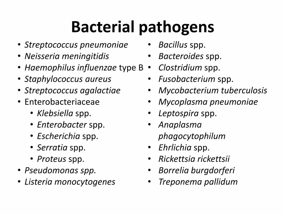
Bacterial pathogens • Streptococcus pneumoniae • Neisseria meningitidis • Haemophilus influenzae type B • Staphylococcus aureus • Streptococcus agalactiae • Enterobacteriaceae • Klebsiella spp. • Enterobacter spp. • Escherichia spp. • Serratia spp. • Proteus spp.
• Pseudomonas spp. • Listeria monocytogenes
• Bacillus spp. • Bacteroides spp. • Clostridium spp. • Fusobacterium spp. • Mycobacterium tuberculosis • Mycoplasma pneumoniae • Leptospira spp. • Anaplasma
phagocytophilum • Ehrlichia spp. • Rickettsia rickettsii • Borrelia burgdorferi • Treponema pallidum
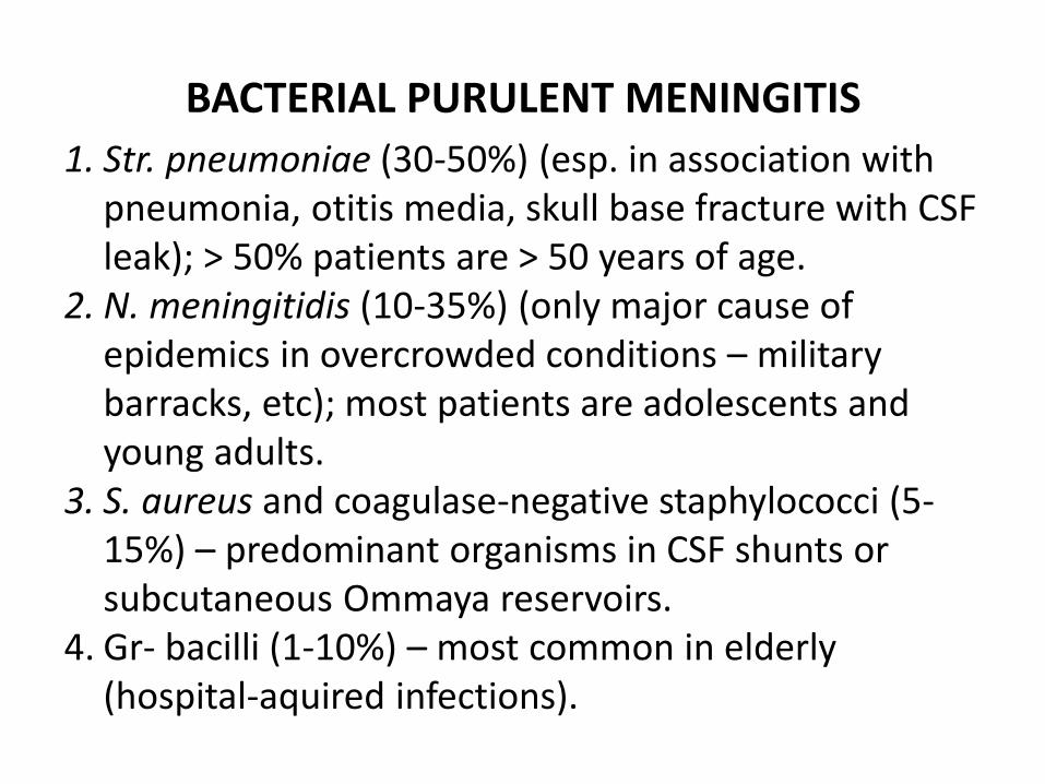
BACTERIAL PURULENT MENINGITIS
1. Str. pneumoniae (30-50%) (esp. in association with pneumonia, otitis media, skull base fracture with CSF leak); > 50% patients are > 50 years of age.
2. N. meningitidis (10-35%) (only major cause of epidemics in overcrowded conditions – military barracks, etc); most patients are adolescents and young adults.
3. S. aureus and coagulase-negative staphylococci (5-15%) – predominant organisms in CSF shunts or subcutaneous Ommaya reservoirs.
4. Gr- bacilli (1-10%) – most common in elderly (hospital-aquired infections).
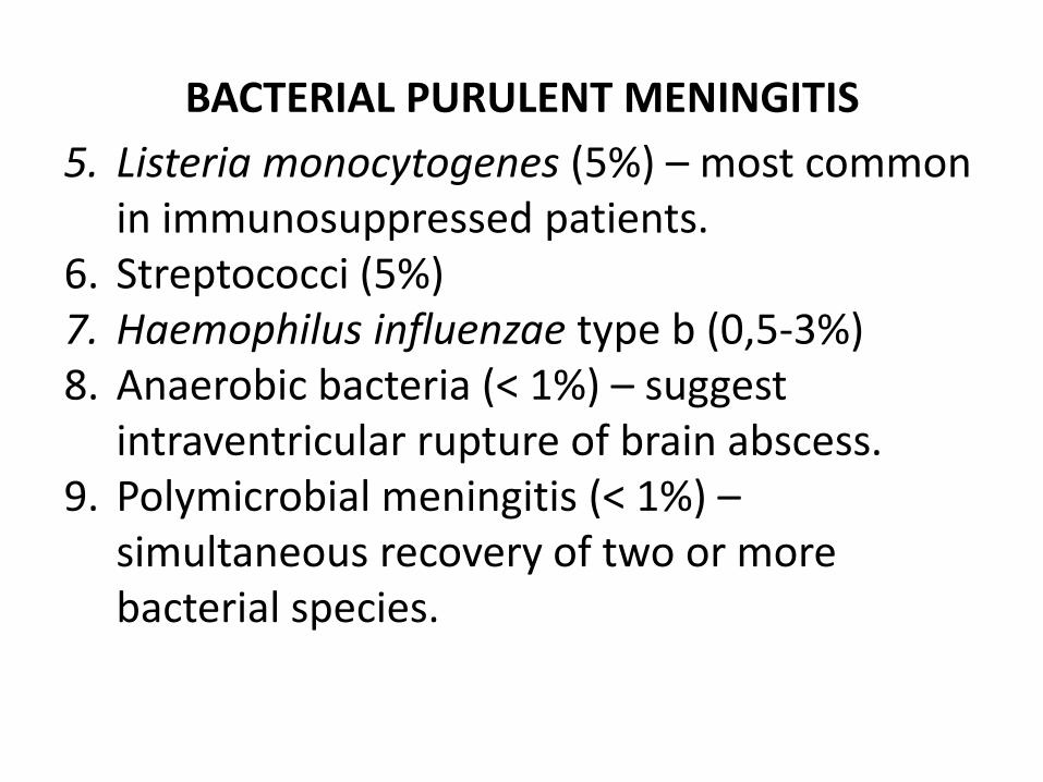
BACTERIAL PURULENT MENINGITIS
5. Listeria monocytogenes (5%) – most common in immunosuppressed patients.
6. Streptococci (5%) 7. Haemophilus influenzae type b (0,5-3%) 8. Anaerobic bacteria (< 1%) – suggest
intraventricular rupture of brain abscess. 9. Polymicrobial meningitis (< 1%) –
simultaneous recovery of two or more bacterial species.
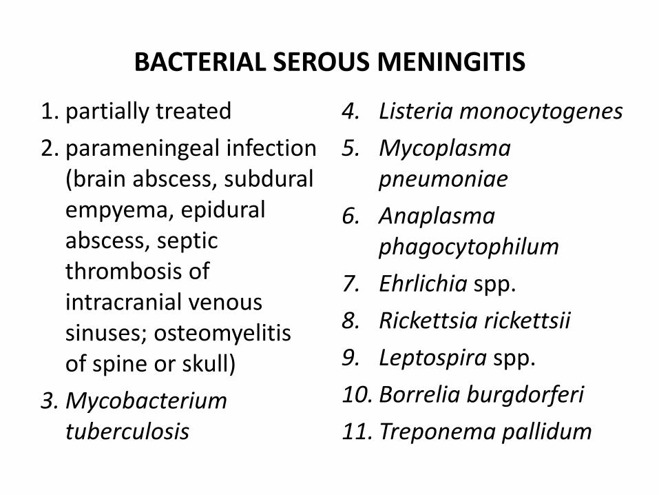
BACTERIAL SEROUS MENINGITIS
1. partially treated
2. parameningeal infection (brain abscess, subdural empyema, epidural abscess, septic thrombosis of intracranial venous sinuses; osteomyelitis of spine or skull)
3. Mycobacterium tuberculosis
4. Listeria monocytogenes
5. Mycoplasma pneumoniae
6. Anaplasma phagocytophilum
7. Ehrlichia spp.
8. Rickettsia rickettsii
9. Leptospira spp.
10. Borrelia burgdorferi
11. Treponema pallidum
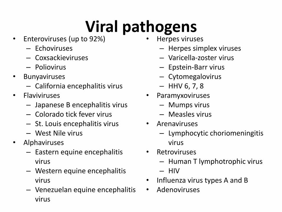
Viral pathogens • Enteroviruses (up to 92%)
– Echoviruses – Coxsackieviruses – Poliovirus
• Bunyaviruses – California encephalitis virus
• Flaviviruses – Japanese B encephalitis virus – Colorado tick fever virus – St. Louis encephalitis virus – West Nile virus
• Alphaviruses – Eastern equine encephalitis
virus – Western equine encephalitis
virus – Venezuelan equine encephalitis
virus
• Herpes viruses – Herpes simplex viruses – Varicella-zoster virus – Epstein-Barr virus – Cytomegalovirus – HHV 6, 7, 8
• Paramyxoviruses – Mumps virus – Measles virus
• Arenaviruses – Lymphocytic choriomeningitis
virus • Retroviruses
– Human T lymphotrophic virus – HIV
• Influenza virus types A and B • Adenoviruses

Fungal and Protozoal • Candida albicans
• Cryptococcus neoformans
• Coccidioides immitis
• Histoplasma capsulatum
• Blastomyces dermatitidis
• Paracoccidioides brasiliensis
• Cladosporium trichoides
• Aspergillus fumigatus
• Toxoplasma Gondii
• Naegleria fowleri
• Acanthamoeba spp.
• Taenia solium cysticercosis
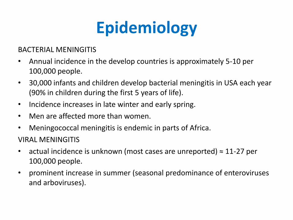
Epidemiology BACTERIAL MENINGITIS
• Annual incidence in the develop countries is approximately 5-10 per 100,000 people.
• 30,000 infants and children develop bacterial meningitis in USA each year (90% in children during the first 5 years of life).
• Incidence increases in late winter and early spring.
• Men are affected more than women.
• Meningococcal meningitis is endemic in parts of Africa.
VIRAL MENINGITIS
• actual incidence is unknown (most cases are unreported) ≈ 11-27 per 100,000 people.
• prominent increase in summer (seasonal predominance of enteroviruses and arboviruses).
Epidemiology
Routes of bacterial infection:
• Hematogenous spread to CNS.
• Direct extension from nearly focus (mastoiditis, sinusitis).
• Direct invasion (dermoid sinus tract, head trauma, meningomyelocele).
Pathogenesis
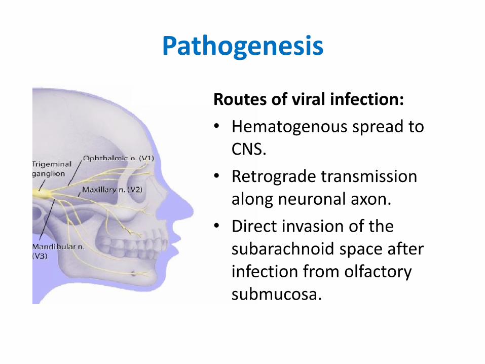
Routes of viral infection:
• Hematogenous spread to CNS.
• Retrograde transmission along neuronal axon.
• Direct invasion of the subarachnoid space after infection from olfactory submucosa.
Pathogenesis PREDISPOSING HOST FACTORS 1) MECHANICAL DISTURBANCES (neurosurgical procedures, basilar
skull fractures). 2) CONGENITAL DEFECTS (dermoid sinus tracts,
meningomyeloceles). 3) IMMUNOLOGIC DEFICIENCIES:
a) cell-mediated immunity (lymphoma, organ transplant recipients, corticosteroid therapy, AIDS) → intracellular bacteria (esp. M. tuberculosis, L. monocytogenes).
b) humoral immunity (splenectomy, chronic lymphocytic leukemia, multiple myeloma, Hodgkin's disease after radiotherapy or chemotherapy) → encapsulated bacteria (S. pneumoniae, H. influenzae type b, N. meningitidis).
c) neutropenia → P. aeruginosa, Enterobacteriaceae.
Pathogenesis
• Nasopharyngeal colonization and mucosae invasion.
• Evasion of the complement pathway. • Bacterial cross the BBB to CSF. • Host defense mechanism within the CSF are
often ineffective (Ig and complement activity are virtually absent; opsonic activity is often undetectable, phagocytosis is inefficient).
• Bacterial proliferation stimulate a convergence of leucocyte into the CSF.

Pathogenesis • release of toxic factors form bacteria → activation of
neutrophils → release of TNF-α, IL-1, 8, platelet activating factor: • cytotoxic cerebral edema. • increase in BBB permeability → vasogenic edema.
• large numbers of leukocytes in subarachnoid space contribute to purulent exudate and impair CSF absorption by arachnoid villi → COMMUNICATING HYDROCEPHALUS.
• pia-arachnoid becomes thickened → adhesions → interfere with CSF flow from 4th ventricle → OBSTRUCTIVE HYDROCEPHALUS.
• hydrocephalus causes transependymal movement of fluid from ventricular system into brain parenchyma (interstitial edema).
Pathogenesis • cerebral edema causes ICP↑. • meningeal and superficial cortical vessels are engorged and
stand out prominently. • since subarachnoid space is continuous over brain, spinal
cord, and optic nerves, infection in this space extends throughout cerebrospinal axis unless there is obstruction of subarachnoid space.
• ventriculitis is nearly uniformly present. • brain parenchyma (cerebritis → abscess), dura
(pachymeningitis), subdural and epidural spaces may be secondarily involved.
• when patient recovers, phagocytes completely clear subarachnoid space; if low-grade infection persists, adhesions and leptomeningeal fibrosis develops.
Classification • ACUTE MENINGITIS – symptoms evolving over 1-24
hours after onset (most cases are bacterial).
• SUBACUTE MENINGITIS – symptoms evolving over 1-7 days (includes virtually all cases of viral meningitis, along with some of fungal etiologies).
• CHRONIC MENINGITIS – symptoms evolving over more than 1 week (causes are fungi, tuberculosis, syphilis, malignancy, systemic collagenoses, sarcoidosis, some viruses).
• RECURRENT MENINGITIS – bouts of acute meningitis with complete resolution between episodes.
Classification RECURRENT BACTERIAL MENINGITIS signals host defect in:
– Immunologic defenses
– Local anatomy - usually after trauma.
RECURRENT NON-BACTERIAL MENINGITIS:
• herpes simplex viruses
• chemical meningitis
• primary inflammatory conditions
• drug hypersensitivity (with repeated administration).
Clinical Manifestation
• toxic syndrome (hyperpyrexia, vomiting without nausea, headache, seizures, muscle and back pain)
• meningeal syndrome – hyperesthesia, photophobia, irritability
– suppression of skin rephlexes
– tenderness in sites of trigeminal nerve endings
– stiff neck, positive Kernig’s and Brudzinski’s signs
• lethargy and obtundation progress to stupor or even coma
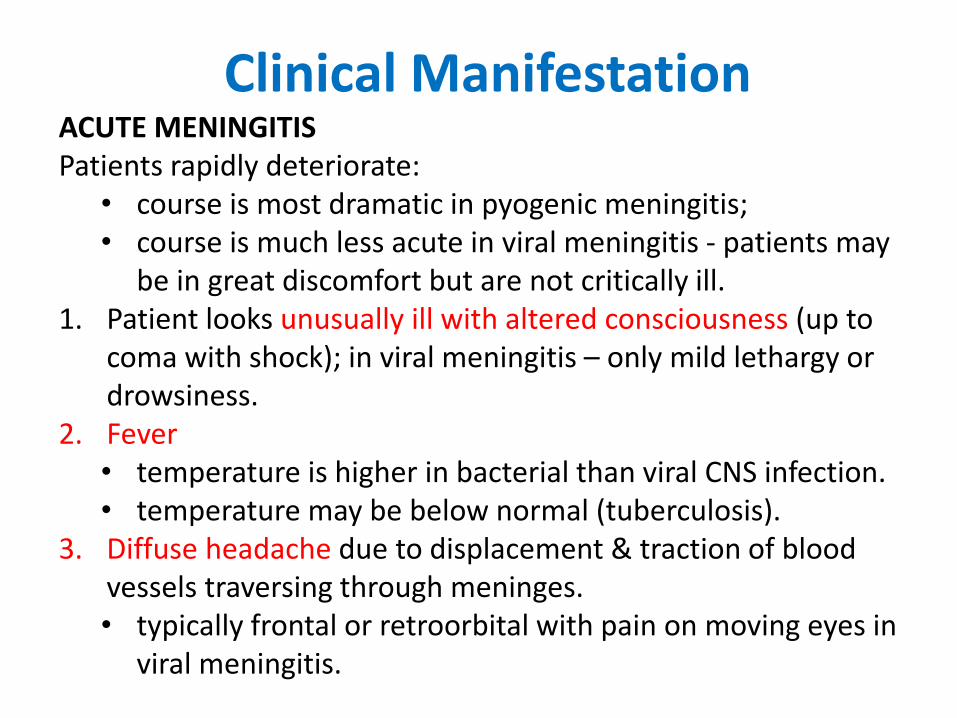
Clinical Manifestation ACUTE MENINGITIS Patients rapidly deteriorate: • course is most dramatic in pyogenic meningitis; • course is much less acute in viral meningitis - patients may
be in great discomfort but are not critically ill. 1. Patient looks unusually ill with altered consciousness (up to
coma with shock); in viral meningitis – only mild lethargy or drowsiness.
2. Fever • temperature is higher in bacterial than viral CNS infection. • temperature may be below normal (tuberculosis).
3. Diffuse headache due to displacement & traction of blood vessels traversing through meninges. • typically frontal or retroorbital with pain on moving eyes in
viral meningitis.
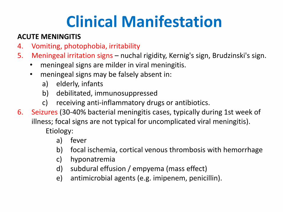
Clinical Manifestation ACUTE MENINGITIS 4. Vomiting, photophobia, irritability 5. Meningeal irritation signs – nuchal rigidity, Kernig's sign, Brudzinski's sign.
• meningeal signs are milder in viral meningitis. • meningeal signs may be falsely absent in:
a) elderly, infants b) debilitated, immunosuppressed c) receiving anti-inflammatory drugs or antibiotics.
6. Seizures (30-40% bacterial meningitis cases, typically during 1st week of illness; focal signs are not typical for uncomplicated viral meningitis). Etiology:
a) fever b) focal ischemia, cortical venous thrombosis with hemorrhage c) hyponatremia d) subdural effusion / empyema (mass effect) e) antimicrobial agents (e.g. imipenem, penicillin).
Clinical Manifestation
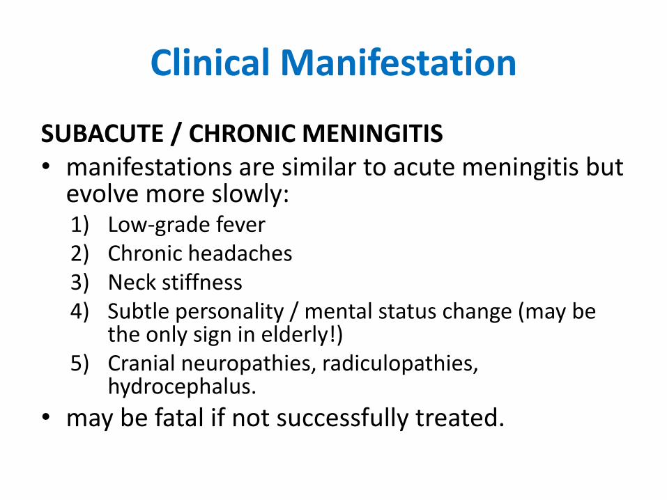
SUBACUTE / CHRONIC MENINGITIS • manifestations are similar to acute meningitis but
evolve more slowly: 1) Low-grade fever 2) Chronic headaches 3) Neck stiffness 4) Subtle personality / mental status change (may be
the only sign in elderly!) 5) Cranial neuropathies, radiculopathies,
hydrocephalus.
• may be fatal if not successfully treated.
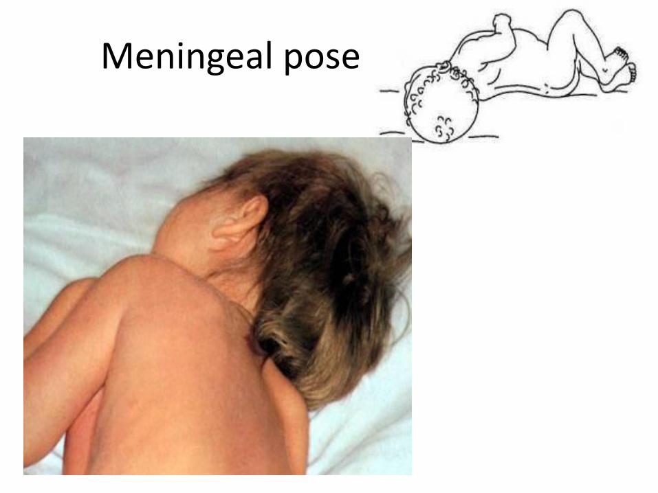
Meningeal pose
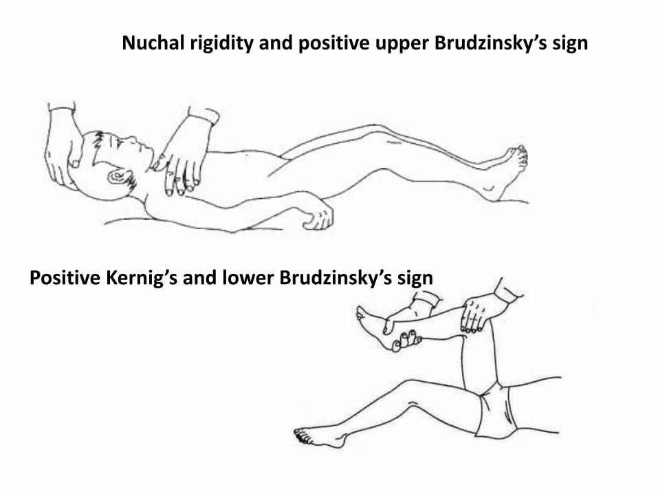
Nuchal rigidity and positive upper Brudzinsky’s sign
Positive Kernig’s and lower Brudzinsky’s sign
Meningism • The brain membranes irritation with appearing
positives meningeal signs without inflammatory data in CSF
Clinical manifestation • fever,
• headache,
• vomiting,
• irritability,
• stiff neck.
• upper Brudzinski sign positive.
• Kernig and lower Brudzinski signs are negative
Clinical Manifestation
Syndrome of consciousness disturbances includes
• excitement with euphoric,
• excitement with negativism,
• somnolence,
• stupor,
• sopor,
• coma (I, II degree).

Glasgow Coma Scale Activity Best Response Score
Eye
opening
Spontaneous
To verbal stimuli
To pain
None
4
3
2
1
Verbal Oriented
Confused
Inappropriate words
Nonspecific sounds
None
5
4
3
2
1
Motor Follows commands
Localizes pain
Withdraws in response to pain
Flexion in response to pain
Extension in response to pain
None
6
5
4
3
2
1
• Favorable prognosis for recovering
13-15 points
• Doubtful prognosis
9-12 points
• Unfavorable prognosis
less 8 points

Complications BACTERIAL MENINGITIS
1. Seizures
2. DIC, shock
3. Subdural effusions - usually in infants as self-limited process (as inflammatory process subsides, subdural fluid is reabsorbed);
4. Brain abscess, subdural empyema
5. Cerebral thrombophlebitis
6. Stroke:
a) vasospasm caused by subarachnoid infection
b) loss of cerebral autoregulation + hypotension
c) inflammatory infiltration of arterial wall (vasculitis).
7. Cranial nerve palsies (esp. sensorineural hearing loss; oculomotor paresis)
8. Consequences of ICP↑ (incl. brain herniation)
9. Chronic adhesive arachnoiditis, hydrocephalus
Complications
VIRAL MENINGITIS
• Various complication related to the systemic effect - orchitis, parotitis, pancreatitis
• Usually all of these complication resolve without sequelae.
Diagnosis
Plan of examination
• Ophthalmoscopy (papilledema)
• Lumbar puncture and examination of CSF
• Blood cultures
• Cultures of the nose and throat
• CT scan or MRI

Diagnosis • Lumbar Puncture Contraindication
– Present of infection in the skin, soft tissue at the puncture site.
– Likelihood of brain herniation.
• Indication for contrast-enhanced CT scan or MRI before LP in suspected bacterial meningitis – Immunocompromised state – History of Stroke, Mass lesion, Focal infection, Head trauma – Seizure within last 7 days – Abnormal level of consciousness – Inability to answer question or follow command appropiately – Abnormal visual fields or paresis of gaze – Focal weakness or neurologic deficit – Abnormal speech – Papilledema
Diagnosis
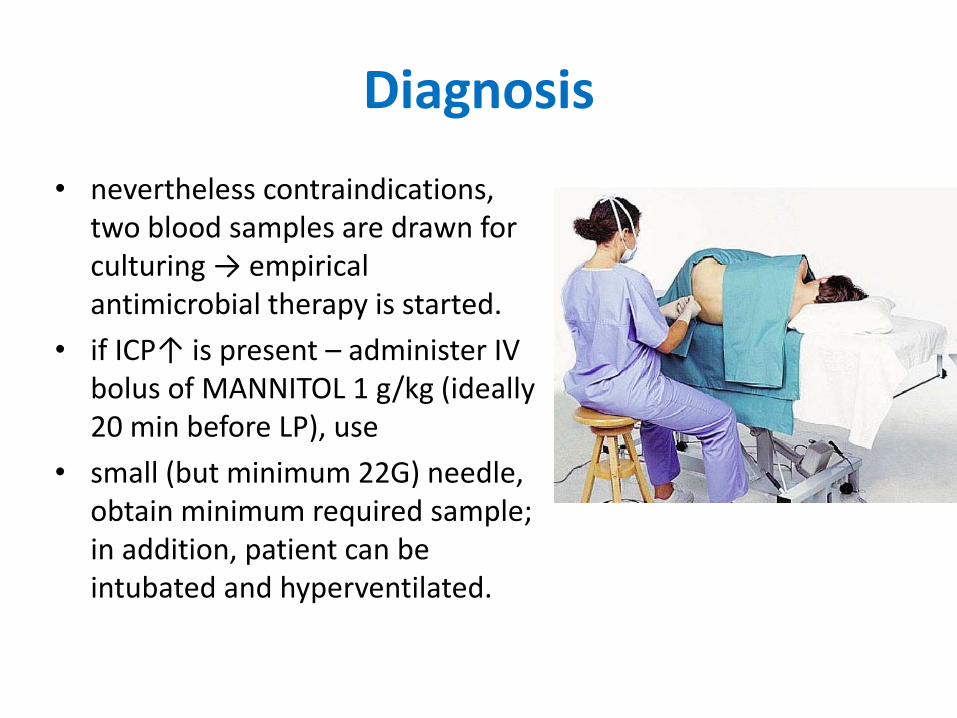
• nevertheless contraindications, two blood samples are drawn for culturing → empirical antimicrobial therapy is started.
• if ICP↑ is present – administer IV bolus of MANNITOL 1 g/kg (ideally 20 min before LP), use
• small (but minimum 22G) needle, obtain minimum required sample; in addition, patient can be intubated and hyperventilated.
Diagnosis
Antimicrobial therapy:
• will not significantly alter CSF profile (WBC count, glucose & lactate concentration, antigen test results) for at least 2-3 days.
• will decrease sensitivity of Gram's stain & culture (window of 2-3 hours after giving parenteral antibiotics when CSF cultures are not adversely affected).
– Gram's stain and culture should be negative in CSF obtained 24 hours after initiation of IV antimicrobial therapy, if organism is sensitive to that antibiotic.
Diagnosis
CSF Analysis
• Opening pressure - 50-180 mmH2O.
–elevated in bacterial, TB, fungal.
–moderately elevated (bacterial meningitis > viral meningitis).
– falsely elevated in tense, obese, marked muscle contraction.
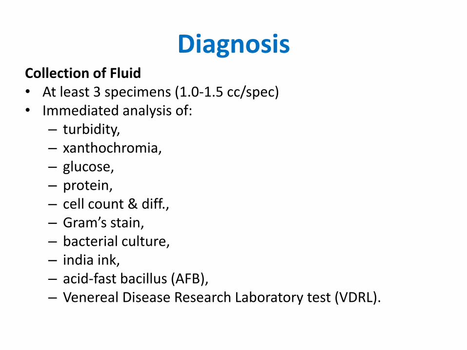
Diagnosis Collection of Fluid • At least 3 specimens (1.0-1.5 cc/spec) • Immediated analysis of:
– turbidity, – xanthochromia, – glucose, – protein, – cell count & diff., – Gram’s stain, – bacterial culture, – india ink, – acid-fast bacillus (AFB), – Venereal Disease Research Laboratory test (VDRL).
Diagnosis
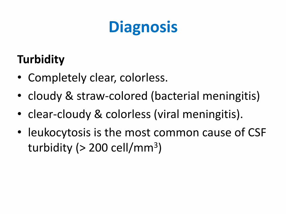
Turbidity
• Completely clear, colorless.
• cloudy & straw-colored (bacterial meningitis)
• clear-cloudy & colorless (viral meningitis).
• leukocytosis is the most common cause of CSF turbidity (> 200 cell/mm3)
Diagnosis
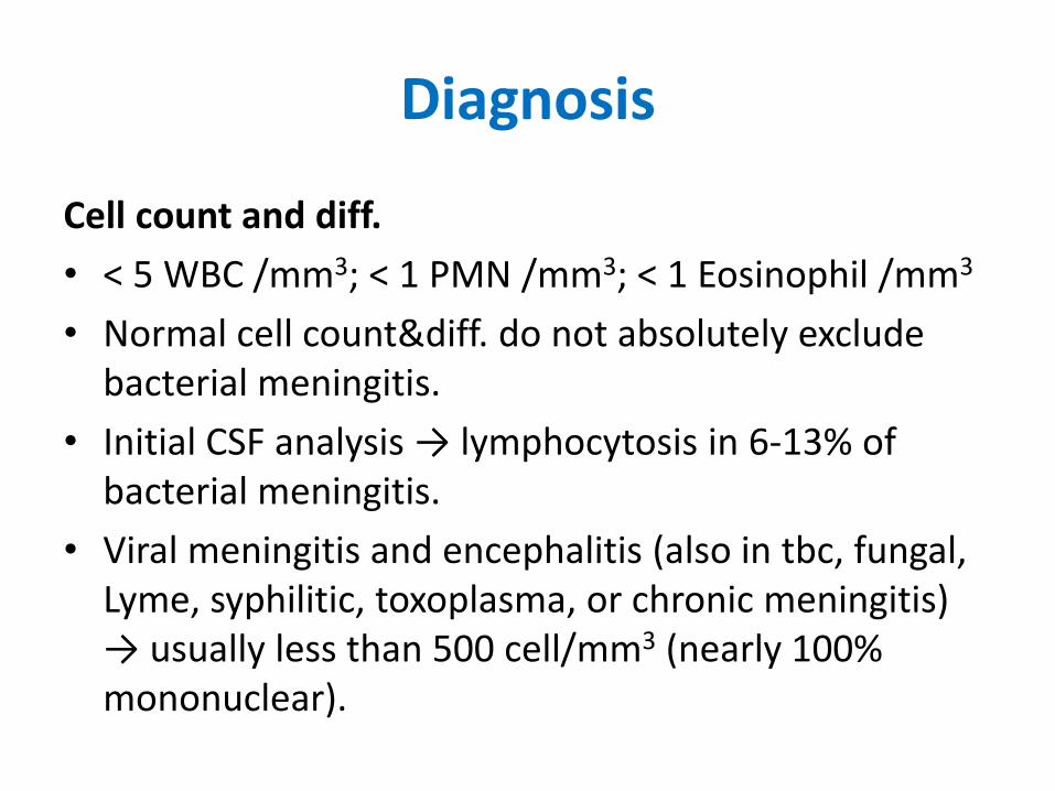
Cell count and diff.
• < 5 WBC /mm3; < 1 PMN /mm3; < 1 Eosinophil /mm3
• Normal cell count&diff. do not absolutely exclude bacterial meningitis.
• Initial CSF analysis → lymphocytosis in 6-13% of bacterial meningitis.
• Viral meningitis and encephalitis (also in tbc, fungal, Lyme, syphilitic, toxoplasma, or chronic meningitis) → usually less than 500 cell/mm3 (nearly 100% mononuclear).
Diagnosis
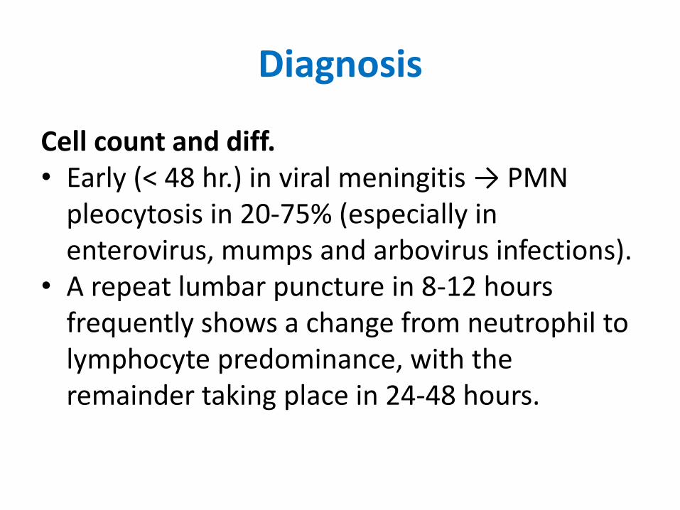
Cell count and diff. • Early (< 48 hr.) in viral meningitis → PMN
pleocytosis in 20-75% (especially in enterovirus, mumps and arbovirus infections).
• A repeat lumbar puncture in 8-12 hours frequently shows a change from neutrophil to lymphocyte predominance, with the remainder taking place in 24-48 hours.
Diagnosis
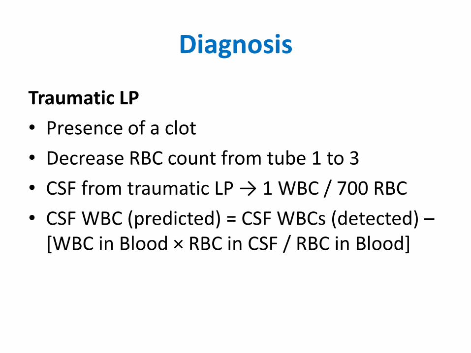
Traumatic LP
• Presence of a clot
• Decrease RBC count from tube 1 to 3
• CSF from traumatic LP → 1 WBC / 700 RBC
• CSF WBC (predicted) = CSF WBCs (detected) – [WBC in Blood × RBC in CSF / RBC in Blood]

Diagnosis
Xanthrochromia
• Lysis of RBC and release of breakdown pigments, oxyhemoglobin, bilirubin and methemoglobin into the CSF.
• Begin within 2 hr. → persist up to 30 days.
• Traumatic tap → ↑ CSF protein 150mg/dl or more
• Subarachnoid hemorrhage.
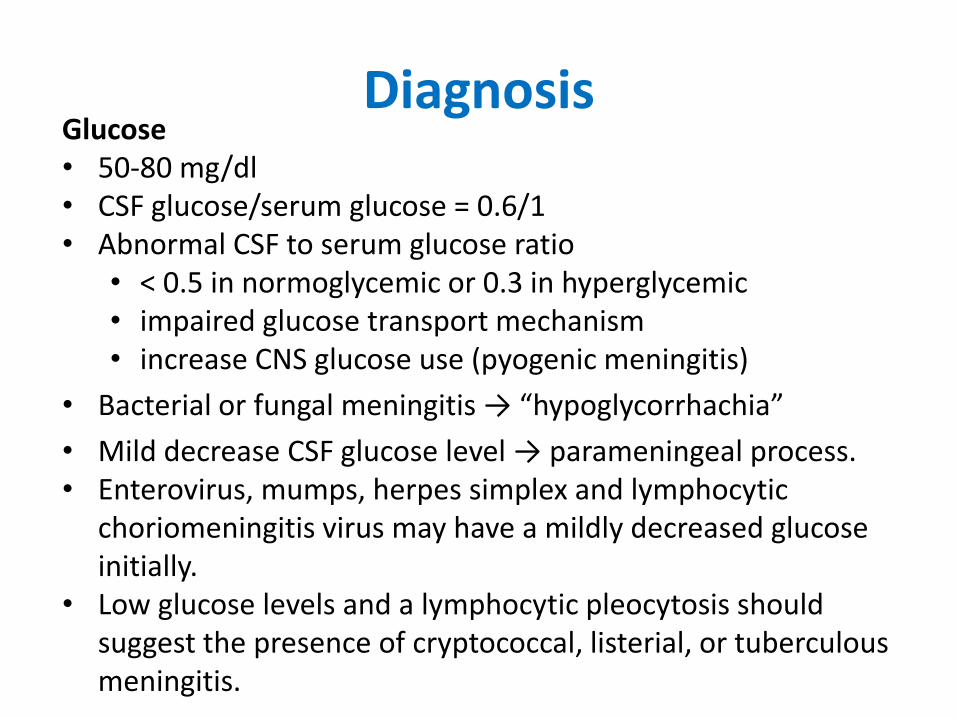
Diagnosis Glucose • 50-80 mg/dl • CSF glucose/serum glucose = 0.6/1 • Abnormal CSF to serum glucose ratio
• < 0.5 in normoglycemic or 0.3 in hyperglycemic • impaired glucose transport mechanism • increase CNS glucose use (pyogenic meningitis)
• Bacterial or fungal meningitis → “hypoglycorrhachia”
• Mild decrease CSF glucose level → parameningeal process. • Enterovirus, mumps, herpes simplex and lymphocytic
choriomeningitis virus may have a mildly decreased glucose initially.
• Low glucose levels and a lymphocytic pleocytosis should suggest the presence of cryptococcal, listerial, or tuberculous meningitis.
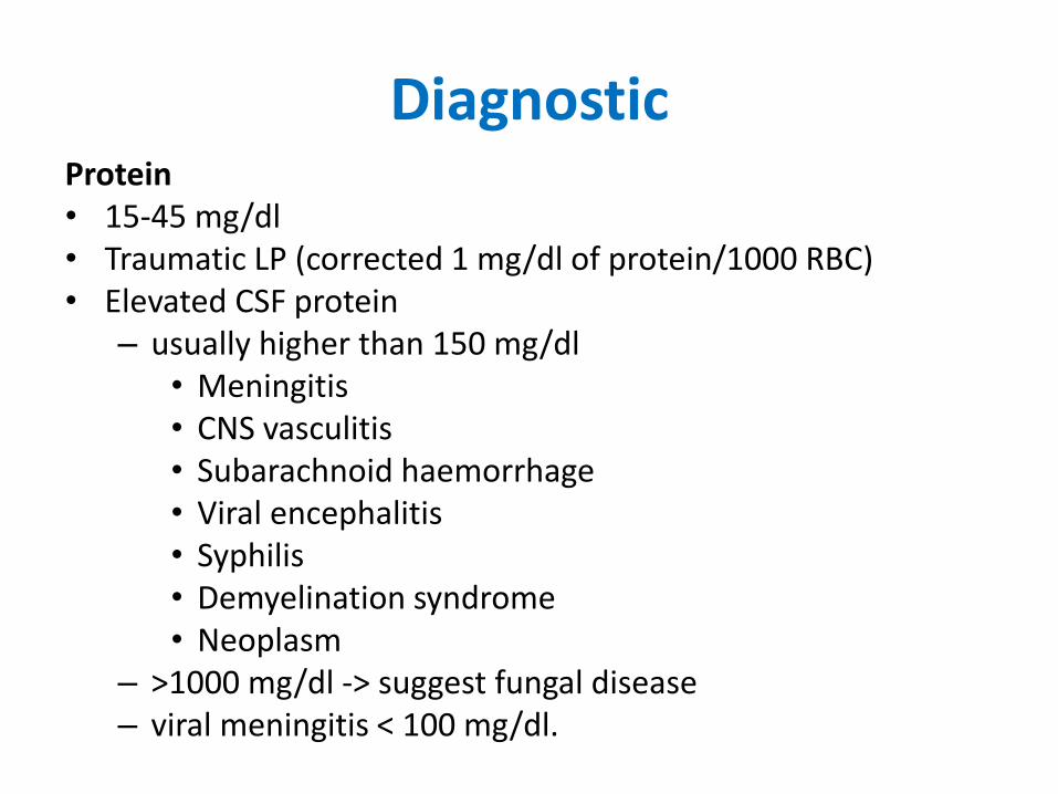
Diagnostic Protein • 15-45 mg/dl • Traumatic LP (corrected 1 mg/dl of protein/1000 RBC) • Elevated CSF protein
– usually higher than 150 mg/dl • Meningitis • CNS vasculitis • Subarachnoid haemorrhage • Viral encephalitis • Syphilis • Demyelination syndrome • Neoplasm
– >1000 mg/dl -> suggest fungal disease – viral meningitis < 100 mg/dl.
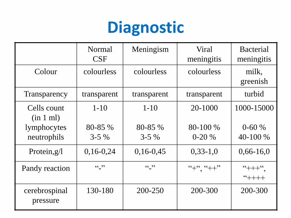
Diagnostic Normal
CSF
Meningism Viral
meningitis
Bacterial
meningitis
Colour colourless colourless colourless
milk,
greenish
Transparency transparent transparent transparent turbid
Cells count
(in 1 ml)
lymphocytes
neutrophils
1-10
80-85 %
3-5 %
1-10
80-85 %
3-5 %
20-1000
80-100 %
0-20 %
1000-15000
0-60 %
40-100 %
Protein,g/l 0,16-0,24 0,16-0,45 0,33-1,0 0,66-16,0
Pandy reaction “-” “-” “+“, “++” “+++“,
“++++
cerebrospinal
pressure
130-180 200-250 200-300 200-300
Diagnosis
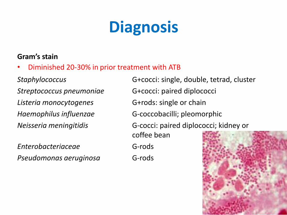
Gram’s stain
• Diminished 20-30% in prior treatment with ATB
Staphylococcus G+cocci: single, double, tetrad, cluster
Streptococcus pneumoniae G+cocci: paired diplococci
Listeria monocytogenes G+rods: single or chain
Haemophilus influenzae G-coccobacilli; pleomorphic
Neisseria meningitidis G-cocci: paired diplococci; kidney or coffee bean
Enterobacteriaceae G-rods
Pseudomonas aeruginosa G-rods
Diagnosis
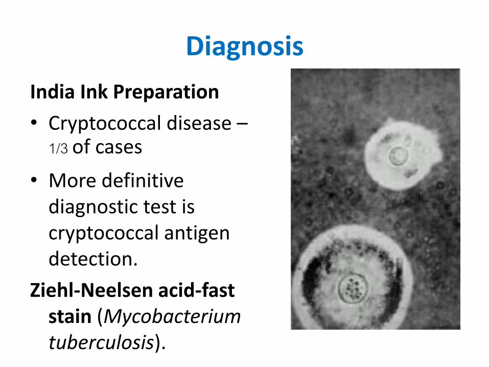
India Ink Preparation
• Cryptococcal disease – 1/3 of cases
• More definitive diagnostic test is cryptococcal antigen detection.
Ziehl-Neelsen acid-fast stain (Mycobacterium tuberculosis).
Diagnosis
• CSF/serum antigen/antibody detection (for viruses, syphilis, Lyme disease) – Counterimmunoelectrophoresis (CIE) – Latex particle agglutination – Coagglutination – Immunofluorescence
• Unfortunately, antibodies appear in CSF too late to aid in any therapeutic decisions (used only for retrospective diagnosis).
• Useful in receiving ATB before CSF sampling • Result - vary • Presence only an antigen, not viable organism • Serum antibody titers↑ → fourfold rise in paired sera (for
viruses).

Diagnosis Bacteriologic culture
– N. meningitis 37-55%
– H. influenzae 50%
Diagnosis
• PCR → resumed for less clear presentation, pretreat with ATB, care in which concern exists for TB, cryptococcal, treatable viral CNS infection.
• Sensitivities of detection in CSF by PCR for
– M. tuberculosis 80-85% (specificity 97-100%)
– N. meningitidis 88%,
– H. influenzae 100%,
– S. pneumoniae 92% (nearly 100% specificity)

Diagnosis Organism detection in other fluids:
• Blood culture (positive in 80-90% patients with bacterial meningitis). Identify causative organism more often when the meningitis is caused by pneumococcus than meningococcus.
• Stool specimen may be better source of viral isolate (enteroviruses), but is not diagnostic of meningitis.
• Virus isolation from saliva, throat washing (mumps).
• Meningococci may be found in skin lesions, nasopharyngeal secretions.
– In general, cultures of body surfaces and orifices are not helpful in identifying causative pathogen!
Diagnosis
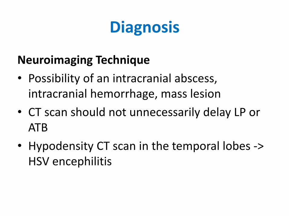
Neuroimaging Technique
• Possibility of an intracranial abscess, intracranial hemorrhage, mass lesion
• CT scan should not unnecessarily delay LP or ATB
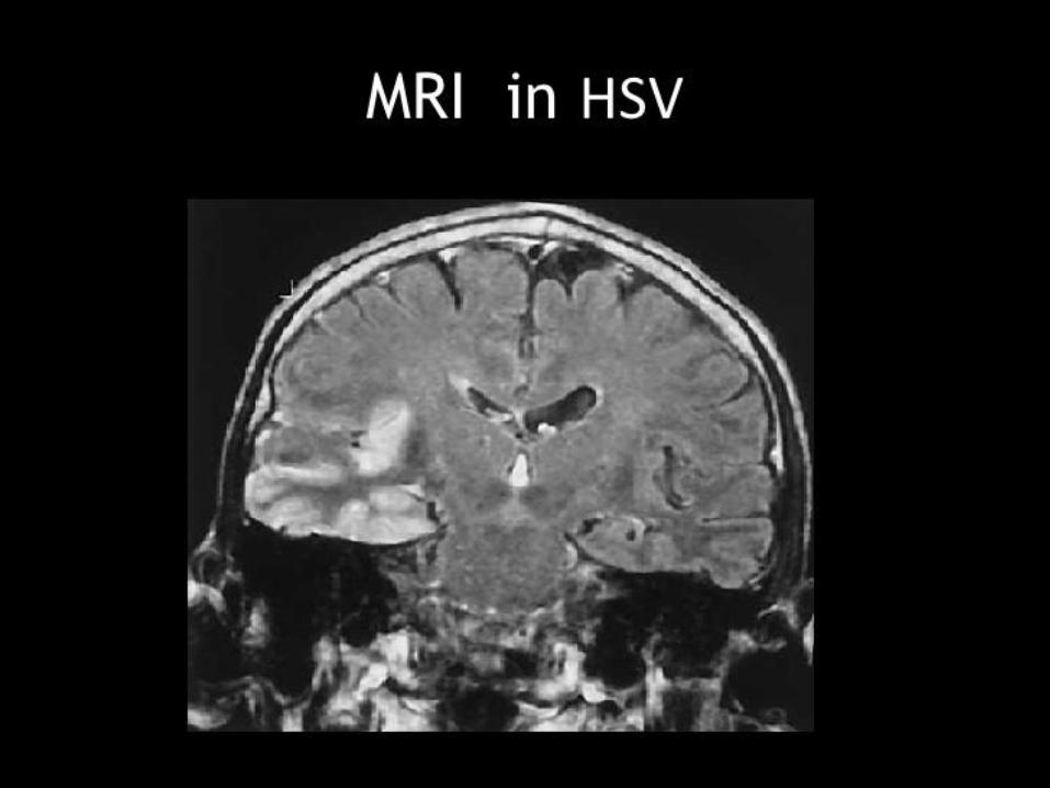
• Hypodensity CT scan in the temporal lobes -> HSV encephilitis
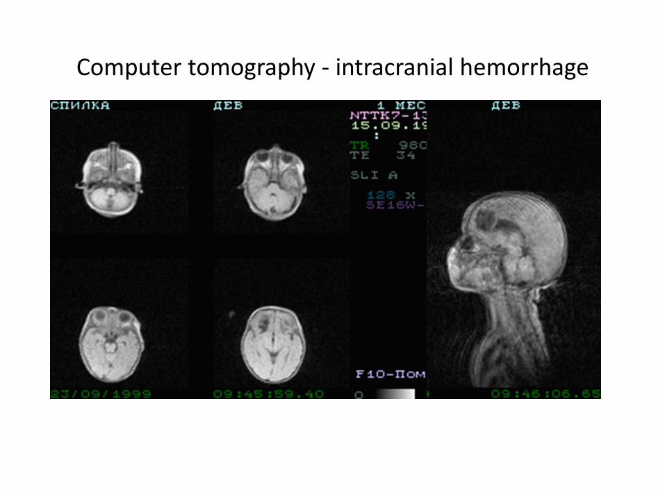
Computer tomography - intracranial hemorrhage
Diagnosis
Additional Investigation • WBC count is markedly elevated in
bacterial meningitis (mildly in viral meningitis).
• CBC may show leukopenia in elderly, immunosuppressed person.
Diagnosis
Additional Investigation • 50% of patients with pneumococcal
meningitis -> evidence of pneumonia on an initial chest x-ray.
Diagnosis
Additional Investigation • EEG is usually normal or slightly slow. • Focal or lateralized EEG abn. →
associated with HSV encephalitis (strong evidence).

Electroencephalography

Ultrasound investigation “Light- brain” – is a sing of meningitis
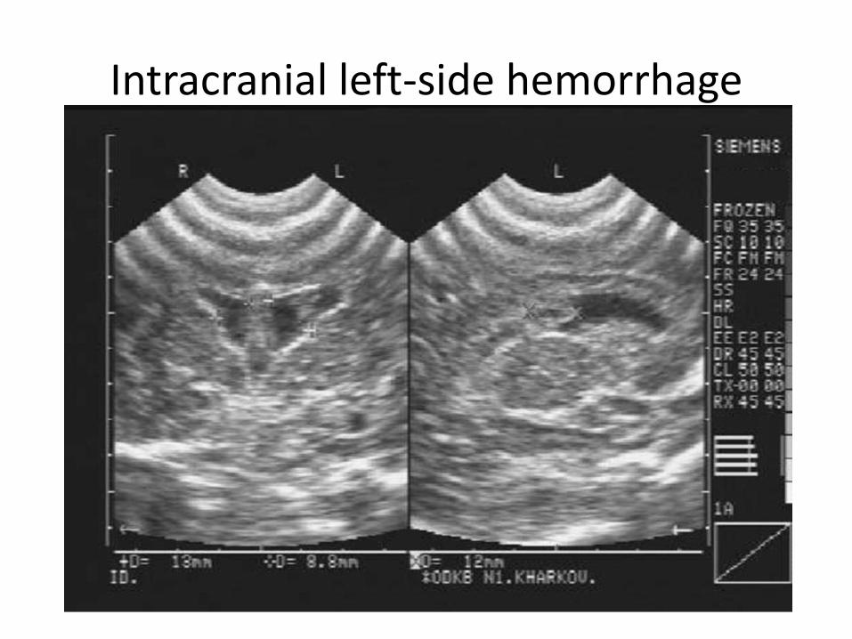
Intracranial left-side hemorrhage
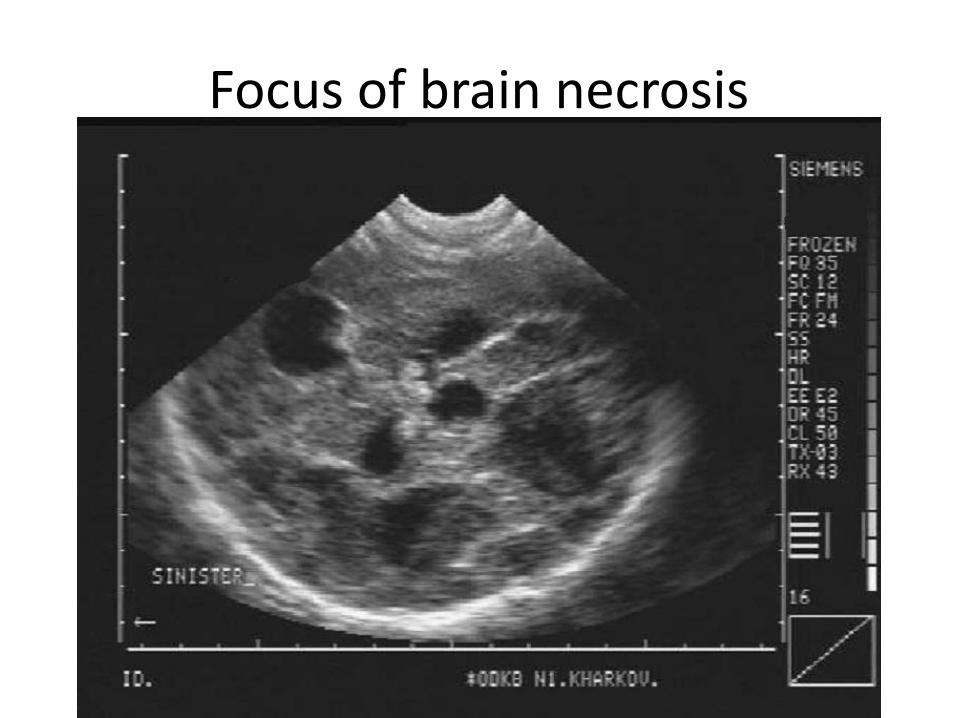
Focus of brain necrosis
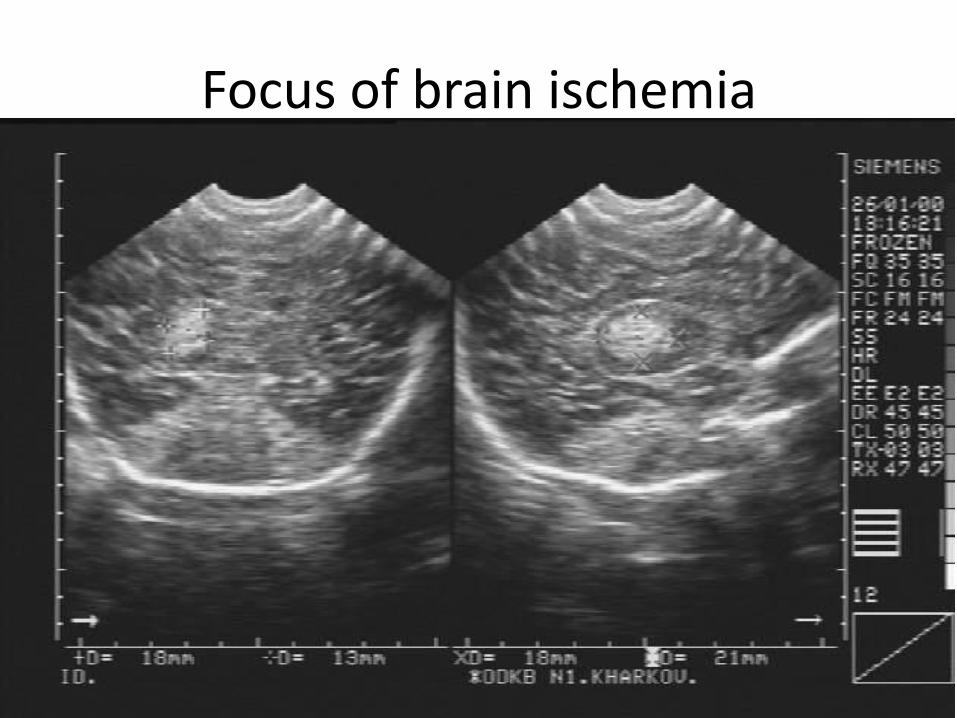
Focus of brain ischemia
Diagnosis
• Etiologic diagnosis in chronic meningitis may require MENINGEAL BIOPSY (→ histology, electron microscopy, PCR, cultures).
• Target regions that enhance with contrast on MRI or CT.
• With current microsurgical techniques, most areas of basal meninges can be accessed via limited craniotomy.
• Most common conditions identified – sarcoid (31%), metastatic adenocarcinoma (25%).

Treatment Patients prefer quiet, darkened room.
ANALGESICS - to relieve headache (often reduced by initial diagnostic lumbar puncture).
ANTIPYRETICS - to reduce fever.
Indications for hospitalization:
1. severe cases
2. deficient humoral immunity (trial of IVIG)
3. herpes meningitis (intravenous ACYCLOVIR)
4. potential nonviral causes.
FIELD STABILIZATION, TRANSPORT.
Treatment
ANTIMICROBIAL THERAPY
• Must be bactericidal in CSF – i.e. maximum tolerated doses!
• Intravenously (intrathecal / intraventricular therapy is not effective).
• Crucial step is to initiate antimicrobial therapy immediately!
If you suspect meningococcus, give PENICILLIN G before transporting to hospital!
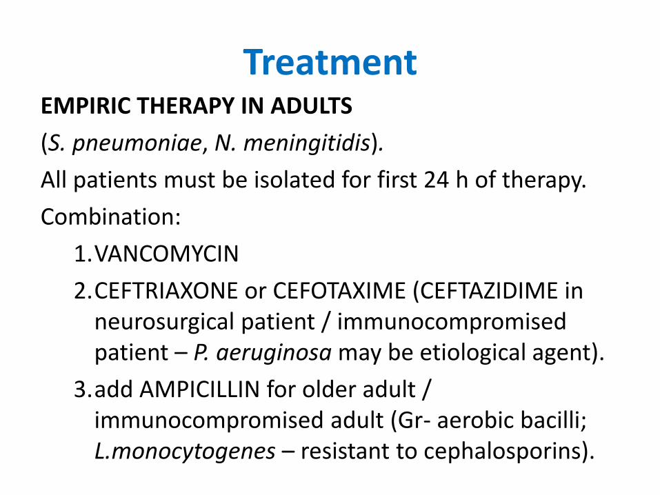
Treatment EMPIRIC THERAPY IN ADULTS
(S. pneumoniae, N. meningitidis).
All patients must be isolated for first 24 h of therapy.
Combination:
1.VANCOMYCIN
2.CEFTRIAXONE or CEFOTAXIME (CEFTAZIDIME in neurosurgical patient / immunocompromised patient – P. aeruginosa may be etiological agent).
3.add AMPICILLIN for older adult / immunocompromised adult (Gr- aerobic bacilli; L.monocytogenes – resistant to cephalosporins).
Treatment
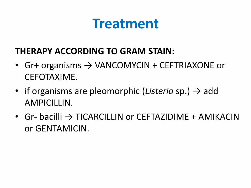
THERAPY ACCORDING TO GRAM STAIN:
• Gr+ organisms → VANCOMYCIN + CEFTRIAXONE or CEFOTAXIME.
• if organisms are pleomorphic (Listeria sp.) → add AMPICILLIN.
• Gr- bacilli → TICARCILLIN or CEFTAZIDIME + AMIKACIN or GENTAMICIN.

Treatment IF CAUSATIVE ORGANISM HAS BEEN IDENTIFIED: N. meningitidis: PENICILLIN G or AMPICILLIN or CEFOTAXIME or CHLORAMPHENICOL + (at end of therapy to eradicate nasopharyngeal carriage) oral RIFAMPIN for 2 d. S. pneumoniae: VANCOMYCIN + CEFTRIAXONE or CEFOTAXIME Enteric Gr- bacilli: CEFTRIAXONE or CEFOTAXIME + AMIKACIN or GENTAMICIN P. aeruginosa: CEFTAZIDIME or CIPROFLOXACIN or TICARCILLIN ± GENTAMICIN L. monocytogenes: AMPICILLIN or TMP-SMX H. influenzae type b: CEFTRIAXONE or CEFOTAXIME or CHLORAMPHENICOL (with AMPICILLIN) S. aureus (methicillin-sensitive): OXACILLIN S. aureus (methicillin-resistant): VANCOMYCIN S. epidermidis: VANCOMYCIN ± RIFAMPIN
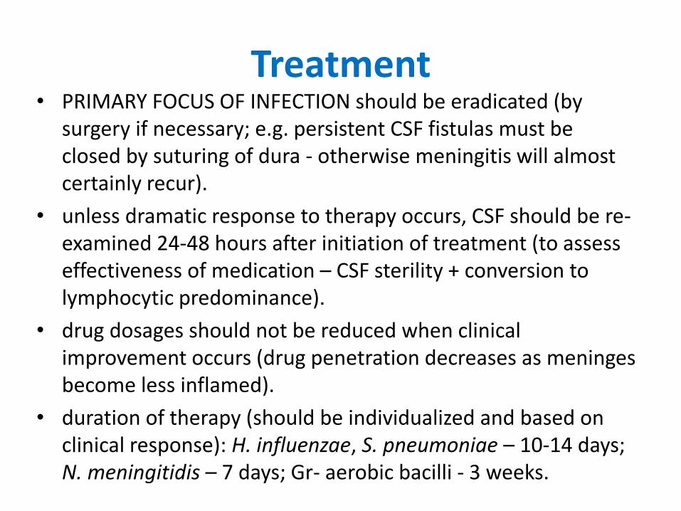
Treatment • PRIMARY FOCUS OF INFECTION should be eradicated (by
surgery if necessary; e.g. persistent CSF fistulas must be closed by suturing of dura - otherwise meningitis will almost certainly recur).
• unless dramatic response to therapy occurs, CSF should be re-examined 24-48 hours after initiation of treatment (to assess effectiveness of medication – CSF sterility + conversion to lymphocytic predominance).
• drug dosages should not be reduced when clinical improvement occurs (drug penetration decreases as meninges become less inflamed).
• duration of therapy (should be individualized and based on clinical response): H. influenzae, S. pneumoniae – 10-14 days; N. meningitidis – 7 days; Gr- aerobic bacilli - 3 weeks.
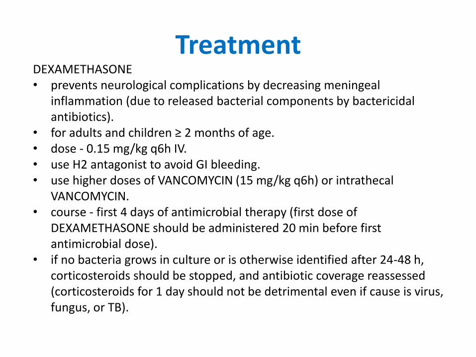
Treatment DEXAMETHASONE • prevents neurological complications by decreasing meningeal
inflammation (due to released bacterial components by bactericidal antibiotics).
• for adults and children ≥ 2 months of age. • dose - 0.15 mg/kg q6h IV. • use H2 antagonist to avoid GI bleeding. • use higher doses of VANCOMYCIN (15 mg/kg q6h) or intrathecal
VANCOMYCIN. • course - first 4 days of antimicrobial therapy (first dose of
DEXAMETHASONE should be administered 20 min before first antimicrobial dose).
• if no bacteria grows in culture or is otherwise identified after 24-48 h, corticosteroids should be stopped, and antibiotic coverage reassessed (corticosteroids for 1 day should not be detrimental even if cause is virus, fungus, or TB).
Treatment
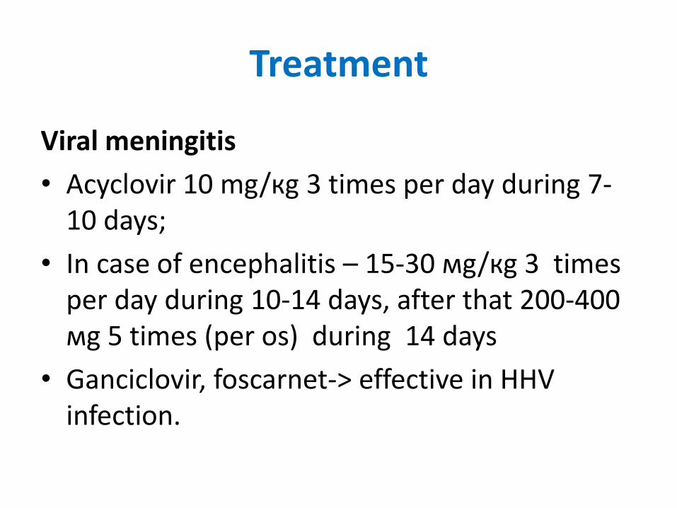
Viral meningitis
• Acyclovir 10 mg/кg 3 times per day during 7-10 days;
• In case of encephalitis – 15-30 мg/кg 3 times per day during 10-14 days, after that 200-400 мg 5 times (per os) during 14 days
• Ganciclovir, foscarnet-> effective in HHV infection.
Treatment
Fungal meningitis
• Amphotericin B
• Fluconazole
• Miconazole
• Flucytosine
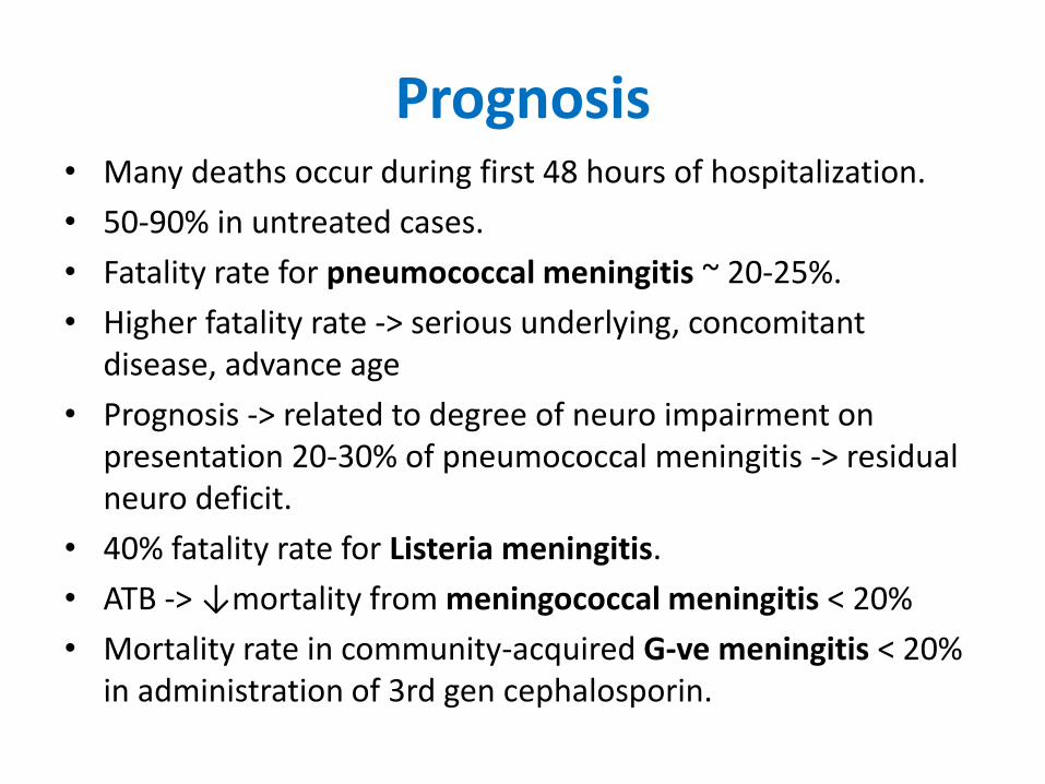
Prognosis • Many deaths occur during first 48 hours of hospitalization.
• 50-90% in untreated cases.
• Fatality rate for pneumococcal meningitis ~ 20-25%.
• Higher fatality rate -> serious underlying, concomitant disease, advance age
• Prognosis -> related to degree of neuro impairment on presentation 20-30% of pneumococcal meningitis -> residual neuro deficit.
• 40% fatality rate for Listeria meningitis.
• ATB -> ↓mortality from meningococcal meningitis < 20%
• Mortality rate in community-acquired G-ve meningitis < 20% in administration of 3rd gen cephalosporin.
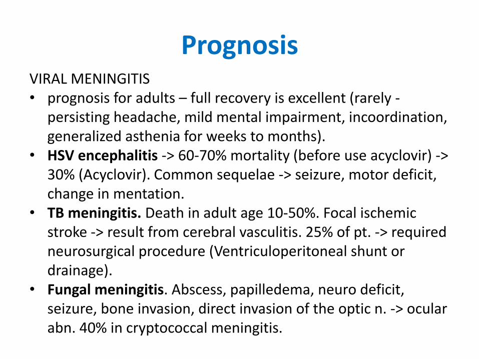
Prognosis VIRAL MENINGITIS • prognosis for adults – full recovery is excellent (rarely -
persisting headache, mild mental impairment, incoordination, generalized asthenia for weeks to months).
• HSV encephalitis -> 60-70% mortality (before use acyclovir) -> 30% (Acyclovir). Common sequelae -> seizure, motor deficit, change in mentation.
• TB meningitis. Death in adult age 10-50%. Focal ischemic stroke -> result from cerebral vasculitis. 25% of pt. -> required neurosurgical procedure (Ventriculoperitoneal shunt or drainage).
• Fungal meningitis. Abscess, papilledema, neuro deficit, seizure, bone invasion, direct invasion of the optic n. -> ocular abn. 40% in cryptococcal meningitis.


Chemoprophylaxis
• Incidence of transmission of meningococcus is ~ 5%
• Household contact – Rifampin adult 600 mg child > 1 mo 10mg/kg child < 1 mo 5mg/kg oral q 12 hr. for a total of 4 doses.
• Health care worker -> do not required prophylaxis.
Chemoprophylaxis
• Directed contact (mouth to mouth, ET tube, nasotrachial suction) – Ciprofloxacin 500 mg oral or Ceftriaxone 250 mg im. (<15 yr. 125 mg im.)
• No indication for chemoprophylaxis in pneumococcal meningitis.

Immunoprophylaxis
• Vaccination is also available to confer immune protection against
– JE virus
–H. influenzae type B (use in pediatrics).

"Only the prepared mind can help the impaired host.“
Dr. Libero Ajello