KEYWORDS PRESSURE ULCERS | ASSESSMENT | WOUND IMAGING Skin ... · and tactile cues to identify...
2
practice guided learning Nursing Times 3 August 2010 Vol 106 No 30 www.nursingtimes.net 16 100OF NURSING JOBS Introducing online learning logs in education Innovative intermediate care for older people HOW TO IDENTIFY PRESSURE DAMAGE IN DARK SKIN Laura Serrant-Green We must act on the findings of the PM’s commission Opinion skin assessment, outlined in Box 1 (NPUAP and EPUAP, 2009). The majority are basedonexpertopinionwith two supported by research evidence. Many of the recommendations for skin assessmentdependonvisual and tactile cues to identify changes in skin appearance – for example, changes in colour, temperature and hardness may all mark early signs of pressure damage. Indeed, for many years the starting point for pressure ulceration has been considered to be an area of erythema that would not blanch when subjected to light pressure (non-blanching erythema). Educational material on this can be found at tinyurl.com/non-blanching. Visual cues for changes in skin appearance may be relatively easy to observe in Caucasian skin but with darker pigmentation it may be harder to spot visual signs of early changes that are due to pressure damage (NPUAP and EPUAP, 2009). FAILURE TO IDENTIFY EARLY SIGNS OF PRESSURE DAMAGE Using visual cues to recognise pressure damagemaydisadvantagethosewithdarker skin pigmentation. Rosen et al (2006) reported the results of a quality improvement project conducted in a single nursing home in the US. Its purpose wastodeterminewhetherthe provision of educational material to nursing home staff improved pressure ulcer prevention. The researchers collectedinformationonnew pressure ulcers; before introducing their educationalprogramme,nograde1pressure ulcers (non-blanching erythema) were reported among black residents while 31.6% of all pressure ulcers in white residents were classified as grade 1. Failure to recognise the early stages of damage may have serious consequences as the presence of non-blanching erythema is a risk factor for the development of more severe forms of pressure damage (Nixon et al, 2007). The difficulty in spotting non- blanching erythema in dark skin may help explain why Rosen et al (2006) found black residents were more likely to have grade 2-4 ulcers (Fig 1) than white residents. This difference was also reported by Baumgarten et al (2004), who looked at 59 nursing homes in the US and found black residents were more likely to develop grade 2-4 ulcers. The data indicated that, for a 50 bed nursing home with equal numbers of black and white residents, 14 black people and nine white people would develop grade 2-4 ulcers. This suggests failing to identify early stages of pressure damage results in greater numbers of people with dark pigmented skin developing more severe pressure damage. While these studies are from the US and represent populations of African Americans, the problem of identifying early stages of pressure damage is not confined to Black Caribbean, Black African or other black populations in the UK. Any variations in skin pigmentation may obscure the first signs of pressure damage; for example it may be easier to identify changes in skin redness in a Nordic compared with a Mediterranean based population. The classic signs of skin damage are different in Caucasian and dark pigmented skin. Advice on how to identify pressure ulcers in dark skin is provided Skin assessment in dark pigmented skin: a challenge in pressure ulcer prevention KEYWORDS PRESSURE ULCERS | ASSESSMENT | WOUND IMAGING AUTHOR Michael Clark, PhD, is manager, Welsh Wound Network. ABSTRACT Clark M (2010) Skin assessment in dark pigmented skin: a challenge in pressure ulcer prevention. Nursing Times; 106: 30, 16-17. Skin assessment is a vital element in the prevention of pressure ulcers, and many recommendations for skin assessment depend on visual and tactile cues to identify changes in skin appearance. These visual cues that indicate changes in skin appearance may be easily observed in Caucasian skin but, with darker pigmentation, it may be harder to spot visual signs of early changes caused by pressure damage. This article outlines how nurses can address this problem in clinical practice and the technological developments that may help to solve this clinical issue. There are five main elements to successful pressure ulcer prevention: l Risk assessment; l Skin assessment; l Nutrition interventions; l Repositioning interventions; l Support surface interventions (National Pressure Ulcer Advisory Panel and European Pressure Ulcer Advisory Panel, 2009). Pressure ulcer guidelines published last year provide seven recommendations on Understand the problems of identifying pressure damage in non-Caucasian skin. Be able to identify a grade 1 pressure ulcer. LEARNING OBJECTIVES Ensure that a complete skin assessment is part of the risk assessment screening policy in place in all healthcare settings. Educate professionals to carry out a comprehensive skin assessment, including techniques for identifying blanching response, localised heat, oedema and induration (hardness).* Inspect skin regularly for signs of redness in people identified as being at risk of pressure ulceration. The frequency of inspection may need to be increased in response to any deterioration in the overall condition.* Skin inspection should include assessments for localised heat, oedema or induration, especially in people who have dark pigmented skin. Ask patients to identify areas of discomfort or pain that could be attributed to pressure damage. Observe the skin for pressure damage caused by medical devices (for example, catheters and cervical collars). Document all skin assessments, noting details of any pain that could possibly be related to pressure damage. *Supported by research evidence Source: NPUAP and EPUAP (2009) BOX 1. SKIN ASSESSMENT
Transcript of KEYWORDS PRESSURE ULCERS | ASSESSMENT | WOUND IMAGING Skin ... · and tactile cues to identify...
practice guided learning
Nursing Times 3 August 2010 Vol 106 No 30 www.nursingtimes.net16
100S OF
NURSING
JOBS
Introducing online learning logs in education
Innovative intermediate care for older people
HOW TO IDENTIFY PRESSURE DAMAGE
IN DARK SKIN
3-9 AUGUST 2010
£1.80 Vol 106 No 30
Laura Serrant-Green We must act on the findings of the PM’s commissionOpinion
2010 £1.80 Vol 106
No 30
We must act on the findings of the PM’s commission
skin assessment, outlined in Box 1 (NPUAP and EPUAP, 2009). The majority are based on expert opinion with two supported by research evidence.
Many of the recommendations for skin assessment depend on visual and tactile cues to identify changes in skin appearance – for example, changes in colour, temperature and hardness may all mark early signs of pressure damage. Indeed, for many years the starting point for pressure ulceration has been considered to be an area of erythema that would not blanch when subjected to light pressure (non-blanching erythema). Educational material on this can be found at tinyurl.com/non-blanching.
Visual cues for changes in skin appearance may be relatively easy to observe in Caucasian skin but with darker pigmentation it may be harder to spot visual signs of early changes that are due to pressure damage (NPUAP and EPUAP, 2009).
FAILURE TO IDENTIFY EARLY SIGNS OF PRESSURE DAMAGE Using visual cues to recognise pressure damage may disadvantage those with darker skin pigmentation.
Rosen et al (2006) reported the results of a
quality improvement project conducted in a single nursing home in the US. Its purpose was to determine whether the provision of educational material to nursing home staff improved pressure ulcer prevention. The researchers collected information on new
pressure ulcers; before introducing their educational programme, no grade 1 pressure ulcers (non-blanching erythema) were reported among black residents while 31.6% of all pressure ulcers in white residents were classified as grade 1.
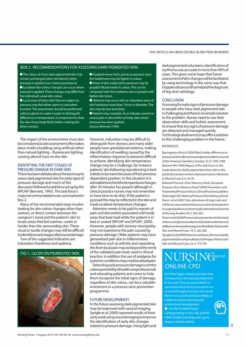
Failure to recognise the early stages of damage may have serious consequences as the presence of non-blanching erythema is a risk factor for the development of more severe forms of pressure damage (Nixon et al, 2007). The difficulty in spotting non-blanching erythema in dark skin may help explain why Rosen et al (2006) found black residents were more likely to have grade 2-4 ulcers (Fig 1) than white residents.
This difference was also reported by Baumgarten et al (2004), who looked at 59 nursing homes in the US and found black residents were more likely to develop grade 2-4 ulcers. The data indicated that, for a 50 bed nursing home with equal numbers of black and white residents, 14 black people and nine white people would develop grade 2-4 ulcers. This suggests failing to identify early stages of pressure damage results in greater numbers of people with dark pigmented skin developing more severe pressure damage.
While these studies are from the US and represent populations of African Americans, the problem of identifying early stages of pressure damage is not confined to Black Caribbean, Black African or other black populations in the UK. Any variations in skin pigmentation may obscure the first signs of pressure damage; for example it may be easier to identify changes in skin redness in a Nordic compared with a Mediterranean based population.
The classic signs of skin damage are different in Caucasian and dark pigmented skin. Advice on how to identify pressure ulcers in dark skin is provided
Skin assessment in dark pigmented skin: a challenge in pressure ulcer prevention
KEYWORDS PRESSURE ULCERS | ASSESSMENT | WOUND IMAGING
AUTHOR Michael Clark, PhD, is manager, Welsh Wound Network.ABSTRACT Clark M (2010) Skin assessment in dark pigmented skin: a challenge in pressure ulcer prevention. Nursing Times; 106: 30, 16-17.Skin assessment is a vital element in the prevention of pressure ulcers, and many recommendations for skin assessment depend on visual and tactile cues to identify changes in skin appearance. These visual cues that indicate changes in skin appearance may be easily observed in Caucasian skin but, with darker pigmentation, it may be harder to spot visual signs of early changes caused by pressure damage. This article outlines how nurses can address this problem in clinical practice and the technological developments that may help to solve this clinical issue.
There are five main elements to successful pressure ulcer prevention: l Risk assessment;l Skin assessment; l Nutrition interventions; l Repositioning interventions; l Support surface interventions (National Pressure Ulcer Advisory Panel and European Pressure Ulcer Advisory Panel, 2009).
Pressure ulcer guidelines published last year provide seven recommendations on
Understand the problems of identifying pressure damage in non-Caucasian skin.
Be able to identify a grade 1 pressure ulcer.
LEARNING OBJECTIVES
Ensure that a complete skin assessment is part of the risk assessment screening policy in place in all healthcare settings.
Educate professionals to carry out a comprehensive skin assessment, including techniques for identifying blanching response, localised heat, oedema and induration (hardness).*
Inspect skin regularly for signs of redness in people identified as being at risk of pressure ulceration. The frequency of inspection may need to be increased in response to any deterioration in the overall condition.*
Skin inspection should include assessments for localised heat, oedema or induration, especially in people who have dark pigmented skin.
Ask patients to identify areas of discomfort or pain that could be attributed to pressure damage.
Observe the skin for pressure damage caused by medical devices (for example, catheters and cervical collars).
Document all skin assessments, noting details of any pain that could possibly be related to pressure damage.*Supported by research evidenceSource: NPUAP and EPUAP (2009)
BOX 1. SKIN ASSESSMENT
practice guided learning
17Nursing Times 3 August 2010 Vol 106 No 30 www.nursingtimes.net
The impact of the environment must also be considered as skin assessment often takes place inside a building using artificial rather than natural lighting – fluorescent lighting causing altered hues on the skin.
IDENTIFYING THE FIRST STAGES OF PRESSURE DAMAGE IN DARK SKINThere has been debate about the best way to assess dark pigmented skin for early signs of pressure damage and much of this discussion followed a task force set up by the NPUAP (Bennett, 1995). The task force’s major recommendations are summarised in Box 2.
Many of the recommended steps involve looking for skin colour changes other than redness, or direct contact between the caregiver’s hand and the patient’s skin to locate areas that feel warmer, cooler or harder than the surrounding skin. These visual or tactile changes may still be difficult to identify as early stages of pressure damage.
Two of the suggested indicators are induration (hardness) and oedema.
However, induration may be difficult to distinguish from dryness and many older people have gravitational oedema, making identification of swelling caused by the inflammatory response to pressure difficult to achieve. Identifying skin temperature change may be a challenge, for instance patients’ skin following repositioning in bed is likely to be warm because of their previous dependent position. In this situation it is advisable to recheck for temperature changes after 30 minutes has passed (although in clinical practice nurses may not remember or have time to do this). If the patient is pyrexial this may be reflected in the skin and mask localised temperature changes.
Attention needs to be paid to reports of pain and discomfort associated with body areas that bear load while the patient is in bed or seated (NPUAP and EPUAP, 2009). However, people with sensory neuropathy may not experience the pain caused by pressure damage. Older patients may have generalised pain due to inflammatory conditions such as arthritis and separating this from local pain may be beyond the remit of the validated pain tools used in clinical practice. In addition the use of analgesia for systemic conditions may mask localised pain.
Detecting early pressure damage is not the sole responsibility of healthcare professionals and educating patients and carers to help them recognise the initial signs of damage, regardless of skin colour, can be a valuable investment in a pressure ulcer prevention programme.
FUTURE DEVELOPMENTSIn the future assessing dark pigmented skin may be improved with wound imaging. Sprigle et al (2009) reported results of their early work using wound imaging to improve the identification of early skin changes related to pressure damage. Using light and
THIS ARTICLE HAS BEEN DOUBLE-BLIND PEER-REVIEWED
Portfolio Pages contain activities that correspond to the learning objectives in the unit. They are presented in a convenient format for you to print out or work through on screen and can be filed in your professional portfolio as evidence of your learning and professional development.
For the Portfolio Pages corresponding to this unit, and for other Guided Learning units, go to tinyurl.com/GL-archive
ONLINE CPD
REFERENCES
Baumgarten M et al (2004) Black/white differences in pressure ulcer incidence in nursing home residents. Journal of the American Geriatrics Society; 52: 8, 1293-1298.Bennett MA (1995) Report of the task force on the implications for darkly pigmented intact skin in the prediction and prevention of pressure ulcers. Advances in Wound Care; 8: 6, 34-35.National Pressure Ulcer Advisory Panel, European Pressure Ulcer Advisory Panel (2009) Prevention and Treatment of Pressure Ulcers: Clinical Practice Guideline. Washington DC: National Pressure Ulcer Advisory Panel.Nixon J et al (2007) Skin alterations of intact skin and risk factors associated with pressure ulcer development in surgical patients: a cohort study. International Journal of Nursing Studies; 44: 5, 655-663.Rosen J et al (2006) Pressure ulcer prevention in black and white nursing home residents: A QI initiative of enhanced ability, incentives and management feedback. Advances in Skin and Wound Care; 19: 5, 262-268.Sprigle S et al (2009) Detection of skin erythema in darkly pigmented skin using multispectral images. Advances in Skin and Wound Care; 22: 4, 172-179.
dark pigmented volunteers, identification of erythema was accurate in more than 90% of cases. This gives some hope that future assessment of skin changes will be facilitated by using technology in the same way that Doppler ultrasound has helped the diagnosis of leg ulcer aetiology.
CONCLUSIONAssessing for early signs of pressure damage in people who have dark pigmented skin is challenging and there is no simple solution to the problem. Nurses need to use their observation skills and holistic assessment to ensure that any signs of pressure damage are detected and managed quickly. Technological advances may offer a solution to this challenging problem in the future. l
SPL
The colour of intact dark pigmented skin may remain unchanged (does not blanch) when pressure is applied over a bony prominence.
Localised skin colour changes can occur where pressure is applied. These changes may differ from the individual’s usual skin colour.
Local areas of intact skin that are subject to pressure may feel either warm or cool when touched. This assessment should be performed without gloves to make it easier to distinguish differences in temperature. It is important to clean the skin of any body fluids before making this direct contact.
If patients have had a previous pressure ulcer, the healed area may be lighter in colour.
Areas of skin subjected to pressure may be purplish/bluish/violet in colour. This can be compared with the erythema seen in people with lighter skin tones.
Oedema may occur with an induration (area of skin hardness) more than 15mm in diameter. The skin may be taut and shiny.
Patients may complain of, or indicate, current or recent pain or discomfort at body sites where pressure has been applied. Source: Bennett (1995)
BOX 2. RECOMMENDATIONS FOR ASSESSING DARK PIGMENTED SKIN
FIG 1. ULCER ON PIGMENTED SKIN