
Management of Keloids By: Thad Riley Advisor: Bill Grimes March 24, 2006.
Upload
menkantozzCategory
view
370download
2
KELOID DISEASE (Case Presentation)
PRESENTED BY
Antony Mamati.

We see what we want?
Common things are usually very rare to spot.

History
A 23 year old female was referred by plastic surgeons for radiotherapy to the posterior ear lobe, following the development of a Keloid Scar, three years after an ear piercing
No family history of keloids

Pathology
Keloid is a unique human dermal fibroproliferative disorder that occurs after injury, inflammation, surgery,and burn.
Commonly causes of keloids include acne, folliculitis, chicken pox, vaccinations and trauma (such as, earlobe piercing, lacerations, or surgical wounds).
It is a benign growth, well-demarcated area of fibrous tissue overgrowth that extends beyond the original defect

Baron Jean-Louis Alibert (1768-1837)
Described appearance “Chele” (crab-claw) (also means: to grab as in “chelation”)
Crab-claw-like Cheloid Keloid

Effects of keloids
Compromise aesthetic Impairment of function

Itchy Pain Prurutic

How best can they be treated?

Treatment Options
Surgery
Intralesional Steroids
Radiation
Laser
Cryotherapy
Pressure
Multimodality Therapy

Still Trying

Surgery Drawbacks
Painful
Difficult reconstruction with large keloids
Utilizes normal surrounding tissue – limiting later reconstructive options
Low long term success as monotherapy

Steroids
Triamcinolone
Hydrocortisone
Dexamethasone
Methylprednisone


Laser 1980s in vogue
Proposed Mechanism
No knife Less tissue trauma

Cryotherapy- cold treatment

Diminishes size and induration (HTS >Keloid) when used as monotherapy
<10% Recurrence when combined with surgery
Photos Courtesy of Dr. Redett
Pressure Therapy

Radiation
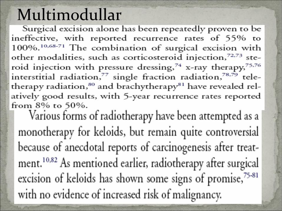
Multimodullar

In this case… Plastic surgery was carried out to remove the Keloid
Scar. The patient attended the following day for a single dose of radiotherapy to the scar, plus a small margin of 5mm. The pinna was taped forward to expose the effected posterior section of the lobe.

Radiotherapy prescription
12Gy in single fraction.
HVL 0.2mm Cu
Energy used was:- 100kv.
Actual Field size:- 3.5 x 1.5 cm –margins of
2mm
Cone size used:- 4 X 6cm
Shielding :-A lead cut out was used to define the field size and protect the surrounding
tissue.

Conclusion
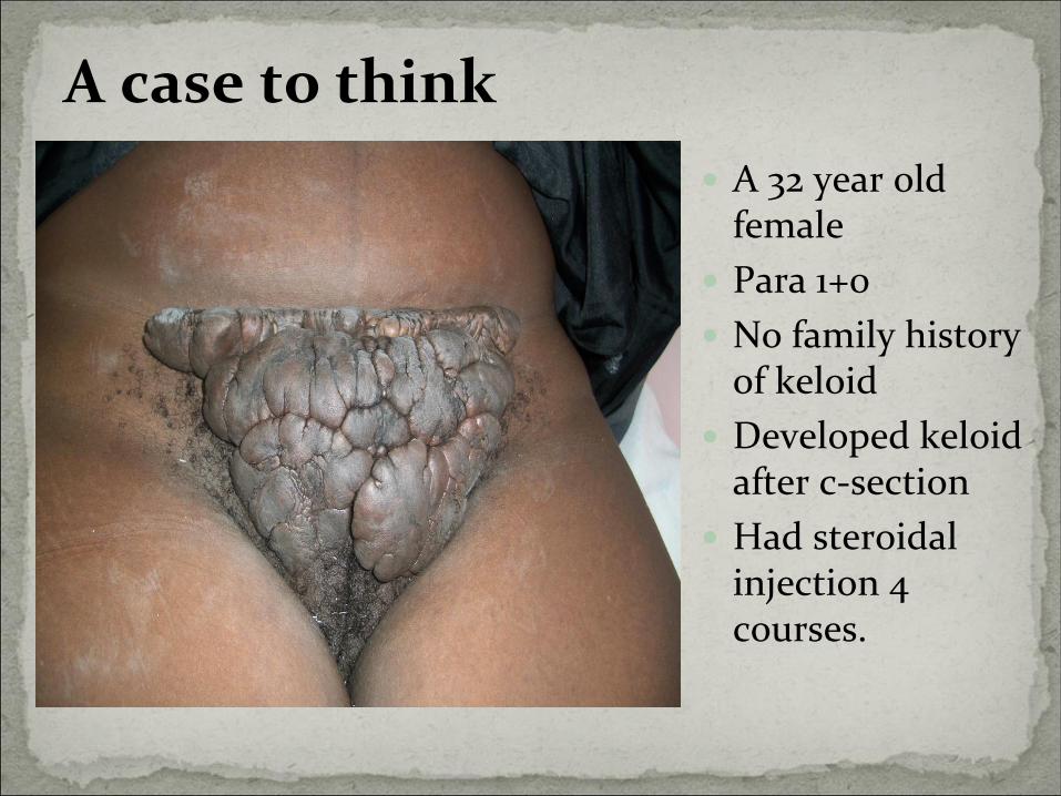
A case to think
A 32 year old female
Para 1+0
No family history of keloid
Developed keloid after c-section
Had steroidal injection 4 courses.

Thank You


![...Vitiligo Acne Bleeding, Excessive Diabetes Heart Problems C] High Blood Pressure Liver Disease Mitral Valve Prolapse SCarring/Keloids Arthritis Blood Clots Eczema Hepatitis HIV/AIDS](https://static.fdocuments.in/doc/165x107/5e30a8c54b0276386f14420e/-vitiligo-acne-bleeding-excessive-diabetes-heart-problems-c-high-blood-pressure.jpg)


